Embed Size (px)
Citation preview

General Pathology VPM 152
Enrique Aburto http://people.upei.ca/eaburto Winter 2015
Lecture 3 Neoplasia: Malignant tumors
Disorders of Cell Growth & Neoplasia
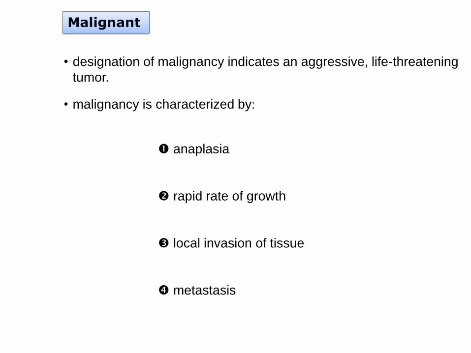
Malignant
• designation of malignancy indicates an aggressive, life-threatening
tumor.
• malignancy is characterized by:
anaplasia
rapid rate of growth
local invasion of tissue
metastasis
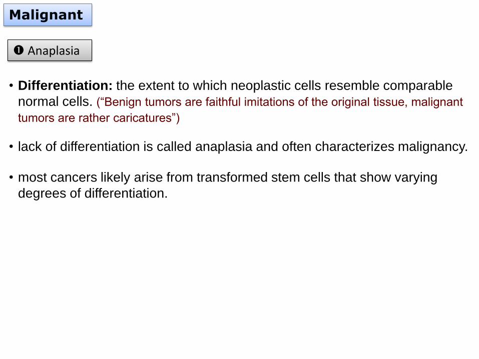
Malignant
• Differentiation: the extent to which neoplastic cells resemble comparable
normal cells. (“Benign tumors are faithful imitations of the original tissue, malignant
tumors are rather caricatures”)
• lack of differentiation is called anaplasia and often characterizes malignancy.
• most cancers likely arise from transformed stem cells that show varying
degrees of differentiation.
Anaplasia
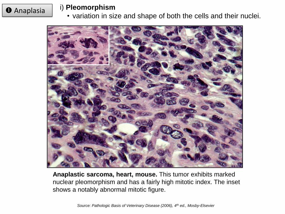
i) Pleomorphism
• variation in size and shape of both the cells and their nuclei. Anaplasia
Anaplastic sarcoma, heart, mouse. This tumor exhibits marked
nuclear pleomorphism and has a fairly high mitotic index. The inset
shows a notably abnormal mitotic figure.
Source: Pathologic Basis of Veterinary Disease (2006), 4th ed., Mosby-Elsevier
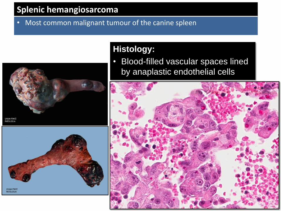
Splenic hemangiosarcoma
• Most common malignant tumour of the canine spleen
Histology:
• Blood-filled vascular spaces lined
by anaplastic endothelial cells
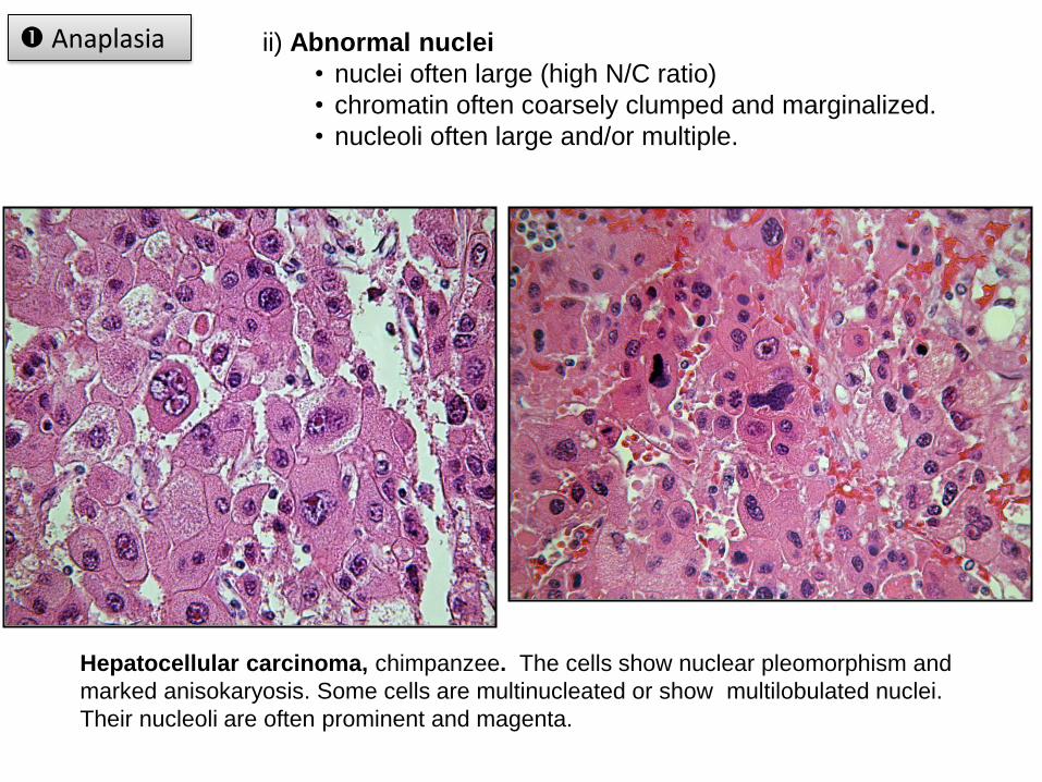
ii) Abnormal nuclei
• nuclei often large (high N/C ratio)
• chromatin often coarsely clumped and marginalized.
• nucleoli often large and/or multiple.
Hepatocellular carcinoma, chimpanzee. The cells show nuclear pleomorphism and
marked anisokaryosis. Some cells are multinucleated or show multilobulated nuclei.
Their nucleoli are often prominent and magenta.
Anaplasia
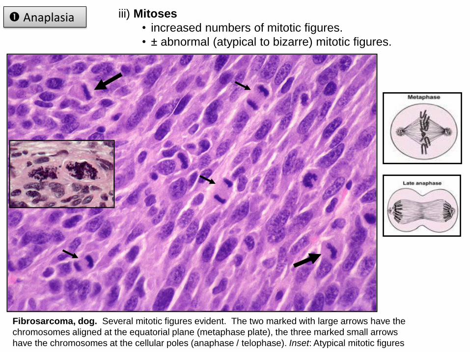
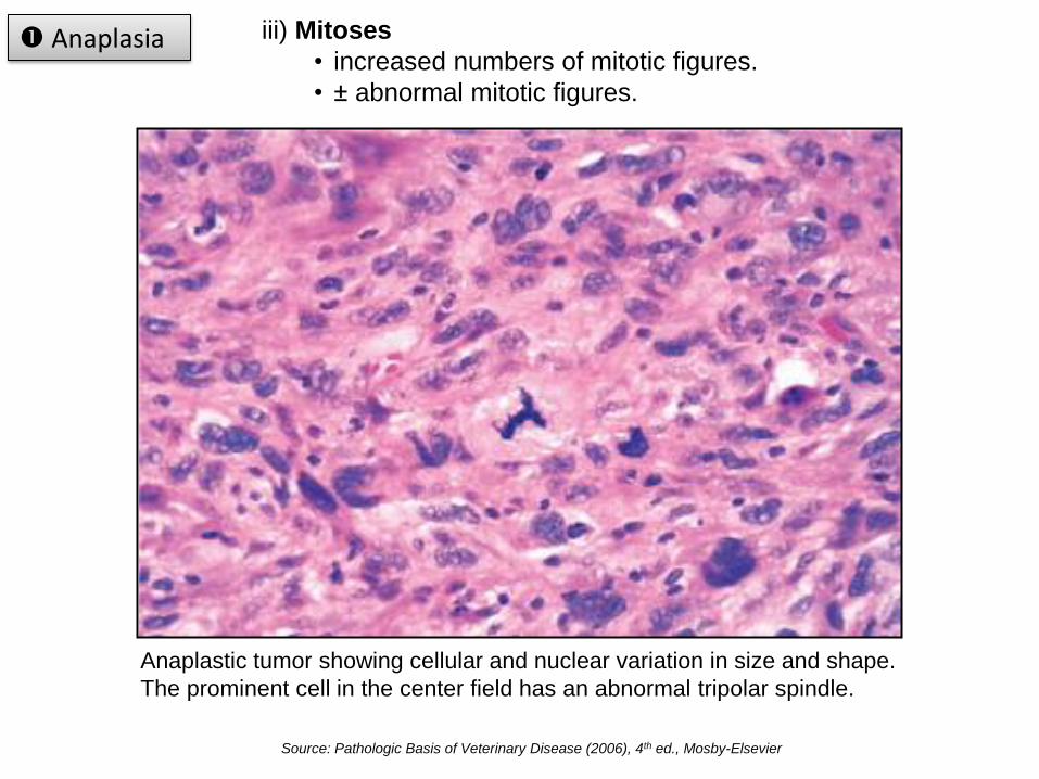
iii) Mitoses
• increased numbers of mitotic figures.
• ± abnormal (atypical to bizarre) mitotic figures.
Fibrosarcoma, dog. Several mitotic figures evident. The two marked with large arrows have the
chromosomes aligned at the equatorial plane (metaphase plate), the three marked small arrows
have the chromosomes at the cellular poles (anaphase / telophase). Inset: Atypical mitotic figures
Anaplasia
iii) Mitoses
• increased numbers of mitotic figures.
• ± abnormal mitotic figures.
Anaplasia
Anaplastic tumor showing cellular and nuclear variation in size and shape.
The prominent cell in the center field has an abnormal tripolar spindle.
Source: Pathologic Basis of Veterinary Disease (2006), 4th ed., Mosby-Elsevier
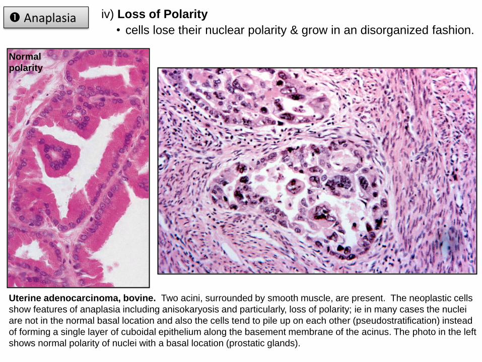
iv) Loss of Polarity
• cells lose their nuclear polarity & grow in an disorganized fashion.
Uterine adenocarcinoma, bovine. Two acini, surrounded by smooth muscle, are present. The neoplastic cells
show features of anaplasia including anisokaryosis and particularly, loss of polarity; ie in many cases the nuclei
are not in the normal basal location and also the cells tend to pile up on each other (pseudostratification) instead
of forming a single layer of cuboidal epithelium along the basement membrane of the acinus. The photo in the left
shows normal polarity of nuclei with a basal location (prostatic glands).
Anaplasia
Normal
polarity
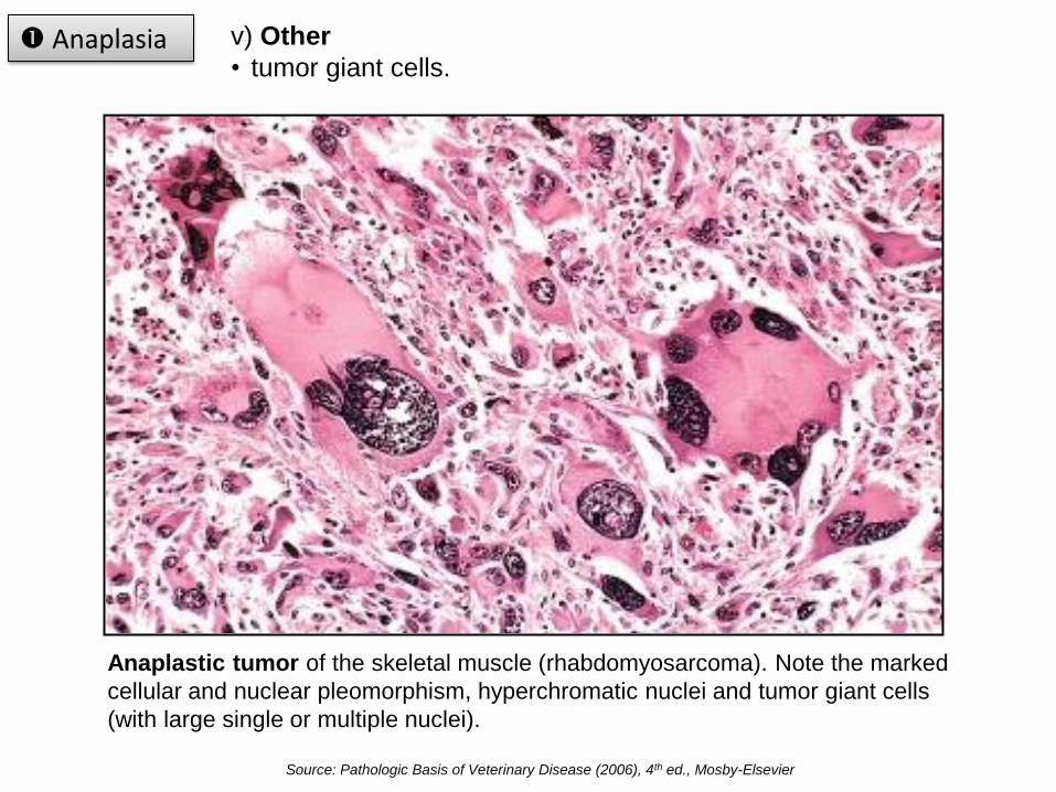
v) Other
• tumor giant cells.
Anaplastic tumor of the skeletal muscle (rhabdomyosarcoma). Note the marked
cellular and nuclear pleomorphism, hyperchromatic nuclei and tumor giant cells
(with large single or multiple nuclei).
Anaplasia
Source: Pathologic Basis of Veterinary Disease (2006), 4th ed., Mosby-Elsevier
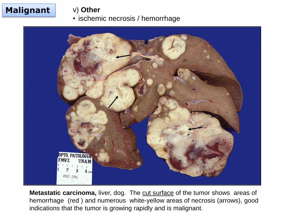
v) Other
• ischemic necrosis / hemorrhage
Metastatic carcinoma, liver, dog. The cut surface of the tumor shows areas of
hemorrhage (red ) and numerous white-yellow areas of necrosis (arrows), good
indications that the tumor is growing rapidly and is malignant.
Malignant
Malignant
• not all features of anaplasia are necessarily seen in a given malignant tumor.
• anaplastic features usually call for a poor or guarded prognosis.
• some malignant neoplasms are composed of well differentiated cells.
• the whole clinical, morphologic (gross & microscopic) and epidemiologic
aspects of each tumor must be evaluated in order to provide an accurate
prognosis.
Anaplasia
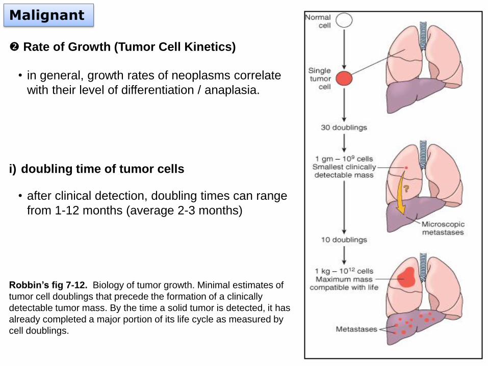
Rate of Growth (Tumor Cell Kinetics)
• in general, growth rates of neoplasms correlate
with their level of differentiation / anaplasia.
i) doubling time of tumor cells
• after clinical detection, doubling times can range
from 1-12 months (average 2-3 months)
Malignant
Robbin’s fig 7-12. Biology of tumor growth. Minimal estimates of
tumor cell doublings that precede the formation of a clinically
detectable tumor mass. By the time a solid tumor is detected, it has
already completed a major portion of its life cycle as measured by
cell doublings.
Malignant
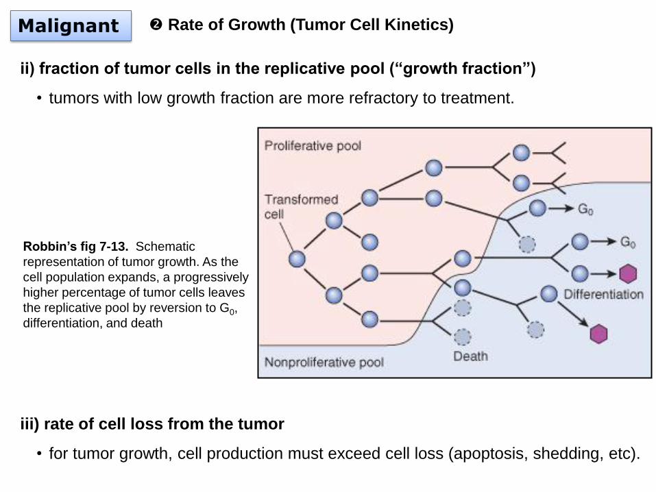
ii) fraction of tumor cells in the replicative pool (“growth fraction”)
• tumors with low growth fraction are more refractory to treatment.
iii) rate of cell loss from the tumor
• for tumor growth, cell production must exceed cell loss (apoptosis, shedding, etc).
Rate of Growth (Tumor Cell Kinetics)
Robbin’s fig 7-13. Schematic
representation of tumor growth. As the
cell population expands, a progressively
higher percentage of tumor cells leaves
the replicative pool by reversion to G0,
differentiation, and death
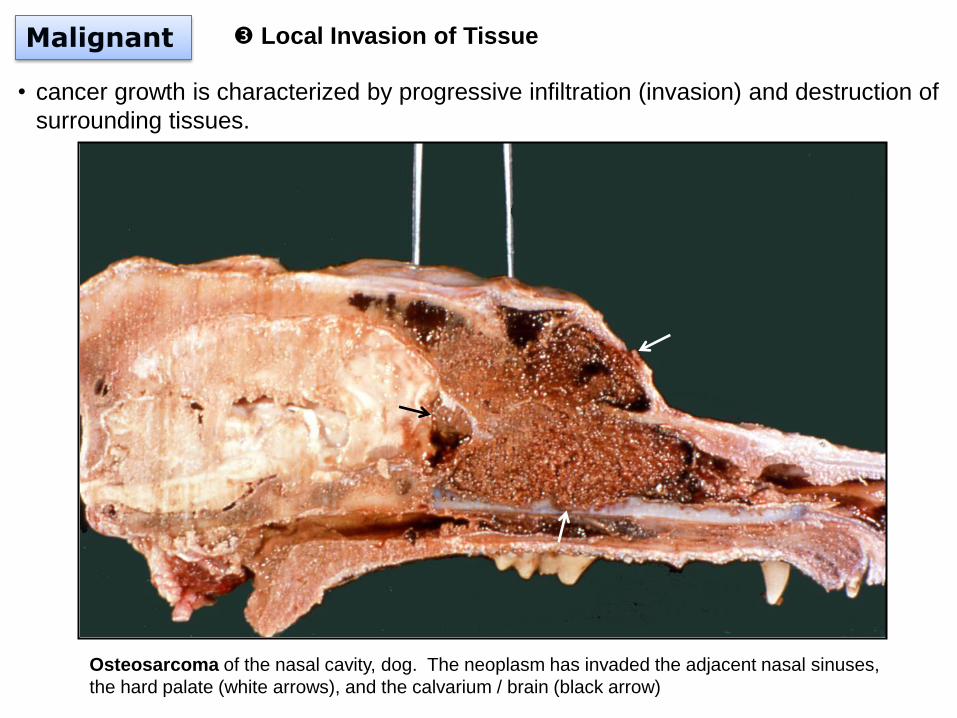
Malignant Local Invasion of Tissue
• cancer growth is characterized by progressive infiltration (invasion) and destruction of
surrounding tissues.
Osteosarcoma of the nasal cavity, dog. The neoplasm has invaded the adjacent nasal sinuses,
the hard palate (white arrows), and the calvarium / brain (black arrow)
Malignant
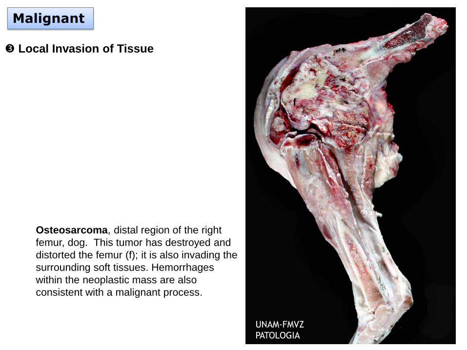
Local Invasion of Tissue
Osteosarcoma, distal region of the right
femur, dog. This tumor has destroyed and
distorted the femur (f); it is also invading the
surrounding soft tissues. Hemorrhages
within the neoplastic mass are also
consistent with a malignant process.
f
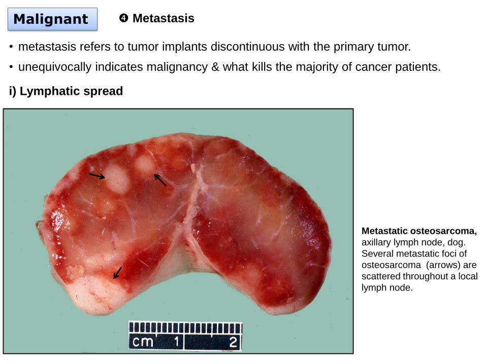
Malignant Metastasis
• metastasis refers to tumor implants discontinuous with the primary tumor.
• unequivocally indicates malignancy & what kills the majority of cancer patients.
i) Lymphatic spread
Metastatic osteosarcoma,
axillary lymph node, dog.
Several metastatic foci of
osteosarcoma (arrows) are
scattered throughout a local
lymph node.
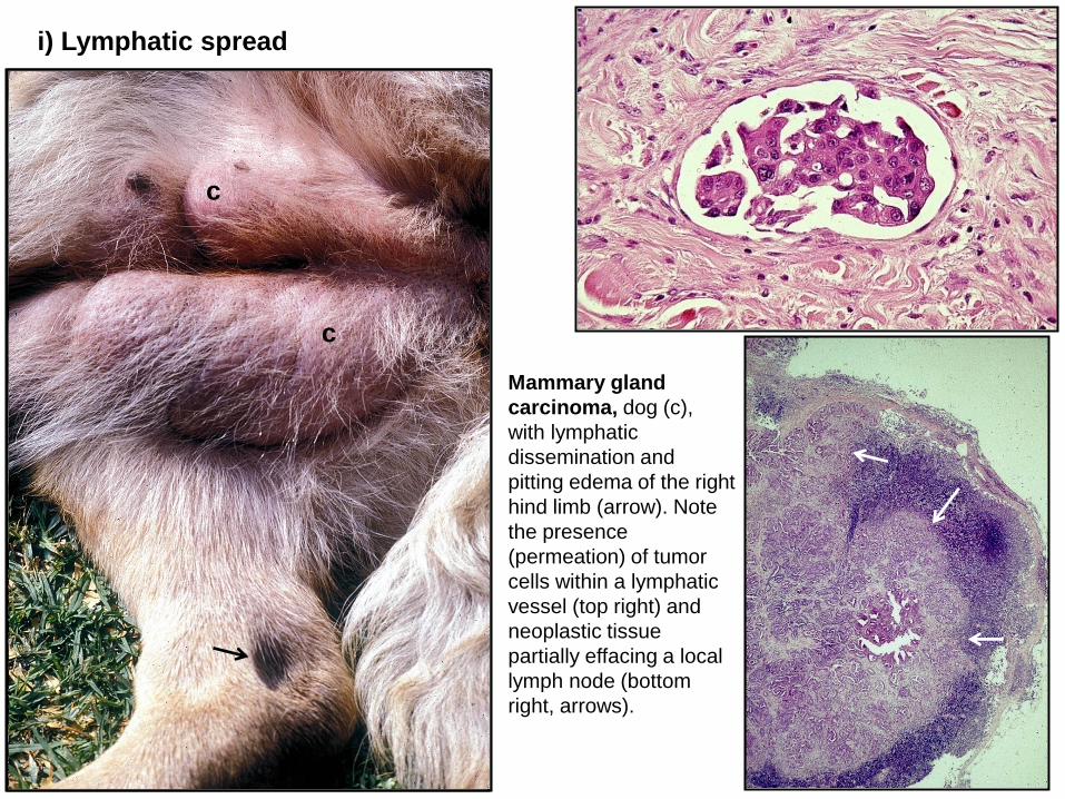
Mammary gland
carcinoma, dog (c),
with lymphatic
dissemination and
pitting edema of the right
hind limb (arrow). Note
the presence
(permeation) of tumor
cells within a lymphatic
vessel (top right) and
neoplastic tissue
partially effacing a local
lymph node (bottom
right, arrows).
i) Lymphatic spread
c
c
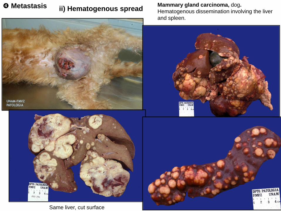
Mammary gland carcinoma, dog.
Hematogenous dissemination involving the liver
and spleen.
.
Same liver, cut surface
ii) Hematogenous spread Metastasis
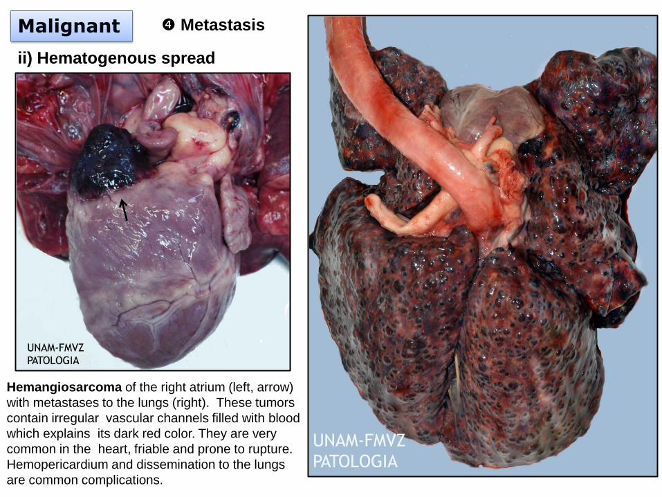
ii) Hematogenous spread
Malignant Metastasis
Hemangiosarcoma of the right atrium (left, arrow)
with metastases to the lungs (right). These tumors
contain irregular vascular channels filled with blood
which explains its dark red color. They are very
common in the heart, friable and prone to rupture.
Hemopericardium and dissemination to the lungs
are common complications.
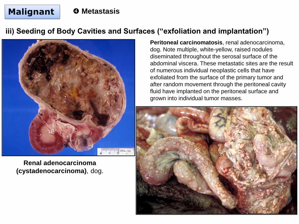
iii) Seeding of Body Cavities and Surfaces (“exfoliation and implantation”)
Malignant Metastasis
Peritoneal carcinomatosis, renal adenocarcinoma,
dog. Note multiple, white-yellow, raised nodules
diseminated throughout the serosal surface of the
abdominal viscera. These metastatic sites are the result
of numerous individual neoplastic cells that have
exfoliated from the surface of the primary tumor and
after random movement through the peritoneal cavity
fluid have implanted on the peritoneal surface and
grown into individual tumor masses.
Renal adenocarcinoma
(cystadenocarcinoma), dog.
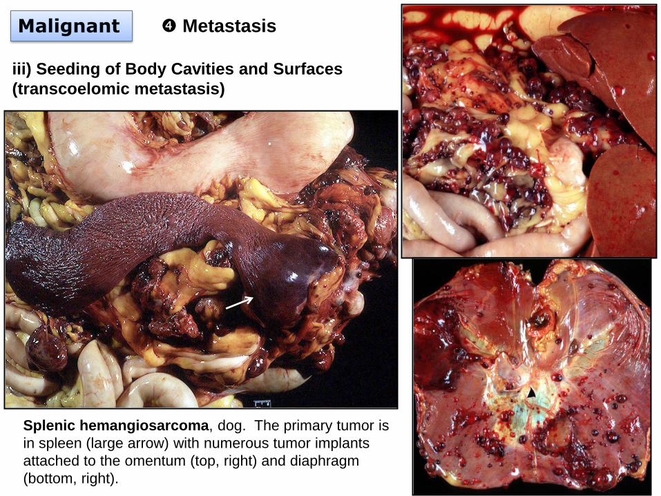
Malignant Metastasis
iii) Seeding of Body Cavities and Surfaces
(transcoelomic metastasis)
Splenic hemangiosarcoma, dog. The primary tumor is
in spleen (large arrow) with numerous tumor implants
attached to the omentum (top, right) and diaphragm
(bottom, right).
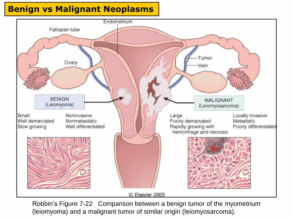
Benign vs Malignant Neoplasms
Robbin’s Figure 7-22 Comparison between a benign tumor of the myometrium
(leiomyoma) and a malignant tumor of similar origin (leiomyosarcoma).