Embed Size (px)
Citation preview
DISCUSSION SESSION: GROSS ANATOMY
ONN BLOCK
Feb 5, 2021
Discuss Face, Embryology Cranial Nerves with Practice Questions
FACE- Arteries, Pulses- Venous Drainage – Spread of Infection- Bell’s Palsy – Facial nerve paralysis, clinical tests, practice question- Embryology - Cleft Lip. Nasolacrimal duct; practice question
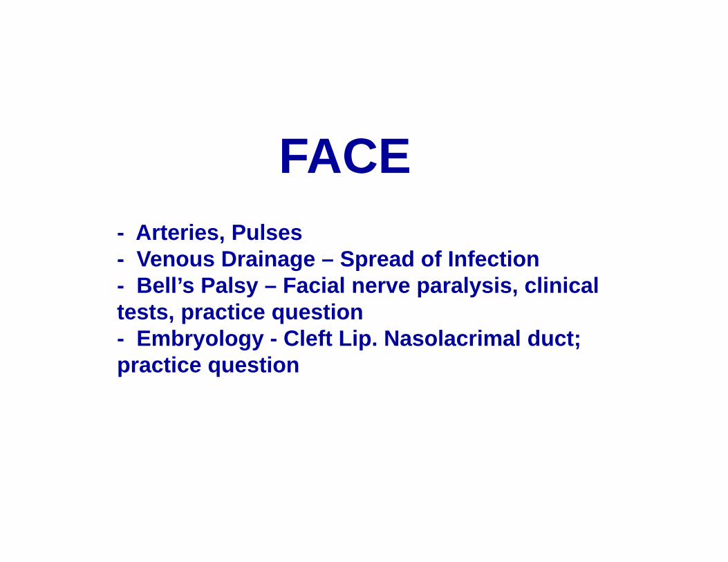
SUPERFICIAL TEMPORAL ARTERY
ARTERIAL SUPPLY TO FACE: CAROTID ARTERY
TAKE PULSE HERE
FACIAL ARTERY- extremely windingand tortuous course(skin moves) –
DESCRIPTIVE TERM –wiggle, wiggle, wiggle
COMMON CAROTIDARTERY
INTERNAL CAROTIDARTERY
EXTERNAL CAROTIDARTERY
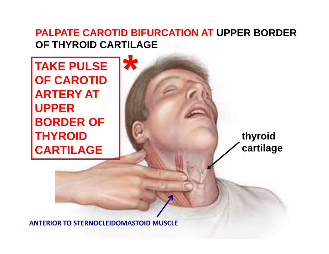
PALPATE CAROTID BIFURCATION AT UPPER BORDER OF THYROID CARTILAGE
thyroidcartilage
TAKE PULSE OF CAROTID ARTERY AT UPPER BORDER OF THYROID CARTILAGE
*
ANTERIOR TO STERNOCLEIDOMASTOID MUSCLE
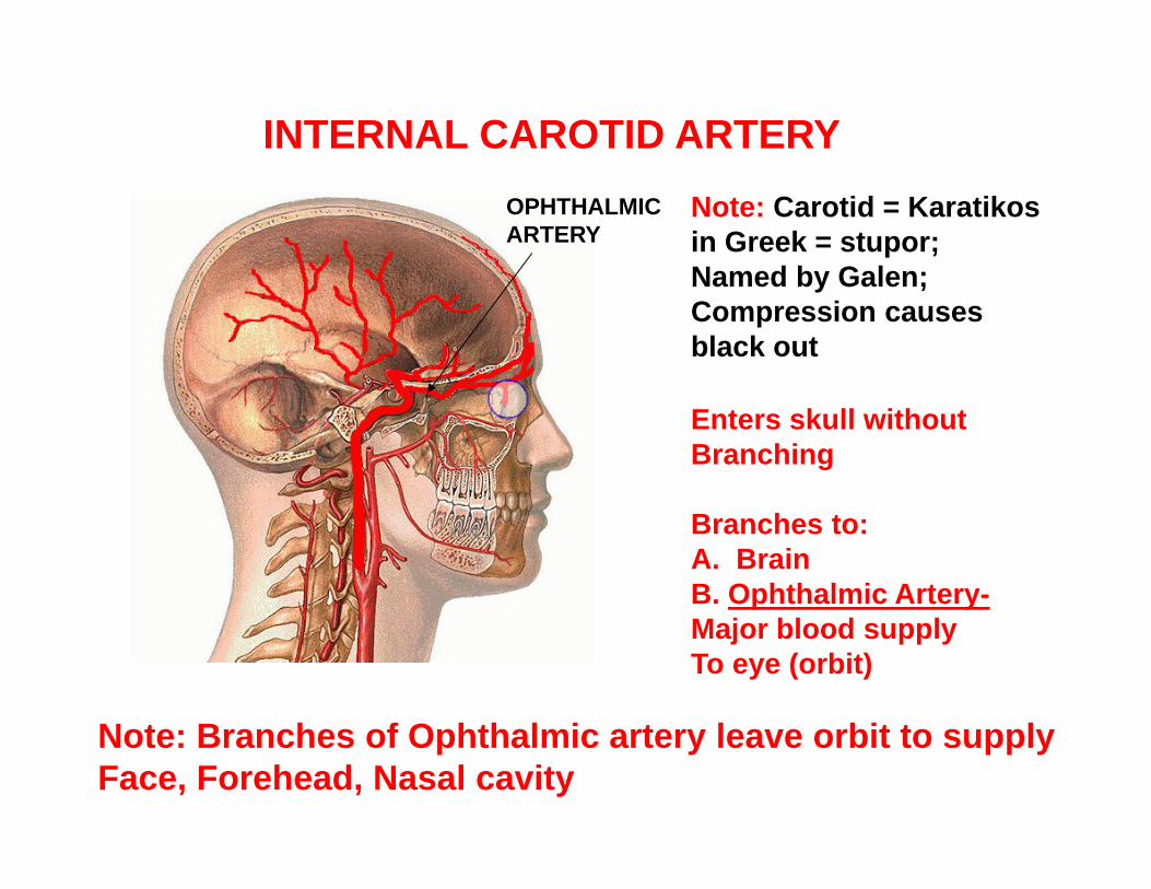
Note: Carotid = Karatikos in Greek = stupor; Named by Galen; Compression causes black out
Enters skull without Branching
Branches to:A. BrainB. Ophthalmic Artery-Major blood supplyTo eye (orbit)
Note: Branches of Ophthalmic artery leave orbit to supply Face, Forehead, Nasal cavity
INTERNAL CAROTID ARTERYOPHTHALMICARTERY
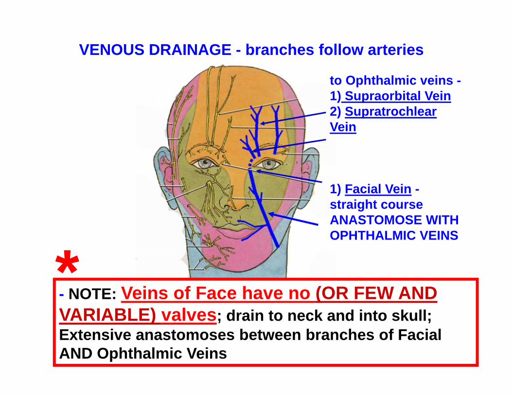
- NOTE: Veins of Face have no (OR FEW AND VARIABLE) valves; drain to neck and into skull; Extensive anastomoses between branches of Facial AND Ophthalmic Veins
VENOUS DRAINAGE - branches follow arteries
to Ophthalmic veins -1) Supraorbital Vein2) SupratrochlearVein
1) Facial Vein -straight courseANASTOMOSE WITH OPHTHALMIC VEINS
*
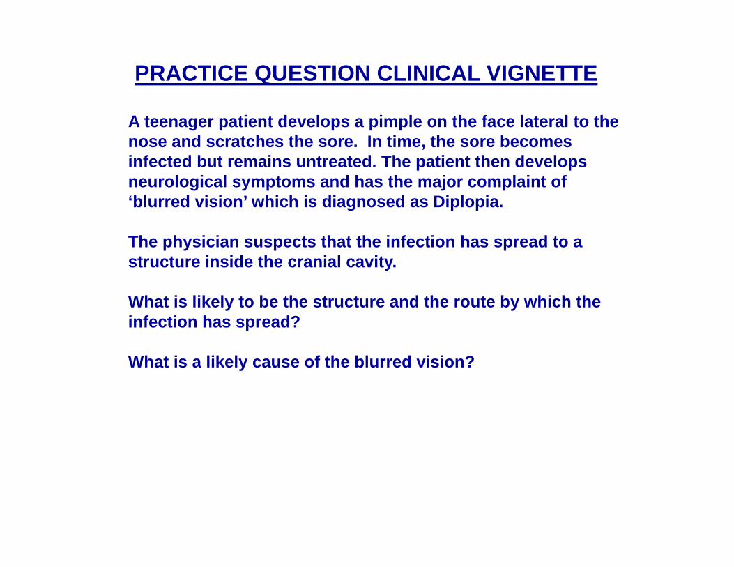
A teenager patient develops a pimple on the face lateral to the nose and scratches the sore. In time, the sore becomes infected but remains untreated. The patient then develops neurological symptoms and has the major complaint of ‘blurred vision’ which is diagnosed as Diplopia.
The physician suspects that the infection has spread to a structure inside the cranial cavity.
What is likely to be the structure and the route by which the infection has spread?
What is a likely cause of the blurred vision?
PRACTICE QUESTION CLINICAL VIGNETTE
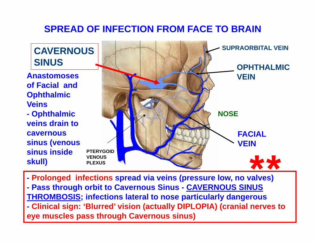
Anastomoses of Facial and Ophthalmic Veins- Ophthalmic veins drain to cavernous sinus (venous sinus inside skull)
OPHTHALMICVEIN
- Prolonged infections spread via veins (pressure low, no valves)- Pass through orbit to Cavernous Sinus - CAVERNOUS SINUS THROMBOSIS; infections lateral to nose particularly dangerous- Clinical sign: ‘Blurred’ vision (actually DIPLOPIA) (cranial nerves to eye muscles pass through Cavernous sinus)
NOSE
PTERYGOID VENOUS PLEXUS
FACIALVEIN
SPREAD OF INFECTION FROM FACE TO BRAIN
CAVERNOUSSINUS
SUPRAORBITAL VEIN
**
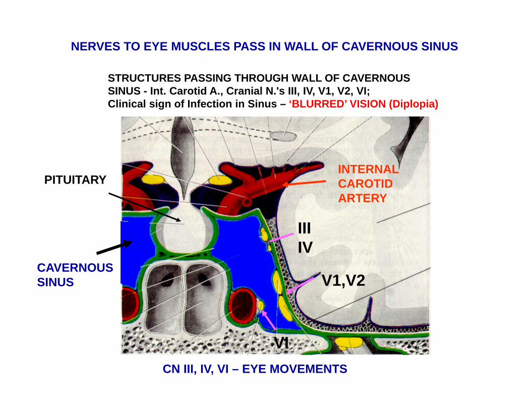
PITUITARY
CAVERNOUSSINUS
INTERNALCAROTIDARTERY
IIIIV
V1,V2
VI
STRUCTURES PASSING THROUGH WALL OF CAVERNOUSSINUS - Int. Carotid A., Cranial N.'s III, IV, V1, V2, VI;Clinical sign of Infection in Sinus – ‘BLURRED’ VISION (Diplopia)
NERVES TO EYE MUSCLES PASS IN WALL OF CAVERNOUS SINUS
CN III, IV, VI – EYE MOVEMENTS
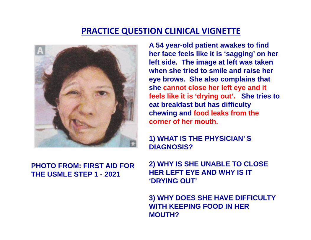
PHOTO FROM: FIRST AID FOR THE USMLE STEP 1 - 2021
PRACTICE QUESTION CLINICAL VIGNETTEA 54 year-old patient awakes to find her face feels like it is ‘sagging’ on her left side. The image at left was taken when she tried to smile and raise her eye brows. She also complains that she cannot close her left eye and it feels like it is ‘drying out’. She tries to eat breakfast but has difficulty chewing and food leaks from the corner of her mouth.
1) WHAT IS THE PHYSICIAN’ S DIAGNOSIS?
2) WHY IS SHE UNABLE TO CLOSE HER LEFT EYE AND WHY IS IT ‘DRYING OUT’
3) WHY DOES SHE HAVE DIFFICULTY WITH KEEPING FOOD IN HER MOUTH?
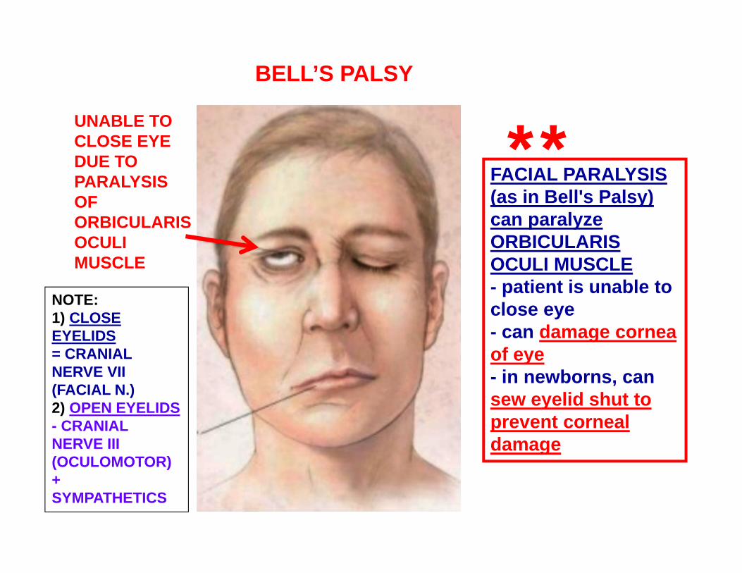
FACIAL PARALYSIS (as in Bell's Palsy) can paralyze ORBICULARISOCULI MUSCLE- patient is unable to close eye- can damage cornea of eye- in newborns, can sew eyelid shut to prevent corneal damage
UNABLE TOCLOSE EYE DUE TO PARALYSIS OF ORBICULARIS OCULI MUSCLE
BELL’S PALSY
**
NOTE:1) CLOSE EYELIDS= CRANIAL NERVE VII (FACIAL N.)2) OPEN EYELIDS- CRANIAL NERVE III (OCULOMOTOR) +SYMPATHETICS
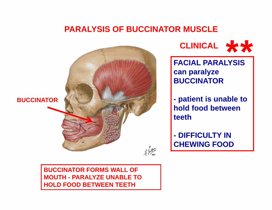
FACIAL PARALYSIS can paralyze BUCCINATOR
- patient is unable to hold food between teeth
- DIFFICULTY IN CHEWING FOOD
BUCCINATOR FORMS WALL OF MOUTH - PARALYZE UNABLE TO HOLD FOOD BETWEEN TEETH
PARALYSIS OF BUCCINATOR MUSCLE
CLINICAL
BUCCINATOR
**
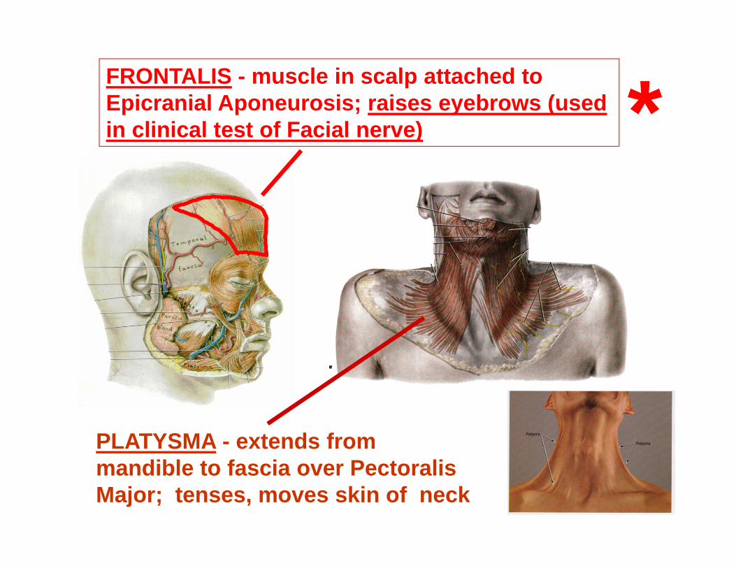
PLATYSMA - extends from mandible to fascia over Pectoralis Major; tenses, moves skin of neck
FRONTALIS - muscle in scalp attached to Epicranial Aponeurosis; raises eyebrows (used in clinical test of Facial nerve) *
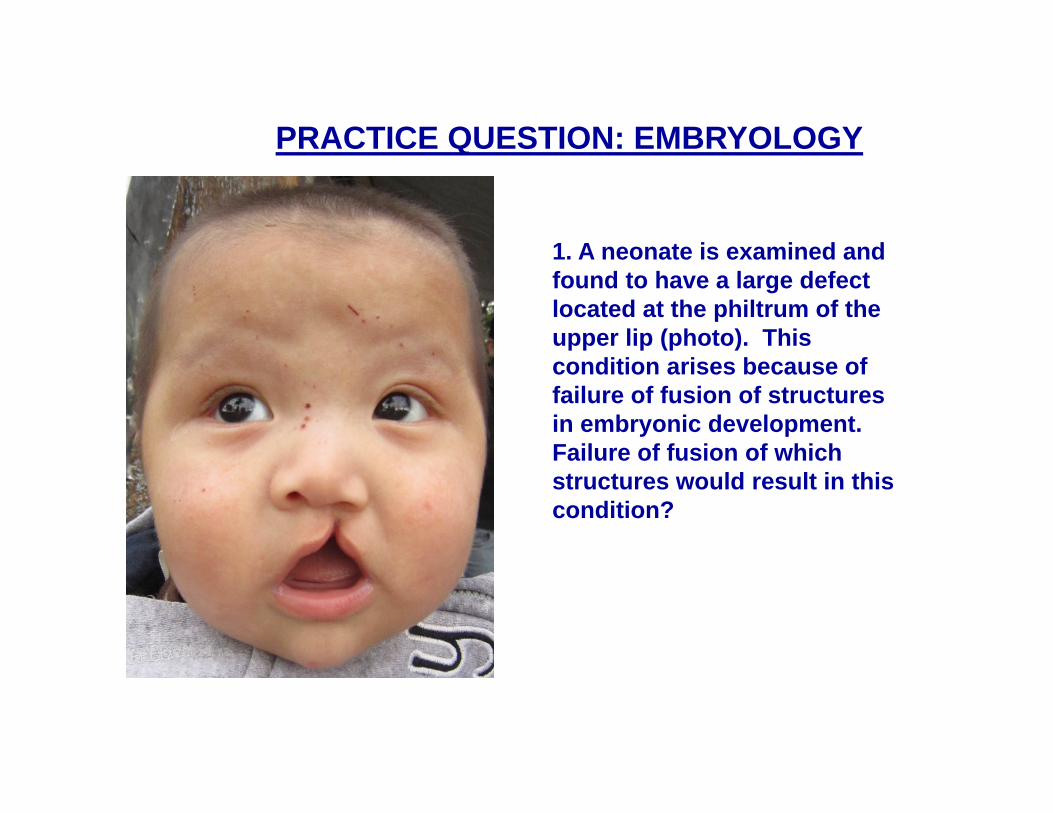
PRACTICE QUESTION: EMBRYOLOGY
1. A neonate is examined and found to have a large defect located at the philtrum of the upper lip (photo). This condition arises because of failure of fusion of structures in embryonic development. Failure of fusion of which structures would result in this condition?
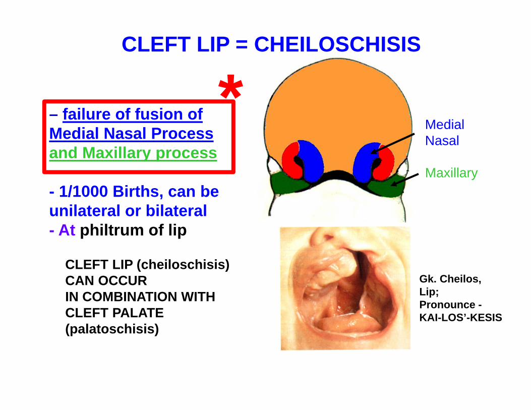
– failure of fusion of Medial Nasal Processand Maxillary process
- 1/1000 Births, can be unilateral or bilateral- At philtrum of lip
MedialNasal
Maxillary
CLEFT LIP = CHEILOSCHISIS
CLEFT LIP (cheiloschisis) CAN OCCURIN COMBINATION WITHCLEFT PALATE (palatoschisis)
Gk. Cheilos,Lip;Pronounce -KAI-LOS’-KESIS
*
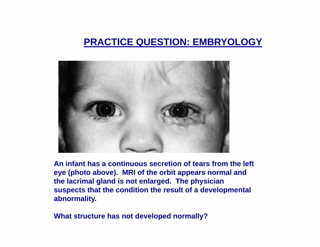
An infant has a continuous secretion of tears from the left eye (photo above). MRI of the orbit appears normal and the lacrimal gland is not enlarged. The physician suspects that the condition the result of a developmental abnormality.
What structure has not developed normally?
PRACTICE QUESTION: EMBRYOLOGY
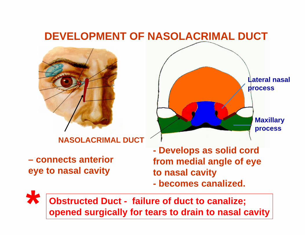
– connects anterior eye to nasal cavity
- Develops as solid cord from medial angle of eye to nasal cavity- becomes canalized.
Obstructed Duct - failure of duct to canalize; opened surgically for tears to drain to nasal cavity
DEVELOPMENT OF NASOLACRIMAL DUCT
NASOLACRIMAL DUCT
*
Lateral nasalprocess
Maxillaryprocess
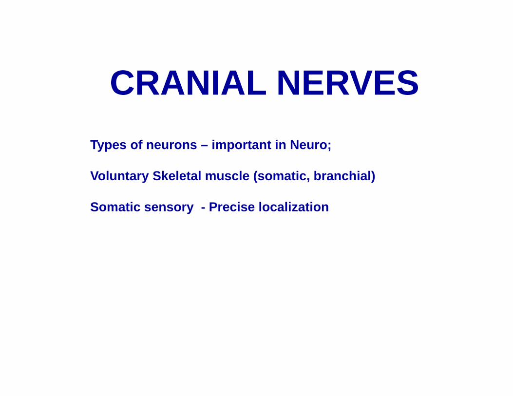
CRANIAL NERVESTypes of neurons – important in Neuro;
Voluntary Skeletal muscle (somatic, branchial)
Somatic sensory - Precise localization
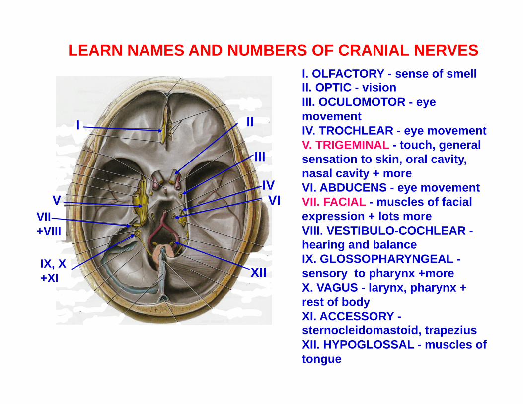
I. OLFACTORY - sense of smellII. OPTIC - visionIII. OCULOMOTOR - eye movementIV. TROCHLEAR - eye movementV. TRIGEMINAL - touch, general sensation to skin, oral cavity, nasal cavity + moreVI. ABDUCENS - eye movementVII. FACIAL - muscles of facial expression + lots moreVIII. VESTIBULO-COCHLEAR -hearing and balanceIX. GLOSSOPHARYNGEAL -sensory to pharynx +moreX. VAGUS - larynx, pharynx + rest of bodyXI. ACCESSORY -sternocleidomastoid, trapeziusXII. HYPOGLOSSAL - muscles of tongue
I II
III
IVV VI
VII+VIII
IX, X+XI XII
LEARN NAMES AND NUMBERS OF CRANIAL NERVES
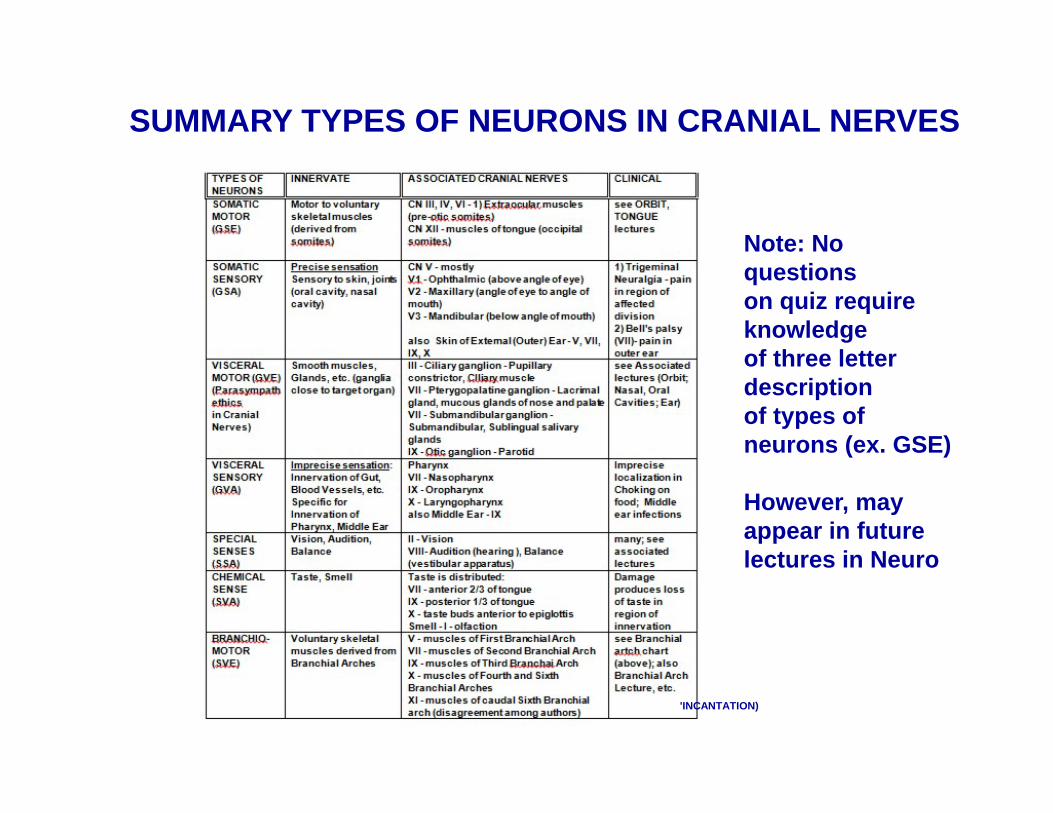
SUMMARY TYPES OF NEURONS IN CRANIAL NERVES
'INCANTATION)
Note: No questionson quiz require knowledgeof three letter descriptionof types of neurons (ex. GSE)
However, may appear in future lectures in Neuro
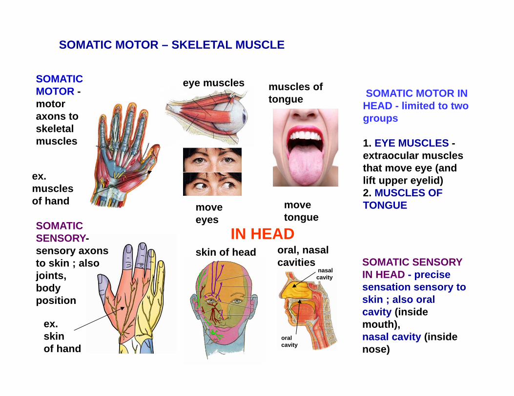
SOMATIC MOTOR IN HEAD - limited to two groups
1. EYE MUSCLES -extraocular muscles that move eye (and lift upper eyelid)2. MUSCLES OF TONGUE
SOMATIC MOTOR – SKELETAL MUSCLE
SOMATIC MOTOR -motoraxons to skeletal muscles
ex.musclesof hand
ex.skinof hand
SOMATIC SENSORY-sensory axons to skin ; also joints, body position
eye muscles
skin of headIN HEAD
muscles oftongue
movetongue
moveeyes
SOMATIC SENSORY IN HEAD - precise sensation sensory to skin ; also oral cavity (inside mouth),nasal cavity (inside nose)
oral, nasalcavities
oralcavity
nasalcavity
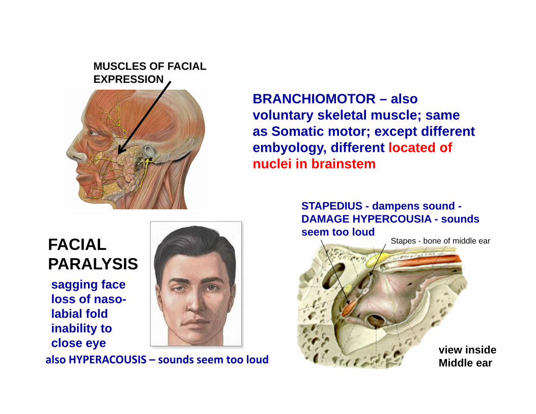
BRANCHIOMOTOR – also voluntary skeletal muscle; same as Somatic motor; except different embyology, different located of nuclei in brainstem
MUSCLES OF FACIAL EXPRESSION
FACIALPARALYSIS
STAPEDIUS - dampens sound -DAMAGE HYPERCOUSIA - sounds seem too loud
sagging faceloss of naso-labial foldinability to close eye view inside
Middle ear
Stapes - bone of middle ear
also HYPERACOUSIS – sounds seem too loud
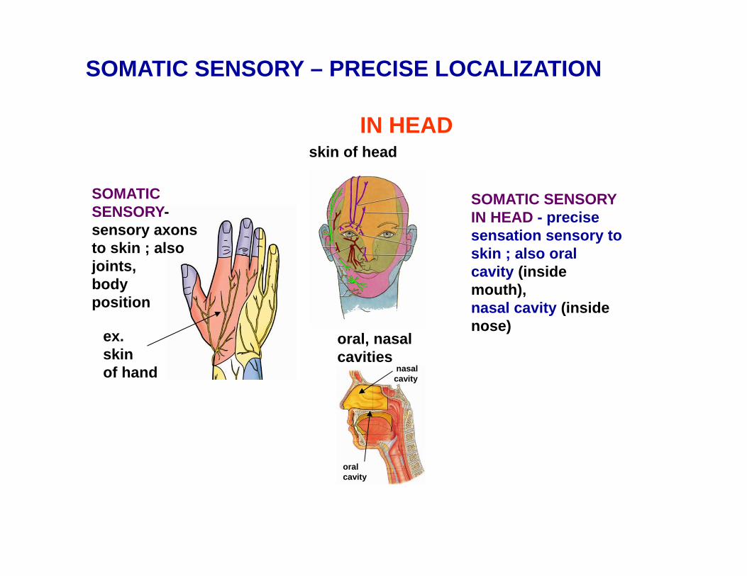
SOMATIC SENSORY – PRECISE LOCALIZATION
ex.skinof hand
SOMATIC SENSORY-sensory axons to skin ; also joints, body position
skin of headIN HEAD
SOMATIC SENSORY IN HEAD - precise sensation sensory to skin ; also oral cavity (inside mouth),nasal cavity (inside nose)
oral, nasalcavities
oralcavity
nasalcavity
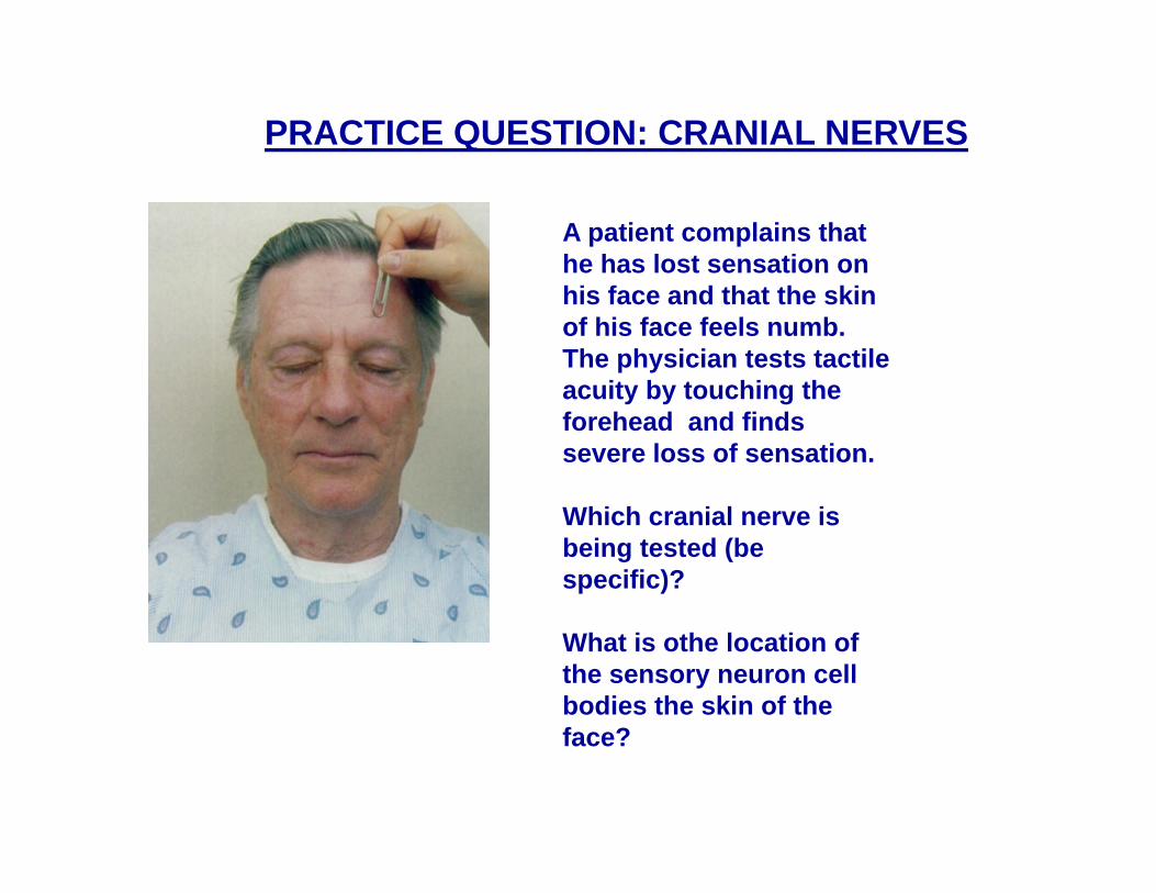
A patient complains that he has lost sensation on his face and that the skin of his face feels numb. The physician tests tactile acuity by touching the forehead and finds severe loss of sensation.
Which cranial nerve is being tested (be specific)?
What is othe location of the sensory neuron cell bodies the skin of the face?
PRACTICE QUESTION: CRANIAL NERVES
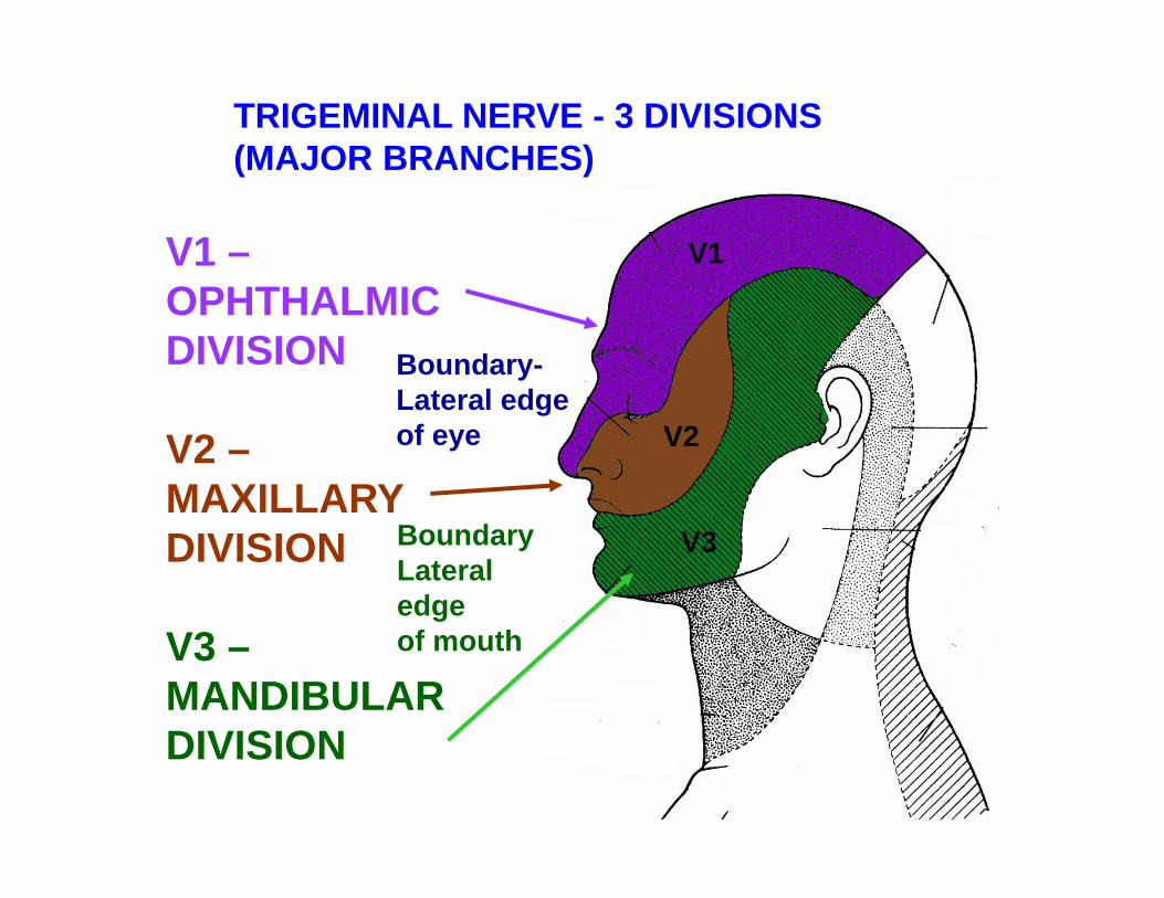
Boundary-Lateral edgeof eye
Boundary Lateral edgeof mouth
TRIGEMINAL NERVE - 3 DIVISIONS (MAJOR BRANCHES)
V1 –OPHTHALMICDIVISION
V2 –MAXILLARYDIVISION
V3 –MANDIBULARDIVISION
V1
V2
V3
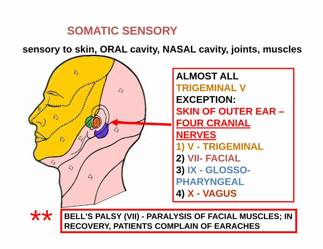
SOMATIC SENSORYsensory to skin, ORAL cavity, NASAL cavity, joints, muscles
ALMOST ALLTRIGEMINAL VEXCEPTION:SKIN OF OUTER EAR –FOUR CRANIAL NERVES 1) V - TRIGEMINAL2) VII- FACIAL3) IX - GLOSSO-PHARYNGEAL4) X - VAGUS
BELL'S PALSY (VII) - PARALYSIS OF FACIAL MUSCLES; IN RECOVERY, PATIENTS COMPLAIN OF EARACHES**
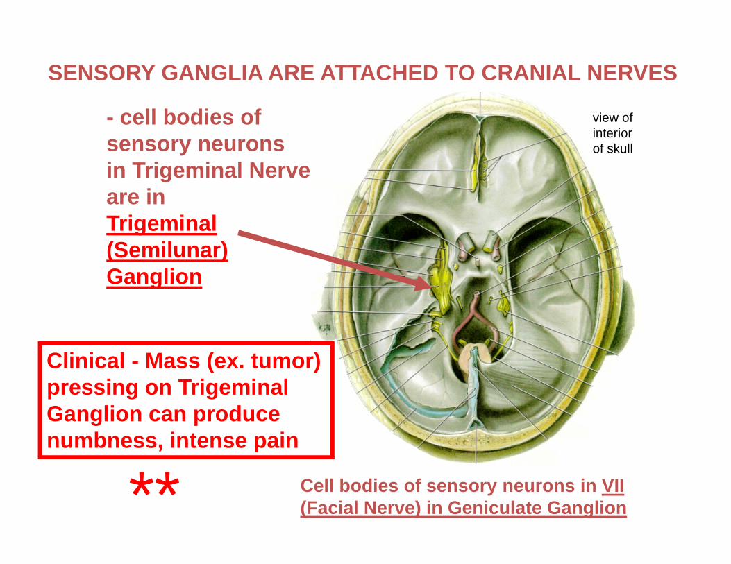
SENSORY GANGLIA ARE ATTACHED TO CRANIAL NERVES
- cell bodies of sensory neuronsin Trigeminal Nerve are inTrigeminal (Semilunar) Ganglion
Cell bodies of sensory neurons in VII (Facial Nerve) in Geniculate Ganglion
view ofinteriorof skull
Clinical - Mass (ex. tumor) pressing on Trigeminal Ganglion can produce numbness, intense pain
**