Embed Size (px)
Citation preview
biomolecules
Article
Discovery of Orexant and Anorexant Agents withIndazole Scaffold Endowed with PeripheralAntiedema Activity
Marilisa P. Dimmito 1, Azzurra Stefanucci 1,*, Stefano Pieretti 2 , Paola Minosi 2,Szabolcs Dvorácskó 3, Csaba Tömböly 3, Gokhan Zengin 4 and Adriano Mollica 1
1 Department of Pharmacy, University of Chieti-Pescara “G. d’Annunzio”, Via dei Vestini 31, 66100 Chieti,Italy; [email protected] (M.P.D.); [email protected] (A.M.)
2 Istituto Superiore di Sanità, Centro Nazionale Ricerca e Valutazione Preclinica e Clinica dei Farmaci,Viale Regina Elena 299, 00161 Rome, Italy; [email protected] (S.P.); [email protected] (P.M.)
3 Institute of Biochemistry, Biological Research Centre of the Hungarian Academy of Sciences, Temesvári krt.62, 6726 Szeged, Hungary; [email protected] (S.D.); [email protected] (C.T.)
4 Department of Biology, Science Faculty, Selcuk University, 42005 Konya, Turkey;[email protected]
* Correspondence: [email protected]; Tel.: +39-0871-3554-482
Received: 7 August 2019; Accepted: 11 September 2019; Published: 16 September 2019�����������������
Abstract: The endocannabinoid system represents an integrated neuronal network involved inthe control of several organisms’ functions, such as feeding behavior. A series of hybrids of5-(4-chlorophenyl)-1-(2,4-dichloro-phenyl)-4-methyl-N-(piperidin-1-yl)-1H-pyrazole-3-carboxamide(mimonabant), a well-known inverse agonist of the type-1 cannabinoid receptor (CB1), once used asan antiobesity drug, and the N-(2S)-substitutes of 1-[(4-fluorophenyl)methyl]indazole-3-carboxamidewith 1-amino-3-methyl-1-oxobutane (AB-Fubinaca), 1-amino-3,3-dimethyl-1-oxobutane(ADB-Fubinaca), and 3-methylbutanoate (AMB-Fubinaca), endowed with potent agonistic activitytowards cannabinoid receptors CB1 and CB2 were in solution as C-terminal amides, acids, methylesters and N-methyl amides. These compounds have been studied by binding assays to cannabinoidreceptors and by functional receptor assays, using rat brain membranes in vitro. The most active amongthem as an agonist, (S)-1-(2,4-dichlorobenzyl)-N-(3,3-dimethyl-1-(methylamino)-1-oxobutan-2-yl)-1H-indazole-3-carboxamide (LONI11), and an antagonist, (S)-2-(1-(2,4-dichlorobenzyl)-1H-indazole-3-carboxamido)-3-methylbutanoic acid (LONI4), were tested in vivo in mic, to evaluate theirability to stimulate or suppress feeding behavior after intraperitoneal (i.p.) administration.For a LONI11 formalin test and a tail flick test after an administration by the subcutaneous(s.c.) and intracerebroventricular (i.c.v.) routes, respectively, were also carried out in vivoin mice to investigate the antinociceptive property at the central and peripheral levesl. Weobserved a significant orexant effect for LONI11 and an intense anorexant effect for (S)-methyl2-(1-(2,4-dichlorobenzyl)-1H-indazole-3-carboxamido)-3,3-dimethylbutanoate (LONI2) and LONI4.In zymosan-induced edema and hyperalgesia, LONI11 reduced the percent of paw volumeincrease and paw latency after s.c. administration, also suggesting a possible peripheralanti-inflammatory activity.
Keywords: cannabinoid receptor; rimonabant; food intake; anorexant agent; edema
1. Introduction
Metabolic syndrome is the result of a group of multifactorial conditions, characterized by the lossof balance between energy income and caloric needs. An efficacious equilibrium between orexigenic
Biomolecules 2019, 9, 492; doi:10.3390/biom9090492 www.mdpi.com/journal/biomolecules
Biomolecules 2019, 9, 492 2 of 20
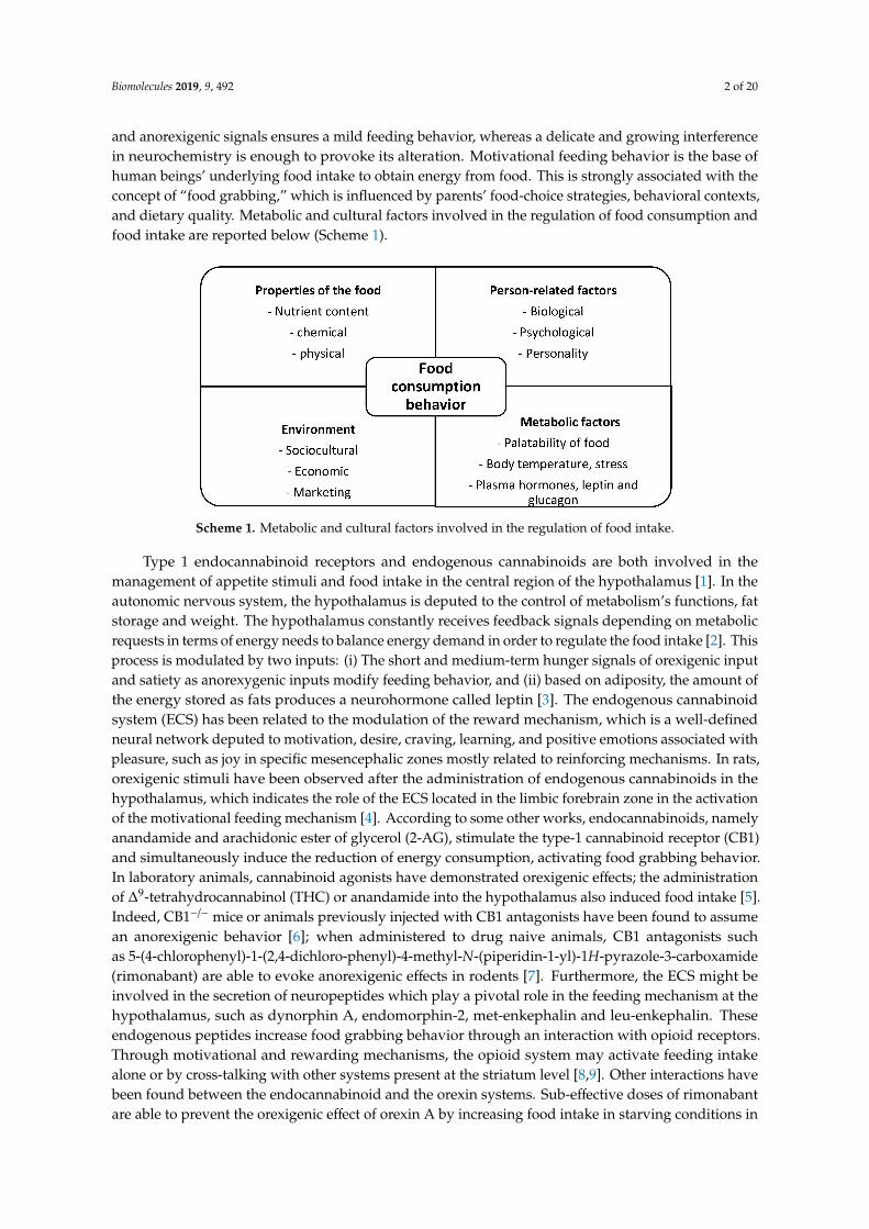
and anorexigenic signals ensures a mild feeding behavior, whereas a delicate and growing interferencein neurochemistry is enough to provoke its alteration. Motivational feeding behavior is the base ofhuman beings’ underlying food intake to obtain energy from food. This is strongly associated with theconcept of “food grabbing,” which is influenced by parents’ food-choice strategies, behavioral contexts,and dietary quality. Metabolic and cultural factors involved in the regulation of food consumption andfood intake are reported below (Scheme 1).
Scheme 1. Metabolic and cultural factors involved in the regulation of food intake.
Type 1 endocannabinoid receptors and endogenous cannabinoids are both involved in themanagement of appetite stimuli and food intake in the central region of the hypothalamus [1]. In theautonomic nervous system, the hypothalamus is deputed to the control of metabolism’s functions, fatstorage and weight. The hypothalamus constantly receives feedback signals depending on metabolicrequests in terms of energy needs to balance energy demand in order to regulate the food intake [2]. Thisprocess is modulated by two inputs: (i) The short and medium-term hunger signals of orexigenic inputand satiety as anorexygenic inputs modify feeding behavior, and (ii) based on adiposity, the amount ofthe energy stored as fats produces a neurohormone called leptin [3]. The endogenous cannabinoidsystem (ECS) has been related to the modulation of the reward mechanism, which is a well-definedneural network deputed to motivation, desire, craving, learning, and positive emotions associated withpleasure, such as joy in specific mesencephalic zones mostly related to reinforcing mechanisms. In rats,orexigenic stimuli have been observed after the administration of endogenous cannabinoids in thehypothalamus, which indicates the role of the ECS located in the limbic forebrain zone in the activationof the motivational feeding mechanism [4]. According to some other works, endocannabinoids, namelyanandamide and arachidonic ester of glycerol (2-AG), stimulate the type-1 cannabinoid receptor (CB1)and simultaneously induce the reduction of energy consumption, activating food grabbing behavior.In laboratory animals, cannabinoid agonists have demonstrated orexigenic effects; the administrationof ∆9-tetrahydrocannabinol (THC) or anandamide into the hypothalamus also induced food intake [5].Indeed, CB1−/− mice or animals previously injected with CB1 antagonists have been found to assumean anorexigenic behavior [6]; when administered to drug naive animals, CB1 antagonists suchas 5-(4-chlorophenyl)-1-(2,4-dichloro-phenyl)-4-methyl-N-(piperidin-1-yl)-1H-pyrazole-3-carboxamide(rimonabant) are able to evoke anorexigenic effects in rodents [7]. Furthermore, the ECS might beinvolved in the secretion of neuropeptides which play a pivotal role in the feeding mechanism at thehypothalamus, such as dynorphin A, endomorphin-2, met-enkephalin and leu-enkephalin. Theseendogenous peptides increase food grabbing behavior through an interaction with opioid receptors.Through motivational and rewarding mechanisms, the opioid system may activate feeding intakealone or by cross-talking with other systems present at the striatum level [8,9]. Other interactions havebeen found between the endocannabinoid and the orexin systems. Sub-effective doses of rimonabantare able to prevent the orexigenic effect of orexin A by increasing food intake in starving conditions in
Biomolecules 2019, 9, 492 3 of 20
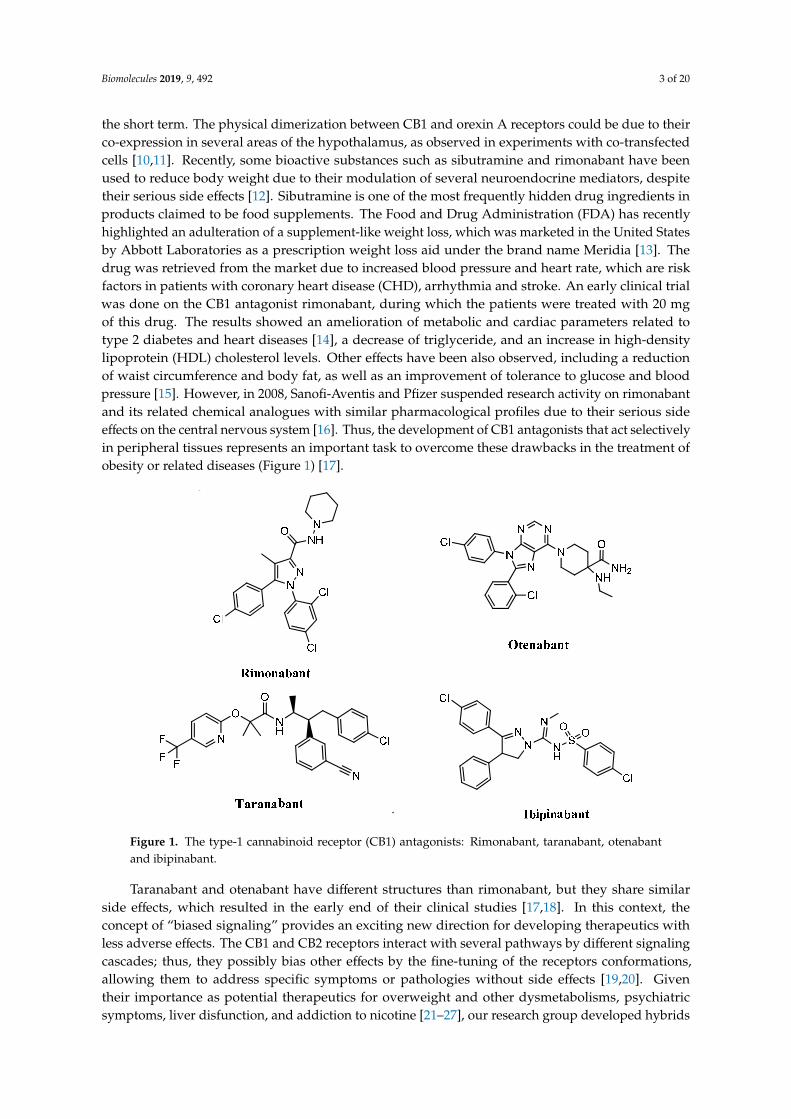
the short term. The physical dimerization between CB1 and orexin A receptors could be due to theirco-expression in several areas of the hypothalamus, as observed in experiments with co-transfectedcells [10,11]. Recently, some bioactive substances such as sibutramine and rimonabant have beenused to reduce body weight due to their modulation of several neuroendocrine mediators, despitetheir serious side effects [12]. Sibutramine is one of the most frequently hidden drug ingredients inproducts claimed to be food supplements. The Food and Drug Administration (FDA) has recentlyhighlighted an adulteration of a supplement-like weight loss, which was marketed in the United Statesby Abbott Laboratories as a prescription weight loss aid under the brand name Meridia [13]. Thedrug was retrieved from the market due to increased blood pressure and heart rate, which are riskfactors in patients with coronary heart disease (CHD), arrhythmia and stroke. An early clinical trialwas done on the CB1 antagonist rimonabant, during which the patients were treated with 20 mgof this drug. The results showed an amelioration of metabolic and cardiac parameters related totype 2 diabetes and heart diseases [14], a decrease of triglyceride, and an increase in high-densitylipoprotein (HDL) cholesterol levels. Other effects have been also observed, including a reductionof waist circumference and body fat, as well as an improvement of tolerance to glucose and bloodpressure [15]. However, in 2008, Sanofi-Aventis and Pfizer suspended research activity on rimonabantand its related chemical analogues with similar pharmacological profiles due to their serious sideeffects on the central nervous system [16]. Thus, the development of CB1 antagonists that act selectivelyin peripheral tissues represents an important task to overcome these drawbacks in the treatment ofobesity or related diseases (Figure 1) [17].
Biomolecules 2019, 9, x 3 of 20
feeding intake alone or by cross-talking with other systems present at the striatum level [8,9]. Other interactions have been found between the endocannabinoid and the orexin systems. Sub-effective doses of rimonabant are able to prevent the orexigenic effect of orexin A by increasing food intake in starving conditions in the short term. The physical dimerization between CB1 and orexin A receptors could be due to their co-expression in several areas of the hypothalamus, as observed in experiments with co-transfected cells [10,11]. Recently, some bioactive substances such as sibutramine and rimonabant have been used to reduce body weight due to their modulation of several neuroendocrine mediators, despite their serious side effects [12]. Sibutramine is one of the most frequently hidden drug ingredients in products claimed to be food supplements. The Food and Drug Administration (FDA) has recently highlighted an adulteration of a supplement-like weight loss, which was marketed in the United States by Abbott Laboratories as a prescription weight loss aid under the brand name Meridia [13]. The drug was retrieved from the market due to increased blood pressure and heart rate, which are risk factors in patients with coronary heart disease (CHD), arrhythmia and stroke. An early clinical trial was done on the CB1 antagonist rimonabant, during which the patients were treated with 20 mg of this drug. The results showed an amelioration of metabolic and cardiac parameters related to type 2 diabetes and heart diseases [14], a decrease of triglyceride, and an increase in high-density lipoprotein (HDL) cholesterol levels. Other effects have been also observed, including a reduction of waist circumference and body fat, as well as an improvement of tolerance to glucose and blood pressure [15]. However, in 2008, Sanofi-Aventis and Pfizer suspended research activity on rimonabant and its related chemical analogues with similar pharmacological profiles due to their serious side effects on the central nervous system [16]. Thus, the development of CB1 antagonists that act selectively in peripheral tissues represents an important task to overcome these drawbacks in the treatment of obesity or related diseases (Figure 1) [17].
.
Figure 1. The type-1 cannabinoid receptor (CB1) antagonists: Rimonabant, taranabant, otenabant and ibipinabant.
Taranabant and otenabant have different structures than rimonabant, but they share similar side effects, which resulted in the early end of their clinical studies [17,18]. In this context, the concept of “biased signaling” provides an exciting new direction for developing therapeutics with less adverse effects. The CB1 and CB2 receptors interact with several pathways by different signaling cascades; thus, they possibly bias other effects by the fine-tuning of the receptors conformations, allowing them to address specific symptoms or pathologies without side effects [19,20]. Given their importance as potential therapeutics for
Figure 1. The type-1 cannabinoid receptor (CB1) antagonists: Rimonabant, taranabant, otenabantand ibipinabant.
Taranabant and otenabant have different structures than rimonabant, but they share similarside effects, which resulted in the early end of their clinical studies [17,18]. In this context, theconcept of “biased signaling” provides an exciting new direction for developing therapeutics withless adverse effects. The CB1 and CB2 receptors interact with several pathways by different signalingcascades; thus, they possibly bias other effects by the fine-tuning of the receptors conformations,allowing them to address specific symptoms or pathologies without side effects [19,20]. Giventheir importance as potential therapeutics for overweight and other dysmetabolisms, psychiatricsymptoms, liver disfunction, and addiction to nicotine [21–27], our research group developed hybrids
Biomolecules 2019, 9, 492 4 of 20
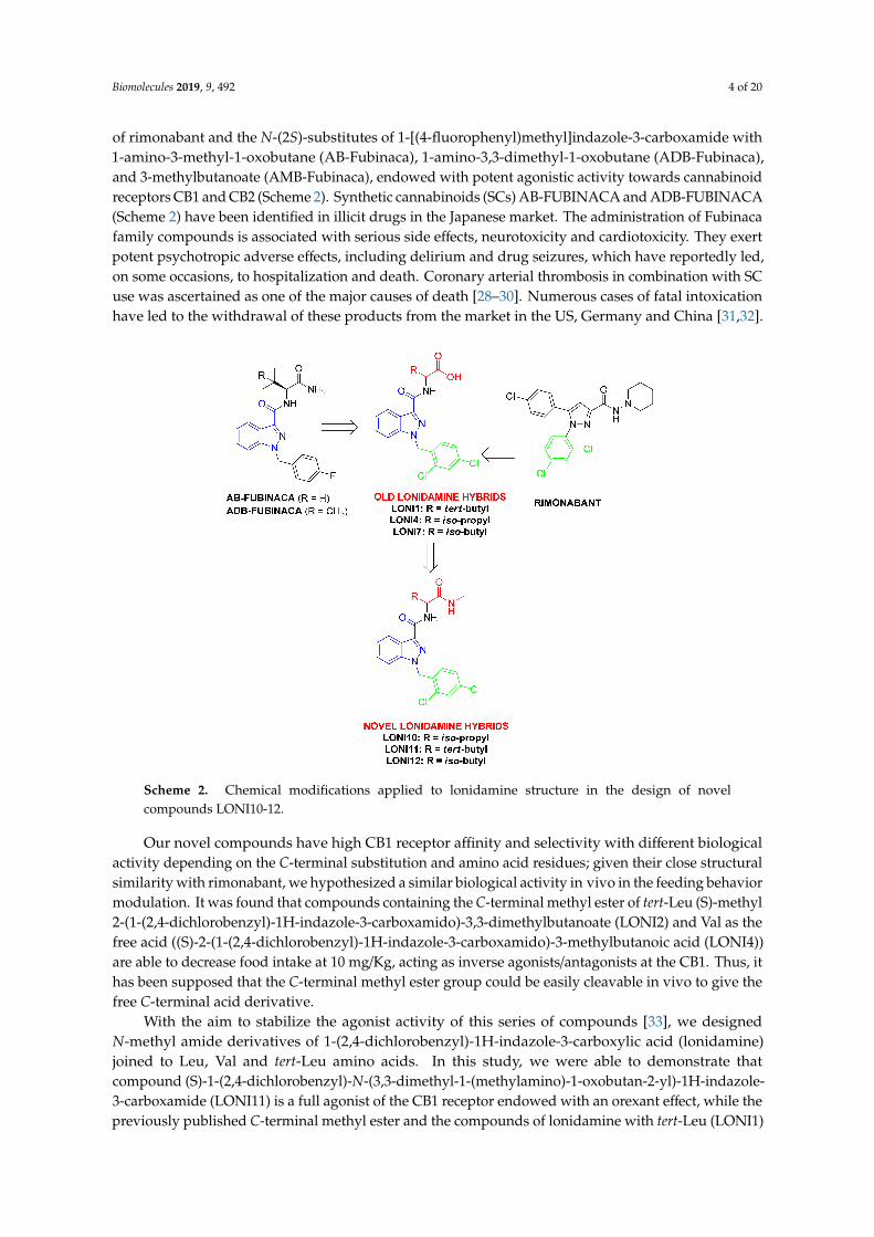
of rimonabant and the N-(2S)-substitutes of 1-[(4-fluorophenyl)methyl]indazole-3-carboxamide with1-amino-3-methyl-1-oxobutane (AB-Fubinaca), 1-amino-3,3-dimethyl-1-oxobutane (ADB-Fubinaca),and 3-methylbutanoate (AMB-Fubinaca), endowed with potent agonistic activity towards cannabinoidreceptors CB1 and CB2 (Scheme 2). Synthetic cannabinoids (SCs) AB-FUBINACA and ADB-FUBINACA(Scheme 2) have been identified in illicit drugs in the Japanese market. The administration of Fubinacafamily compounds is associated with serious side effects, neurotoxicity and cardiotoxicity. They exertpotent psychotropic adverse effects, including delirium and drug seizures, which have reportedly led,on some occasions, to hospitalization and death. Coronary arterial thrombosis in combination with SCuse was ascertained as one of the major causes of death [28–30]. Numerous cases of fatal intoxicationhave led to the withdrawal of these products from the market in the US, Germany and China [31,32].
Scheme 2. Chemical modifications applied to lonidamine structure in the design of novelcompounds LONI10-12.
Our novel compounds have high CB1 receptor affinity and selectivity with different biologicalactivity depending on the C-terminal substitution and amino acid residues; given their close structuralsimilarity with rimonabant, we hypothesized a similar biological activity in vivo in the feeding behaviormodulation. It was found that compounds containing the C-terminal methyl ester of tert-Leu (S)-methyl2-(1-(2,4-dichlorobenzyl)-1H-indazole-3-carboxamido)-3,3-dimethylbutanoate (LONI2) and Val as thefree acid ((S)-2-(1-(2,4-dichlorobenzyl)-1H-indazole-3-carboxamido)-3-methylbutanoic acid (LONI4))are able to decrease food intake at 10 mg/Kg, acting as inverse agonists/antagonists at the CB1. Thus, ithas been supposed that the C-terminal methyl ester group could be easily cleavable in vivo to give thefree C-terminal acid derivative.
With the aim to stabilize the agonist activity of this series of compounds [33], we designedN-methyl amide derivatives of 1-(2,4-dichlorobenzyl)-1H-indazole-3-carboxylic acid (lonidamine)joined to Leu, Val and tert-Leu amino acids. In this study, we were able to demonstrate thatcompound (S)-1-(2,4-dichlorobenzyl)-N-(3,3-dimethyl-1-(methylamino)-1-oxobutan-2-yl)-1H-indazole-3-carboxamide (LONI11) is a full agonist of the CB1 receptor endowed with an orexant effect, while thepreviously published C-terminal methyl ester and the compounds of lonidamine with tert-Leu (LONI1)
Biomolecules 2019, 9, 492 5 of 20
and Val (LONI4) as free acids showed an anorexic profile in vivo. Furthermore, several antinociceptionmodels were also studied to characterize the analgesic potential of this novel compound (LONI11).
2. Materials and Methods
2.1. Chemistry
Lonidamine, solvents, reagents and amino acids Boc-tert-Leu-OH, Boc-Val-OH, and Boc-Leu-OH,are commercially available and were acquired from Sigma-Aldrich (Milano, Italy). The intermediatecompounds LONI1-4,7 were synthetized, as previously published by Stefanucci et al. [33]. Thestructures of the intermediates and the final compounds were confirmed by 1H-NMR and 13C-NMRspectra recorded on a 300 MHz Varian Inova spectrometer (Varian Inc., Palo Alto, CA). Chemical shiftswere reported in parts per million (δ) downfield from the internal standard tetramethylsilane (Me4Si).The purity of each final product was established by analytical reverse phase-high performance liquidchromatography (RP-HPLC) (C18-bonded 4.6 × 150 mm) at a flow rate of 1 mL/min by using (as aneluent) a gradient of H2O/ACN 0.1% TFA ranging from 10% ACN to 90% ACN for 30 min; it wasfound to be >95% (see Supplementary Materials). UV detection at 254 nm was chosen for analyticalHPLC. Mass spectra were performed on an LCQ (Finnigan–Mat) ion trap mass spectrometer (San Jose,CA, USA) equipped with an electrospray ionization (Supplementary Materials) source. The capillarytemperature was set at 300 ◦C, and the spray voltage was set at 3.5 kV. The fluid was nebulized usingnitrogen as both the sheath gas and the auxiliary gas [34–36].
General Procedure for the N-Methyl Amide Formation
HOBt anhydrous (1.1 eq.) in DMF (3 mL), EDC.HCl (1.1 eq.) and NMM (1 eq.) were addedTo a stirred solution of lonidamine-amino acid compound (200 mg) at 0 ◦C; this was followed bythe addition of a solution of methylamine 40% in water (2 eq.) and NMM (2 eq.) in DMF (3 mL).After 10 min at 0 ◦C, the reaction was stirred at room temperature (r.t.) overnight. Later, the reactionmixture was evaporated to dryness, and the residue taken up in ethyl acetate (EtOAc). The organicphase was washed with 5% citric acid, NaHCO3 saturated solution (s.s.) and NaCl s.s., dried andevaporated in a high vacuum. The crude compound was triturated in Et2O two times to give thedesired white solid product. The characterization of the final LONI10-12 compound is reported in theSupplementary Materials.
(S)-2-(1-(2,4-dichlorobenzyl)-1H-indazole-3-carboxamido)-3,3-dimethylbutanoic acid (LONI1).Boc-tert-Leu-OH was coupled to lonidamine according to Stefanucci et al. [33].
(S)-methyl-2-(1-(2,4-dichlorobenzyl)-1H-indazole-3-carboxamido)-3,3-dimethylbutanoate (LONI2).Boc-tert-Leu-OH was transformed in its methyl ester derivative and coupled to lonidamine accordingto Stefanucci et al. [33].
(S)-N-(1-amino-3,3-dimethyl-1-oxobutan-2-yl)-1-(2,4-dichlorobenzyl)-1H-indazole-3-carboxamide(LONI3). Boc-L-tert-Leu-OH was converted into the amide derivative and coupled to lonidamineaccording to Stefanucci et al. [33].
(S)-2-(1-(2,4-dichlorobenzyl)-1H-indazole-3-carboxamido)-3-methylbutanoic acid (LONI4).Boc-Val-OH was coupled to lonidamine according to Stefanucci et al. [33].
(S)-1-(2,4-dichlorobenzyl)-N-(3-methyl-1-(methylamino)-1-oxobutan-2-yl)-1H-indazole-3-carboxamide(LONI10). The LONI4 compound was transformed in the N-methyl amide derivative LONI 10following the general procedure. The desired compound was obtained in a 96% yield after reactionwork up.
(S)-1-(2,4-dichlorobenzyl)-N-(3, 3-dimethyl-1-(methylamino)-1-oxobutan-2-yl)-1H-indazole-3-carboxamide (LONI11). The LONI1 compound was transformed in the N-methyl amide derivative
Biomolecules 2019, 9, 492 6 of 20
LONI 11 following the general procedure. The desired compound was obtained in a 97% yield afterreaction work up.
(S)-1-(2,4-dichlorobenzyl)-N-(4-methyl-1-(methylamino)-1-oxopentan-2-yl)-1H-indazole-3-carboxamide(LONI12). The LONI7 compound was transformed in the N-methyl amide derivative LONI12following the general procedure. The desired compound was obtained in a quantitative yield afterreaction work up.
2.2. In Vitro Biological Assays
2.2.1. Preparation of Brain Membrane Homogenates
Wistar rats were locally bred and handled according to the EU Directive 2010/63/EU and to theRegulations on Animal Protection (40/2013. (II. 14.) Korm. r.) of Hungary. Crude membrane fractionswere prepared from the brain. Brains were quickly removed from the euthanized rats and directlyput in an ice-cold 50 mM Tris–HCl buffer (pH 7.4). The collected tissue was then homogenized in 30volumes (v/w) of an ice-cold buffer with a Braun Teflon-glass homogenizer at the highest rpm. Thehomogenate was centrifuged at 20,000× g for 25 min, and the resulting pellet was suspended in thesame volume of a cold buffer followed by incubation at 37 ◦C for 30 min to remove endogenous ligands.Centrifugation was then repeated. The final pellets were taken up in five volumes of a 50 mM Tris–HCl(pH 7.4) buffer containing 0.32 M sucrose and stored at −80 ◦C. Prior to the experiment, aliquots werethawed and centrifuged at 20,000× g for 25 min and then they were resuspended in 50 mM Tris–HCl(pH 7.4), homogenized with a Douncer, followed by the determination of the protein concentration bythe method of Bradford. The membrane suspensions were immediately used either in radioligandbinding experiments or in [35S]GTPγS functional assays.
2.2.2. Radioligand Competition Binding Assay
Binding experiments were performed at 30 ◦C for 60 min in a 50 mM Tris–HCl binding buffer(pH 7.4) containing 2.5 mM of EGTA, 5 mM of MgCl2 and 0.5 mg/mL of fatty acid-free BSA in plastictubes in a total assay volume of 1 mL that contained 0.3–0.5 mg/mL of a membrane protein [33,37].Competition binding experiments were carried out by incubating rat brain membranes with 5 nM of[3H]WIN55212-2 (Kd: 10.1 nM) in the presence of increasing concentrations (10−11–10−5 M) of variouscompeting unlabeled ligands. Non-specific binding was determined in the presence of 10 µM of WIN55212-2. The incubation was terminated by diluting the samples with an ice-cold wash buffer (50 mMof Tris–HCl, 2.5 mM of EGTA, 5 mM of MgCl2, 0.5% fatty acid free BSA, pH 7.4), followed by repeatedwashing and rapid filtration through Whatman GF/B glass fiber filters (Whatman Ltd., Maidstone, UK)presoaked with 0.1% polyethyleneimine (30 min before the filtration). Filtration was performed with a24-well Brandel Cell Harvester (Gaithersburg, MD, USA). Filters were air-dried and immersed intoUltima Gold MV scintillation cocktail, and then radioactivity was measured with a TRI-CARB 2100TRliquid scintillation analyzer (Packard, Perkin Elmer, Waltham, MA, USA).
2.2.3. Ligand Stimulated [35S]GTPγS Binding Assay
Rat brain membranes (30 µg protein/tube), prepared as described above, were incubated with0.05 nM of [35S]GTPγS (PerkinElmer) and 10−10–10−5 M unlabeled ligands in the presence of 30 µM ofGDP, 100 mM of NaCl, 3 mM of MgCl2 and 1 mM of EGTA in a 50 mM Tris–HCl buffer (pH 7.4) for60 min at 30 ◦C. Basal [35S]GTPγS binding was measured in the absence of ligands and set as 100%.Nonspecific binding was determined by the addition of 10 µM unlabeled GTPγS and subtracted fromtotal binding. Incubation, filtration and radioactivity measurements of the samples were carried out asdescribed above.
Biomolecules 2019, 9, 492 7 of 20
2.2.4. Data Analysis
The results of the competition binding studies are reported as means ±S.E.M. of at least threeindependent experiments each performed in duplicate. In competition binding studies, the inhibitoryconstants (Ki) were calculated from the inflection points of the displacement curves using non-linearleast-square curve fitting and the Cheng–Prusoff equation, Ki= EC50/(1 + [ligand]/Kd).
In [35S]GTPγS binding studies, data were expressed as the percentage stimulation of the specific[35S]GTPγS binding over the basal activity and are given as means ±S.E.M. Each experiment wasperformed in triplicate and analyzed with sigmoid dose–response curve fitting to obtain potency (EC50)and efficacy (Emax) values. All data and curves were analyzed by GraphPad Prism 5.0 (San Diego,CA, USA).
2.3. In Vivo Biological Assays
2.3.1. Animals
The international and national law and policies approved by Italian Ministry of Health wereused to comply with all animal care and experimental procedures. Animal studies were advisedin compliance with the ARRIVE guidelines and with the recommendations made by EU Directive2010/63/EU for animal experiments and the Basel declaration including the 3Rs concept [38,39]. CD-1male mice (10–14 weeks of age, 25–30 g of weight) were bought from Charles River (Milan, Italy).Shortly after their arrival and for at least one week, they were kept in an animal care facility undercontrolled standard conditions of temperature (21 ± 1 ◦C), light (from 7:00 AM to 7:00 PM), and relativehumidity (60 ± 10%). Access to drinking water and food was assured. All procedures were performedto decrease the number of animals used (n = 6 per group) and their distress.
2.3.2. Feeding Test
The test was carried out as previously described [40]. At 24 h before the start of a feeding test,all food was removed from the home cages of mice to be tested. The next day and at least 1 h beforethe feeding test began, the mice were transported to the laboratory. On test days, the animals wereplaced in the home cages for 30 min of drug assimilation, during which food was not available. Thencompounds were intraperitoneally administered (10 mg/kg). Mice were transferred into transparentand individual plastic cages with thick white paper lining the bottom and access to a pre-measuredamount of their regular lab chow (2 gr) for the 1-h test. At the end of 1 h, mice were repositionedinto their home cage. The amount of food left in the trial cage, including crumbs, was measured, andthe amount consumed was calculated. Feeding trials normally happened on Tuesdays and Fridaysbetween 12:00 and 14:00 h.
2.3.3. Tail Flick Test
The tail flick test was used to determinate antinociceptive responses [41]. Tail flick latency(Ugo Basile, Varese, Italy) consists of an infrared radiant light source (100 W, 15 V bulb) targeted ona photocell utilizing an aluminum parabolic mirror. During the trials, the mice were gently handrestrained with a glove. Radiant heat was targeted 5–6 cm from the tip of the tail, and the latency (s) ofthe tail withdrawal recorded. The measurement was disconnected if the latency crossed the cutoff
time. A cutoff time of 15 s was imposed, and data were expressed as time course of the percentage ofmaximum effect (% MPE) = (post drug latency/baseline latency)/(cutoff time baseline latency) × 100.In all experiments, the baseline was calculated as the mean of three readings recorded before testingat intervals of 10 min, and the time course of latency was determined 10–120 min after compoundtreatment. Compounds were freshly diluted in saline 0.1% v/v DMSO and were injected at 10 µg/10 µLfor intracerebroventricular (i.c.v.) administrations, as previously reported [42,43].
Biomolecules 2019, 9, 492 8 of 20
2.3.4. Formalin Test
The method utilized was comparable to the one previously described by Pieretti et al. [44]. Micewere located to adapt into the transparent cages individually (30× 14× 12 cm) for at least an hour beforetesting. They were injected with 20 µL of a 1% solution of formalin in saline. Then, the compoundswere administered subcutaneously in the dorsal surface of the right hind paw of the mouse using amicrosyringe with a 27-gauge needle for 15 min before. Compounds were prepared by freshly dilutingsaline containing 0.9% NaCl in the ratio DMSO:saline 1:3 (v/v). Then, these solutions were injectedfor subcutaneous (s.c.) administrations in doses of 30–100 µg/20 µL. The total time the animal spentlicking or biting its paw was calculated.
2.3.5. Edema Induced by Zymosan
In this test, 100 µg of the compounds were administered subcutaneously in a volume of 20 µL inthe dorsal surface of mice hind paw; this was done 15 min before a subcutaneous injection (20 µL/paw)of zymosan A (2.5% w/v in saline) into the same paw. Then, paw edema was calculated as formerlydescribed [45]. The percentage difference between the paw volume at each time point and the basalpaw volume was used as an index of the increase in paw volume. Paw volume was quantified using ahydroplethysmometer modified for small volumes (Ugo Basile, Varese, Italy) three times before theinjections and at 1, 2, 3, 4 and 24 h thereafter.
2.3.6. Zymosan-Induced Hyperalgesia
The compounds (100 µg) were administered subcutaneously in a volume of 20 µL in the dorsalsurface of mice hind paw; this was done 15 min before a subcutaneous injection (20 µL/paw) ofzymosan A (2.5% w/v in saline) into the same paw before the measurement of hyperalgesia [45].The sensitivity to a noxious heat stimulus was measured by the plantar test (Ugo Basile, Italy), inorder to evaluate thermal hyperalgesia after the zymosan-induced inflammation of the mouse hindpaw. Mice were allocated in clear plastic boxes with a glass floor and acclimatized for at least 1 hin a temperature-controlled (21 ◦C) experimental room for three consecutive days prior to testing.Furthermore, on the test day, the animals were acclimatized to the experimental room 1 h before pawwithdrawal latency (PWL) was calculated. Using a timer and in term of seconds, yhe paw withdrawallatency was measured automatically after placing the mouse footpad in contact with a radiant heatsource. A timer initiated automatically when the heat source was activated, and a photocell stoppedthe timer when the mouse withdrew its hind paw. An intensity of 30 and a cut-off time of 15 s wereused for the heat source on the plantar apparatus to avoid tissue damage. Animals were first tested todefine their baseline PWL in terms of seconds against 1, 2, 3, 4, 5 and 24 h after zymosan A injection.
2.3.7. Data Analysis and Statistics
The mean ±S.E.M. was used to explain the results obtained. Statistically significant differencesbetween groups were measured with an analysis of variance (ANOVA) followed by Tukey’s post-hoccomparisons or the Mann–Whitney test when the comparison was restricted to two groups. GraphPadPrism 6.0 software (San Diego, CA, USA) was used to analyze the data. Data were consideredstatistically significant when a value of p < 0.05 was performed. The data and statistical analysesrespected the recommendations on experimental design and analysis [46].
2.4. In Silico Experiments
2.4.1. Receptor Preparation
The crystal structure of the human CB1 receptor co-crystallized with an MDMB-Fubinaca agonistwas downloaded from the RSCB database (pdb id: 6N4B) [47]. The raw file was prepared for thedocking experiment by the PrepWizard module of Maestro 10.2 [48]. Briefly, the missing chains were
Biomolecules 2019, 9, 492 9 of 20
added automatically by Prime [49], and the protonation state was calculated by PropKa at pH = 7.4 [48].Finally the receptor–ligand complex was minimized by OPLS-3 force field following a well-establishedprotocol reported by our research group [42,50].
2.4.2. Docking Grid Generation
The docking grid was generated by the Glide module of Maestro [51]. The grid was centered onthe MDMB-Fubinaca ligand present in the crystal structure and extended to a space of 20 × 20 × 20Angstrom. The generated grid was used for the docking experiments.
2.4.3. Self-Docking and Validation Procedure
In order to validate the docking procedure, a self-docking experiment was conducted. Thecrystallographic ligand MDMB-Fubinaca was removed from the receptor, prepared, and minimized bythe LigPrep module [52] of Maestro using EpiK at pH = 7.4 [48]. The software generated 32 minimizedstructures, and the best ranked structure was used for the self-docking procedure. The obtained ligandwas submitted to a first round of docking by using Glide at Standard Precision (Glide-SP) accuracy.The best raked pose was subjected to a second round of docking by using Glide in eXtra Precision(Glide-XP) mode. Then, the RMSD of the best ranked pose was measured as 0.5 Å below the originalcrystallographic pose (see Figure S1). This procedure was applied to the docking experiments of thenovel compounds.
2.4.4. Ligands Preparation
LONI4 and LONI11 were drawn by the 2D editor embedded in Maestro and prepared by theLigPrep module following the same procedure applied to MDMB-Fubinaca. Then, the minimizedstructures were submitted to the docking experiments without further modifications.
2.4.5. Ligand Docking Experiments
The prepared molecules LONI4 and LONI11 were docked to the CB1 receptor. The first roundof docking was performed by Glide in Standard Precision mode. The best pose generated fromthe first step was then submitted to the second round of docking by using Glide in eXtra Precisionaccuracy. The best ranked poses are depicted in Figure S2 as a bi-dimensional interaction diagram (seeSupplementary Materials).
2.4.6. Molecular Dynamic
The LONI4-CB1, LONI11-CB1 and MDMB-Fubinaca-CB1 complexes obtained from self-dockingwere also submitted to molecular dynamic (MD) experiments by the Desmond module embedded inMaestro 12.0 [53]. The MD simulation system was composed of the receptor–ligand complex embeddedinto a dipalmitoylphosphatidylcholine (DPPC) membrane-bilayer surrounded by water (Figure S3, seeSupplementary Materials). Firstly, each complex was positioned in the membrane bilayer of DPPClipids and then inserted into a water box to simulate a model of cellular bilayer system. The waterbox had a minimum size as to contain the receptor complex embedded in the membrane, ensuring adistance of 10 Å from the edge of the box and the protein. In order to neutralize the system, 0.15 Mof NaCl was added. The OPLS-3 force field was used for all the experiments, and the TIP4P modelwas used for the water [54]. The system was minimized up to 2000 steps, holding all the protein andligand atoms. Then, the minimized system was subjected to MD simulations, using the NPT ensembleand periodic boundary conditions for 20 ns. The Martyna–Tobias–Klein algorithm [55] was used tokeep the pressure of the system at 1.01 bar by using the isotropic coupling method. The Nose–Hooverthermostat was applied to control the temperature at 310K [56]. The trajectories and other parameterswere saved every 20 and 1.2 ps, respectively, to return 1000 frames. The simulation analysis was doneby the simulation interactive diagram (Supplementary Materials, to visualize the RMSD fluctuations of
Biomolecules 2019, 9, 492 10 of 20
the ligand and the receptor, the hydrogen bonds stability, the water network formation and the overallstability of the secondary structure of the enzyme.
3. Results
The LONI1-4,7 compounds were prepared by a well-established procedure previously reportedby Stefanucci et al. [33]. The LONI10-12 novel chemical entities were efficiently recovered in excellentyields by standard solution phase peptide synthesis using EDC/HOBt coupling reagents, NMM as abase, and a solution of methylamine 40% in water (Scheme 3). All the final compounds were trituratedin diethyl ether two times and then characterized by low resolution mass spectroscopy (LRMS), 1H-and 13C-NMR; the purity of the final products was determined by analytical RP-HPLC and found tobe >95% (see Supplementary Materials).
Scheme 3. Reagents and Conditions: (a) EDC·HCl (1.1 eq.), HOBt an. (1.1. eq.), NMM (3 eq.),methylamine 40% in water (2 eq.), DMF (6 mL), r.t. 12 h (LONI10: 96% yield; LONI11: 97% yield;LONI12: Quantitative).
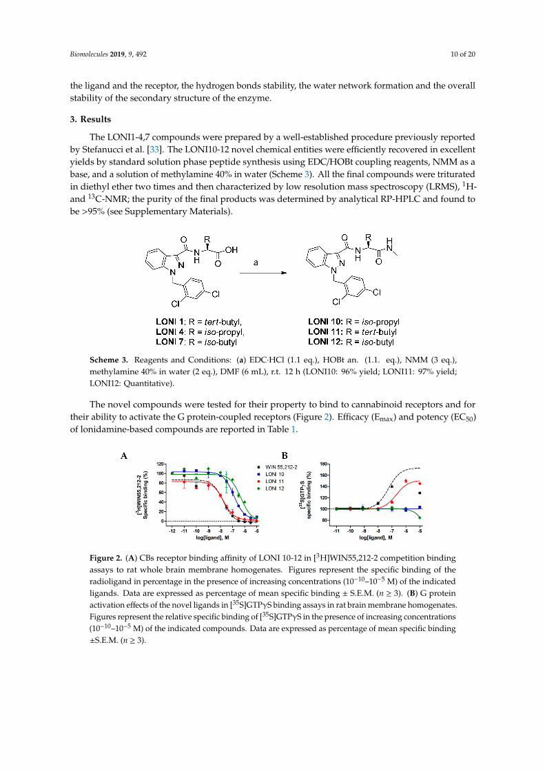
The novel compounds were tested for their property to bind to cannabinoid receptors and fortheir ability to activate the G protein-coupled receptors (Figure 2). Efficacy (Emax) and potency (EC50)of lonidamine-based compounds are reported in Table 1.
Figure 2. (A) CBs receptor binding affinity of LONI 10-12 in [3H]WIN55,212-2 competition bindingassays to rat whole brain membrane homogenates. Figures represent the specific binding of theradioligand in percentage in the presence of increasing concentrations (10−10–10−5 M) of the indicatedligands. Data are expressed as percentage of mean specific binding ± S.E.M. (n ≥ 3). (B) G proteinactivation effects of the novel ligands in [35S]GTPγS binding assays in rat brain membrane homogenates.Figures represent the relative specific binding of [35S]GTPγS in the presence of increasing concentrations(10−10–10−5 M) of the indicated compounds. Data are expressed as percentage of mean specific binding±S.E.M. (n ≥ 3).
Biomolecules 2019, 9, 492 11 of 20
Table 1. Binding affinity (Ki) and signal properties efficacy (Emax) and potency (EC50) oflonidamine-based compounds.
Compounds Sequence Ki (nM) [35S]GTPγS Binding
Emax (%) EC50 (nM)
WIN55,212-2 10 ± 1 173 ± 11 56 ± 3.8JWH-018 [33] 3.5 ± 1 163 ± 5.2 16 ± 3LONI1 [33] Lonidamine-tert-Leu-OH 0.08 * 84 ± 6.6 >1 µMLONI2 [33] Lonidamine-tert-Leu-OCH3 3.1 * 143 ± 5.7 8.4LONI3 [33] Lonidamine-tert-Leu-NH2 17 * 139 ± 4.5 126LONI4 [33] Lonidamine-Val-OH 2.6 * 82 ± 10.6 >1 µM
LONI10 Lonidamine-Val-NHMe 84 ± 3.4 100 ± 1.7 n.r.LONI11 Lonidamine-tert-Leu-NHMe 11 ± 1.2 151 ± 3 200 ± 13LONI12 Lonidamine-Leu-NHMe 320 ± 16 101 ± 1 n.r.
Ki values were calculated from the corresponding displacement curves of Figure 2A. The Emax and EC50 wereextrapolated from the dose–response curves of Figure 2B. Data represent the mean ±S.E.M. from least threeindependent experiments. n.r.: Not relevant. * Mean of three independent experiments (S.D. values are in the rangeof 5%–10%).
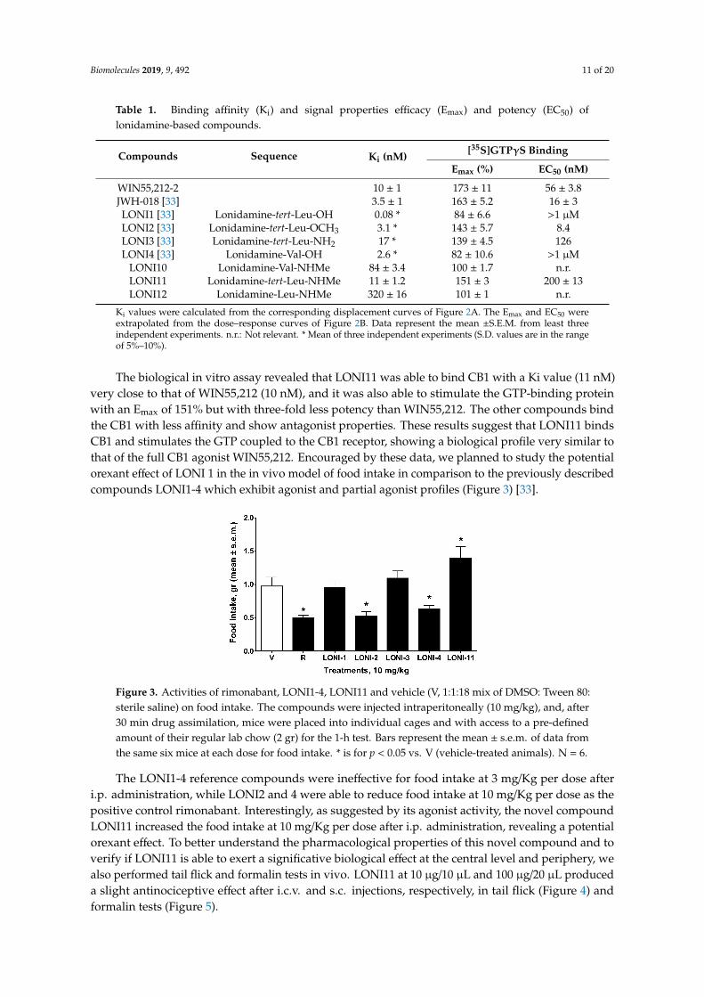
The biological in vitro assay revealed that LONI11 was able to bind CB1 with a Ki value (11 nM)very close to that of WIN55,212 (10 nM), and it was also able to stimulate the GTP-binding proteinwith an Emax of 151% but with three-fold less potency than WIN55,212. The other compounds bindthe CB1 with less affinity and show antagonist properties. These results suggest that LONI11 bindsCB1 and stimulates the GTP coupled to the CB1 receptor, showing a biological profile very similar tothat of the full CB1 agonist WIN55,212. Encouraged by these data, we planned to study the potentialorexant effect of LONI 1 in the in vivo model of food intake in comparison to the previously describedcompounds LONI1-4 which exhibit agonist and partial agonist profiles (Figure 3) [33].
Figure 3. Activities of rimonabant, LONI1-4, LONI11 and vehicle (V, 1:1:18 mix of DMSO: Tween 80:sterile saline) on food intake. The compounds were injected intraperitoneally (10 mg/kg), and, after30 min drug assimilation, mice were placed into individual cages and with access to a pre-definedamount of their regular lab chow (2 gr) for the 1-h test. Bars represent the mean ± s.e.m. of data fromthe same six mice at each dose for food intake. * is for p < 0.05 vs. V (vehicle-treated animals). N = 6.
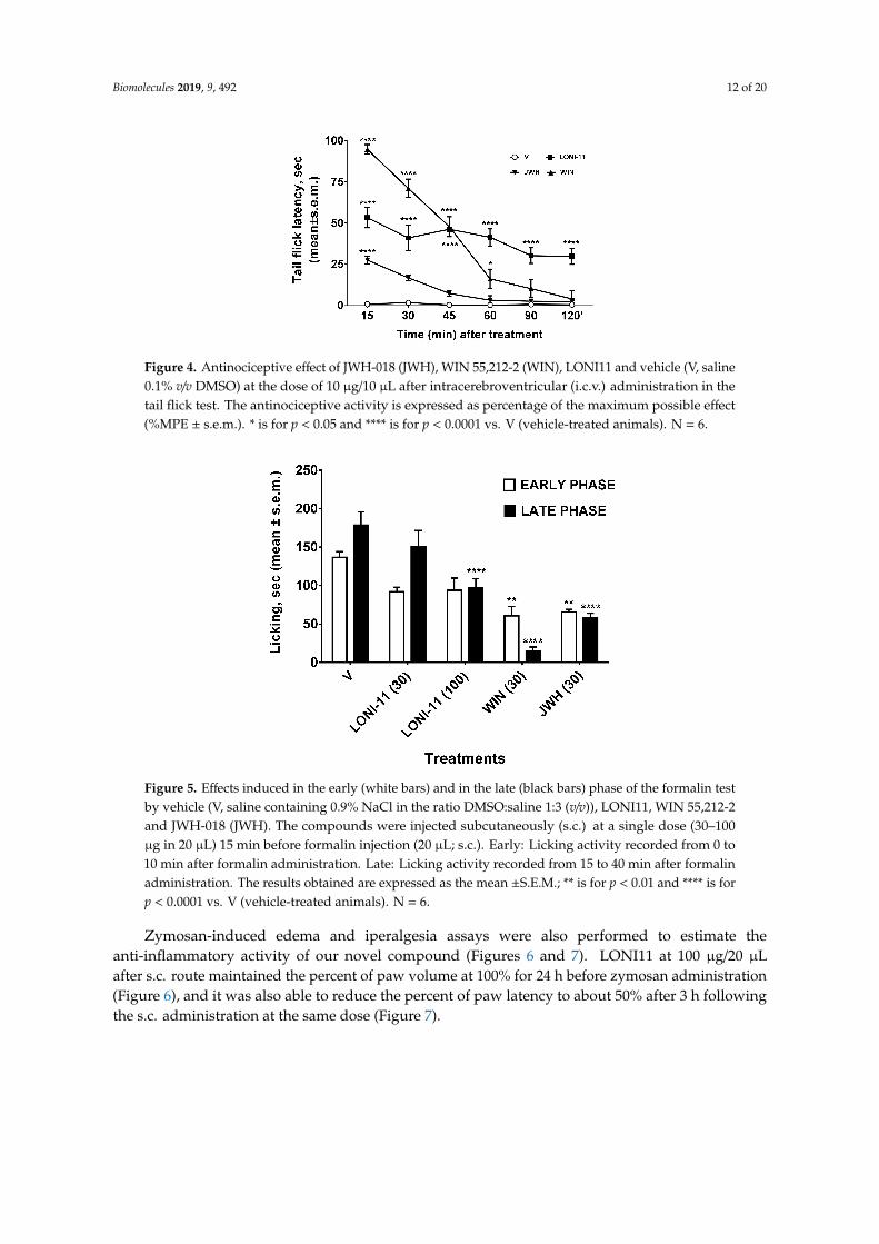
The LONI1-4 reference compounds were ineffective for food intake at 3 mg/Kg per dose afteri.p. administration, while LONI2 and 4 were able to reduce food intake at 10 mg/Kg per dose as thepositive control rimonabant. Interestingly, as suggested by its agonist activity, the novel compoundLONI11 increased the food intake at 10 mg/Kg per dose after i.p. administration, revealing a potentialorexant effect. To better understand the pharmacological properties of this novel compound and toverify if LONI11 is able to exert a significative biological effect at the central level and periphery, wealso performed tail flick and formalin tests in vivo. LONI11 at 10 µg/10 µL and 100 µg/20 µL produceda slight antinociceptive effect after i.c.v. and s.c. injections, respectively, in tail flick (Figure 4) andformalin tests (Figure 5).
Biomolecules 2019, 9, 492 12 of 20
Figure 4. Antinociceptive effect of JWH-018 (JWH), WIN 55,212-2 (WIN), LONI11 and vehicle (V, saline0.1% v/v DMSO) at the dose of 10 µg/10 µL after intracerebroventricular (i.c.v.) administration in thetail flick test. The antinociceptive activity is expressed as percentage of the maximum possible effect(%MPE ± s.e.m.). * is for p < 0.05 and **** is for p < 0.0001 vs. V (vehicle-treated animals). N = 6.
Figure 5. Effects induced in the early (white bars) and in the late (black bars) phase of the formalin testby vehicle (V, saline containing 0.9% NaCl in the ratio DMSO:saline 1:3 (v/v)), LONI11, WIN 55,212-2and JWH-018 (JWH). The compounds were injected subcutaneously (s.c.) at a single dose (30–100µg in 20 µL) 15 min before formalin injection (20 µL; s.c.). Early: Licking activity recorded from 0 to10 min after formalin administration. Late: Licking activity recorded from 15 to 40 min after formalinadministration. The results obtained are expressed as the mean ±S.E.M.; ** is for p < 0.01 and **** is forp < 0.0001 vs. V (vehicle-treated animals). N = 6.
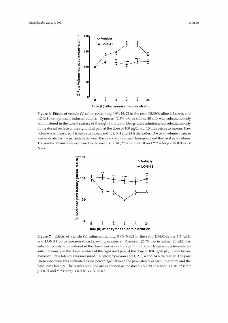
Zymosan-induced edema and iperalgesia assays were also performed to estimate theanti-inflammatory activity of our novel compound (Figures 6 and 7). LONI11 at 100 µg/20 µLafter s.c. route maintained the percent of paw volume at 100% for 24 h before zymosan administration(Figure 6), and it was also able to reduce the percent of paw latency to about 50% after 3 h followingthe s.c. administration at the same dose (Figure 7).
Biomolecules 2019, 9, 492 13 of 20
Figure 6. Effects of vehicle (V, saline containing 0.9% NaCl in the ratio DMSO:saline 1:3 (v/v)), andLONI11 on zymosan-induced edema. Zymosan (2.5% w/v in saline, 20 µL) was subcutaneouslyadministered in the dorsal surface of the right hind paw. Drugs were administered subcutaneouslyin the dorsal surface of the right hind paw at the dose of 100 µg/20 µL, 15 min before zymosan. Pawvolume was measured 1 h before zymosan and 1, 2, 3, 4 and 24 h thereafter. The paw volume increasewas evaluated as the percentage between the paw volume at each time-point and the basal paw volume.The results obtained are expressed as the mean ±S.E.M.; ** is for p < 0.01 and **** is for p < 0.0001 vs. V.N = 6.
Figure 7. Effects of vehicle (V, saline containing 0.9% NaCl in the ratio DMSO:saline 1:3 (v/v)),and LONI11 on zymosan-induced paw hyperalgesia. Zymosan (2.5% w/v in saline, 20 µL) wassubcutaneously administered in the dorsal surface of the right hind paw. Drugs were administeredsubcutaneously in the dorsal surface of the right hind paw at the dose of 100 µg/20 µL, 15 min beforezymosan. Paw latency was measured 1 h before zymosan and 1, 2, 3, 4 and 24 h thereafter. The pawlatency decrease was evaluated as the percentage between the paw latency at each time-point and thebasal paw latency. The results obtained are expressed as the mean ±S.E.M.; * is for p < 0.05, ** is forp < 0.01 and **** is for p < 0.0001 vs. V. N = 6.
Biomolecules 2019, 9, 492 14 of 20
4. Discussion
Compounds LONI2 and LONI4, namely lonidamine-tert-LeuOCH3 and lonidamine-Val-OH,respectively, were found to inhibit food intake, consistent with an inverse agonism at CB1 receptors(Table 1) [33].
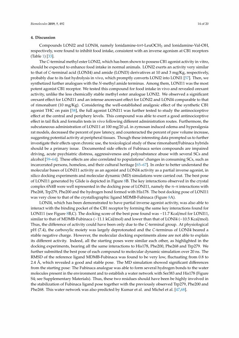
The C-terminal methyl ester LONI2, which has been shown to possess CB1 agonist activity in vitro,should be expected to enhance food intake in normal animals. LONI2 exerts an activity very similarto that of C-terminal acid (LONI4) and amide (LONI3) derivatives at 10 and 3 mg/Kg, respectively,probably due to its fast hydrolysis in vivo, which promptly converts LONI2 into LONI1 [57]. Then, wesynthetized further analogues with the N-methyl amide terminus. Among them, LONI11 was the mostpotent agonist CB1 receptor. We tested this compound for food intake in vivo and revealed orexantactivity, unlike the less chemically stable methyl ester analogue LONI2. We observed a significantorexant effect for LONI11 and an intense anorexant effect for LONI2 and LONI4 comparable to thatof rimonabant (10 mg/Kg). Considering the well-established analgesic effect of the synthetic CB1agonist THC on pain [58], the full agonist LONI11 was further tested to study the antinociceptiveeffect at the central and periphery levels. This compound was able to exert a good antinociceptiveeffect in tail flick and formalin tests in vivo following different administration routes. Furthermore, thesubcutaneous administration of LONI11 at 100 mg/20 µL in zymosan induced edema and hyperalgesiarat models, decreased the percent of paw latency, and counteracted the percent of paw volume increase,suggesting potential activity at peripheral tissues. Though these interesting data prompted us to furtherinvestigate their effects upon chronic use, the toxicological study of these rimonabant/Fubinaca hybridsshould be a primary issue. Documented side effects of Fubinaca series compounds are impaireddriving, acute psychiatric distress, aggressiveness and polysubstance abuse with several SCs andalcohol [59–64]. These effects are also correlated to populations’ changes in consuming SCs, such asincarcerated persons, homeless, and their cultural heritage [65–67]. In order to better understand themolecular bases of LONI11 activity as an agonist and LONI4 activity as a partial inverse agonist, insilico docking experiments and molecular dynamic (MD) simulations were carried out. The best poseof LONI11 generated by Glide is depicted in Figure 8B. The key interactions observed in the crystalcomplex 6N4B were well represented in the docking pose of LONI11, namely the π–π interactions withPhe268, Trp279, Phe200 and the hydrogen bond formed with His178. The best docking pose of LONI11was very close to that of the crystallographic ligand MDMB-Fubinaca (Figure 8A).
LONI4, which has been demonstrated to have partial inverse agonist activity, was also able tointeract with the binding pocket of the CB1 receptor by forming the same key interactions found forLONI11 (see Figure 8B,C). The docking score of the best pose found was −11.7 Kcal/mol for LONI11,similar to that of MDMB-Fubinaca (−11.1 kCal/mol) and lower than that of LONI4 (−10.5 Kcal/mol).Thus, the difference of activity could have been only due to the C-terminal group. At physiologicalpH (7.4), the carboxylic moiety was largely deprotonated and the C-terminus of LONI4 beared astable negative charge. However, the molecular docking experiments alone are not able to explainits different activity. Indeed, all the starting poses were similar each other, as highlighted in thedocking experiments, bearing all the same interactions to His178, Phe200, Phe268 and Trp279. Wefurther submitted the best pose of each compound to molecular dynamic simulation over 20 ns. TheRMSD of the reference ligand MDMB-Fubinaca was found to be very low, fluctuating from 0.8 to2.4 Å, which revealed a good and stable pose. The MD simulation showed significant differencesfrom the starting pose: The Fubinaca analogue was able to form several hydrogen bonds to the watermolecules present in the environment and to establish a water network with Ser383 and His178 (FigureS4; see Supplementary Materials). Thus, these two residues should have been be highly involved inthe stabilization of Fubinaca ligand pose together with the previously observed Trp279, Phe200 andPhe268. This water network was also predicted by Kumar et al. and Michel et al. [47,68].
Biomolecules 2019, 9, 492 15 of 20
Figure 8. Best ranked pose of MDMB-Fubinaca (A), LONI11 (B) and LONI4 (C) docked to theCB1 receptor.
During the MD simulation, LONI11 maintained the π–π stack with the aromatic residues Phe268,Trp279 and Phe379 (Figure S4; see Supplementary Materials). However, the polar portion of themolecule largely lost the initial hydrogen bond to His178, and a water network between one waterand the residue Ser383 was formed, as in the case of Fubinaca ligand. The presence of the freecarboxylic group in LONI4 deeply influenced the stabilization of the starting docked pose during theMD simulation. Indeed, the RMSD plot of LONI4 ranged from 0.8 to 4.5 Å, demonstrating that thestarting pose was scarcely stable (Figure S5; see Supplementary Materials). During the MD simulation,several novel interactions were formed, along with a strong hydration of the free carboxylic terminus,higher than that found for the methyl ester group of Fubinaca and for the N-methylamide groupof LONI11.
The water permanency of LONI4 was more than 10% of the time, whereas for MDMB-Fubinacaand LONI11, it reached only 5%. The authors Michel et al. and Kumar et al. previously predicted thepossible involvement of a water network formed by a water molecule strongly bound to the receptor,mediating the interactions among the ligand, His178 and Ser383 [47,68]. The capacity of the ligand toactivate or deactivate the receptor could be largely due to its ability to form a specific water network.However, in the case of LONI4, the MD experiments demonstrated that the presence of a permanentnegative charge allowed the carboxylic group to form strong polar interactions with three positivecenters in the binding pocket represented by Arg182, Lys192 and Lys376 residues. This binding modeseemed to be preferred over the water network and the related interactions with Ser383 and His178.This difference is fundamental to explain the inverse agonist activity of LONI4. Moreover, the aromaticportion of the molecule seems to prefer a different accommodation in the receptor pocket. Thus, thenovel compound LONI11 bearing the N-methyl-amide C-terminus and the 2,4-dichloro-benzyl ringlargely interacted with the CB1 binding cavity, as in the case of MDMB-Fubinaca. On the contrary,LONI4 (bearing a free carboxylic acid) was not able to strongly interact with the polar key residuesHis178 and Ser383 either directly or through a water network; instead, it may have been able toestablish a whole new set of polar interactions with the positive residues present at the receptor bindingcavity (Lys376, Lys192 and Arg182), leading to the inactivation of the CB1 receptor.
Biomolecules 2019, 9, 492 16 of 20
5. Conclusions
In the last two decades, obesity has assumed the form of a pandemic, often related to thedevelopment of cardiovascular diseases (CVD) and strokes. There is a worldwide emergency thatrequires the development of novel and safer anti-obesity treatments. Rimonabant and sibutramineassociated with lifestyle modifications were successfully engaged in the control of obesity by reducingappetite and weight gain. Their complex action mechanism promotes monoamine reuptake in thecentral nervous system (CNS), the secretion of neuropeptides with anorexigenic activity and thelowering of those with orexigenic properties, the elevation of metabolism, and the improvement of theperipheral sympathetic activity. Though those drugs have shown to be effective to control body weightand to manage obesity, they have been withdrawn from the market due to their important side effects.Thus, a novel drug able to provide the same therapeutic efficacy without the dangerous side effects is anurgent need. An agonist able to bind a specific receptor that preferentially drives a single downstreamsignaling cascade could represent a possible solution. However, the broad distribution of cannabinoidreceptors in different areas of the body allows them to interact with a variety of system networks. TheCB1 receptors are connected with different receptor-activity modulatory proteins, and the possibilityto assume a selectively targetable ligand-binding conformation is still uncertain; currently, there arefew process and site-specific CB1 targeted drugs.
Our study identified a series of lonidamine joined Leu, tert-Leu and Val amino acids with differentC-terminal functional groups (LONI1-4,11) as novel compounds endowed with orexant/anorexantactivity. They are structurally related to the Fubinaca series and follow the same general structurereported by Schoeder et al. [30]. However, in this study, novel features were identified. In particular,the four structural features described by Huffman et al. [69,70] were well defined in our compounds,namely: (i) An heterocyclic scaffold represented by an indole, indazole or imidazole ring with severalsubstitutions; (ii) a dipolar linker such as an amide or ester; (iii) a lipophilic moiety such as the sidechain of an amino acid or a lipophilic chain; and (iv) a hydrophobic side chain bound to the heterocyclicscaffold. This general structure was only able to establish the structural requirements for CB1 receptoraffinity without taking intrinsic activity into consideration. In this work, we have demonstrated theimportance of the C-terminus group, which is shown in the general structure depicted in Figure 9.Polar and uncharged C-terminus groups such as methyl ester and methyl amide moiety are able to actas H bond donors and are required for agonist activity, while a more polar C-terminal amide groupis detrimental, giving weak agonists [33]. The free carboxylic group at C-terminus is able to shiftagonist to antagonist activity. However, a large structure-activity relationships (SAR) study should bedesigned in order to verify our hypothesis.
Figure 9. Structural features of the synthetic cannabinoids reported in this study.
Since one of them (LONI11) exhibited a potent agonist activity at CB1, the antinociceptive activitywas also analyzed and revealed a good analgesic effect after sub-cutaneous and intracerebroventricularadministration in vivo and a significative anti-inflammatory effect after s.c. administration, suggestingpotential activity at the periphery. Even if the preliminary biological data in vivo and in vitro of these
Biomolecules 2019, 9, 492 17 of 20
novel compounds were promising, a deep investigation on animal models is necessary to assess theirselectivity, potency and long-term effects upon chronic and acute administration. These results could beuseful as a starting point to optimize the pharmacological properties of rimonabant/Fubinaca hybridsand to better understand the molecular mechanism below their biological profile. An understanding ofall the neuroendocrine networks and their roles in the hypothalamus to regulate the feeding behaviorcould lead to the development of novel and safer approaches to manage the main nutritional disorders.
6. Patents
This work is part of the European patent EP18170728, deposited on 4th May 2018 by some of theauthors involved in this work with the approval of all the inventors.
Supplementary Materials: The following are available online at http://www.mdpi.com/2218-273X/9/9/492/s1,RP-HPLC analytical traces, 1H NMR and LRMS for the final compounds, molecular modeling and MD details.
Author Contributions: Conceptualization, A.M. and A.S.; methodology, M.P.D.; formal analysis, G.Z.;investigation, P.M.; data curation, S.P. and S.D.; validation, C.T.; writing original draft preparation, A.S.;writing, review and editing, A.S., M.P.D., P.M., S.P.; supervision, A.M.
Funding: This research received no external funding.
Conflicts of Interest: The authors declare no conflict of interest.
References
1. Carella, A.M.; Conte, M.; Melfitano, A.; Ponziano, E.; Benvenuto, A. Neuroendocrine Mediators, Food Intakeand Obesity: A Narrative Review. Int. J. Cardiol. Lipidol. Res. 2014, 1, 18–32. [CrossRef]
2. Thomas, B.F. Neuroanatomical basis for the therapeutic applications of cannabinoid receptor 1 antagonists.Drug Dev. Res. 2009, 70, 527–554. [CrossRef]
3. Foster-Schubert, K.E.; Cummings, D.E. Emerging Therapeutic Strategies for Obesity. Endocr. Rev. 2006, 27,779–793. [CrossRef] [PubMed]
4. Soria-Gomez, E.; Matias, I.; Rueda-Orozco, P.E.; Cisneros, M.; Petrosino, S.; Navarro, L.; di Marzo, V.;Prospéro-García, O. Pharmacological enhancement of the endocannabinoid system in the nucleus accumbensshell stimulates food intake and increases c-Fos expression in the hypothalamus. Br. J. Pharmacol. 2007, 151,1109–1116. [CrossRef] [PubMed]
5. Kirkham, T.C.; Williams, C.M.; Fezza, F.; di Marzo, V. Endocannabinoid levels in rat limbic forebrainandhypothalamus in relation to fasting, feeding and satiation: Stimulation of eating by 2-arachidonoyl glycerol.Br. J. Pharmacol. 2002, 136, 550–557. [CrossRef] [PubMed]
6. Di Marzo, V.; Matias, I. Endocannabinoid control of food intake and energy balance. Nat. Neurosci. 2005, 8,585–589. [CrossRef]
7. Colombo, G.; Agabio, R.; Diaz, G.; Lobina, C.; Reali, R.; Gessa, G.L. Appetite suppression and weight lossafter the cannabinoid antagonist SR141716. Life Sci. 1998, 63, PL113–PL117. [CrossRef]
8. Lambert, P.; Wilding, J.; Al-Dokhayel, A.; Gilbey, S.; Bloom, S. The effect of central blockade of kappa-opioidreceptors on neuropeptide Y-induced feeding in the rat. Brain Res. 1993, 629, 146–148. [CrossRef]
9. Brunetti, L.; Ferrante, C.; Orlando, G.; Recinella, L.; Leone, S.; Chiavaroli, A.; di Nisio, C.; Shohreh, R.;Manippa, F.; Ricciuti, A.; et al. Orexigenic effects of endomorphin-2 (EM-2) related to decreased CRH geneexpression and increased dopamine and norepinephrine activity in the hypothalamus. Peptides 2013, 48,83–88. [CrossRef]
10. Crespo, I.; De Heras, R.G.; de Fonseca, F.R.; Navarro, M. Pretreatment with subeffective doses of Rimonabantattenuates orexigenic actions of orexin A-hypocretin 1. Neuropharmacology 2008, 54, 219–225. [CrossRef]
11. Hilairet, S.; Bouaboula, M.; Carrière, D.; le Fur, G.; Casellas, P. Hypersensitization of the Orexin 1 Receptorby the CB1 Receptor: Evidence For Cross-Talk Blocked by the Specific Cb1 Antagonist, SR141716. J. Biol.Chem. 2003, 278, 23731–23737. [CrossRef]
12. Scheen, A.J. Sibutramine on Cardiovascular Outcome. Diabetes Care 2001, 34, 114–119. [CrossRef]13. Zhang, W.; Roederer, M.W.; Chen, W.-Q.; Fan, L.; Zhou, H.-H. Pharmacogenetics of drugs withdrawn from
the market. Pharmacogenomics 2012, 13, 223–231. [CrossRef]
Biomolecules 2019, 9, 492 18 of 20
14. Després, J.-P.; Golay, A.; Sjöstrom, L. Effects of Rimonabant on Metabolic Risk Factors in Overweight Patientswith Dyslipidemia. N. Engl. J. Med. 2005, 353, 2121–2134. [CrossRef]
15. Després, J.P.; Ross, R.; Boka, G.; Alméras, N.; Lemieux, I. Effect of rimonabant on the high-triglyceride/
low-HDL-cholesterol dyslipidemia, intra-abdominal adiposity, and liver fat: The ADAGIO-Lipids trial.Arterioscler. Thromb. Vasc. Biol. 2009, 29, 416–423. [CrossRef]
16. Sam, A.H.; Salem, V.; Ghatei, M.A. Rimonabant: From RIO to Ban. J. Obes. 2011, 2011, 432607. [CrossRef]17. Erkekoglu, P.; Giray, B.; Sahin, G. Toxicological evaluation of Rimonabant, Taranabant, Surinabant and
Otenabant in the treatment of obesity: Why the trials on endocannabinoid receptor antagonists and inverseagonists are suspended? FABAD J. Pharm. Sci. 2008, 33, 95–108.
18. Janero, D.R.; Makriyannis, A. Cannabinoid receptor antagonists: Pharmacological opportunities, clinicalexperience, and translational prognosis. Expert Opin. Emerg. Drugs 2009, 14, 43–65. [CrossRef]
19. Ibsen, M.S.; Connor, M.; Glass, M. Cannabinoid CB1 and CB2 Receptor Signaling and Bias.Cannabis Cannabinoid Res. 2017, 2, 48–60. [CrossRef]
20. Al-Zoubi, R.; Morales, P.; Reggio, P.H. Structural Insights into CB1 Receptor Biased Signaling. Int. J. Mol. Sci.2019, 20, 1837. [CrossRef]
21. Al-Zoubi, W.; Salih, A.A.; Duraid, S.A.; Basheer, H.M.; Awad Al-Luhaibi, R.S.; Dib, A.; Young, G.K. Synthesis,characterization, and antioxidant activities ofimine compounds. J. Phys. Org. Chem. 2019, 32, 3916.
22. Ibsen, D.B.; Laursen, A.S.D.; Lauritzen, L.; Tjønneland, A.; Overvad, K.; Jakobsen, M.U. Substitutionsbetween dairy product subgroups and risk of type 2 diabetes: The Danish Diet, Cancer and Health cohort.Br. J. Nutr. 2017, 118, 989–997. [CrossRef]
23. Mazier, W.; Saucisse, N.; Gatta-Cherifi, B.; Cota, D. The Endocannabinoid System: Pivotal Orchestrator ofObesity and Metabolic Disease. Trends Endocrinol. Metab. 2015, 26, 524–537. [CrossRef]
24. Black, A.D.; Car, J.; Pagliari, C.; Anandan, C.; Cresswell, K.; Bokun, T.; McKinstry, B.; Procter, R.; Majeed, A.;Sheikh, A. The Impact of eHealth on the Quality and Safety of Health Care: A Systematic Overview. PLoS Med.2011, 8, e1000387. [CrossRef]
25. Rubino, F.; Nathan, D.M.; Eckel, R.H.; Schauer, P.R.; Alberti, K.G.M.; Zimmet, P.Z.; Del Prato, S.; Ji, L.;Sadikot, S.M.; Herman, W.H.; et al. Metabolic Surgery in the Treatment Algorithm for Type 2 Diabetes: AJoint Statement by International Diabetes Organizations. Diabetes Care 2016, 39, 861–877. [CrossRef]
26. Mallat, S.G.; Abu Samra, S.; Younes, F.; Sawaya, M.-T. Identifying predictors of blood pressure control inthe Lebanese population—A national, multicentric survey—I-PREDICT. BMC Public Health 2014, 14, 1142.[CrossRef]
27. Schindler, K.; Themessl-Huber, M.; Hiesmayr, M.; Kosak, S.; Lainscak, M.; Laviano, A.; Ljungqvist, O.;Mouhieddine, M.; Schneider, S.; de van der Schueren, M.; et al. To eat or not to eat? Indicators for reducedfood intake in 91,245 patients hospitalized on nutritionDays 2006–2014 in 56 countries worldwide: Adescriptive analysis. Am. J. Clin. Nutr. 2016, 104, 1393–1402. [CrossRef]
28. Banister, S.D.; Longworth, M.; Kevin, R.; Sachdev, S.; Santiago, M.; Stuart, J.; Mack, J.B.; Glass, M.;McGregor, I.S.; Connor, M.; et al. Pharmacology of Valinate and tert-Leucinate synthetic cannabinoids5F-AMBICA, 5F-AMB, 5F-ADB, AMB-FUBINACA, MDMB-FUBINACA, MDMB-CHMICA, and theiranalogues. ACS Chem. Neurosci. 2016, 7, 1241–1254. [CrossRef]
29. Shanks, K.G.; Clark, W.; Behonick, G. Death Associated With the Use of the Synthetic CannabinoidADB-FUBINACA. J. Anal. Toxicol. 2016, 40, 236–239. [CrossRef]
30. Schoeder, C.T.; Hess, C.; Madea, B.; Meiler, J.; Muller, C.E. Pharmacological evaluation of new constituentsof “Spice”: Synthetic cannabinoids based on indole, indazole, benzimidazole and carbazole scaffolds.Forensic Toxicol. 2018, 36, 385–403. [CrossRef]
31. Adamowicz, P.; Meissner, E.; Maslanka, M. Fatal intoxication with new synthetic cannabinoidsAMB-FUBINACA and EMB-FUBINACA. Clin. Toxicol. 2019, 26, 1–6. [CrossRef]
32. Scourfield, A.; Flick, C.; Ross, J.; Wood, D.M.; Thurtle, N.; Stellmach, D.; Dargan, P.I. Synthetic cannabinoidavailability on darknet drug markets—Changes during 2016–2017. Toxicol. Commun. 2019, 3, 7–15. [CrossRef]
33. Stefanucci, A.; Macedonio, G.; Dvorácskó, S.; Tömböly, C.; Mollica, A. Novel Fubinaca/Rimonabant hybridsas endocannabinoid system modulators. Amino Acids 2018, 50, 1595–1605. [CrossRef]
34. Mollica, A.; Costante, R.; Akdemir, A.; Carradori, S.; Stefanucci, A.; Macedonio, G.; Ceruso, M.; Supuran, C.T.Exploring new Probenecid-based carbonic anhydrase inhibitors: Synthesis, biological evaluation and dockingstudies. Bioorganic Med. Chem. 2015, 23, 5311–5318. [CrossRef]
Biomolecules 2019, 9, 492 19 of 20
35. Mollica, A.; Pelliccia, S.; Famiglini, V.; Stefanucci, A.; Macedonio, G.; Chiavaroli, A.; Orlando, G.; Brunetti, L.;Ferrante, C.; Pieretti, S.; et al. Exploring the first Rimonabant analog-opioid peptide hybrid compound, asbivalent ligand for CB1 and opioid receptors. J. Enzym. Inhib. Med. Chem. 2017, 32, 444–451. [CrossRef]
36. Mollica, A.; Pinnen, F.; Stefanucci, A.; Costante, R. The evolution of peptide synthesis: From early days tosmall molecular machines. Curr. Bioact. Comp. 2013, 9, 184–202. [CrossRef]
37. Dvorácskó, S.; Keresztes, A.; Mollica, A.; Stefanucci, A.; Macedonio, G.; Pieretti, S.; Zádor, F.; Walter, F.R.;Deli, M.A.; Kékesi, G.; et al. Preparation of bivalent agonists for targeting the mu opioid and cannabinoidreceptors. Eur. J. Med. Chem. 2019, 178, 571–588. [CrossRef]
38. Kilkenny, C.; Browne, W.; Cuthill, I.C.; Emerson, M.; Altman, D.G. Animal research: Reporting in vivoexperiments: The ARRIVE guidelines. Br. J. Pharmacol. 2010, 160, 1577–1579. [CrossRef]
39. McGrath, J.C.; Lilley, E. Implementing guidelines on reporting research using animals (ARRIVE etc.): Newrequirements for publication in BJP. Br. J. Pharmacol. 2015, 172, 3189–3193. [CrossRef]
40. Wiley, J.L.; Burston, J.J.; Leggett, D.C.; Alekseeva, O.O.; Razdan, R.K.; Mahadevan, A.; Martin, B.R. CB1cannabinoid receptor-mediated modulation of food intake in mice. Br. J. Pharmacol. 2005, 145, 293–300.[CrossRef]
41. Pieretti, S.; di Giannuario, A.; de Felice, M.; Perretti, M.; Cirino, G. Stimulus-dependent specificity for annexin1 inhibition of the inflammatory nociceptive response: The involvement of the receptor for formylatedpeptides. Pain 2004, 109, 52–63. [CrossRef]
42. Stefanucci, A.; Lei, W.; Pieretti, S.; Novellino, E.; Dimmito, M.P.; Marzoli, F.; Streicher, J.M.; Mollica, A. Onresin click-chemistry-mediated synthesis of novel enkephalin analogues with potent anti-nociceptive activity.Sci. Rep. 2019, 9, 5771. [CrossRef]
43. Maione, F.; Minosi, P.; di Giannuario, A.; Raucci, F.; Chini, M.G.; de Vita, S.; Bifulco, G.; Mascolo, N.;Pieretti, S. Long-Lasting Anti-Inflammatory and Antinociceptive Effects of Acute Ammonium GlycyrrhizinateAdministration: Pharmacological, Biochemical, and Docking Studies. Molecules 2019, 24, 2453. [CrossRef]
44. Pieretti, S.; Dominici, L.; di Giannuario, A.; Cesari, N.; Piaz, V.D. Local anti-inflammatory effect and behavioralstudies on new PDE4 inhibitors. Life Sci. 2006, 79, 791–800. [CrossRef]
45. Niederberger, E.; Schmidtko, A.; Gao, W.; Kühlein, H.; Ehnert, C.; Geisslinger, G. Impaired acute andinflammatory nociception in mice lacking the p50 subunit of NF-kappaB. Eur. J. Pharmacol. 2007, 559, 55–60.[CrossRef]
46. Curtis, M.J.; Bond, R.A.; Spina, D.; Ahluwalia, A.; Alexander, S.P.A.; Giembycz, M.A.; Gilchrist, A.; Hoyer, D.;Insel, P.A.; Izzo, A.A.; et al. Experimental design and analysis and their reporting: New guidance forpublication in BJP. Br. J. Pharmacol. 2015, 172, 3461–3471. [CrossRef]
47. Kumar, K.K.; Shalev-Benami, M.; Robertson, M.J.; Hu, H.; Banister, S.D.; Hollingsworth, S.A.; Latorraca, N.R.;Kato, H.E.; Hilger, D.; Maeda, S.; et al. Structure of a Signaling Cannabinoid Receptor 1-G Protein Complex.Cell 2019, 176, 448–458.e12. [CrossRef]
48. Schrödinger Release 2019-3: Schrödinger Suite 2019-2 Protein Preparation Wizard; Epik, Schrödinger, LLC:New York, NY, USA; Impact, Schrödinger, LLC: New York, NY, USA; Prime, Schrödinger, LLC: New York,NY, USA, 2019.
49. Jacobson, M.P.; Pincus, D.L.; Rapp, C.S.; Day, T.J.; Honig, B.; Shaw, D.E.; Friesner, R.A. A hierarchicalapproach to all-atom protein loop prediction. Proteins Struct. Funct. Bioinform. 2004, 55, 351–367. [CrossRef]
50. Stefanucci, A.; Novellino, E.; Mirzaie, S.; Macedonio, G.; Pieretti, S.; Minosi, P.; Szucs, E.; Erdei, A.I.; Zádor, F.;Benyhe, S.; et al. Opioid Receptor Activity and Analgesic Potency of DPDPE Peptide Analogues Containinga Xylene Bridge. ACS Med. Chem. Lett. 2017, 8, 449–454. [CrossRef]
51. Schrödinger Release 2019-3: Glide; Schrödinger, LLC: New York, NY, USA, 2019.52. Schrödinger Release 2019-3: LigPrep; Schrödinger, LLC: New York, NY, USA, 2019.53. Schrödinger Release 2019-3: Desmond Molecular Dynamics System; D. E. Shaw Research: New York, NY, USA;
Maestro-Desmond Interoperability Tools, Schrödinger: New York, NY, USA, 2019.54. Chandrasekhar, J.; Impey, R.W.; Jorgensen, W.L.; Madura, J.D.; Klein, M.L. Comparison of simple potential
functions for simulating liquid water. J. Chem. Phys. 1983, 79, 926.55. Martyna, G.J.; Tobias, D.J.; Klein, M.L. Constant pressure molecular dynamics algorithms. J. Chem. Phys.
1994, 101, 4177–4189. [CrossRef]56. Hünenberger, P.H. Thermostat Algorithms for Molecular Dynamics Simulations. Adv. Polym. Sci. 2005, 173,
105–149.
Biomolecules 2019, 9, 492 20 of 20
57. Matsubara, K.; Kagawa, M.; Fukui, Y. In vivo and in vitro studies on cocaine metabolism: Ecgonine methylester as a major metabolite of cocaine. Forensic Sci. Int. 1984, 26, 169–180. [CrossRef]
58. Slomski, A. THC for Chronic Pain. JAMA 2018, 320, 1631. [CrossRef]59. Kaneko, S. Motor vehicle collisions caused by the ‘super-strength’ synthetic cannabinoids, MAM-2201,
5F-PB-22, 5F-AB-PINACA, 5F-AMB and 5F-ADB in Japan experienced from 2012 to 2014. Forensic Toxicol.2017, 35, 244–251. [CrossRef]
60. Castaneto, M.S.; Gorelick, D.A.; Desrosiers, N.A.; Hartman, R.L.; Pirard, S.; Huestis, M.A. Syntheticcannabinoids: Epidemiology, pharmacodynamics, and clinical implications. Drug Alcohol Depend. 2014, 144,12–41. [CrossRef]
61. Deng, H.; Verrico, C.D.; Kosten, T.R.; Nielsen, D.A. Psychosis and synthetic cannabinoids. Psychiatry Res.2018, 268, 400–412. [CrossRef]
62. Abouchedid, R.; Ho, J.H.; Hudson, S.; Dines, A.; Archer, J.R.H.; Wood, D.M.; Dargan, P.I. Acute ToxicityAssociated with Use of 5F-Derivations of Synthetic Cannabinoid Receptor Agonists with AnalyticalConfirmation. J. Med. Toxicol. 2016, 12, 396–401. [CrossRef]
63. Langford, A.M.; Bolton, J.R. Synthetic cannabinoids: Variety is definitely not the spice of life. J. ForensicLeg. Med. 2018, 59, 36–38. [CrossRef]
64. European Monitoring Centre for Drugs and Drug Addiction. EMCDDA–Europol Joint Reporton a New Psychoactive Substance: 1-(4-Cyanobutyl)-N-(2-Phenylpropan-2-yl)Indazole-3-Carboxamide(CUMYL-4CN-BINACA); Joint Reports; Publications Office of the European Union: Luxembourg, 2017.
65. Van Hout, M.C.; Benschop, A.; Bujalski, M.; Dabrowska, K.; Demetrovics, Z.; Felvinczi, K.; Herne, E.;Henriques, S.; Kaló, Z.; Kamphausen, G.; et al. Health and social problems associated with recent novelpsychoactive substance (NPS) use amongst marginalised, nightlife and online users in six European countries.Int. J. Ment. Health Addict. 2018, 16, 480–495. [CrossRef]
66. European Monitoring Centre for Drugs and Drug Addiction. Report on the Risk Assessment of1-(4-Cyanobutyl)-N-(2-Phenylpropan-2-yl)-1H-Indazole-3-Carboxamide (CUMYL-4CN-BINACA) in the Frameworkof the Council Decision on New Psychoactive Substances; Risk Assessments; Publications Office of the EuropeanUnion: Luxembourg, 2018.
67. Drug Enforcement Administration, Department of Justice. Schedules of controlled substances:Temporary placement of six synthetic cannabinoids (5F-ADB, 5F-AMB, 5F-APINACA, ADB-FUBINACA,MDMB-CHMICA and MDMB-FUBINACA) into Schedule I. Fed. Regist. 2017, 82, 17119–17124.
68. Michel, J.; Tirado-Rives, J.; Jorgensen, W.L. Prediction of the Water Content in Protein Binding Sites. J. Phys.Chem. B 2009, 113, 13337–13346. [CrossRef]
69. Huffman, J.W.; Dai, D.; Martin, B.R.; Compton, D.R. Design, Synthesis and Pharmacology of CannabimimeticIndoles. Bioorganic Med. Chem. Lett. 1994, 4, 563–566. [CrossRef]
70. Huffman, J.W.; Zengin, G.; Wu, M.J.; Lu, J.; Hynd, G.; Bushell, K.; Thompson, A.L.; Bushell, S.; Tartal, C.;Hurst, D.P.; et al. Structure-activity relationships for 1-alkyl-3-(1-naphthoyl)indoles at the cannabinoid CB(1)and CB(2) receptors: Steric and electronic effects of naphthoyl substituents. New highly selective CB(2)receptor agonists. Bioorg. Med. Chem. 2005, 13, 89–112. [CrossRef]
© 2019 by the authors. Licensee MDPI, Basel, Switzerland. This article is an open accessarticle distributed under the terms and conditions of the Creative Commons Attribution(CC BY) license (http://creativecommons.org/licenses/by/4.0/).


![Current Perspective in the Discovery of Anti-aging Agents ... · Current Perspective in the Discovery of Anti-aging Agents from Natural Products ... [37], and sirtu-ins [38]. These](https://img.pdfslide.us/doc/110x75/5ccb6c0e88c993b16c8d5119/current-perspective-in-the-discovery-of-anti-aging-agents-current-perspective.jpg)