Embed Size (px)
Citation preview
LETTERdoi:10.1038/nature12576
Discovery of new enzymes and metabolic pathwaysby using structure and genome contextSuwen Zhao1*, Ritesh Kumar2*, Ayano Sakai2*, Matthew W. Vetting3*, B. McKay Wood2*, Shoshana Brown4, Jeffery B. Bonanno3,Brandan S. Hillerich3, Ronald D. Seidel3, Patricia C. Babbitt4, Steven C. Almo3, Jonathan V. Sweedler2,5, John A. Gerlt2,5,6,John E. Cronan2,6,7 & Matthew P. Jacobson1
Assigning valid functions to proteins identified in genome projectsis challenging: overprediction and database annotation errors arethe principal concerns1. We and others2 are developing computation-guided strategies for functional discovery with ‘metabolite docking’to experimentally derived3 or homology-based4 three-dimensionalstructures. Bacterial metabolic pathways often are encoded by ‘genomeneighbourhoods’ (gene clusters and/or operons), which can provideimportant clues for functional assignment. We recently demonstratedthe synergy of docking and pathway context by ‘predicting’ the inter-mediates in the glycolytic pathway in Escherichia coli5. Metabolitedocking to multiple binding proteins and enzymes in the same path-way increases the reliability of in silico predictions of substrate speci-ficities because the pathway intermediates are structurally similar.Here we report that structure-guided approaches for predicting thesubstrate specificities of several enzymes encoded by a bacterial genecluster allowed the correct prediction of the in vitro activity of astructurally characterized enzyme of unknown function (PDB 2PMQ),2-epimerization of trans-4-hydroxy-L-proline betaine (tHyp-B) andcis-4-hydroxy-D-proline betaine (cHyp-B), and also the correct identi-fication of the catabolic pathway in which Hyp-B 2-epimerase par-ticipates. The substrate-liganded pose predicted by virtual libraryscreening (docking) was confirmed experimentally. The enzymaticactivities in the predicted pathway were confirmed by in vitro assaysand genetic analyses; the intermediates were identified by metabo-lomics; and repression of the genes encoding the pathway by highsalt concentrations was established by transcriptomics, confirmingthe osmolyte role of tHyp-B. This study establishes the utility ofstructure-guided functional predictions to enable the discovery ofnew metabolic pathways.
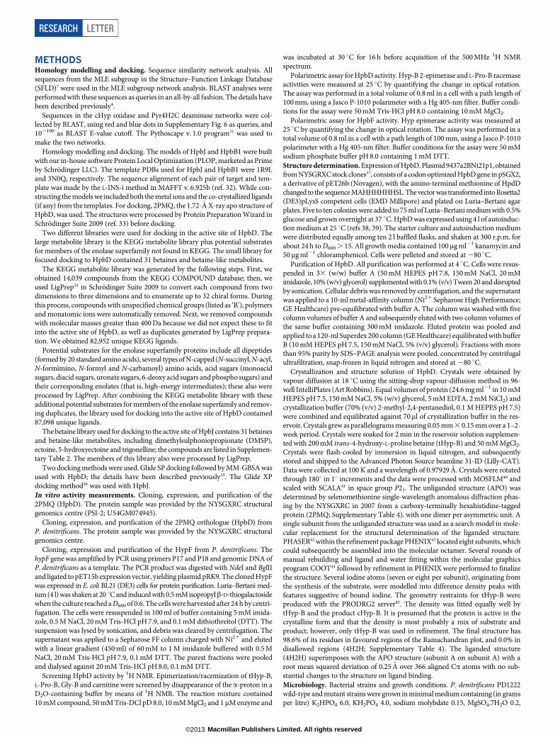
We have applied a structure-guided strategy using metabolite dockingto multiple proteins and enzymes in a metabolic pathway to discover apreviously undocumented reaction, 4R-hydroxyproline betaine 2-epimerase(Hyp-B 2-epimerase; Fig. 1a), as well as the catabolic pathway by whichtHyp-B is converted to a-ketoglutarate. The crystallographically deter-mined unliganded structure of the uncharacterized ‘target’ as well ashomology models for a binding protein and a second enzyme encodedby its genome neighbourhood were used to predict the Hyp-B 2-epimeraseactivity as well as those of downstream enzymes of the pathway.
The marine bacterium Pelagibaca bermudensis encodes an unchar-acterized member of the enolase superfamily (National Center for Bio-technology Information GI number 114543141) in which two lysineresidues of the TIM-barrel domain are positioned to function as acid–base catalysts6–8. The New York SGX Research Consortium determinedits structure (PDB 2PMQ) because it shared less than 30% sequenceidentity with structurally characterized enolase superfamily members.The only ligand was the Mg21 that stabilizes the enolate anion inter-mediate obtained by abstraction of thea-proton of a carboxylate substrate.
The active site is sequestered from solvent by two closed loops and wastherefore suitable for virtual metabolite docking for substrate predic-tion (Supplementary Fig. 1).
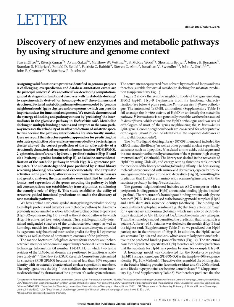
Figure 2 shows the genome neighbourhoods of the gene encoding2PMQ (hpbD; Hyp-B 2-epimerase from its functional characte-rization (see below)) plus a putative Paracoccus denitrificans ortholo-gue. The automated TrEMBL annotations (Supplementary Table 1)fail to assign the in vitro activity of HpbD or to identify the metabolicpathway. P. bermudensis is not genetically tractable; we therefore studiedP. denitrificans, which encodes one HpbD orthologue and two sets oforthologues of most of the genes neighbouring the P. bermudensishpbD gene. Genome neighbourhoods are ‘conserved’ for other putativeorthologues (about 20 can be identified in the sequence databases athttp://sfld.rbvi.ucsf.edu/).
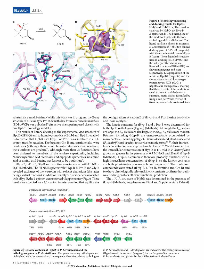
The in silico ligand docking library (87,098 members) included theKEGG metabolite library9 as well as other potential enolase superfamilysubstrates such as dipeptides, N-acylated amino acids, acid sugars andthe enolate anions obtained by abstraction of thea-proton (high-energyintermediates10) (Methods). The library was docked in the active site ofHpbD by using Glide SP, and energy scoring functions rank-orderedthe members of the library according to binding affinity. The best-scoringmolecules were enriched with amino acid derivatives, especially prolineanalogues and N-capped amino acid derivatives (Fig. 3), permitting theprediction that HpbD is an amino acid racemase/epimerase, with thesubstrate probably having N-substitution.
The genome neighbourhood includes an ABC transporter with aperiplasmic binding protein (HpbJ) annotated as binding ‘glycine betaine/L-proline’. The structure of a homologous binding protein with glycinebetaine11 (PDB 1R9L) was used as the homology model template (HpbJand 1R9L share 48% sequence identity) (Methods). The binding sitecontains three tryptophan residues (Fig. 1b) that form a p-cation ‘cage’for a quaternary ammonium (betaine), which may also be electrosta-tically stabilized by Glu 42, located 5.4 A from the quaternary nitrogen.Thus, the homology model permitted the prediction that its ligand is abetaine. A library of 31 betaines was docked to the model; tHyp-B hadthe highest rank (Supplementary Table 2), so we predicted that HpbJparticipates in the transport of tHyp-B. In addition, the HpbD activesite contains Trp 320 and Asp 292, which are similarly positioned rela-tive to the predicted binding pose of betaines (Fig. 1c). The structuralbasis for the predicted specificity of HpbJ therefore refined the predictionthat the substrate for HpbD is a proline betaine, for example tHyp-B.
A homology model was constructed for the Rieske-type protein(HpbB1) using a homologue (PDB 3N0Q) as the template (60% sequenceidentity; Fig. 1d) (Methods). The active site resembled the binding sitesin the betaine-binding proteins (aromatic residues and Glu 200); indeed,some Rieske-type proteins are betaine demethylases12–14 (Supplemen-tary Fig. 2 and Supplementary Table 3). We therefore predicted that the
*These authors contributed equally to this work.
1Department of Pharmaceutical Chemistry, University of California, San Francisco, California 94143, USA. 2Institute for Genomic Biology, University of Illinois at Urbana-Champaign, Urbana, Illinois 61801,USA. 3Department of Biochemistry, Albert Einstein College of Medicine, Bronx, New York 10461, USA. 4Department of Bioengineering and Therapeutic Sciences, University of California, San Francisco,California 94143, USA. 5Department of Chemistry, University of Illinois at Urbana-Champaign, Urbana, Illinois 61801, USA. 6Department of Biochemistry, University of Illinois at Urbana-Champaign,Urbana, Illinois 61801, USA. 7Department of Microbiology, University of Illinois at Urbana-Champaign, Urbana, Illinois 61801, USA.
0 0 M O N T H 2 0 1 3 | V O L 0 0 0 | N A T U R E | 1
Macmillan Publishers Limited. All rights reserved©2013
substrate is a small betaine. (While this work was in progress, the X-raystructure of a Rieske-type Pro-B demethylase from Sinorhizobium meliloti(PDB 3VCP) was published15; its active site superimposed closely withour HpbB1 homology model.)
The results of library docking to the experimental apo structure ofHpbD (2PMQ) and to homology models of HpbJ and HpbB1 enabledus to predict that HpbD uses Hyp-B or Pro-B as a substrate in a 1,1-proton transfer reaction. The betaines Gly-B and carnitine also werecandidates (although these would be substrates for virtual reactions;the a-carbons are prochiral). Although more than 25 functions havebeen assigned to members of the enolase superfamily, includingN-succinylamino acid racemases and dipeptide epimerases, no aminoacid or amino acid betaine was known to be a substrate8.
tHyp-B, L-Pro-B, Gly-B and carnitine were incubated with HpbD inD2O (Methods). The 1H NMR spectra with tHyp-B, D-Pro-B and Gly-Brevealed exchange of the a-proton with solvent deuterium (the latterbeing a virtual reaction); in addition, for tHyp-B, resonances associatedwith cHyp-B, the 2-epimer, were observed (Supplementary Fig. 3). Theseresults are expected for a 1,1-proton transfer reaction that equilibrates
the configurations at carbon 2 of tHyp-B and Pro-B using two lysineacid–base catalysts.
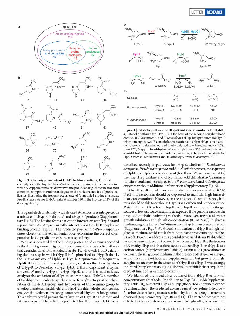
The kinetic constants for tHyp-B and L-Pro-B were determined forboth HpbD orthologues (Fig. 4b) (Methods). Although the kcat valuesare large, the Km values are also large, so the kcat/Km values are modest.Betaines, including tHyp-B, are osmoprotectants accumulated bymany bacteria, including pelagic (P. bermudensis) and plant-associated(P. denitrificans) species, to survive osmotic stress16–20; their intracel-lular concentrations can approach molar levels21,22. We determined thatthe intracellular concentration of Hyp-B is 170 mM in P. denitrificansgrown on glucose in the presence of 0.5 M NaCl and 20 mM tHyp-B(Methods). Hyp-B 2-epimerase therefore probably functions with ahigh intracellular concentration of tHyp-B, so the kinetic constantsare both physiologically reasonable and expected23. That only fourcompounds were tested (tHyp-B, L-Pro-B, carnitine and Gly-B) andtwo have physiologically relevant kinetic constants confirms that path-way docking enables efficient functional prediction.
The 1.70-A structure of HpbD was determined in the presence oftHyp-B (Methods, Supplementary Fig. 4 and Supplementary Table 4).
Val 209
Phe 224
Phe 215
Cys 204
Cys 201 Ala 206
Asp 205
Glu 200
N+
CO2–
OH
tHyp-B
H
N+
H
OH
–O2C
HpbD
cHyp-B
a
d
Trp 87
Glu 42
Trp 209
Trp 161
Gln 91
Ile 68
Trp 320
Lys 163
Asp 193
Glu 218
Asp 241
Lys 265
Asp 292
b
c
Figure 1 | Homology modellingand docking results for HpbD,HpbJ and HpbB1. a, The reactioncatalysed by HpbD, the Hyp-B2-epimerase. b, The binding site ofthe model of HpbJ, with the top-ranked ligand tHyp-B docked. Theligand surface is shown in magenta.c, Comparison of HpbD top-rankeddocking pose of D-Pro-B (magenta)with the experimental pose of tHyp-B (cyan). The unliganded structureused in docking (PDB 2PMQ) andthe subsequently determinedliganded structure (PDB 4H2H) areshown in magenta and cyan,respectively. d, Superposition of themodel of HpbB1 (magenta) and theclosest characterized Rieske-typeprotein (cyan; PDB 1O7G, anaphthalene dioxygenase), showingthat the active site of the model is toosmall to accept naphthalene as asubstrate. Steric clashes identified byusing a van der Waals overlap of0.6 A or more are shown in red lines.
hpbA hpbB1 hpbC1 hpbD hpbE hpbF hpbG hpbH hpbI hpbJ hpbK hpbL hpbC2 hpbB2 hpbR
1187 1188 1189 1190 ... 1193 1194 1195 2831 2832
4859 4860 4865 ... 4867
1197
4746 4747 4748 4749 4751 4752 4753 ...
...
Paracoccus denitrificans PD1222
hpbK hpbL hpbM hpbD hpbC1 hpbB1 hpbR hpbX hpbY hpbZ hpbA hpbC2 hpbB2
hpbN hpbO hpbH hpbJ hpbE hpbF hpbG hypF hypO hypH hypD
58%
78% 84% 64% 61% 59% 59% 83% 62% 58%
22% 58% 76% 68%
...1186 ...0015 0016
...
1613 1623 1628 1638 1643 1648 1653 1663 1668 1673 4863 4943 18653 18658 18663... ... ............
...
Pelagibaca bermudensis HTCC2601
Figure 2 | Genome contexts of HpbD in P. bermudensis and theorthologous genes in P. denitrificans. The genes encoding orthologues arehighlighted with the same colour; the sequence identities relating orthologues
in P. bermudensis and P. denitrificans are indicated. The ecological sources oftHyp-B would be seaweed (sargasso) for the Sargasso Sea bacteriumP. bermudensis, and plants for the soil bacterium P. denitrificans.
RESEARCH LETTER
2 | N A T U R E | V O L 0 0 0 | 0 0 M O N T H 2 0 1 3
Macmillan Publishers Limited. All rights reserved©2013
The ligand electron density, with elevated B-factors, was interpreted asa mixture of tHyp-B (substrate) and cHyp-B (product) (Supplemen-tary Fig. 5). The betaine forms a p-cation interaction with Trp 320 andis proximal to Asp 292, similar to the interactions in the Gly-B periplasmicbinding protein (Fig. 1c). The predicted pose with D-Pro-B superim-poses closely on the experimental pose, explaining the correct com-putation-based prediction of substrate specificity.
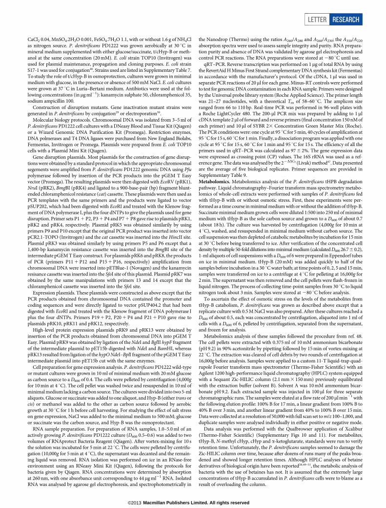
We also speculated that the binding proteins and enzymes encodedin the HpbD genome neighbourhoods constitute a catabolic pathwaythat degrades tHyp-B to a-ketoglutarate (Fig. 4a), with HpbD catalys-ing the first step in which tHyp-B is 2-epimerized to cHyp-B; that is,the in vivo activity of HpbD is Hyp-B 2-epimerase. Subsequently,HpbB1/HpbC1, the Rieske-type protein, catalyses the demethylationof cHyp-B to N-methyl cHyp; HpbA, a flavin-dependent enzyme,converts N-methyl cHyp to cHyp; HpbE, a D-amino acid oxidase,catalyses the oxidation of cHyp to its imino acid; HpbG, a memberof the dihydrodipicolinate synthase superfamily24, catalyses the dehyd-ration of the 4-OH group and ‘hydrolysis’ of the 5-amino group toa-ketoglutarate semialdehyde; and HpbF, an aldehyde dehydrogenase,catalyses the oxidation ofa-ketoglutarate semialdehyde to a-ketoglutarate.This pathway would permit the utilization of tHyp-B as a carbon andnitrogen source. The activities predicted for HpbE and HpbG were
described recently in pathways for tHyp catabolism in Pseudomonasaeruginosa, Pseudomonas putida and S. meliloti25,26; however, the sequencesof HpbE and HpbG are so divergent (less than 35% sequence identity)that the cHyp oxidase and cHyp imino acid dehydratase/deaminasefunctions could not be assigned to the P. bermudensis and P. denitrificansenzymes without additional information (Supplementary Fig. 6).
When tHyp-B is used as an osmoprotectant (sea water is about 0.6 MNaCl), its catabolism should be depressed to maintain high intracel-lular concentrations. However, in the absence of osmotic stress, bac-teria should be able to catabolize tHyp-B as a carbon and nitrogen source.P. denitrificans utilizes both tHyp-B and cHyp-B as carbon and nitrogensources at low salt concentrations, as expected if the genome encodes theproposed catabolic pathway (Methods). Moreover, tHyp-B alleviatesgrowth inhibition at high salt concentration (0.5 M NaCl) in glucosemedium, arguing that P. denitrificans uses tHyp-B as an osmoprotectant(Supplementary Figs 7–9). Growth stimulation by tHyp-B in high-saltglucose medium could result from both osmoprotection and catabo-lism of tHyp-B. To address this possibility we used strain RPd4, whichlacks the demethylases that convert the isomers of Hyp-B to the isomersof N-methyl Hyp and therefore cannot utilize tHyp-B or cHyp-B as acarbon source (Supplementary Table 8). Strain RPd4 grew almost aswell on high-salt glucose medium in the presence of tHyp-B or cHyp-Bas did the culture without salt supplementation, but growth on high-salt glucose medium in the absence of tHyp-B or cHyp-B was stronglyinhibited (Supplementary Fig. 8). The results establish that tHyp-B andcHyp-B function as osmoprotectants.
We identified the metabolites obtained from tHyp-B at low saltconcentrations (Methods). In addition to Hyp-B (21 mM; Supplemen-tary Table 10), N-methyl Hyp and Hyp (the carbon-2 epimers cannotbe distinguished), the predicted downstreamD1-pyrroline-4-hydroxy-2-carboxylate, a-ketoglutarate semialdehyde and a-ketoglutarate wereobserved (Supplementary Figs 10 and 11). The metabolites were notdetected with succinate as a carbon source. In high-salt glucose medium
a
b
N+
–O2C
OH
CO2–
OO
–O2C CO2–
O
H2O NH3 H2O, H+,
NADP+ NADPH
tHyp-B
α-KGSA α-KG
cHyp-B
O2 HCHO
Pyr4H2C
N+
–O2C
OHHpbD HpbB1, HpbC1
NH+
–O2C
OH
N-methyl cHyp
+H2N
–O2C
OH
HpbA
cHyp
NH+OH
–O2CHpbE
O2 HCHO O2 HOOH
HpbG HpbF
kcat
(s–1)
Km
(mM)
kcat /Km
(s–1 M–1)
tHyp-B 330 ± 30 42 ± 10 7,800
L-Pro-B 5.3 ± 0.3 8 ± 1 700
tHyp-B 110 ± 9 64 ± 9 1,700
L-Pro-B 68 ± 10 34 ± 10 2,000
P. bermudensis
P. denitrificans
Species
Figure 4 | Catabolic pathway for tHyp-B and kinetic constants for HpbD.a, Catabolic pathway for tHyp-B. On the basis of the genome neighbourhoodcontexts in P. bermudensis and P. denitrificans, tHyp-B is epimerized to cHyp-Bwhich undergoes two N-demethylation reactions to cHyp; cHyp is oxidized,dehydrated and deaminated, and finally oxidized to a-ketoglutarate (a-KG).Pyr4H2C, D1-pyrroline-4-hydroxy-2-carboxylate; a-KGSA, a-ketoglutaratesemialdehyde. The enzymes are coloured as in Fig. 2. b, Kinetic constants forHpbD from P. bermudensis and its orthologue from P. denitrificans.
Top 120 hits
Amino acid derivatives
114
N-capped amino
acid derivatives
40
Proline
analogues
37
N-capped
proline
analogues
16
a
b
NH2+
CO2–
HO NH
N
CO2–
NH+
123
N
NH2+
N
O
NH3+
N
O
NH3+
37 42
25
NH
N
CO2–
+H2N
10
NH2+
CO2–
HO
51
N+
CO2–
HO
56
NH2+
CO2–
CO2–
+H3N
70 71
NH+
CO2–
HO
73
NH+
CO2–
79
NH2+
CO2–
N+
CO2–
11098
N
N NH3+
CO2–
81
NH
N
CO2–
NH+
53
–OO–
–OO–
–OO–
Figure 3 | Chemotype analysis of HpbD docking results. a, Enrichedchemotypes in the top 120 hits. Most of them are amino acid derivatives, inwhich N-capped amino acid derivatives and proline analogues are the two mostcommon subtypes. b, Proline analogues in the rank-ordered list of predictedligands, illustrating the frequent occurrence of N-modified proline analogues.Pro-B, a substrate for HpbD, ranks at number 110 in the list (top 0.12% of thedocking library).
LETTER RESEARCH
0 0 M O N T H 2 0 1 3 | V O L 0 0 0 | N A T U R E | 3
Macmillan Publishers Limited. All rights reserved©2013
containing tHyp-B, the intracellular concentration of Hyp-B was170 mM (as expected for an osmolyte; Supplementary Table 10); how-ever, its downstream metabolites were not detected. Thus, the fluxthrough the pathway is regulated so that tHyp-B is not catabolizedwhen it is needed as osmoprotectant19,20,27. No Hyp-B was detectedin cells grown on high-salt glucose medium, establishing that P. deni-trificans lacks an anabolic pathway for tHyp-B.
We used quantitative PCR with reverse transcription (qRT–PCR) toinvestigate expression of the genes encoding the catabolic pathway(Methods and Supplementary Table 6). P. denitrificans encodes oneorthologue of Hyp-B 2-epimerase (HpbD) and the FAD-dependentN-methyl Hyp demethylase but two orthologues of the remainingproteins and enzymes involved in the transport of tHyp-B and itscatabolism (Fig. 2). The genes encoding the pathway are upregulatedby tHyp-B and cHyp-B, as expected if their encoded proteins areinvolved in the catabolic pathway.
The effects of high salt concentration were determined using equi-molar concentrations of glucose and either tHyp-B or cHyp-B. Salt(0.5 M NaCl) enhanced the expression of the transporters (HpbN/HpbO/HpbH/HpbJ and HpbX/HpbY/HpbZ). In contrast, salt decreasedthe expression of the genes encoding Hyp-B 2-epimerase (HpbD), bothHyp-B demethylases (HpbB1/HpbC1 and HpbB2/HpbC2) and thesingle N-methyl Hyp demethylase (HpbA) (Supplementary Table 6).Transport of tHyp-B/cHyp-B is required for uptake as osmolytes aswell as carbon and nitrogen sources; expression of their transporters isenhanced, whereas epimerization and demethylation are suppressed,thereby allowing tHyp-B/cHyp-B to be retained as osmolytes.
The genes encoding the P. denitrificans pathway were individuallydisrupted by the insertion of antibiotic-resistance cassettes (Methodsand Supplementary Table 7). The growth phenotypes are consistentwith the predicted functions (Supplementary Discussion).
Here we have used homology modelling and metabolite docking toseveral proteins encoded by a gene cluster to guide the in vitro assign-ment of the previously undocumented Hyp-B 2-epimerase activity to2PMQ, a structure determined by the Protein Structure Initiative.With knowledge of the catalytic capabilities of enzyme superfamilies,we also predicted the pathway that catabolizes cHyp-B toa-ketoglutarate.These predictions were verified by metabolomics and genetics. Finally,we used transcriptomics to demonstrate that Hyp-B 2-epimerase is a‘switch’ that determines whether the tHyp-B is accumulated as anosmolyte or catabolized as carbon and nitrogen source.
Orthologues of HpbD can be identified in 20 microbial species(http://sfld.rbvi.ucsf.edu/), so both the in vitro activity and the in vivofunctional assignments identified in this study can be extended to theseproteins and organisms. Moreover, we expect that the Hyp-B 2-epimeraseactivity assigned to HpbD will be used to facilitate the discovery of thein vitro activities and in vivo functions of uncharacterized homologuesin the enolase superfamily.
We propose pathway docking as an efficient strategy for predictingin vitro enzymatic activities and in vivo physiological functions. Addi-tional refinements and applications of this strategy are in progress.
METHODS SUMMARYThe metabolite library was docked into the unliganded structure of HpbD (2PMQ)with program Glide SP followed by rescoring with MM-GBSA. Homology modelsfor HpbJ and HpbB1 were made by PLOP v. 25.0; the betaine library was dockedinto the model of HpbJ with Glide XP. Kinetic constants for HpbD were measuredby quantifying the change in optical rotation. HpbD was expressed with a carboxy-terminal hexahistidine tag; crystals were grown by sitting-drop vapour diffusionand flash-cooled in liquid nitrogen. Data were collected at the Advanced PhotonSource beamline 31-ID (Lilly-CAT); the structure was determined by molecularreplacement using the unliganded structure (2PMQ).
Metabolomics analyses by liquid chromatography Fourier transform mass spec-trometry were performed with tHyp-B either as a sole source of carbon or as bothan osmoregulant and the sole source of carbon28. Gene expression profiles werestudied with a Roche LightCycler 480. P. denitrificans gene disruption mutantswere generated by conjugation29 or electroporation30.
Online Content Any additional Methods, Extended Data display items and SourceData are available in the online version of the paper; references unique to thesesections appear only in the online paper.
Received 22 January; accepted 15 August 2013.
Published online 22 September 2013.
1. Schnoes, A. M., Brown, S. D., Dodevski, I. & Babbitt, P. C. Annotation error in publicdatabases: misannotation of molecular function in enzyme superfamilies. PLOSComput. Biol. 5, e1000605 (2009).
2. Gerlt, J. A. et al. The Enzyme Function Initiative. Biochemistry 50, 9950–9962(2011).
3. Hermann, J. C. et al. Structure-based activity prediction for an enzyme of unknownfunction. Nature 448, 775–779 (2007).
4. Song, L. et al. Prediction and assignment of function for a divergent N-succinylamino acid racemase. Nature Chem. Biol. 3, 486–491 (2007).
5. Kalyanaraman, C. & Jacobson, M. P. Studying enzyme-substrate specificity insilico: a case study of the Escherichia coli glycolysis pathway. Biochemistry 49,4003–4005 (2010).
6. Babbitt, P. C. et al. The enolase superfamily: a general strategy for enzyme-catalyzed abstraction of the a-protons of carboxylic acids. Biochemistry 35,16489–16501 (1996).
7. Gerlt, J. A., Babbitt, P. C. & Rayment, I. Divergent evolution in the enolasesuperfamily: the interplay of mechanism and specificity. Arch. Biochem. Biophys.433, 59–70 (2005).
8. Gerlt, J. A., Babbitt, P. C., Jacobson, M. P. & Almo, S. C. Divergent evolutionin enolase superfamily: strategies for assigning functions. J. Biol. Chem. 287, 29–34(2012).
9. Tanabe, M. & Kanehisa, M. Using the KEGG database resource. Curr. ProtocolsBioinformatics 38, 1.12.1–1.12.43 (2012).
10. Hermann, J.C.et al. Predictingsubstratesbydockinghigh-energy intermediates toenzyme structures. J. Am. Chem. Soc. 128, 15882–15891 (2006).
11. Schiefner, A. et al. Cation-p interactions as determinants for binding of thecompatible solutes glycine betaine and proline betaine by the periplasmic ligand-binding protein ProX from Escherichia coli. J. Biol. Chem. 279, 5588–5596 (2004).
12. Goldmann, A. et al. Symbiotic plasmid genes essential to the catabolism of prolinebetaine, or stachydrine, are also required for efficient nodulation by Rhizobiummeliloti. FEMS Microbiol. Lett. 115, 305–311 (1994).
13. Burnet, M. W. et al. The stachydrine catabolism region in Sinorhizobium melilotiencodes a multi-enzyme complex similar to the xenobiotic degrading systems inother bacteria. Gene 244, 151–161 (2000).
14. Wargo, M. J., Szwergold, B. S. & Hogan, D. A. Identification of two gene clusters anda transcriptional regulator required for Pseudomonas aeruginosa glycine betainecatabolism. J. Bacteriol. 190, 2690–2699 (2008).
15. Daughtry, K. D. et al. Quaternary ammonium oxidative demethylation: X-raycrystallographic, resonance Raman, and UV-visible spectroscopic analysis of aRieske-type demethylase. J. Am. Chem. Soc. 134, 2823–2834 (2012).
16. Larsen, P. I., Sydnes, L. K., Landfald, B. & Strom, A. R. Osmoregulation in Escherichiacoli by accumulation of organic osmolytes: betaines, glutamic acid, and trehalose.Arch. Microbiol. 147, 1–7 (1987).
17. Hanson, A. D. et al. Osmoprotective compounds in the Plumbaginaceae: a naturalexperiment in metabolic engineering of stress tolerance. Proc. Natl Acad. Sci. USA91, 306–310 (1994).
18. Amin, U. S., Lash, T. D. & Wilkinson, B. J. Proline betaine is a highly effectiveosmoprotectant for Staphylococcus aureus. Arch. Microbiol. 163, 138–142(1995).
19. Bernard, T., Pocard, J.-A., Berrould, B. & Le Rudulier, D. Variations in the responseof salt-stressed Rhizobium strains to betaines. Arch. Microbiol. 143, 359–364(1986).
20. Alloing, G., Travers, I., Sagot, B., Le Rudulier, D. & Dupont, L. Proline betaineaccumulation and metabolism in alfalfa plants under sodium chloride stress.Exploring its compartmentalization in nodules. J. Bacteriol. 188, 6308–6317(2006).
21. Burg, M. B., Kwon, E. D. & Kultz, D. Regulation of gene expression by hypertonicity.Annu. Rev. Physiol. 59, 437–455 (1997).
22. Kempf, B. & Bremer, E. Uptake and synthesis of compatible solutes as microbialstress responses to high-osmolality environments. Arch. Microbiol. 170, 319–330(1998).
23. Bar-Even, A. et al. The moderately efficient enzyme: evolutionary andphysicochemical trends shaping enzyme parameters. Biochemistry 50,4402–4410 (2011).
24. Babbitt, P. C. & Gerlt, J. A. Understanding enzyme superfamilies. Chemistry as thefundamental determinant in the evolution of new catalytic activities. J. Biol. Chem.272, 30591–30594 (1997).
25. Watanabe, S. et al. Identification and characterization of D-hydroxyprolinedehydrogenase andD1-pyrroline-4-hydroxy-2-carboxylate deaminase involved innovel L-hydroxyproline metabolism of bacteria: metabolic convergent evolution.J. Biol. Chem. 287, 32674–32688 (2012).
26. White, C. E., Gavina, J. M., Morton, R., Britz-McKibbin, P. & Finan, T. M. Control ofhydroxyproline catabolism in Sinorhizobium meliloti. Mol. Microbiol. 85,1133–1147 (2012).
27. Gloux, K. & Le Rudulier, D. Transport and catabolism of proline betaine in salt-stressed Rhizobium meliloti. Arch. Microbiol. 151, 143–148 (1989).
28. Lenky, C. C., McEntyre, C. J. & Lever, M. Measurement of marine osmolytes inmammalian serum by liquid chromatography–tandem mass spectrometry. Anal.Biochem. 420, 7–12 (2012).
RESEARCH LETTER
4 | N A T U R E | V O L 0 0 0 | 0 0 M O N T H 2 0 1 3
Macmillan Publishers Limited. All rights reserved©2013
29. Van Spanning, R. J. et al. A method for introduction of unmarked mutations in thegenome of Paracoccus denitrificans: construction of strains with multiplemutations in the genes encoding periplasmic cytochromes c550, c551i, and c553i.J. Bacteriol. 173, 6962–6970 (1991).
30. Matsson, M., Ackrell, B. A., Cochran, B. & Hederstedt, L. Carboxin resistancein Paracoccus denitrificans conferred by a mutation in the membrane-anchordomain of succinate:quinone reductase. Arch. Microbiol. 170, 27–37(1998).
Supplementary Information is available in the online version of the paper.
Acknowledgements This research was supported bycooperative agreements fromtheUS National Institutes of Health (U54GM093342, U54GM074945 andU54GM094662). Molecular graphics and analyses were performed with the Universityof California, San Francisco (UCSF) Chimera package. Chimera is developed by theResource for Biocomputing, Visualization, and Informatics at UCSF (supported byNational Institutes of Health P41-GM103311). Use of the Advanced Photon Source, anOffice ofScience User Facility operated for the USDepartment of Energy (DOE) Office ofScience by Argonne National Laboratory, was supported by the US DOEunder contractno. DE-AC02-06CH11357. Use of the Lilly Research Laboratories Collaborative Access
Team (LRL-CAT) beamline at Sector 31 of the Advanced Photon Source was providedby Eli Lilly Company, which operates the facility.
Author Contributions S.Z., R.K., A.S., M.W.V., B.M.W., S.B., J.B.B., B.S.H., R.D.S., P.C.B.,S.C.A., J.V.S., J.A.G., J.E.C. and M.P.J. designed the research. S.Z., R.K., A.S., M.W.V.,B.M.W., J.B.B., B.S.H. and R.D.S. performed the research. S.Z., R.K., A.S., M.W.V., B.M.W.,S.B., J.B.B., B.S.H., R.D.S., P.C.B., S.C.A., J.V.S., J.A.G., J.E.C. and M.P.J. analysed data. S.Z.,R.K., A.S., M.W.V., B.M.W., S.B., J.B.B., B.S.H., R.D.S., P.C.B., S.C.A., J.V.S., J.A.G., J.E.C. andM.P.J. wrote the paper.
Author Information The atomic coordinates and structure factors for APO Hyp-B2-epimerase (HpbD) and tHyp-B-liganded HpbD are deposited in the Protein DataBank under accession numbers 2PMQ and 4H2H, respectively. Reprints andpermissions information is available at www.nature.com/reprints. The authors declarecompeting financial interests: details accompany the paper on www.nature.com/nature. Readers are welcome to comment on the online version of the paper.Correspondence and requests for materials should be addressed to P.C.B.([email protected]), S.C.A. ([email protected]), J.V.S.([email protected]), J.A.G. ([email protected]), J.E.C. ([email protected]) orM.P.J. ([email protected]).
LETTER RESEARCH
0 0 M O N T H 2 0 1 3 | V O L 0 0 0 | N A T U R E | 5
Macmillan Publishers Limited. All rights reserved©2013
METHODSHomology modelling and docking. Sequence similarity network analysis. Allsequences from the MLE subgroup in the Structure–Function Linkage Database(SFLD)7 were used in the MLE subgroup network analysis. BLAST analyses wereperformed with these sequences as queries in an all-by-all fashion. The details havebeen described previously4.
Sequences in the cHyp oxidase and Pyr4H2C deaminase networks were col-lected by BLAST, using red and blue dots in Supplementary Fig. 6 as queries, and102100 as BLAST E-value cutoff. The Pythoscape v. 1.0 program31 was used tomake the two networks.
Homology modelling and docking. The models of HpbJ and HpbB1 were builtwith our in-house software Protein Local Optimization (PLOP, marketed as Primeby Schrodinger LLC). The template PDBs used for HpbJ and HpbB1 were 1R9Land 3N0Q, respectively. The sequence alignment of each pair of target and tem-plate was made by the L-INS-i method in MAFFT v. 6.925b (ref. 32). While con-structing the models we included both the metal ions and the co-crystallized ligands(if any) from the templates. For docking, 2PMQ, the 1.72-A X-ray apo structure ofHpbD, was used. The structures were processed by Protein Preparation Wizard inSchrodinger Suite 2009 (ref. 33) before docking.
Two different libraries were used for docking in the active site of HpbD. Thelarge metabolite library is the KEGG metabolite library plus potential substratesfor members of the enolase superfamily not found in KEGG. The small library forfocused docking to HpbD contained 31 betaines and betaine-like metabolites.
The KEGG metabolite library was generated by the following steps. First, weobtained 14,039 compounds from the KEGG COMPOUND database; then, weused LigPrep34 in Schrodinger Suite 2009 to convert each compound from twodimensions to three dimensions and to enumerate up to 32 chiral forms. Duringthis process, compounds with unspecified chemical groups (listed as ‘R’), polymersand monatomic ions were automatically removed. Next, we removed compoundswith molecular masses greater than 400 Da because we did not expect these to fitinto the active site of HpbD, as well as duplicates generated by LigPrep prepara-tion. We obtained 82,952 unique KEGG ligands.
Potential substrates for the enolase superfamily proteins include all dipeptides(formed by 20 standard amino acids), several types of N-capped (N-succinyl, N-acyl,N-formimino, N-formyl and N-carbamoyl) amino acids, acid sugars (monoacidsugars, diacid sugars, uronate sugars, 6-deoxy acid sugars and phospho sugars) andtheir corresponding enolates (that is, high-energy intermediates); these also wereprocessed by LigPrep. After combining the KEGG metabolite library with theseadditional potential substrates for members of the enolase superfamily and remov-ing duplicates, the library used for docking into the active site of HpbD contained87,098 unique ligands.
The betaine library used for docking to the active site of HpbJ contains 31 betainesand betaine-like metabolites, including dimethylsulphoniopropionate (DMSP),ectoine, 5-hydroxyectoine and trigonelline; the compounds are listed in Supplemen-tary Table 2. The members of this library also were processed by LigPrep.
Two docking methods were used. Glide SP docking followed by MM-GBSA wasused with HpbD; the details have been described previously35. The Glide XPdocking method36 was used with HpbJ.In vitro activity measurements. Cloning, expression, and purification of the2PMQ (HpbD). The protein sample was provided by the NYSGXRC structuralgenomics centre (PSI-2; U54GM074945).
Cloning, expression, and purification of the 2PMQ orthologue (HpbD) fromP. denitrificans. The protein sample was provided by the NYSGXRC structuralgenomics centre.
Cloning, expression and purification of the HypF from P. denitrificans. ThehypF gene was amplified by PCR using primers P17 and P18 and genomic DNA ofP. denitrificans as a template. The PCR product was digested with NdeI and BglIIand ligated to pET15b expression vector, yielding plasmid pRK9. The cloned HypFwas expressed in E. coli BL21 (DE3) cells for protein purification. Luria–Bertani med-ium (4 l) was shaken at 20 uC and induced with 0.5 mM isopropylb-D-thiogalactosidewhen the culture reached a D600 of 0.6. The cells were harvested after 24 h by centri-fugation. The cells were resuspended in 100 ml of buffer containing 5 mM imida-zole, 0.5 M NaCl, 20 mM Tris-HCl pH 7.9, and 0.1 mM dithiothreitol (DTT). Thesuspension was lysed by sonication, and debris was cleared by centrifugation. Thesupernatant was applied to a Sepharose FF column charged with Ni21 and elutedwith a linear gradient (450 ml) of 60 mM to 1 M imidazole buffered with 0.5 MNaCl, 20 mM Tris-HCl pH 7.9, 0.1 mM DTT. The purest fractions were pooledand dialysed against 20 mM Tris-HCl pH 8.0, 0.1 mM DTT.
Screening HpbD activity by 1H NMR. Epimerization/racemization of tHyp-B,L-Pro-B, Gly-B and carnitine were screened by disappearance of the a-proton in aD2O-containing buffer by means of 1H NMR. The reaction mixture contained10 mM compound, 50 mM Tris-DCl pD 8.0, 10 mM MgCl2 and 1mM enzyme and
was incubated at 30 uC for 16 h before acquisition of the 500 MHz 1H NMRspectrum.
Polarimetric assay for HpbD activity. Hyp-B 2-epimerase and L-Pro-B racemaseactivities were measured at 25 uC by quantifying the change in optical rotation.The assay was performed in a total volume of 0.8 ml in a cell with a path length of100 mm, using a Jasco P-1010 polarimeter with a Hg 405-nm filter. Buffer condi-tions for the assay were 50 mM Tris-HCl pH 8.0 containing 10 mM MgCl2.
Polarimetric assay for HpbF activity. Hyp epimerase activity was measured at25 uC by quantifying the change in optical rotation. The assay was performed in atotal volume of 0.8 ml in a cell with a path length of 100 mm, using a Jasco P-1010polarimeter with a Hg 405-nm filter. Buffer conditions for the assay were 50 mMsodium phosphate buffer pH 8.0 containing 1 mM DTT.Structure determination. Expression of HpbD. Plasmid 9437a2BNt21p1, obtainedfrom NYSGRXC stock clones37, consists of a codon optimized HpbD gene in pSGX2,a derivative of pET26b (Novagen), with the amino-terminal methionine of HpdDchanged to the sequence MAHHHHHHSL. The vector was transformed into Rosetta2(DE3)pLysS competent cells (EMD Millipore) and plated on Luria–Bertani agarplates. Five to ten colonies were added to 75 ml of Luria–Bertani medium with 0.5%glucose and grown overnight at 37 uC. HpbD was expressed using 4 l of autoinduc-tion medium at 25 uC (refs 38, 39). The starter culture and autoinduction mediumwere distributed equally among ten 2 l baffled flasks, and shaken at 300 r.p.m. forabout 24 h to D600 . 15. All growth media contained 100mg ml21 kanamycin and50mg ml21 chloramphenicol. Cells were pelleted and stored at 280 uC.
Purification of HpbD. All purification was performed at 4 uC. Cells were resus-pended in 33 (w/w) buffer A (50 mM HEPES pH 7.8, 150 mM NaCl, 20 mMimidazole, 10% (w/v) glycerol) supplemented with 0.1% (v/v) Tween 20 and disruptedby sonication. Cellular debris was removed by centrifugation, and the supernatantwas applied to a 10-ml metal-affinity column (Ni21 Sepharose High Performance;GE Healthcare) pre-equilibrated with buffer A. The column was washed with fivecolumn volumes of buffer A and subsequently eluted with two column volumes ofthe same buffer containing 300 mM imidazole. Eluted protein was pooled andapplied to a 120-ml Superdex 200 column (GE Healthcare) equilibrated with bufferB (10 mM HEPES pH 7.5, 150 mM NaCl, 5% (v/v) glycerol). Fractions with morethan 95% purity by SDS–PAGE analysis were pooled, concentrated by centrifugalultrafiltration, snap-frozen in liquid nitrogen and stored at 280 uC.
Crystallization and structure solution of HpbD. Crystals were obtained byvapour diffusion at 18 uC using the sitting-drop vapour-diffusion method in 96-well IntelliPlates (Art Robbins). Equal volumes of protein (24.6 mg ml21 in 10 mMHEPES pH 7.5, 150 mM NaCl, 5% (w/v) glycerol, 5 mM EDTA, 2 mM NiCl2) andcrystallization buffer (70% (v/v) 2-methyl-2,4-pentanediol, 0.1 M HEPES pH 7.5)were combined and equilibrated against 70ml of crystallization buffer in the res-ervoir. Crystals grew as parallelograms measuring 0.05 mm 3 0.15 mm over a 1–2-week period. Crystals were soaked for 2 min in the reservoir solution supplemen-ted with 200 mM trans-4-hydroxy-L-proline betaine (tHyp-B) and 50 mM MgCl2.Crystals were flash-cooled by immersion in liquid nitrogen, and subsequentlystored and shipped to the Advanced Photon Source beamline 31-ID (Lilly-CAT).Data were collected at 100 K and a wavelength of 0.97929 A. Crystals were rotatedthrough 180u in 1u increments and the data were processed with MOSFLM40 andscaled with SCALA41 in space group P21. The unliganded structure (APO) wasdetermined by selenomethionine single-wavelength anomalous diffraction phas-ing by the NYSGXRC in 2007 from a carboxy-terminally hexahistidine-taggedprotein (2PMQ; Supplementary Table 4), with one dimer per asymmetric unit. Asingle subunit from the unliganded structure was used as a search model in mole-cular replacement for the structural determination of the liganded structure.PHASER42 within the refinement package PHENIX43 located eight subunits, whichcould subsequently be assembled into the molecular octamer. Several rounds ofmanual rebuilding and ligand and water fitting within the molecular graphicsprogram COOT44 followed by refinement in PHENIX were performed to finalizethe structure. Several iodine atoms (seven or eight per subunit), originating fromthe synthesis of the substrate, were modelled into difference density peaks withfeatures suggestive of bound iodine. The geometry restraints for tHyp-B wereproduced with the PRODRG2 server45. The density was fitted equally well bytHyp-B and the product cHyp-B. It is presumed that the protein is active in thecrystalline form and that the density is most probably a mix of substrate andproduct; however, only tHyp-B was used in refinement. The final structure has98.6% of its residues in favoured regions of the Ramachandran plot, and 0.0% indisallowed regions (4H2H; Supplementary Table 4). The liganded structure(4H2H) superimposes with the APO structure (subunit A on subunit A) with aroot mean squared deviation of 0.25 A over 366 aligned Ca atoms with no sub-stantial changes to the structure on ligand binding.Microbiology. Bacterial strains and growth conditions. P. denitrificans PD1222wild-type and mutant strains were grown in minimal medium containing (in gramsper litre) K2HPO4 6.0, KH2PO4 4.0, sodium molybdate 0.15, MgSO4.7H2O 0.2,
RESEARCH LETTER
Macmillan Publishers Limited. All rights reserved©2013
CaCl2 0.04, MnSO4.2H2O 0.001, FeSO4.7H2O 1.1, with or without 1.6 g of NH4Clas nitrogen source. P. denitrificans PD1222 was grown aerobically at 30 uC inmineral medium supplemented with either glucose/succinate, t/cHyp-B or meth-anol at the same concentration (20 mM). E. coli strain TOP10 (Invitrogen) wasused for plasmid maintenance, propagation and cloning purposes. E. coli strainS17-1 was used for conjugation46. Strains used are listed in Supplementary Table 7.To study the role of t/cHyp-B in osmoprotection, cultures were grown in minimalmedium with glucose, in the presence or absence of 500 mM NaCl. E. coli cultureswere grown at 37 uC in Luria–Bertani medium. Antibiotics were used at the fol-lowing concentrations (inmg ml21): kanamycin sulphate 50, chloramphenicol 35,sodium ampicillin 100.
Construction of disruption mutants. Gene inactivation mutant strains weregenerated in P. denitrificans by conjugation29 or electroporation30.
Molecular biology protocols. Chromosomal DNA was isolated from 3–5 ml ofP. denitrificans PD1222 cell cultures with a DNeasy Blood and Tissue Kit (Qiagen)or a Wizard Genomic DNA Purification Kit (Promega). Restriction enzymes,DNA polmerases and T4 DNA ligases were purchased from New England Biolabs,Fermentas, Invitrogen or Promega. Plasmids were prepared from E. coli TOP10cells with a Plasmid Mini Kit (Qiagen).
Gene disruption plasmids. Most plasmids for the construction of gene disrup-tions were obtained by a standard protocol in which the appropriate chromosomalsegements were amplified from P. denitrificans PD1222 genomic DNA using Pfupolymerase followed by insertion of the PCR products into the pGEM T Easyvector (Promega). The resulting plasmids were then digested with EcoRV (pRK1),NruI (pRK2), BmgBI (pRK4) and ligated to a 900-base-pair (bp) fragment blunt-ended chloramphenicol resistance (cat) cassette. These plasmids were then used asPCR templates with the same primers and the products were ligated to vectorpSUP202, which had been digested with EcoRI and treated with the Klenow frag-ment of DNA polymerase I, plus the four dNTPs to give the plasmids used for genedisruption. Primer sets P1 1 P2, P3 1 P4 and P7 1 P8 gave rise to plasmids pRK1,pRK2 and pRK4, respectively. Plasmid pRK5 was obtained similarily by usingprimers P9 and P10 except that the original PCR product was inserted into vectorpCR2.1-TOPO (Invitrogen) and the cat cassette was inserted into the HincII site.Plasmid pRK3 was obtained similarly by using primers P5 and P6 except that a1,400-bp kanamycin resistance cassette was inserted into the BmgBI site of theintermediate pGEM T Easy construct. For plasmids pRK6 and pRK8, the productsof PCR (primers P11 1 P12 and P15 1 P16, respectively) amplification fromchromosomal DNA were inserted into pETBlue-1 (Novagen) and the kanamycinresisance cassette was inserted into the SfoI site of this plasmid. Plasmid pRK7 wasobtained by the same manipulations with primers 13 and 14 except that thechloramphenicol cassette was inserted into the SfoI site.
Expression plasmids. These plasmids were constructed as above except that thePCR products obtained from chromosomal DNA contained the promoter andcoding sequences and were directly ligated to vector pSUP404.2 that had beendigested with EcoRI and treated with the Klenow fragment of DNA polymerase Iplus the four dNTPs. Primers P19 1 P2, P20 1 P4 and P21 1 P10 gave rise toplasmids pRK10, pRK11 and pRK12, respectively.
High-level protein expression plasmids pRK9 and pRK13 were obtained byinsertion of the PCR products obtained from chromosomal DNA into pGEM TEasy. Plasmid pRK9 was obtained by ligation of the NdeI and BglII hypF fragmentof the intermediate plasmid to pET15b digested with NdeI and BamHI, whereaspRK13 resulted from ligation of the hypO NdeI–BplI fragment of the pGEM T Easyintermediate plasmid into pET15b cut with the same enzymes.
Cell preparation for gene expression analysis. P. denitrificans PD1222 wild-typeor mutant cultures were grown in 10 ml of minimal medium with 20 mM glucoseas carbon source to a D600 of 0.4. The cells were pelleted by centrifugation (4,000gfor 10 min at 4 uC). The cell pellet was washed twice and resuspended in 10 ml ofminimal medium lacking a carbon source. The cultures were divided into two 5-mlaliquots. Glucose or succinate was added to one aliquot, and Hyp-B (either trans orcis) or methanol was added to the other as carbon source followed by aerobicgrowth at 30 uC for 1 h before cell harvesting. For studying the effect of salt stresson gene expression, NaCl was added to the minimal medium to 500 mM; glucoseor succinate was the carbon source, and Hyp-B was the osmoprotectant.
RNA sample preparation. For preparation of RNA samples, 1.0–5.0 ml of anactively growing P. denitrificans PD1222 culture (D600 0.5–0.6) was added to twovolumes of RNAprotect Bacteria Reagent (Qiagen). After vortex-mixing for 10 sthe solution was incubated for 5 min at 22 uC. The cells were pelleted by centrifu-gation (10,000g for 5 min at 4 uC), the supernatant was decanted and the remain-ing liquid was removed. RNA isolation was performed on ice in an RNase-freeenvironment using an RNeasy Mini Kit (Qiagen), following the protocols forbacteria given by Qiagen. RNA concentrations were determined by absorptionat 260 nm, with one absorbance unit corresponding to 44mg ml21 RNA. IsolatedRNA was analysed by agarose gel electrophoresis, and spectrophotometrically in
the Nanodrop (Thermo) using the ratios A260/A280 and A260/A230; the A350/A220
absorption spectra were used to assess sample integrity and purity. RNA prepara-tion purity and absence of DNA was validated by agarose gel electrophoresis andcontrol PCR reactions. The RNA preparations were stored at 280 uC until use.
qRT–PCR. Reverse transcription was performed on 1mg of total RNA by usingthe RevertAid H Minus First Strand complementary DNA synthesis kit (Fermentas)in accordance with the manufacturer’s protocol. Of the cDNA, 1ml was used inseparate PCR reactions of 20ml for each gene. Minus-RT controls were performedto test for genomic DNA contamination in each RNA sample. Primers were designedby the Universal probe library system (Roche Applied Science). The primer lengthwas 21–27 nucleotides, with a theoretical Tm of 58–60 uC. The amplicon sizeranged from 66 to 110 bp. Real-time PCR was performed in 96-well plates witha Roche LightCycler 480. The 200-ml PCR mix was prepared by adding to 1mlcDNA template 2ml of forward and reverse primers (final concentration 150 nM ofeach primer) and 10ml of SYBR 23 Concentration Green Master Mix (Roche).The PCR conditions were: one cycle at 95 uC for 5 min, 40 cycles of amplification at95 uC for 15 s, 60 uC for 1 min. Finally, a dissociation program was applied with onecycle at 95 uC for 15 s, 60 uC for 1 min and 95 uC for 15 s. The efficiency of all theprimers used in qRT–PCR was calculated as 97 6 2%. The gene expression datawere expressed as crossing point (CP) values. The 16S rRNA was used as a ref-erence gene. The data was analysed by the 2{DDCT (Livak) method47. Data presentedare the average of five biological replicates. Primer sequences are provided inSupplementary Table 9.Metabolomics. Metabolomics analysis of the P. denitrificans tHPB degradationpathway. Liquid chromatography–Fourier transform mass spectrometry metabo-lomics of whole-cell extracts were performed with samples of P. denitrificans fedwith tHyp-B with or without osmotic stress. First, these experiments were per-formed as a time course in minimal medium with or without the addition of tHyp-B.Succinate minimal medium grown cells were diluted 1:500 into 250 ml of minimalmedium with tHyp-B as the sole carbon source and grown to a D600 of about 0.7(about 18 h). The culture was harvested by centrifugation (4,000g for 10 min at4 uC), washed, and resuspended in minimal medium without carbon source. Thecell suspension was then depleted of catabolic metabolites by incubation for 15 minat 30 uC before being transferred to ice. After verification of the concentrated celldensity by multiple 50-fold dilutions into minimal medium (calculated D600 26.76 0.2),1-ml aliquots of cell suspensions with a D600 of 6 were prepared in Eppendorf tubeson ice in minimal medium. tHyp-B (20 mM) was added quickly to half of thesamples before incubation in a 30 uC water bath; at time points of 0, 2, 5 and 15 min,samples were transferred on ice to a centrifuge at 4 uC for pelleting at 16,000g for2 min. The supernatant was then removed and the cell pellets were flash-frozen inliquid nitrogen. The process of collecting time-point samples from 30 uC to liquidnitrogen took about 3 min. Samples were stored at 280 uC before analysis.
To ascertain the effect of osmotic stress on the levels of the metabolites fromtHyp-B catabolism, P. denitrificans was grown as described above except that areplicate culture with 0.5 M NaCl was also prepared. After these cultures reached aD600 of about 0.5, each was concentrated by centrifugation, aliquoted into 1 ml ofcells with a D600 of 6, pelleted by centrifugation, separated from the supernatant,and frozen for analysis.
Metabolomics analysis of these samples followed the procedure from ref. 48.The cell pellets were extracted with 0.375 ml of 10 mM ammonium bicarbonate(pH 9.2) in 90% acetonitrile by pipetting followed by 15 min of vortex-mixing at22 uC. The extraction was cleared of cell debris by two rounds of centrifugation at16,000g before analysis. Samples were applied to a custom 11-T liquid-trap quad-rupole Fourier transform mass spectrometer (Thermo-Fisher Scientific) with anAgilent 1200 high-performance liquid chromatography (HPLC) system equippedwith a Sequant Zic-HILIC column (2.1 mm 3 150 mm) previously equilibratedwith the extraction buffer (solvent B). Solvent A was 10 mM ammonium bicar-bonate pH 9.2. Each extracted sample was injected in 100ml for three separatechromatographic runs. The samples were eluted at a flow rate of 200ml min21 withthe following elution profile: 100% B for 17 min, a linear gradient from 100% B to40% B over 3 min, and another linear gradient from 40% to 100% B over 15 min.Data were collected at a resolution of 50,000 with full scan set to m/z 100–1,000, andduplicate samples were analysed individually in either positive or negative mode.
Data analysis was performed with the Qualbrowser application of Xcalibur(Thermo-Fisher Scientific) (Supplementary Figs 10 and 11). For metabolites,tHyp-B, N-methyl cHyp, cHyp and a-ketoglutarate, standards were run to verifyretention time. Unfortunately, the P. denitrificans samples seemed to damage theZic-HILIC column over time, because after dozens of runs many of the peaks broa-dened and showed longer retention times. Although HPLC analyses of betainederivatives of biological origin have been reported28,49–51, the metabolic analysis ofbacteria with the use of betaines has not. It is assumed that the extremely largeconcentrations of tHyp-B accumulated in P. denitrificans cells were to blame as aresult of overloading the column.
LETTER RESEARCH
Macmillan Publishers Limited. All rights reserved©2013
31. Barber, A. E. & Babbitt, P. C. Pythoscape: a framework for generation of largeprotein similarity networks. Bioinformatics 28, 2845–2846 (2012).
32. Katoh, K., Kuma, K., Toh, H.& Miyata, T.MAFFTversion 5: improvement inaccuracyof multiple sequence alignment. Nucleic Acids Res. 33, 511–518 (2005).
33. Suite,S. 2009 ProteinPreparationWizard;Epik version 2.0; Impact version 5.5;Primeversion 2.1 (Schrodinger LLC, 2009).
34. Suite, S. 2009 LigPrep, version 2.3 (Schrodinger LLC, 2009).35. Kalyanaraman, C., Bernacki, K. & Jacobson, M. P. Virtual screening against highly
charged active sites: identifying substrates of a–b barrel enzymes. Biochemistry44, 2059–2071 (2005).
36. Friesner, R. A. et al. Extra precision glide: docking and scoring incorporating amodel of hydrophobic enclosure for protein–ligand complexes. J. Med. Chem. 49,6177–6196 10.1021/jm051256o (2006).
37. Sauder, M. J. et al. High throughput protein production and crystallization atNYSGXRC. Methods Mol. Biol. 426, 561–575 (2008).
38. Fox, B. G. & Blommel, P. G. Autoinduction of protein expression. Curr. ProtocolsProtein Sci. 5.23.1–5.23.18 (2009).
39. Studier, F. W. Protein production by auto-induction in high density shakingcultures. Protein Expr. Purif. 41, 207–234 (2005).
40. Leslie, A. G. The integration of macromolecular diffraction data. ActaCrystallogr. D Biol. Crystallogr. 62, 48–57 (2006).
41. Evans, P.Scaling and assessment ofdataquality.Acta Crystallogr. DBiol.Crystallogr.62, 72–82 (2006).
42. McCoy, A. J. et al. Phaser crystallographic software. J. Appl. Cryst. 40, 658–674(2007).
43. Zwart, P.H.et al. Automated structure solutionwith the PHENIXsuite. Methods Mol.Biol. 426, 419–435 (2008).
44. Emsley, P. & Cowtan, K. Coot: model-building tools for molecular graphics. ActaCrystallogr. D Biol. Crystallogr. D60, 2126–2132 (2004).
45. Schuttelkopf, A. W. & van Aalten, D. M. PRODRG: a tool for high-throughputcrystallography of protein–ligand complexes. Acta Crystallogr. D Biol. Crystallogr.60, 1355–1363 (2004).
46. Simon, R., Priefer, U. & Puhler, A. A broad host range mobilization system for in vivogenetic engineering: transposon mutagenesis in Gram negative bacteria. NatureBiotechnol. 1, 784–791 (1983).
47. Livak, K. J. & Schmittgen, T. D. Analysis of relative gene expression data using real-time quantitative PCR and the 2{DDCT method. Methods 25, 402–408 (2001).
48. Erb, T. J. et al. A RubisCO-like protein links SAM metabolism with isoprenoidbiosynthesis. Nature Chem. Biol. 8, 926–932 (2012).
49. Chambers, S. T.& Kunin, C.M. Isolation of glycine betaine andproline betaine fromhuman urine. Assessment of their role as osmoprotective agents for bacteria andthe kidney. J. Clin. Invest. 79, 731–737 (1987).
50. Oufir, M. et al. Simultaneous measurement of proline and related compounds inoak leaves by high-performance ligand-exchange chromatography andelectrospray ionization mass spectrometry for environmental stress studies.J. Chromatogr. A 1216, 1094–1099 (2009).
51. Li, C., Hill, R. W. & Jones, A. D. Determination of betaine metabolites anddimethylsulfoniopropionate in coral tissues using liquid chromatography–time-of-flight mass spectrometry and stable isotope-labeled internal standards.J. Chromatogr. B Analyt. Technol. Biomed. Life Sci. 878, 1809–1816 (2010).
RESEARCH LETTER
Macmillan Publishers Limited. All rights reserved©2013