Embed Size (px)
Citation preview

Discover a New Dimension in Live Cell Research
Inverted Research System Microscopes
IX71/IX81 IX2 Series

2 INTRODUCTION
EVERY NEW BEGINNING IS A MIRACLEFrom vision to reality: a natural processA living cell in all its wondrous complexity can be the starting point for almost endless discoveries.However, scientists nowadays not only study individual cells and their components. They alsoexamine cellular networks in cultures, tissue and even whole organisms – gaining fundamentalinsights into their distinct functions. The combination of molecular biological techniques, e.g.labelling proteins with GFP, with advanced light microscopy has boosted this field of researchenormously. Olympus supplies scientists all over the world with sophisticated equipment for highly demanding projects – to help them turn their visions into reality.
Your visionIt all starts with an idea. By investigating this idea further, you turn it into a hypothesis – whichneeds to be proved or disproved. To do this, you need to visualise and create the right platformfor your idea – but there is an overwhelming choice of research equipment. How do you find theright set-up?
You are not aloneOur ambition is to understand our customers, their needs and every detail of the work they do.Because we want to offer you the best support possible, consulting and building personal relationships are essential parts of the Olympus philosophy. Our network of in-house and fieldspecialists assists you in finding the right solution for your research – individually and accurately.
Your vision developsStep by step, the development of your idea takes shape. The process might start with discussionsabout basic necessities for frame and optics – and could end with a system solution for multi-dimensional data acquisition and analysis. Tests might be necessary to find the best solution for a specific experiment. Whatever you need, you can count on your partner Olympus to be with you all the way.
Your vision comes trueOur goal is to help you with simple system solutions you can operate intuitively. Nevertheless,modern microscope acquisition and analysis systems can be very complex. This is where theOlympus start-up and after-sales support comes in: it guarantees that you can always collect and analyse as many results as possible with your equipment – helping you to successfully reachyour goals.
Olympus – your partner for successful research.

4 THE OLYMPUS IX71/IX81
YOUR RESEARCH PARTNERS: THE OLYMPUS IX71/IX81 MICROSCOPESReady for live cell actionFlexibility is the word that best summarises the concept behind the Olympus IX71/IX81 microscopes.Outstanding optical performance is only the beginning of what the IX71/IX81 can do to take yourresearch to the next level – and efficient automation for a broad range of tasks is by no means the end.
Working with them, you will soon find out that the Olympus IX71/IX81 are far more than microscopes:they are reliable and competent partners for your research.

6 CONTENT
EVERYTHING YOU NEED:A SUCCESS STORYEverything under control: from basics to systemsThe processes in living cells and organisms are endlessly fascinating – and also very complexand dynamic. Observing and unravelling them requires a microscope system with outstandingcapabilities. To satisfy the complex and diverse needs of live cell research, Olympus microscopesand their accessories are setting a new standard in optical performance, flexibility and systemcompatibility – giving you the freedom to focus on your research rather than on your equipment.
The Basics of Cell Imaging 8–13Every cell imaging system is based on a single crucial component: its optics.The new high-performance Olympus UIS2 optics enable researchers to view the structure and functions of biological systems in ways that were previouslyunimaginable. This means they are now visible in all their details.
The Platform 14–19Expertly constructed, the slim and rigid frame of the IX71/IX81 combines optimalalignment and stability with ergonomic handling – guaranteeing the success ofeven the most complex experiment. The unique multi-port system and modularmotorisation allow maximum flexibility – enabling you to readily perform experiments today and tomorrow.
The Experiment 20–33Olympus offers a range of imaging systems and software to meet all your needs –from routine observations to complicated intracellular imaging of cellular anddynamic molecular processes. Sophisticated microdissection systems enablethe removal of intact organelles for further study while high-content screeningreveals morphology and multiple molecular parameters in parallel – making sureyou always get the most out of your experiments.
Your successful futureAs your partner for advanced cellular research, Olympus is dedicated to making state-of-the-art microscopes and accessories that are the best in their class for live cell experiments. Our capabilities in R&D and quality manufacturing, and our attentive and informed customer support, are totally focused on success for your current and future experiments – turning your visions into reality.

8 CHAPTER I
THE BASICS OF CELL IMAGINGFrom new insights to new ideasThis is where it all begins: a clear image. Fluorescence microscopy has changed the way lifescience research is performed – with Olympus leading the way to new levels of image quality.Because the more you see, the more impressive and reliable the results of your experiments are –and this is the source of our motivation to supply you with the best optical systems possible.

10 THE BASICS OF CELL IMAGING
A Light sourceBright, even illumination
B X-Cite 120With adjustable iris
C HQ fluorescence cubesPrecise signal separation
OUTSTANDING SIGNAL PERFORMANCEAs fluorescence microscopy becomes more and more sophisticated, Olympusis continually improving its products to meet the rising demands. Unrivalledin their sensitivity, protection of live cells and organisms, and flexibility, ouroptical systems offer scientists unmatched quality and performance. However,improvements and advances are not just restricted to the optics, but include allmicroscope system components. Expertly constructed, Olympus microscopesare impressively easy and comfortable to use – while offering maximum cost-effectiveness.
Cell protectionImage noise that overlays the signal information is a major obstacle in fluorescencemicroscopy. Noise originates from several sources including the specimen’s inherentbackground, out-of-focus signals, autofluorescence from optical components andreadout noise from cameras. To solve this problem, Olympus is continually devisinginnovative solutions that focus on noise reduction throughout the entire optical system.
Shorter exposure time – prolonged observations: a high signal-to-noise ratio (S/N ratio) significantly reduces data image acquisition time. Furthermore, this reduction in illumination intensity greatly decreases the damaging phototoxic effects on cells – and allows observation periods to be increased. Superior optics coupledwith high-stability frames and a wide range of accessories like incubators enabledetailed observations of living cells and organisms over prolonged periods.
Characteristics of the HQ filter set for GFP
Reaching the limitTo protect them from harmful light, some sensitive specimens might need the S/Nratio tuning to the uppermost level. Olympus offers high-quality optics, user-friendlyelaborated accessories to meet this requirement.
UIS2 objectivesUsing the latest technologies and materials, UIS2 objectives deliver low
autofluorescence with maximum numerical aperture, resulting in an excellent S/N performance. With high transmission from UV to IR and unparalleled chromatic aberration correction, UIS2 objectives are ready to take research into a new dimension of fluorescence microscopy.
D
Fluorescence filterBenefit from more precise signal separation and higher-contrast images with
the new Olympus HQ filter sets. Thanks to a new ion coating technique, Olympus HQ filters have an accuracy of +/- 2 nm, combined with exceptional edge steepnessand low autofluorescence. To enhance the S/N ratio, the filter cubes of all Olympusfilter sets are also equipped with a stray light noise destructor.
C
Transmittance
Chromatic aberration
Tuned for the best resultsAll components of the light path contribute to the extraordinary performance of theOlympus fluorescence system.
Illumination unitOlympus’s standard light sources deliver high intensity and stability coupled with a
long burner lifetime, while an aspherical fluorescence collector bundles the light withminimum intensity loss.
Illumination sources with pre-aligned lamps are also available which display richspectral excitation and a totally uniform illumination of the field of view. No heat istransferred to the specimen due to external light guide coupling. For live cell experi-ments that require fast wavelength switches, Olympus developed the superior multi-functional illumination systems MT10 and MT20 which are an integral part ofand imaging stations. Fast shutters (< 5 ms /< 1 ms) control the illumination ofthe specimen to avoid photobleaching when no image is acquired.
B
A
D LUCPLFL objektiveExcellent working distance, low autofluorescence
E UPLSAPO 100XOHigh UV and IR transmission without focus shift
E
EE

12 THE BASICS OF CELL IMAGING
A
E Relief contrast condenserOptimised visualisation of cellularmembranes
HIGHEST IMAGE QUALITY FORMORPHOLOGICAL ANALYSIS ANDMICROMANIPULATIONDifferent applications require different contrast methods: while phase contrast iseasy and efficient for cell culture observations, differential interference contrast(DIC) is necessary when high resolution without any compromises is required.Providing DIC-like images, the Olympus relief contrast is the ideal solution forobservation in plastic vessels. Olympus also devotes its creativity to developingsolutions for highly specific applications: enhanced condensers such as the IX2-DICD, for example, excel in the clarity they give to electrophysiology experiments.
From cell to organism: DIC with maximum flexibilityLive cell research is not restricted to individual cells or cell cultures. Observations oftissues and whole organisms such as Caenorhabditis elegans or fruit fly embryos arebecoming increasingly important. Olympus has developed a range of different high-precision DIC prisms that offer superb performance, from cell observation up to thickspecimens.
Contrast tailored to the specimenOlympus has customised its prisms by optimising them for different specimen
types. The high-contrast DIC prism rectifies the low contrast typically observed in thinspecimens to reveal fine structures, while the high-resolution DIC prism producescrisp, clear images without glare and noise in thick specimens. The universal prismscater for these extremes in specimen thickness. They are suitable for a broad rangeof specimen thicknesses to produce images with a good balance between contrastand resolution.
Adding clarity to electrophysiological experimentsThe IX2-DICD slim condenser with its outstanding numerical aperture of 0.9
efficiently collects light to give a better resolution and enhances the clarity in specialised, demanding applications, including micromanipulation and patch clamping.It is suitable for brightfield, phase contrast and DIC observations. To match yourexperimental requirements, front lenses are available for oil and water immersion and for dry observations.
B
A Phase contrast: simply efficientPhase contrast is the standard contrast observation method for cell cultures.
Consequently, all condensers for the IX71/IX81 are suitable for phase contrast observation.
Excellent contrast through incubation chambers and T-flasksCombining a long working distance (27 mm) and an NA of 0.55, the IX2-LWUCD
condenser accommodates most incubation chambers and T-flasks. The five-positionturret provides versatility with DIC or phase inserts.
Extended workingOlympus has designed the 73 mm ultra long working distance universal condenserIX-ULWCD, ensuring excellent image contrast for any sample from thin to thick cellsand easy operation.
Micromanipulation made easy: Olympus relief contrastClear 3-D-like images are crucial for accurate and easy manipulation. The 45 mm
long working distance relief contrast condenser for the IX71/IX81 greatly facilitatesmicromanipulation, providing consistent oblique illumination across all magnificationswith optimised visualisation of cellular membranes. It also gives an excellent contrast-ing performance in brightfield, phase contrast and DIC observations.
To the point with ON3 micromanipulatorsIn vitro fertilisation, patch clamping, injection – modern standards in easy-to-
operate micromanipulators offer the right model to meet every specific need. TheON3 series features pipette holders, microinjectors and both manual and motorised micromanipulators with up to three-axis movement. Different adaptors and joints are available to customise the microscope for micromanipulation.
F
E
D
C
DLong working distance condenserCombines versatility with opticalexcellence
B High-resolution condenserSlim design with outstanding numerical aperture
40°
Gentle but highly precise: intracytoplasmatic sperm injection
Excellent DIC results for various specimens
C
Live cell observation with phase contrast
A
F
(Note: Other micromanipulators on request)
14 CHAPTER II
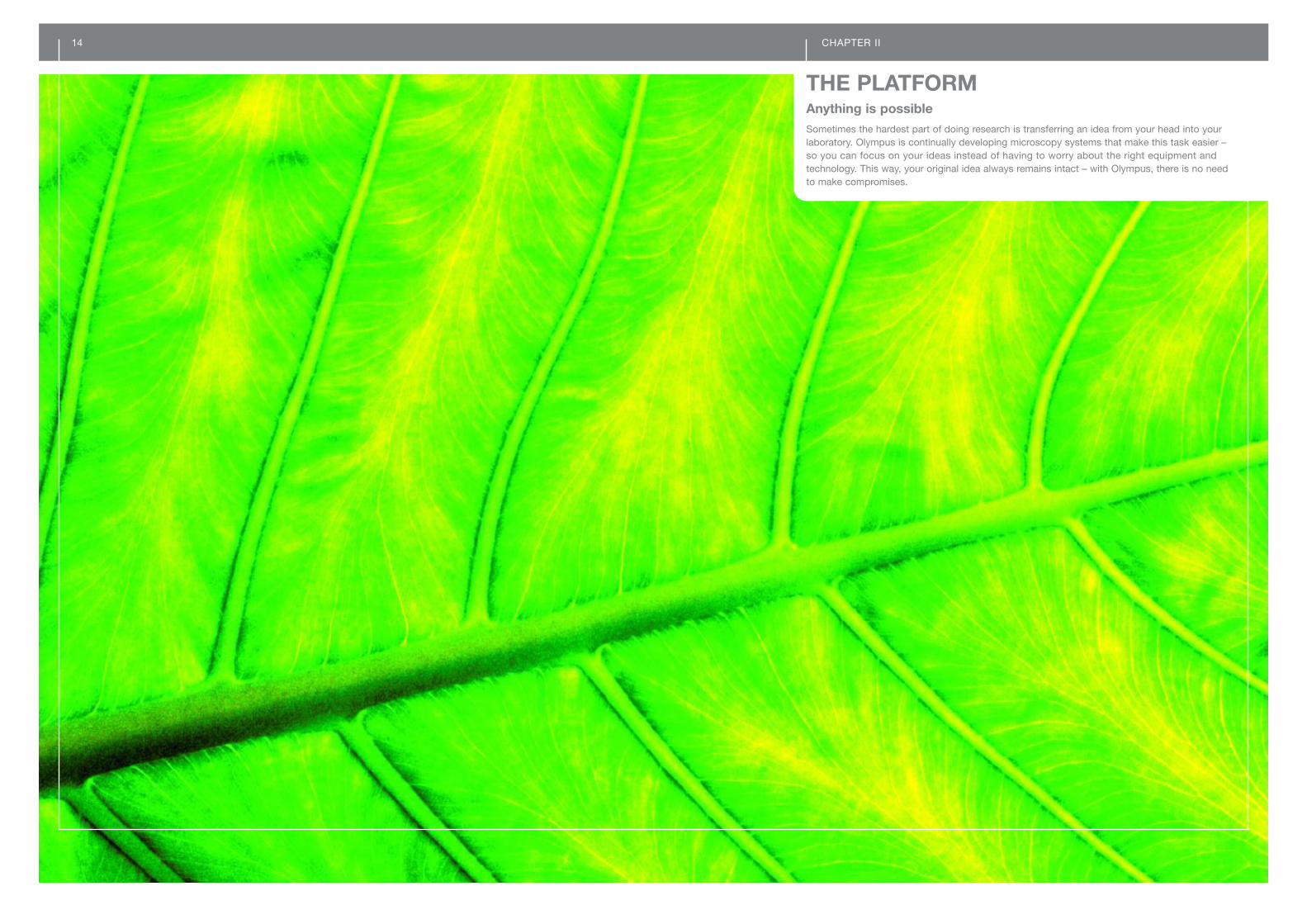
THE PLATFORMAnything is possibleSometimes the hardest part of doing research is transferring an idea from your head into yourlaboratory. Olympus is continually developing microscopy systems that make this task easier – so you can focus on your ideas instead of having to worry about the right equipment and technology. This way, your original idea always remains intact – with Olympus, there is no need to make compromises.

16 THE PLATFORM
B IX71/IX81Light path
C Dual-port C-mountOne port – two cameras
A
D Double lamp house adaptorEasy switching between xenon and mercury
E Nosepiece stageUltimate focus stability
BE INSPIRED BY THE POSSIBILITIESMultidimensional analysis requires a flexible microscope system. But how canyou combine laser illumination with standard fluorescence illumination? Or usethree different detectors at the same time? The unique multi-port concept of theIX71/IX81 microscopes gives an innovative answer to these questions.
Multi-port conceptNo other microscope system offers you greater flexibility: the IX71/IX81 frames
are equipped with input/output ports for a wide variety of light sources and detectors.This unique Olympus design permits more than ten port configurations for eachmicroscope. Our technical sales representatives look forward to advising you on thebest port combination to meet your needs.
A
Thermal stability after switching on the light source
Primary image or parallel light access with the right side portThe right side port offers a variety of possibilities for usage. Together with the includedtelan lens, the port is a fully functional camera port suitable for mounting a secondcamera onto the frame. It also provides parallel light access, for example, enables thecoupling of a light source, e.g. a laser for uncaging experiments, or the placement ofa detector, e.g. a spectrometer, in the light path. The right side port features a fieldnumber of 16 and a 1x C-mount.
Double flexibility
To further increase flexibility and application support, the dual-port C-mountadaptor facilitates the attachment of two cameras. The adaptor has a filter holderthat ensures easy adaptation for different wavelength ranges. The double lamphousing adaptor offers the same flexibility for illumination.
Saving space for your ideas The IX2 L-shaped illuminator is a great space-saving device. The ultra compact
design of both illuminator and microscope offers increased flexibility for configuringperipheral devices such as the lamp house, micromanipulators, incubator and camera.Moreover, the L-shaped illuminator improves ergonomics, providing easy access toburner centration and aperture/field stop.
Everything under controlThe investigation of fast processes in the nano scale, long exposure times to catch theweakest emissions, or time-lapse observations over several hours or days all needa stable platform ensuring that, once defined, target positions will be perfectly keptdespite vibrations and environmental changes.
Thermal stabilitySlim and rigid appear to be mutually exclusive requirements for a light microscope.Olympus successfully met this challenge by using its innovative product engineering in combination with state-of-the-art materials. The result is a compact microscopewith maximum rigidity and minimum thermal expansion – a stable platform for all applications, including the most demanding time-lapse observations.
A
DC
Focus stabilityThe IX2-NPS nosepiece stage (patent pending) provides ultimate focus stability –
even under different environmental conditions. This stability extends to all applications,including TIRF, where even the smallest of changes in the Z-position can significantlyinterfere with obtaining experimental results.
E

D
18 THE PLATFORM
A IX71Frontal control panel
E Fast observation filter wheelWith C-mount adaptor
F Light controlAutomatic light intensity setting
C Motorised nosepieceErgonomy and security
B IX81Programmable frame keys
FROM MANUAL TO FULLY AUTOMATEDMotorisation is essential for full process automation. Consequently, all majormicroscope functions can be motorised – including focus, illumination, objectivechange and optical path selection. In addition, the Olympus modular motorisationallows you to choose a level of automation that perfectly matches your require-ments. The innovative experiment manager of and imaging systemsintegrates the motorised microscope functions with image acquisition controland other automated options, facilitating the automation of both simple and com-plex experimental workflows.
IX71 – nearly autoThe optical specifications of the manual IX71 microscope and the fully automated
IX81 are identical. The main difference is that the IX71 has a lower level of motorisation(optional). Motorised filter wheels, filter cube turret, condenser, light intensity control,shutters and X/Y stage offer a high level of flexibility for a broad range of automatedtasks. Individual user settings can be assigned to the keys of the remote handset.
IX81 – the fully motorised platformHigh-precision Z-motor, motorised six-position nosepiece, light path changer,
intensity control and user-programmable keys for the frame and hand switch are integral parts of the automated IX81. All motorised units are driven by the IX2-UCBcontrol box, which connects to a PC to access software control provided by theRS232 interface.
High-precision internal Z-motor 10 nm step size and 1 µm reproducibility independent of movement direction. Fineand coarse movement with 3 mm/sec. maximum speed.
Motorised nosepiece The integrated motorised six-position nosepiece with a computer-controlled
automated objective escape function maximises security for both specimen andobjective.
Light path changer Switch between binocular observation and camera detection with 8% or 100% of the light being directed to the camera port, depending on the frame configuration.LED indication enables you to readily view the light path status. Motorised bottomport with mounted camera allows convenient control of image acquisition.
Programmable frame keysAll keys for the frame as well as the keys for the handset remote control are user-programmable. Depending on individual preferences and needs, the keys can beprogrammed and used for any desired mircoscope function.
The environment might change – but never the focusA laser-based fast Z-drift compensator (ZDC) system enables long-term observa-
tions, screening of multiwell plates and other specimen containers – allowing a fastadjustment time and specimen photoprotection. The IR laser focused to a small spot(1 to 3 µm) on the glass surface of e.g. a slide is reflected. The spot image is analysedby a photosensor and the focus is automatically adjusted. Individual thresholds fordifferent slide thicknesses allow focusing on the plane of interest.
D
C
B
A
E Filter wheelsFor excitation and emission
G Motorised stagePrecise and convenient
A. Focus position
B. Closer thanfocus position
C. Further away thanfocus position
Objective
Reflection plane
Lightshieldingplate
Split detector
Motorised modules for maximum flexibility
All motorised modules are designed to fit the IX71/IX81 frames perfectly. The easyattachment to the frame offers the option to add new motorised modules to tune thesystem to the latest experiment requirements even later on.
PIFOC nosepieceThis unique piezoelectric nanofocusing device moves the entire objectiverevolver with 10 nm precision and a switching time of 30 ms (for steps smaller than 10 µm) over a total range of 80 µm. What’s more, unlike piezosteppers for single objectives, it is compatible with DIC optics. The PIFOCnosepiece, as an alternative or addition to the motorised Z-drive, is an idealoption for fast and precise Z-stack and 3-D time-lapse acquisition.
Filter wheelsDifferent six-position filter wheels are available for excitation and observation,holding filters of 25 mm or 32 mm in diameter. Switching time between neigh-bouring positions: 0.6 sec. A special fast observation filter wheel enablesrapid wavelength switches in multi-wavelength observations, for use with
and imaging stations, eight positions for filters, 25 mm in diameter,replaces C-mount adaptor; minimum switching time 58 ms.
Fluorescence filter cube turretsSix-position turrets with quick lock mechanism for fast filter cube exchange.A fast filter cube turret (< 300 ms switching time) is available for live cellexperiments in and .
ShutterVibration-free shutter to be mounted in the reflected or transmitted light path.Two shutters can be mounted in parallel.
CondenserSix-position condenser. Designed for brightfield, phase contrast and DICobservations. Switching time between neighbouring positions: 0.6 sec.
Lamp houseAlways the right brightness – automatically. While changing magnification or contrast method, utilising the motorised nosepiece and condenser, the light is automatically set to the desired intensity (PC control).
X/Y stageHigh-precision stages controlled via joystick and/or PC. Different frameinserts for various culture vessels are available.
G
F
E
Laser lightsource(class 1)

20 CHAPTER III
THE EXPERIMENTWhat you get is what you need: reliable resultsWhen it comes to studying the processes in living cells, seeing is believing. As your partner,Olympus makes sure you always get the best images possible. Whatever the goal of your experiment is, our live cell imaging systems not only help you to reach it – they assist you in leaving limitations behind and opening up new horizons. Explore what has never been exploredbefore – with Olympus at your side.

22 THE EXPERIMENT
Multidimensional image formats
High-resolution overview images are easily created with multi-imagealignment (MIA)
Microscope control
EASY ACQUISITION AND ANALYSISWith the imaging software packages of the family, you can concentrate onyour work without worrying about all the overwhelming possibilities of modernmicroscopes and cameras. All technical functions are translated into an intuitiveuser interface which caters for the working process during life science experi-ments. Once an image has been stored, will allow you to discover muchmore detail than you would have imagined before.
Control made easyIntuitive software allows easy control of all microscope and camera functions –
putting you in total control of all experimental parameters while guaranteeing theacquisition of reproducible, high-quality results.
Multidimensional data handlingAll series of acquisitions are saved together with all experimental parameters in
a multidimensional image format. This means that a single image file can consist ofmultiple images taken at e.g. different Z-positions and at different times or acquiredwith different wavelengths. The images can be processed further and visualised asdesired. A specially designed navigation tool is implemented.
The basic image acquisition and documentation toolsand
More than a microscope and camera control, is the solid, comprehensiveentry level to imaging systems for biological microscopy. The basic documentationpackage encompasses all functions of , with the addition of archiving in a structured database, standard measurement functions, as well as a convenientstandards-compliant report generation tool. Acquisition and processing features,such as a panorama function for multi-image alignment (MIA) for creating overviewimages in high resolution, are also available.
C
B
A
Rapid image acquisition with provides documentation and control, representing a comprehensive system
for image acquisition, archiving and documentation in the biological field. offersmore advanced functions, allowing rapid image acquisition, direct Web transfer,numerous processing operations and the full capacity of interactive measurements.
Fluorescence image acquisition and processing with is the optimal system for fluorescence applications. It enables documentation,
visualisation, processing and analysis of multi-channel fluorescence images (mFIP),such as those created using GFP variants. For enhanced spectral resolution of multi-channel images, a spectral unmixing tool is incorporated, as are tools for fluorescenceimage evaluation, such as co-localisation. also features Z-sectioning and imageacquisition at different focal positions to visualise specimens in 3D.
Get more withThe outstanding package offers all the benefits of and more. Dynamic
processes can be registered by the image sequence processing module (ISP), auto-mating the complete process from microscope control through to data archiving.Advanced tools for haze reduction of 3-D images, time-lapse photography and quan-titative image analysis enable the user to carry out and analyse a range of complexand highly sophisticated experiments.
Discover your potentialExperiments often need to be repeated to generate statistically relevant data – this
is why automation considerably increases reliability. Extend your system with a motorised stage and optional software modules for automated stage navigation and screening of culture vessels, multiwell plates and slides to automatically repeatexperiments and collect data conveniently and reproducibly. Using the particle detec-tion module, thousands of objects can be analysed within an image in seconds.Objects can be automatically tracked in time-lapse series with the motion analysismodule TrackIt and 3-D image stacks can be deconvoluted for high-resolution 3-Dreconstruction.
G
F
E
D
1* Courtesy of Dr Jeremy C. Simpson, EMBL, Heidelberg, Germany.
2* Courtesy of Per Homfeldt, Martin Gullberg Laboratory, Molecular Biology Dept., Umea, Sweden.
3* Courtesy of Dr Uwe Walldorf, Homburg/Saar, Germany.
Time-lapse observation of mitosis. Red:DNA with propidium iodide; green: micro-tubules with anti-alpha-tubulin antibodyconjugated with Alexa-488*2
Image stack of Drosophila. Dual-labelledwith Cy3 and FITC; left: original image;right: after deconvolution*3
Acquisition and processing of multicolourfluorescence images*1
A
B
C
E
F
G
Graphical user interface
D

24 THE EXPERIMENT
A
Pseudo-coloured CFP/YFP double- emission image acquired with Dual-ViewTM
Micro-Imager *1
F
NEW INSIGHTS INTO CELLULAR PROCESSESMicroscopy in life science has progressed significantly: from static morphologicalobservations to the characterisation of the 3-D architecture of cellular structuresand the real-time investigation of dynamic molecular processes in living cells.Moreover, new fluorescence methods such as TIRF and fluorescence resonanceenergy transfer (FRET) microscopy or GFP labelling are providing exciting insightsinto the complex dynamic processes in living organisms.
fast and flexible, multi-purpose station
is specifically designed to meet the experimental requirements for multicolourfluorescence time-lapse image acquisition. A key feature is the all-in-one MT10 illumination system for wavelength switch, attenuation and shuttering. The systemcoordinator, a control board solely for controlling hardware, increases imaging speedconsiderably in comparison with systems driven by software alone. ’s intuitivelystructured Experiment Manager is a user-friendly graphical drag and drop interface.This makes setting up even the most complex experiments exceptionally quick andeasy.
real-time imaging stationheads our live cell imaging system family: a fully integrated, modular system
for a broad range of life science experiments – including time-lapse imaging, multidi-mensional imaging, ratio imaging, FRET and TIRF microscopy and spectral unmixing.We listen to the demands of our customers and their needs, constantly enhancingthe system family to match new and emerging applications.
MT20 illumination systemThis multifunctional, all-in-one illumination system for fast wavelength switch
and attenuation is designed to meet the requirements for fast multicolour real-timeacquisition by highly sensitive cameras. Two types of light sources are available:high-stability 150 W Xe or Hg/Xe mixed-gas arc burners. The device provideswavelength switches within 65 ms and shutter times of 1ms. The integrated lightattenuator offers 14 grades of illumination intensity between 1% and 100%. Allmodules operate in parallel to ensure optimised light management. A uniquemechanism (patent pending) facilitates the fast and easy exchange of excitation filters – without requiring any tools.
and imaging softwareThis powerful modular software platform features user-definable database storage
to archive multidimensional data sets. It also includes a comprehensive collection oftools for acquisition, documentation, processing and analysis, and fully supportssophisticated routines for time sequence analysis such as ratio and deltaF/F imagecalculations. The report generator enables automated reporting with layout control oftext, graphics and images.
Hyper precision controlAn additional independent plug-in CPU board ensures interruption-free data
capture by the imaging computer during an experiment. All integrated deviceswork in parallel and are synchronised with sub-millisecond precision for optimisedtiming and minimal photobleaching.
D
C
B
A
B
C
D
E
CFP/YFP channel overlay and pseudo-coloured ratio image *1
F
Cells with GFP-H2B histone protein andYFP-tubulin. Left: original image, acquiredwith an imaging station, filters: GFP andYFP exciters, YFP dichroic mirror andemitter; right: after spectral unmixing *2
F
Application solutionsThe full control of illumination systems, microscope, and signal detection systemsenables Olympus to offer application solutions that transform complex routines intoeasy-to-handle tools.
FRET and spectral unmixingSpectral unmixing is a unique tool of the imaging software for colour
resolution enhancement. It separates the signals of fluorochromes with pronouncedspectral overlap that could otherwise not be distinguished, providing sharply contrasted images for less common fluorescent protein combinations such asGFP/YFP. In FRET studies, the occurrence of molecular interactions becomes moreimmediately obvious even before quantitative analysis. Furthermore, co-localisationstudies become more reliable because bleed-through artefacts are avoided.
TIRFTotal internal reflection fluorescence microscopy enables the investigation of surfaceswith extremely high Z-resolution and without interfering background. Illuminationcombiners for up to three lasers and the MT20 illumination system allow fast switching (1 ms) between the different light sources and thus combined TIRF andwidefield applications.
Ratio imagingThe fluorescence behaviour of many dyes is influenced by the concentration of certain ions such as calcium (FURA-2) or the pH value (BCECF). The detection,quantification and analysis of changes in fluorescence intensity allow the indirectstudy of certain cellular processes such as signal transduction.
*1 HeLa cells labelled with CFP/YFP chameleon, triggered with histamine.Courtesy of Dr Hideaki Mizuno and Dr Atsushi Miyawaki, Brain Science Institute, RIKEN, Wako, Saitama, Japan.
* 2 Courtesy of Dr Paulo Magalhaes and Prof. Dr Tullio Pozzan, University of Padua, Italy.
F
CFP/YFP channel overlay and pseudo-coloured ratio imaging
Experiment ManagerA unique graphical interface and intuitive drag and drop programming make the
design and execution of experiments very easy. The complete experimental design isreadily visible at a glance and is automatically stored together with the captured datain the archive database.
E

26 THE EXPERIMENT
A TIRF objectivesUnprecedented numerical apertures – outstanding visible performance
B TIRF illumination combiner
Conventional fluorescence observation
Total internal reflection fluorescenceobservation; courtesy of M. Faretta, Eur.Inst. Oncology (IEO-IFOM), Milan, Italy
EXCITING THE PERIPHERY WITHVERSATILE TIRFM PLATFORMSTotal Internal Reflection Microscopy (TIRFM) is an elegant optical technique forextremely high-resolution cell surface imaging without the disturbing out-of-focushaze characteristic of widefield fluorescence microscopy. In 1998 we introduceda TIRFM illuminator and an objective with sufficiently high numerical aperture as the first commercial solution for objective-based TIRFM. Now, based on ourlong-standing experience and expertise, we are offering a range of TIRFM object-ives, single- and multi-port illuminators, as well as a choice of lasers to enablelife science researchers to exploit the potential of TIRFM for their application.
Objective-based TIRF microscopyThe basic principle of TIRFM is that a laser beam is focused to the periphery of theback focal plane of the objective. If the objective numerical aperture (NA) is largerthan 1.38, the beam exits the objective at a very shallow angle and is totally reflectedat the glass interface of the sample. The reflected light causes a near-field effect that selectively excites fluorochromes that are within 100 and 700 nm of the surface,yielding images with very high S/N ratios. High-performance objectives and innovativeillumination systems give Olympus a leading position in this cutting-edge field.
and fully integrated turnkey solutions for TIRFMOlympus offers modular illumination combiners to couple up to three laser beamsand an MT10/MT20 white-light source to the microscopes, allowing convenient adjust-ment of the laser beam incident angle. Providing all laser safety features a series of specially designed laser systems is the optimal TIRFM excitation source for theentire range of fluorophores. By integrating the TIRFM equipment with full softwaresupport into the versatile and imaging stations, Olympus offers easy-to-use, turnkey solutions to enable researchers to exploit the entire potential of totalinternal reflection microscopy.
A brilliant solution: the first white-light TIRFM system on the marketAchieving a breakthrough in microscopy, Olympus developed the world’s first fluor-escence excitation light from a standard white-light source (xenon or mercury arclamp) for TIRF. We offer TIRF microscopy at a moderate cost with high flexibility influorochrome selection.
TIRFM specialistsThe new 60x plan apochromat objective with an NA of 1.45 provides optimum
imaging through a temperature correction collar, making it highly responsive to chang-ing environmental conditions.
Our 100x apochromat objective combines an unprecedented NA of 1.65 and a highS/N ratio, guaranteeing an outstanding visible performance. It allows extreme TIRangles and adjustments over a wide angle range. Thus, the depth of excitation canbe lowered down to yield a Z-section as narrow as around 50 nm at short wavelengths.
With its extraordinarily high magnification, the 150x universal apochromat (NA 1.45) is the only TIRFM objective of its kind on the market and was specially developed forsingle-molecule applications. It features a compensation collar for temperature andcover glass thickness.
The 100x plan apochromat TIRFM objective with 1.45 NA guarantees high-resolutionTIRFM images and completes the series of dedicated TIRFM objectives.
BA
C
Time series of Kaede-expressed HeLacells. Photo conversion done via local405 nm laser illumination with the SIMscanner. Confocal image observationdocumented every three seconds with488 nm/543 nm laser excitation and dual-channel (red/green) detection.*2
Montage image of a Brainbow transgenicmouse brain stem, showing large caliberaxons of the auditory pathway. In Brain-bow mice, neurons randomly choosecombinations of red, yellow and cyanfluorescent proteins, so that they eachglow a particular color. This provides away to distinguish neighboring neuronsand visualize brain circuits.*1
OPENING NEW FRONTIERS –FV1000/FV1000MPE LSM-SYSTEMS
The FluoView FV1000 confocal and FV1000MPE multiphoton systems deliverall the key performance functions required from a confocal laser scanningmicroscope – plus the unique dual scanner, allowing stimulation and observationat the same time. It minimises specimen damage during high-speed imaging of living organisms and accurately captures a full range of related information.With high sensitivity, high speed and high precision, the FV1000 is ready to meetthe demands of all your applications.
C
Don’t miss anything – reliable capture of reactionsThe FV1000 with the unique SIM (SIMultaneous) scanner incorporates two inde-
pendent, fully synchronised laser scanners in a single compact design for simultaneouslaser light stimulation and high-resolution confocal observation. This unique scanningcapability ensures that confocal image observation is no longer interrupted during laserlight stimulation, e.g. photoactivation, or laser manipulation, e.g. photobleaching.
BenefitYou will not miss rapid fluorescence changes that occur during or immediately following laser stimulation or manipulation.
Applications The unique SIM concept offers distinct advantages for sophisticated applications –including FRAP (fluorescence recovery after photobleaching), FLIP (fluorescence loss in photobleaching), TIRFM (total internal reflection fluorescence microscopy), FLIM(fluorescence lifetime imaging), photoactivation, photoconversion, uncaging, laserablation and many more.
D
* 1 Image courtesy of Jean Livet, Harvard University, Cambridge, MA, USA.
* 2 Data courtesy of Ms Ryoko Ando, Dr Atsushi Miyawaki, Brain Science Institute, RIKEN, Wako,Saitama, Japan.
Coupling up to three laser lines and an MT10/MT20 widefield illumin-ation system
28 THE EXPERIMENT
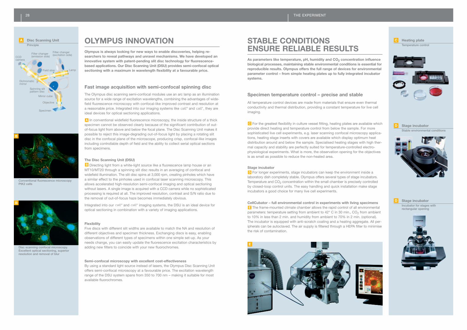
OLYMPUS INNOVATIONOlympus is always looking for new ways to enable discoveries, helping re-searchers to reveal pathways and unravel mechanisms. We have developed aninnovative system with patent-pending slit disc technology for fluorescence-based applications. Our Disc Scanning Unit (DSU) provides semi-confocal opticalsectioning with a maximum in wavelength flexibility at a favourable price.
STABLE CONDITIONS ENSURE RELIABLE RESULTSAs parameters like temperature, pH, humidity and CO2 concentration influencebiological processes, maintaining stable environmental conditions is essential forreproducible results. Olympus offers the full range of devices for environmentalparameter control – from simple heating plates up to fully integrated incubatorsystems.
Specimen temperature control – precise and stable
All temperature control devices are made from materials that ensure even thermalconductivity and thermal distribution, providing a constant temperature for live cellimaging.
For the greatest flexibility in culture vessel fitting, heating plates are available whichprovide direct heating and temperature control from below the sample. For moresophisticated live cell experiments, e.g. laser scanning confocal microscopy applica-tions, heating stage inserts with covers are available which display optimum heat distribution around and below the sample. Specialised heating stages with high ther-mal capacity and stability are perfectly suited for temperature-controlled electro-physiological experiments. What is more, the observation opening for the objectivesis as small as possible to reduce the non-heated area.
Stage incubator For longer experiments, stage incubators can keep the environment inside a
laboratory dish completely stable. Olympus offers several types of stage incubators.Temperature and CO2 concentration within the small chamber is precisely controlledby closed-loop control units. The easy handling and quick installation make stageincubators a good choice for many live cell experiments.
CellCubator – full environmental control in experiments with living specimensThe frame-mounted climate chamber allows the rapid control of all environmental
parameters: temperature setting from ambient to 42° C in 30 min., CO2 from ambientto 10% in less than 2 min. and humidity from ambient to 70% in 2 min. (optional).The incubator is equipped with anti-scratch coating and a heating aggregate. All per-ipherals can be autoclaved. The air supply is filtered through a HEPA filter to minimisethe risk of contamination.
E
D
C
Fast image acquisition with semi-confocal spinning discThe Olympus disc scanning semi-confocal modules use an arc lamp as an illuminationsource for a wide range of excitation wavelengths, combining the advantages of wide-field fluorescence microscopy with confocal-like improved contrast and resolution ata reasonable price. Integrated into our imaging systems like and , they areideal devices for optical sectioning applications.
In conventional widefield fluorescence microscopy, the inside structure of a thickspecimen cannot be observed clearly because of the significant contribution of out-of-focus light from above and below the focal plane. The Disc Scanning Unit makes itpossible to reject this image-degrading out-of-focus light by placing a rotating slitdisc in the confocal plane of the microscope, producing crisp, confocal-like imagesincluding controllable depth of field and the ability to collect serial optical sectionsfrom specimens.
The Disc Scanning Unit (DSU)
Directing light from a white-light source like a fluorescence lamp house or anMT10/MT20 through a spinning slit disc results in an averaging of confocal andwidefield illumination. The slit disc spins at 3,000 rpm, creating pinholes which have a similar effect to the pinholes used in confocal laser scanning microscopy. Thisallows accelerated high-resolution semi-confocal imaging and optical sectioning without lasers. A single image is acquired with a CCD camera while no sophisticatedprocessing is required at all. The improved resolution, contrast and S/N ratio due tothe removal of out-of-focus haze becomes immediately obvious.
Integrated into our and imaging systems, the DSU is an ideal device foroptical sectioning in combination with a variety of imaging applications.
FlexibilityFive discs with different slit widths are available to match the NA and resolution ofdifferent objectives and specimen thickness. Exchanging discs is easy, enablingobservations of different types of specimens within one simple set-up. As yourneeds change, you can easily update the fluorescence excitation characteristics byadding new filters to coincide with your new fluorochromes.
Semi-confocal microscopy with excellent cost-effectiveness By using a standard light source instead of lasers, the Olympus Disc Scanning Unitoffers semi-confocal microscopy at a favourable price. The excitation wavelengthrange of the DSU system spans from 350 to 700 nm – making it suitable for mostavailable fluorochromes.
B
A
A Disc Scanning Unit Principle
CCD camera
Filter changer(emission side)
Filter changer(excitation side)
Lamp
Specimen
Objective
Mirror cube
Spinning slit pattern disc
Dichromaticmirror
Field stop
Conventional fluorescence microscopyPtK2 cells
Disc scanning confocal microscopy.Excellent optical sectioning, superiorresolution and removal of blur
B
B
E
C Heating plateTemperature control
D Stage incubatorStable environmental conditions
D Stage incubatorIncubator for stages with rectangular opening
30 THE EXPERIMENT
MERGING MICROSCOPY WITH MOLECULAR BIOLOGY AND CYTOMETRYCombining the knowledge of different research fields often leads to the break-through that makes real discoveries possible. In the same way, Olympus hascreated systems that merge microscopy with other technologies, allowing newinsights into the mechanisms of life.
Fully automated cellular high-content analysisCellular high-content screening has been an established method in pharmaceuticalcompound screening and drug development for many years. Due to the lack of flexibleand economical technologies, microplate readers or flow cytometers have mostly beenused for cell population assays in basic and applied research. To combine the power-ful possibilities of fluorescent population assays with the outstanding spatial resolutionof microscopy, Olympus has developed an extremely flexible and reliable screeningsystem for a wide range of cellular applications in functional genomics, cancer re-search, neuroscience and drug development.
Automated, high-throughput image acquisition with unrivalled flexibilityIn modern assay development and cellular screening, complex biological reactionshave to be monitored in whole cell populations with high resolution and speed. The system offers a user-friendly image acquisition portal for fast and reliable raw data collection enabling almost every cellular assay type. An unlimited numberof different dye channels, fast and long-term time loop experiments, Z-stacks andparallel differential interference contrast or phase contrast modes are only a few options. A highly stabilised illumination system in combination with fast hardwareand software autofocus routines guarantees the maximum accuracy in quantitativeanalyses. The integration of robotic microplate handlers and dedicated environmentcontrol systems expand ’s possibilities in throughput and live cell applications.
Accurate multi-step cell segmentation and object analyserExtracting reliable quantitative and morphological information from complex cell
culture models is the most challenging step in high-throughput, high-content analysis.With the analysis module, Olympus has succeeded in combining a user-friendlyinterface with the highest level of flexibility. Most standard assay types are predefinedand can be adjusted to new conditions with minimal effort. For special applications,an open graphical interface enables the quick and flexible integration of further objectdetection and object analysing modules from huge libraries.
Cytometry-like data display is combined with image-data linkingAs in flow cytometry, the extracted data are displayed in scatter plots and histo-
grams. Selected data sets can be further investigated using gating procedures, whereonly data fitting a specific criterion are selected. A series of ‘gates’ can thereforebe used as a hierarchical filter to select data points that fall within precise boundaries.Images, objects and sub-objects are linked directly to any data related to them,meaning that by clicking on a data point or image, the reciprocal information is high-lighted alongside. Selected image sets can also be displayed in a gallery window foreasier comparison. In combination with the accurate quantification of fluorescentsignals, the perfectly interconnects the cytometry of whole cell populationswith the high-resolution data of modern microscopy.
C
B
Non-contact laser microdissectionThe CellCut system is the most precise and versatile laser manipulation system,
offering a broad range of applications: laser microdissection, laser-induced micro-injection, cell fusion and “contamination-free” removal of target cells and subcellularstructures from paraffin or cryo sections, smears, cytospins and cell cultures. Preciselaser ablation as a single shot can make holes in cell membranes, organelles or cellwalls, enabling microinjection of drugs or genetic materials. Cell clusters or singlecells can be isolated and even single chromosomes or chromosome parts can be dissected. A cut target element can be easily and quickly removed from the specimento a microfuge tube cap using a unique sandwich preparation in conjunction withCapLift technology. This provides non-contaminating capture without any contact to the target, ready for downstream analysis.
Precise, quick and intuitive
Laser microdissection combined with extraction of target structures offers a preciseand gentle process designed to protect the extracted components. Based on a solid-state bUVa laser with picosecond pulses, cutting of target areas is performed veryquickly and without any negative impact on subsequent DNA and RNA extraction orprotein analysis. The versatile system controlled by system software with intuitivegraphical user interfaces and a unique touch screen is suitable for all specimens, inclu-ding tissue slides, stained specimens, smears, cytospins and living cells.
Fully configurable to meet your needsThe CellCut systems are available in different configurations based on the invertedmicroscope frames IX71 and IX81 – all fully tailored to meet your needs. Easy tohandle, they include a highly precise motorised stage, outstanding UIS2 optics andcomprehensive control software for the system operation and specimen screeningprocess. Fluorescence illumination for simultaneous observation of fluorescence-labelled specimen structures and laser microdissection is possible without any limita-tions due to a dual-level coupling of the cutting laser. Further options are also available,such as MultiSlide, MultiCap and CellExplorer.
A
Microdissection of living cells
Target material from colon tissues withextracted targets
A CellCutLaser microdissection system
Genome wide screen on cell arrays.Detection and separation of labels. BlueDAPI-stained nuclei are circled in cyan;green CFP-tagged VSVG protein in thegolgi is circled in red; red Cy-5 VSVG anti-bodies on the cell surface are circled inblue *
* Courtesy of Dr Rainer Pepperkok,EMBL, Heidelberg, Germany.
Gating, classification and data evaluation *
B
C

32 THE EXPERIMENT
DIGITAL REVOLUTIONIf your aim is to record and analyse microscopy samples on the basis of excellentimages, you need high-resolution digital cameras with sophisticated dynamics,sensitivity and resolution. Olympus supplies both black-and-white cameras forfluorescence and colour cameras for all other applications. All models can befully controlled by the family of imaging software.
XC30 – perfect colour matchThe Olympus XC30 offers a 3.2-megapixel resolution and fast frame rates with
the added benefit of 2x and 3x colour binning. The versatile CCD camera is idealfor brightfield applications and can also be used for high-intensity fluorescence. The Peltier cooling mechanism maintains the CCD chip at a constant 10 ˚C (at 25 ˚Cambient temperature) to guarantee perfect colour images that are rich in contrast,with excellent colour fidelity and extremely low background noise.
The CCD chip offers 14 bits per colour channel and can be switched between variousframe rates for easier use. As well as two binning modes with frame rates up to 35 fps,the XC30 also offers a partial readout mode where a segment of the entire field of viewcan be defined by the user and only this image segment is read out by the camera. This enables faster focusing on and imaging of the features of interest within the field of view.
The unique Olympus True Colour (OTC) system ensures perfect colour fidelity fromthe specimen to the monitor.
DP72 – one for allThe Olympus DP72 digital camera offers unmatched versatility for microscopists,
combining a 12.8-megapixel resolution with the highest sensitivity, very fast imageacquisition rates and colour match performance.
The Olympus DP72 digital camera uses the latest interface technology and a high-sensitivity CCD to provide the fastest image acquisition rates of any comparable camera.In live mode, full-frame images (1,360 x 1,024 pixels) can be viewed at 15 frames persecond, making it ideal for viewing, discussion and documentation. A real-time frame-averaging function minimises noise and optimises the display, resulting in low-noiselive images. Still images, at pinpoint resolutions of up to 12.8 million pixels, can beobtained in less than 2.5 seconds with Olympus’ unique piezo-shift technology.
With superior hardware-driven image processing capabilities, the DP72 is especiallysuitable for applications such as pathology, where faithful colour reproduction is essen-tial. In applications such as fluorescence microscopy, where a specimen is wholly orpartially dark, the DP72 records black-and-white images with exceptional sensitivity.The custom monochrome mode allows the user to optimise the recording sensitivityaccording to the fluorescent dye in the specimen.
XM10 – perfect black and whiteThe XM10 offers all of the properties required to provide dependable fluorescence
microscopy images: high resolution, extremely high sensitivity, a cooled 1.4-mega-pixel CCD chip, variable resolutions and an optional external trigger function.
The XM10 is extremely sensitive, low in electronic noise and supports long integrationtimes of up to 160 seconds. The chip has a pixel size of 6.45 µm x 6.45 µm, which,in combination with the camera cooling, ensures that the XM10 is ideal for recordingeven the faintest fluorescence signals in your specimen. The XM10 uses a CCD chipcooled to 10 ˚C (at 25 ˚C ambient temperature) with a 14-bit dynamic range and 15fps at full resolution. It offers three binning modes, resulting in increased sensitivityand excellent frame rates in live mode.
C
B
A
OBJECTIVESUsing the latest technologies and materials, the new UIS2 optics were designedby Olympus experts to offer contrast and image resolution without compromises.This enables you to capture the most intricate of details from your experiments –making your work more efficient and your results more reliable.
UPLSAPO high fidelity for advancedfluorescence applications
High-performance universal plan wide-rangeapochromatic-corrected objectives with unsurpassednumerical aperture values deliver the best resolution,contrast and field flatness for any microscope technique. In combination with the lowest auto-fluorescence of all objective series, they are the bestchoice for advanced fluorescence applications.
LUCPLFLN objectives for tissuesand cell cultures
Semi-apochromatic objectives, designed for tissue culture observations in flasks and dishes.Excellent contrast and resolution in brightfield,phase contrast, DIC and fluorescence observations.
UPLFLN universal objectivesAffordable semi-apochromatic universal objectives
that deliver superb resolution, contrast and imageflatness with any microscopic technique.
ORC objectivesOlympus relief contrast (ORC) objectives
were designed for observing living cells includingoocytes in glass and plastic vessels, and havefound widespread use in micromanipulation work.
TIRF objectivesWith their extremely high numerical apertures of
1.45, 1.49 or even 1.65, these objectives have beendeveloped especially for total internal reflection fluor-escence (TIRF) microscopy, which enables high-resolution visualisation of cell surface signals on thenanometer scale.
UAPO/340 objectivesHigh transmission in the near-UV wavelength
(340 nm) makes these objectives perfect for applications such as ratio imaging or measurement of intracellular pH with UV-excitable fluorochromes.
A
B
C
D
E
F
Cover glass Objective NA WD (mm) correction (mm)
A UPLSAPO 4x 0.16 13 –
UPLSAPO 10x 0.4 3.1 0.17
UPLAPO 10xO 0.4 0.24 0.17
UPLAPO 10xW 0.4 0.43 0.17
UPLSAPO 20x 0.75 0.6 0.17
UPLSAPO 20xO 0.85 0.17 –
UPLSAPO 40x 0.9 0.18 0.11– 0.23
UPLAPO 40xOI 0.5 –1 0.12 –
UPLSAPO 60xO 1.35 0.15 0.17
PLAPON 60xO 1.42 0.15 0.17
UPLSAPO 60xW 1.2 0.28 0.15 – 0.2
UPLSAPO 100xOI 1.4 0.13 0.17
B CPLN 10xPH 0.25 10 1
LCACHN 20xPH 0.4 3.2 1
LCACHN 40xPH 0.55 2.2 1
CPLFLN 10xPH 0.3 9.5 1
LUCPFLN 20x 0.45 6.6–7.8 0–2
LUCPFLN 20xPH 0.45 6.6–7.8 0–2
LUCPFLN 40x 0.6 2.7–4 0–2
LUCPFLN 40xPH 0.6 3– 4.2 0–2
LUCPFLN 60x 0.7 1.5–2.2 0.1–1.3
LUCPFLN 60xPH 0.7 1.5–2.2 0.1–1.3
C UPLFLN 4x 0.13 17 –
UPLFLN 4xPH 0.13 17 –
UPLFLN 10x 0.3 10 –
UPLFLN 10xPH 0.3 10 –
UPLFLN 20x 0.5 2.1 0.17
UPLFLN 20xPH 0.5 2.1 0.17
UPLFLN 40x 0.75 0.51 0.17
UPLFLN 40xO 1.3 0.2 0.17
UPLFLN 40xPH 0.75 0.51 0.17
UPLFLN 60xO 0.9 0.2 0.17
UPLFLN 60xOI 0.65 –1.25 0.12 0.17
UPLFLN 60xOIPH 0.65 –1.25 0.12 0.17
UPLFLN 100xO 1.3 0.2 0.17
UPLFLN 100xOI 0.6 –1.3 0.2 0.17
UPLFLN 100xOPH 1.3 0.2 0.17
D CPLFLN 10xRC 0.3 9 1.5
CPLN 10xRC 0.25 9.7 1.5
LCPLFLN 20xRC 0.45 6.6–7.8 0–2
LUCPLFLN 40xRC 0.6 3–4.2 0–2
LCACHN 20xRC 0.4 2.8 1.5
LCACHN 40xRC 0.55 1.9 1.5
E PLAPON 60xOTIRFM 1.45 0.1 0.13– 0.19
PLAPO 100xOTIRFM 1.45 0.1 0.17
APO 100xOHR 1.65 0.1 0.15
UAPO 150xOTIRFM 1.45 0.12 0.13– 0..21
APON 60xO 1.49 0.1 0.13– 0.19
F UAPO 20x/340 0.75 0.55 0.17
UAPO 20xW/340 0.7 0.4 0.17
UAPO 40x/340 0.9 0.2 0.11–0.23
UAPO 40xOI/340 0.65–1.35 0.1 0.17
UAPO 40xW/340 1.15 0.25 0.13– 0.25
C XM10Monochrome cooled CCD
B DP72Multi-purpose camera
A XC303-megapixel cooled camera head
For further information on our UIS2 objectives, please visit our homepage www.olympus-europa.com

34 IX71/IX81 SYSTEM DIAGRAM
1 Stages
2 Observation tubes,eyepieces
A B C D Adaptors, video portsF
3 Illumination pillars, condensers
4 Fluorescenceilluminators
F
FF
- -

Item IX71 IX81
UIS2
Vertical stage movement: 9 mm stroke with upper limit stopper, coaxial
coarse and fine focusing knobs (minimum fine focus graduation:
1 µm, full rotation of fine focusing knobs: 100 µm), torque adjustment
for coarse drive controls
Vertical stage movement: 9 mm stroke with upper limit stopper, coaxial
coarse and fine focusing knobs (minimum fine focus graduation:
1 µm, full rotation of fine focusing knobs: 100 µm), torque adjustment
for coarse drive controls
Optical system
Focus
Lower branch: standard left side port: S1F 100% or S8F 80% or lower back port selectable (2-step light path selection),
upper branch: optional right side port or upper back port selectable when built-in magnification changer 1X/1.6X is replaced (2-step light path
selection), bottom port optional
Primary image port
Light path selector, transmitted light intensity control
and light ON/OFF switch, TTL pulse control switch
Light path selector button, transmitted light intensity control buttons
and light ON/OFF switch button, fine/coarse selector button, TTL pulse
control (auxiliary) buttons
Frontal operation
Widefield (FN22)
Microscope body
Sextuple, simple waterproof mechanism incorporated Sextuple motorised with objective lens retraction in PC mode, simple
waterproof mechanism incorporated
Motorised universal long working distance, 6 positions for optical
devices (3 positions for ø 30 mm and 2 position for ø 38 mm), aperture
iris diaphragm adjustable, NA 0.55/WD 27 mm
Motorised turret with 6 positions, built-in shutter
Revolving nosepiece
– 100 W halogen lamp
– Illumination pillar tilt mechanism with 30° inclination angle and vibration-reducing mechanism
– Condenser holder with 50 mm stroke included, swing-in/out mechanism
– Field iris diaphragm adjustable
– 4 filter holders (ø45 mm, t = 6 mm or less)
Transmitted light illuminator
– Widefield tilting binocular, inclined 35–85°, eyepoint height range: 406 mm–471mm, interpupillary distance adjustable between 50–76 mm,
dioptre adjustment function, erect image
– Widefield binocular, with centring telescope, inclined 90°, interpupillary distance adjustable 50–76 mm, dioptre adjustment function
– Widefield binocular, inclined 90°, interpupillary distance adjustable 50–76 mm, dioptre adjustment function
– Widefield trinocular, inclined 30°, 3-step optical path selectable (100:0, 20:80, 0:100), interpupillary distance adjustable 50–76 mm,
dioptre adjustment function
Observation tube
– Ceramic-coated coaxial stage with right-hand drive control, 50 mm (X) x 50 mm (Y) stroke, stage insert plate exchangeable (ø 110 mm)
– Ceramic-coated coaxial stage with short left handle, 50 mm (X) x 43 mm (Y) stroke, stage insert plate exchangeable (ø 110 mm)
– Ceramic-coated plain stage, 232 mm (X) x 240 mm (Y) stage size, stage insert plate exchangeable (ø 110 mm)
– Specimen guide to be used with plain stage, 130 mm (X) x 85 mm (Y) stroke
– Ceramic-coated narrow plain stage, 160 mm (X) x 240 mm (Y) stage size, stage insert plate exchangeable (ø 110 mm)
– Specimen guide to be used with narrow plain stage, 120 mm (X) x 78 mm (Y) stroke
– Ceramic-coated upper circular stage, 360° rotatable, 20 mm (X/Y) travel
Stage
– Universal, 8 positions for optical devices (5 positions for ø 30 mm, 3 positions for ø 30 mm), aperture iris diaphragm adjustable, with dry front
lens NA 0.9/WD 1.5, with oil immersion front lens NA 1.4/WD 0.6
– Long universal working distance, 5 positions for optical devices (3 positions for ø 30 mm and 2 positions for ø 38 mm), aperture iris
diaphragm adjustable, NA 0.55/WD 27 mm
– Long working distance relief contrast, 4 positions for optical devices (for ø 50 mm, relief contrast optical devices rotatable), aperture iris
diaphragm adjustable, NA 0.5/WD 45 mm
– Ultra long working distance phase contrast, 4 positions for optical devices (for ø 29 mm), aperture iris diaphragm adjustable, NA 0.3/WD 73 mm
– DIC, single position for optical device (include 2 optical device holders), 40° injection pipette or electrode insertion angle, aperture iris dia-
phragm adjustable, with water immersion front lens NA 0.9/WD 3.7 mm, with dry front lens NA 0.9/WD 1.5, with oil immersion front lens NA
1.4/WD 0.6
Condenser
– L-shaped fluorescence illuminator, exchangeable F.S. and A.S. modules, 2 filter holder sliders (2 positions, ø 32 mm, t = 6 mm or less)
– Straight fluorescence illuminator with field iris diaphragm, filter holder slider (2 positions, ø 32 mm, t = 6 mm or less)
– Fluorescence cube turret, 6 positions in a rotating turret, built-in shutter
– Fast motorised turret, 6 positions, built-in shutter, switching speed ≤ 300 ms, requires or
– 100 W Hg lamp housing and transformer, or 75 W Xe lamp housing and transformer
Reflected light
fluorescence unit
IX71/IX81 specifications
Interface between air and cover glass
Interface between sample (cultured liquid) and cover glass
Compensation for shift of observation position toward the focusing plane is by Z-axis control
Manual exchange
Dry objective
Oil immersion objective
Controlled by software
Focusing position
Offset method
Observation methods
Dichromatic mirror IN/OUT method for AF laser introduction
Light volume is low at the image perimeter for F.N. 22 when using 2x, 4x, 10x objectivesF.N. limitation
< 1 s from near focusing position (not including offset time through software)
Speed also varies according to the start position of autofocusing and individual PC performance
Focusing speed
± 0.3 µm when environmental temperature change is within 5° C)Focusing accuracy
Class 1 (JISC6802, IEC825, CDRH)Laser safety standard
Front monitor method (laser light volume by special PD)Laser safety function
IX81 ZDC specifications
The manufacturer reserves the right to make technical changes without prior notice www.olympus-europa.com
Art
.co
de:
E04
3038
9•
Prin
ted
inG
erm
any
06/2
009
Postfach 10 49 08, D-20034 Hamburg, GermanyWendenstraße 14–18, D-20097 Hamburg, GermanyPhone: +49 40 23773-0, Fax: +49 40 23773-4647E-mail: [email protected]
IEC60825