Embed Size (px)
Citation preview
1
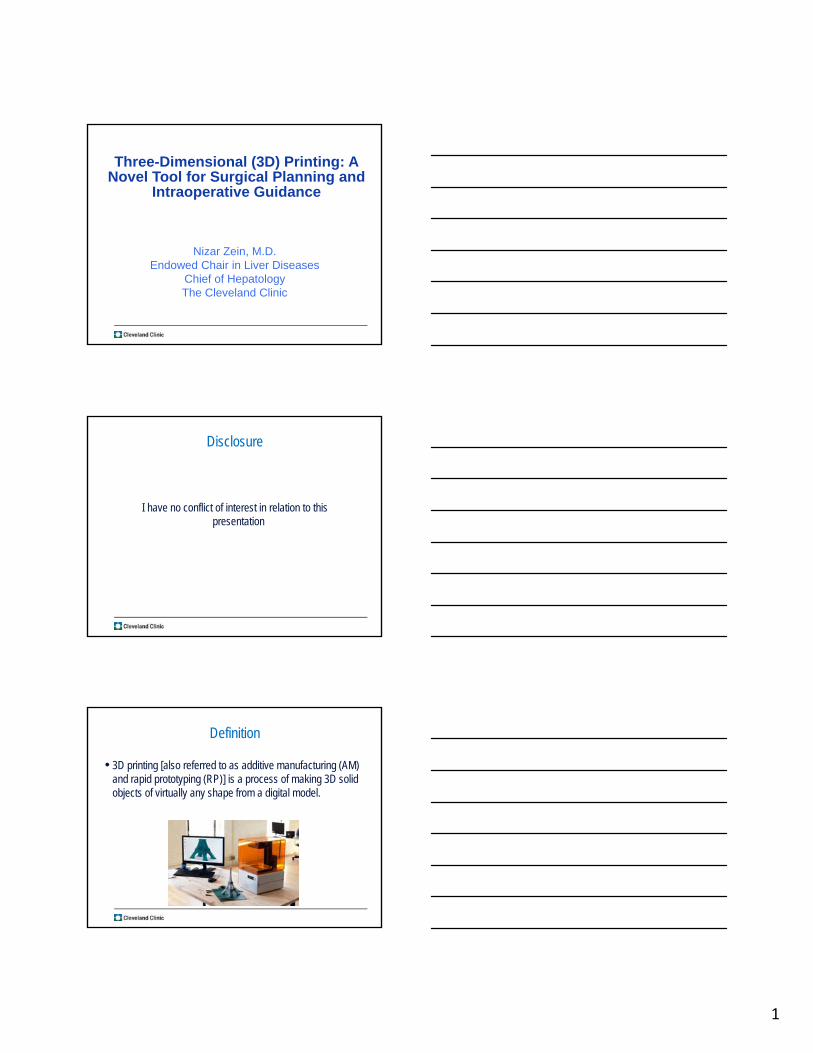
Three-Dimensional (3D) Printing: A Novel Tool for Surgical Planning and
Intraoperative Guidance
Nizar Zein, M.D.Endowed Chair in Liver Diseases
Chief of HepatologyThe Cleveland Clinic
Disclosure
I have no conflict of interest in relation to this presentation
Definition
• 3D printing [also referred to as additive manufacturing (AM) and rapid prototyping (RP)] is a process of making 3D solid objects of virtually any shape from a digital model.

2
The ProcessPrinterController Computer Post
ProcessingComputer Aided Design (CAD) or
CT reconstruction*
STL File
Build Prep Software**-“slicer”
* MEVIS or TeraRecon** Catalyst (FDM) or Objet Studio (polyjet)
Serial, Ethernet or USP port
Mechanics
Electronics
Code Interpretation
Motor control
Fusion control
Removeexcess material
Dying
Clear coating
Stereolithography• Laser cured liquid resin
Selective Laser Sintering• Laser sintered powder (metal, plastic)
Powder/Inkjet• Powder glued selectively via liquid binder
Fused Deposition Modeling• Extruded liquid thermoplastic
Polyjet• UV light cured liquid resin
Selective Laser Melting• Laser melted/fused metal powder
3D Printing Technologies3D Printing Technologies
3D Printers at CCF3D Printers at CCF
Stratasys uPrint SE Plus (“FDM”)
ABS Thermoplastic
Strong parts
Accuracy: +/- 0.4mm
Low cost
Stratasys Connex 350 (“Polyjet”)
UV cured liquid resin
2 materials in same build
Accuracy: +/- 0.04mm
Flexible, Clear materials

3
Stratasys Object500
Stratasys Objet500 (“Polyjet”)
UV cured liquid resin
Multi-materials in same build
Multi-color in the same build
Accuracy: +/- 0.02mm
Flexible, Clear materials
3D Printing Examples in Medicine3D Printing Examples in Medicine
Surgical Planning
• Fact: Great public, governmental and professional interest in improving surgical outcomes
• A wide-range of pre-operative planning techniques have been used to diminish operative time and complications:
–Imaging (CT, MRI, angiogram, biliary imaging, etc.)-2D
–Computer-assisted 3D imaging-viewed through 2D computer screen
–Generic physical models-not patient specific
4
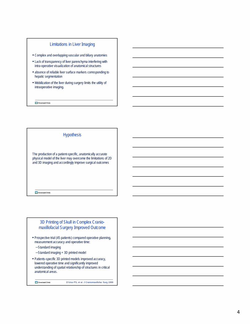
Limitations in Liver Imaging
• Complex and overlapping vascular and biliary anatomies
• Lack of transparency of liver parenchyma interfering with intra-operative visualization of anatomical structures
• absence of reliable liver surface markers corresponding to hepatic segmentation
• Mobilization of the liver during surgery limits the utility of intraoperative imaging.
Hypothesis
The production of a patient-specific, anatomically accurate physical model of the liver may overcome the limitations of 2D and 3D imaging and accordingly improve surgical outcomes
3D Printing of Skull in Complex Cranio-maxillofacial Surgery Improved Outcome
• Prospective trial (45 patients) compared operative planning, measurement accuracy and operative time:
–Standard imaging
–Standard imaging + 3D printed model
• Patients-specific 3D printed models improved accuracy, lowered operative time and significantly improved understanding of spatial relationship of structures in critical anatomical areas.
D’Urso PS, et al. J Craniomaxillofac Surg 1999

5
The Team: A Multidisciplinary Effort
Objectives
1. Create the first patient-specific three 3D printed liver based on standard 2D imaging (CT and MRI)
2. Validate the accuracy of 3D-printed liver models against native resected liver specimens
3. Assess the utility of individualized 3D printed livers in surgical planning and medical education.
CT/MR Imaging
3D Reconstruction
Digital Preparation
3D Printing Post Processing
Objective #1: 3D Liver Model ProductionObjective #1: 3D Liver Model Production
6
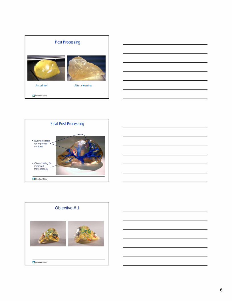
Post Processing
As printed After cleaning
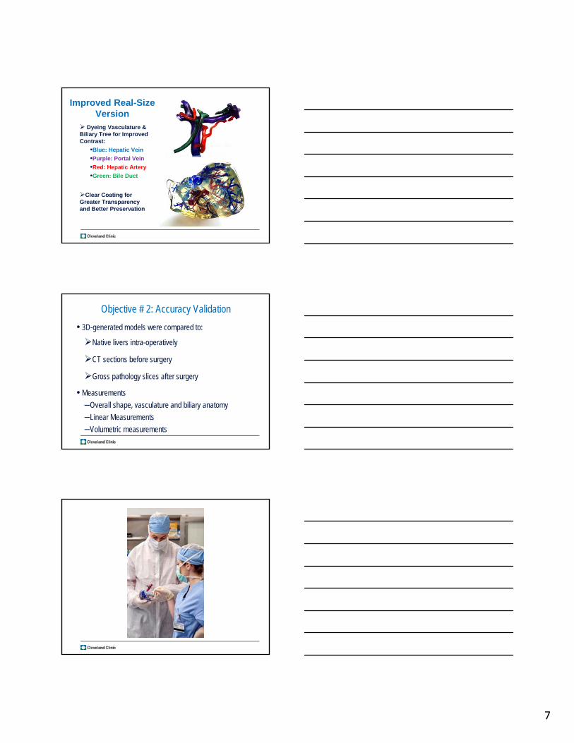
Final Post-Processing
• Dyeing vessels for improved contrast
• Clear-coating for improved transparency
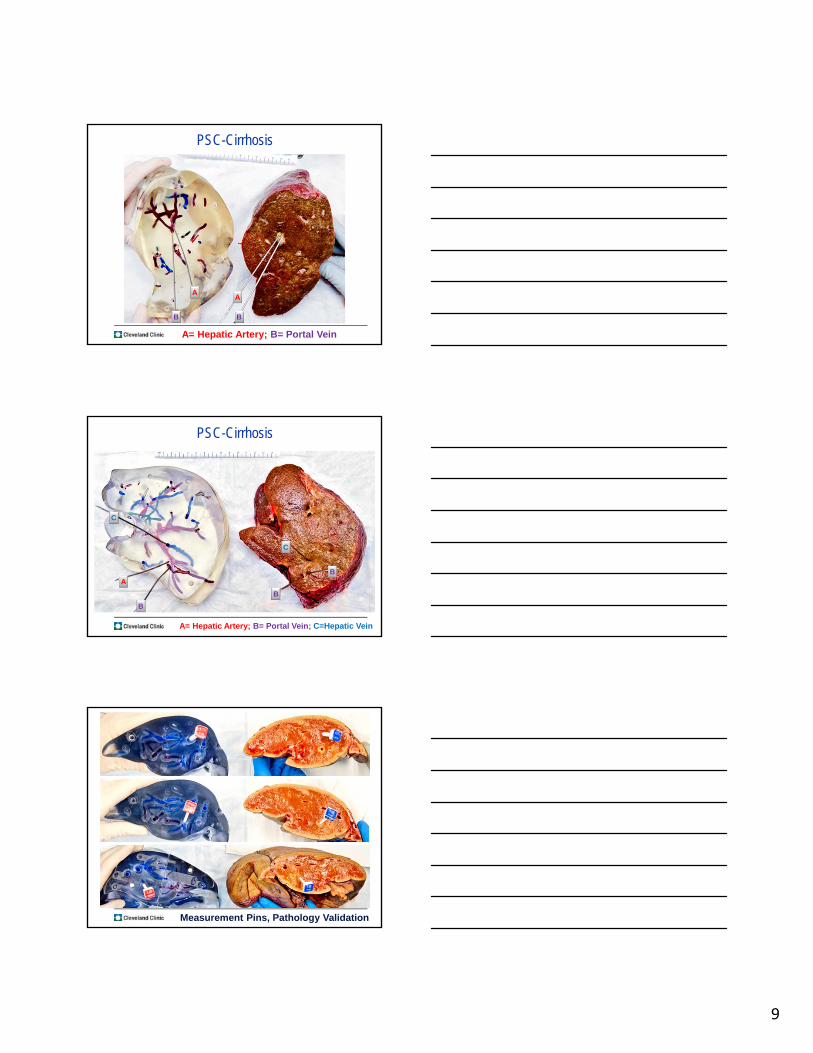
Objective # 1
7
Improved Real-Size Version
Dyeing Vasculature & Biliary Tree for Improved Contrast:
•Blue: Hepatic Vein
•Purple: Portal Vein
•Red: Hepatic Artery
•Green: Bile Duct
Clear Coating for Greater Transparency and Better Preservation
Objective # 2: Accuracy Validation
• 3D-generated models were compared to:
Native livers intra-operatively
CT sections before surgery
Gross pathology slices after surgery
• Measurements
–Overall shape, vasculature and biliary anatomy
–Linear Measurements
–Volumetric measurements

8
LDLT, Total Right LobectomyHealthy Donor to His Brother with Cryptogenic Cirrhosis
Zein NN, Hanouneh IA, Bishop PD, et al. Three-dimensional print of a liver for preoperative planning in living donor liver transplantation. Liver Transpl 2013;19:1304-10.
PSC-Cirrhosis
Pathology Validation
PSC-CirrhosisA A
A= Hepatic Artery Pathology Validation
9
PSC-Cirrhosis
AA
BB
A= Hepatic Artery; B= Portal Vein
PSC-Cirrhosis
AB
B
C
C
A= Hepatic Artery; B= Portal Vein; C=Hepatic Vein
B
Measurement Pins, Pathology Validation

10
APPLYING 3D LIVER MODELS TO CLINICAL PRACTICE
Living Donor Liver Transplantation
Hepatic Tumor Resection
Medical Education
Objective # 3
LDLT
• Case #1: Middle hepatic vein curved and too close to resection plane in the donor.
• Case # 2: Rejected donor based on length of R hepatic artery (too short for anastomosis
11
Resection for HCC
• Hepatic resection is considered the most curative approach for hepatic tumors.
• Characterization of intrahepatic anatomy, lesions size, number, location and proximity to vascular and biliary structures is critical to achieve cure.
• Traditional imaging modalities, including 2D CT & MRI, provide limited information on the tumor’s extent and its relationship with surrounding vessels for complex hepatic resection planning.
Difficult to Resect Liver Tumors
• Defined as:
Extended right/left hepatectomies
Central resections
Polysegmentectiomies
Large atypical resections
• We evaluated the asset of 3D-printed liver models for surgical preplanning and intraoperative guidance.
AIMS
• Compare 2D imaging (CT or MRI) to 3D printed liver models for preoperative surgical planning and intraoperative guidance:
• Determination of resectability
• Changes in operative strategy

12
Patients & Methods
• Prospective study (Jan-Aug 2014) of 6 patients with liver tumors, who underwent high-risk procedures for complex liver tumors.
• 3 patients with central intrahepatic cholangiocarcinoma, 1 patient with Klatskin tumor, and 2 patients with metastatic colon cancer into the liver.
• Median lesion size 7.1 cm.
• In 3 of the 6 cases, the pre-operative plan was modified after review of anatomical spatial relationship of tumor to nearby structures in the 3D model compared to initial plan based on standard imaging alone.
• Changes included:
–resection modification,
–extension and intrahepatic vascular reconstruction.
Results: Pre-Op
AASLD Abstract
• Surgeons reported greater confidence with use of 3D model for identification of intra and extrahepatic structure, segmentation and tumor specific extent.
• Surgeons agreed that 3D model offered a realistic representation that allowed interactive manipulation simulating intraoperative mobilization.
Results: Intra-Op
AASLD Abstract

13
A Case: 56 year old Female
4/2013: Developedpruritis ofextremitiesandtorso.
6/2013: Labwork Transaminitisandelevatedlivertests
Abd MRI Llobehepaticmass(9cm),likelymalignant.
ThemassabuttingtheIVCandhepaticveinswith
encasementofLandmiddlehepaticveins.MarketL
sidedbiliarydilation
7/3/13: CTGuidedBiopsy: poorlydifferentiated
adenocarcinomaconsistentwithprimary
cholangiocarcinoma.
CT diagnostic Pre-chemotherapy
Outside Institution
• Based on all testes, patient was evaluated at Rosewell Park, and tumor was determined unresectable.
14
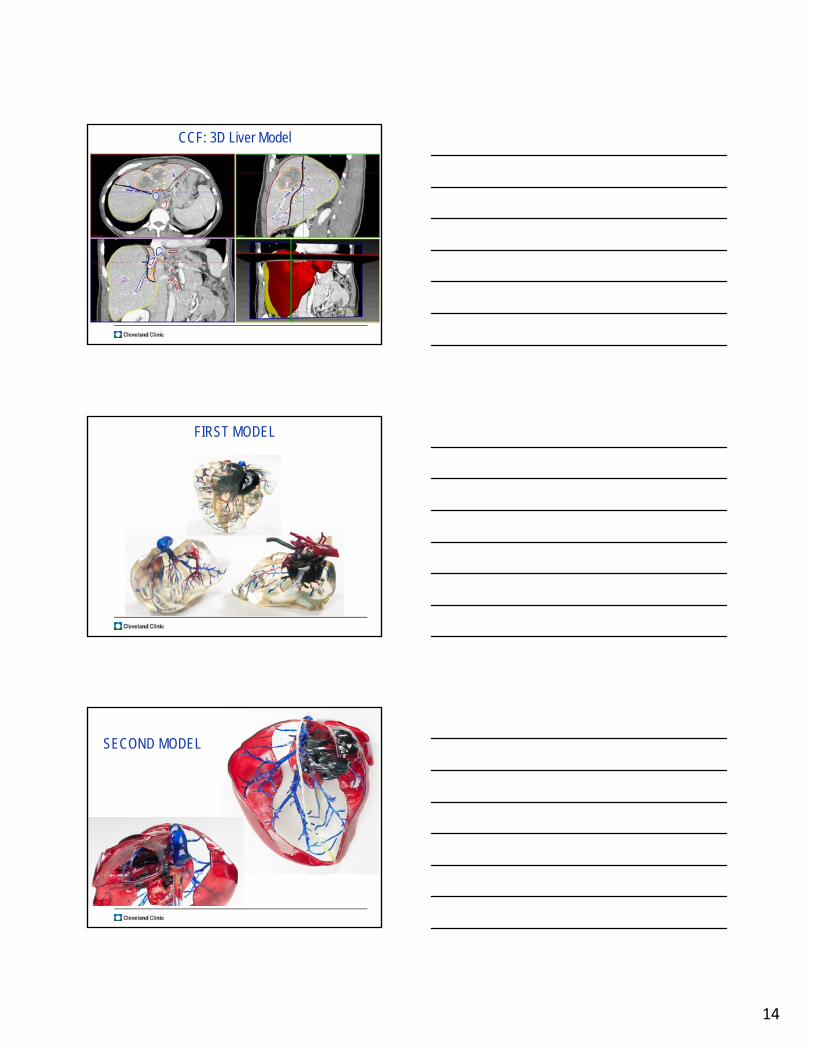
CCF: 3D Liver Model
FIRST MODEL
SECOND MODEL
15
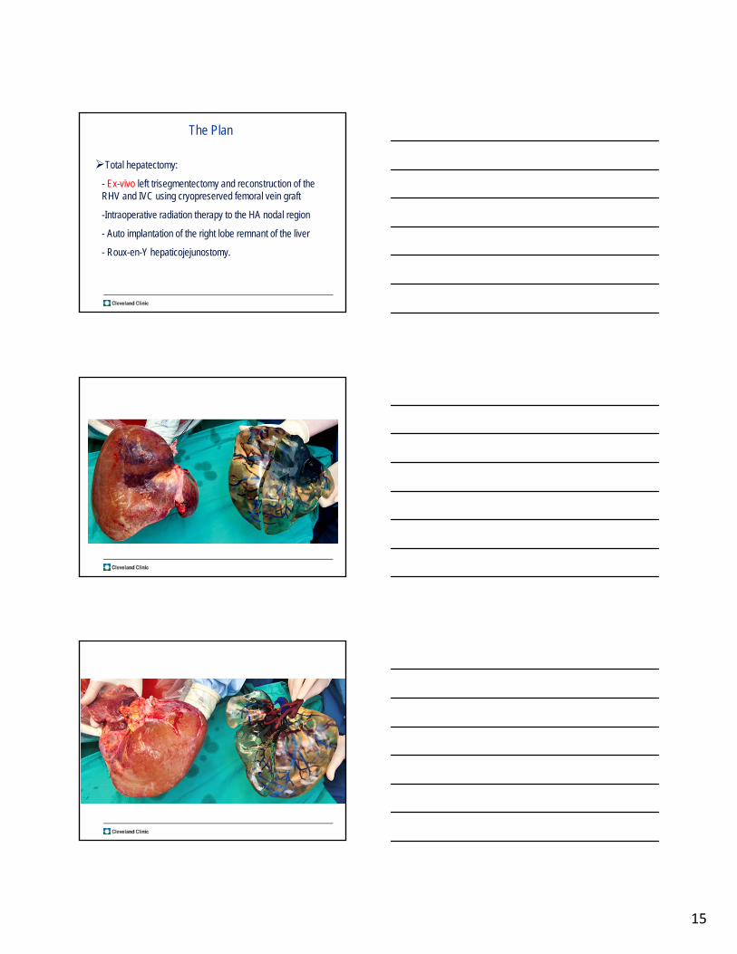
The Plan
Total hepatectomy:
- Ex-vivo left trisegmentectomy and reconstruction of the RHV and IVC using cryopreserved femoral vein graft
-Intraoperative radiation therapy to the HA nodal region
- Auto implantation of the right lobe remnant of the liver
- Roux-en-Y hepaticojejunostomy.
16
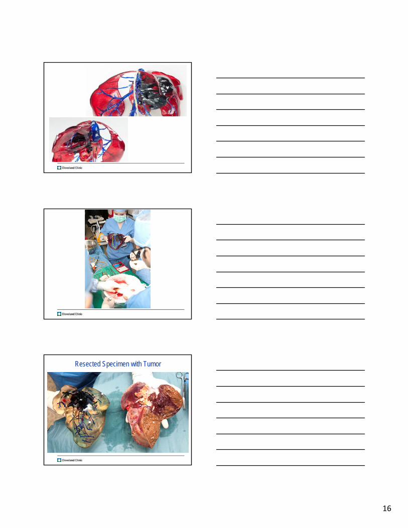
Resected Specimen with Tumor
17
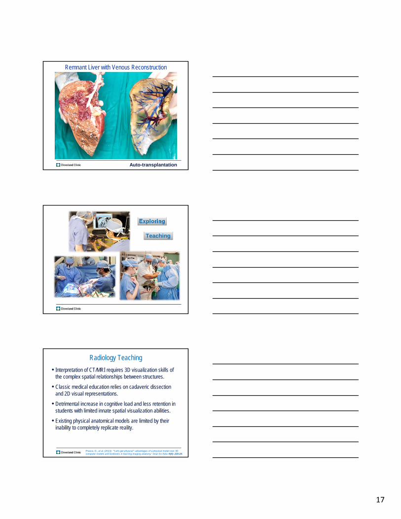
Remnant Liver with Venous Reconstruction
Auto-transplantation
Exploring
Teaching
Radiology Teaching
• Interpretation of CT/MRI requires 3D visualization skills of the complex spatial relationships between structures.
• Classic medical education relies on cadaveric dissection and 2D visual representations.
• Detrimental increase in cognitive load and less retention in students with limited innate spatial visualization abilities.
• Existing physical anatomical models are limited by their inability to completely replicate reality.
Preece, D., et al. (2013). ""Let's get physical": advantages of a physical model over 3D computer models and textbooks in learning imaging anatomy." Anat Sci Educ 6(4): 216-24.
18
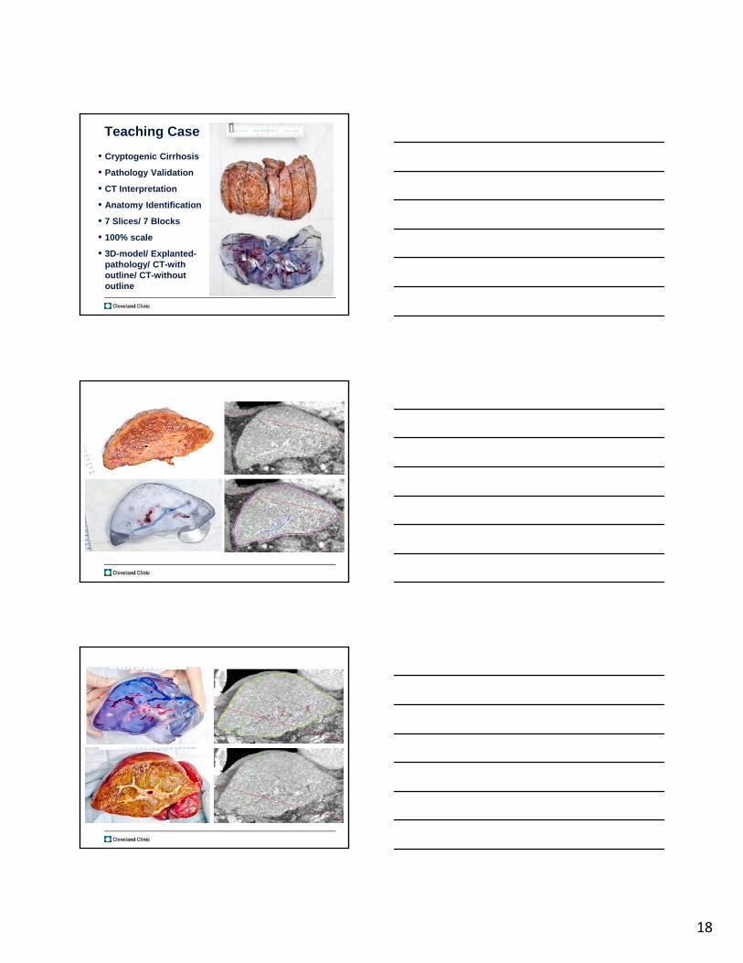
• Cryptogenic Cirrhosis
• Pathology Validation
• CT Interpretation
• Anatomy Identification
• 7 Slices/ 7 Blocks
• 100% scale
• 3D-model/ Explanted-pathology/ CT-with outline/ CT-without outline
Teaching Case

19
Innovations in Medical Education: Case Western Reserve University
Bio-3D Printing?
Conclusions
Transparent 3D-printed models used for surgery granted:
• Easier segmentation
• Better comprehension of spatial relationships
• Higher confidence levels among surgical staff
3D-printed models may provide a novel educational tool

20