Embed Size (px)
Citation preview

Clinical Chest Radiography Interpretation
Part IIAnthony M. Angelow,
PhD(c), MSN, ACNPC, AGACNP-BC, CENAssociate Lecturer,
Fitzgerald Health Education Associates Clinical practice
Division of Trauma Surgery and Surgical Critical CareCooper University Hospital, Camden, NJ
Assistant Clinical Professor, College of Nursing and Health Professions
Drexel University, Philadelphia, PA
Disclosure
• No real or potential conflict of interest to disclose.
• No off-label, experimental or investigational use of drugs or devices will be presented.
Fitzgerald Health Education Associates 84
ObjectivesPart II
• At the end of this presentation, the participant will be able to:– Apply a systematic approach to the
interpretation of clinical chest radiography.– Identify common abnormalities of the chest
radiograph based on patient case studies.– Describe normal and abnormal findings of
the chest radiograph.
Fitzgerald Health Education Associates 85
Before We Begin….
Some Reminders!
Fitzgerald Health Education Associates 86
Chest Radiograph BasicsREMINDER!
• Standard rules of chest radiography– Obtain a thorough history and physical
examination– Always evaluate the entire radiograph– Before confirming a diagnosis re-examine the
patient and the radiograph and be sure the diagnosis fits the clinical picture
– Always confirm the right patient– Develop a systematic approach– Do not get distracted– Why is the plain radiograph appropriate?– Why is a diagnostic study appropriate?
Fitzgerald Health Education Associates 87
Chest Radiograph InterpretationA Systematic (A-I) Approach
• Adequacy and airway
• Bones and soft tissue
• Cardiac• Diaphragm• Edges (pleura)• Fields (lungs)
• Gastric bubble• Hila and
mediastinum• Impression
Fitzgerald Health Education Associates 88

Case Study #1
Fitzgerald Health Education Associates 89
Case Study #1MVC with Chest Pain
• A 35-year-old male presented to the trauma center with a chief complaint of chest pain after being involved in a motor vehicle accident. The patient describes left sided sharp chest pain and also complains of shortness of breath. Physical examination reveals hyperresonance on the right with absent breath sound in the lower right thorax.
Fitzgerald Health Education Associates 90
Case Study #1MVC with Chest Pain (continued)
Source: https://commons.wikimedia.org/wiki/File:Pneumothorax_CXR.jpg
Fitzgerald Health Education Associates 91
Case Study #1Step 1 – Adequacy and Airway
Source: https://commons.wikimedia.org/wiki/File:Pneumothorax_CXR.jpg
AdequacyInspiration?
Penetration?
Rotation?
Fitzgerald Health Education Associates 92
Case Study #1Step 1 – Adequacy and Airway (continued)
Source: https://commons.wikimedia.org/wiki/File:Pneumothorax_CXR.jpg
Airway
Is the trachea midline?
Is the tracheal bifurcation at the level of T4‒T5?
Fitzgerald Health Education Associates 93
Case Study #1Step 2 – Bones and Soft Tissue
Source: https://commons.wikimedia.org/wiki/File:Pneumothorax_CXR.jpg
Bones
Fractured ribs?
Clavicles?
Scapula, humerus?
Fitzgerald Health Education Associates 94

Case Study #1Step 2 – Bones and Soft Tissue (continued)
Source: https://commons.wikimedia.org/wiki/File:Pneumothorax_CXR.jpg
Soft Tissues
Is the soft tissue obscuring any structures?
Is there any subcutaneous air?
Fitzgerald Health Education Associates 95
Case Study #1Step 3 – Cardiac
Source: https://commons.wikimedia.org/wiki/File:Pneumothorax_CXR.jpg
Heart
Is the heart positioned appropriately?
Is the heart a normal size?
Fitzgerald Health Education Associates 96
Case Study #1Step 4 – Diaphragm
Source: https://commons.wikimedia.org/wiki/File:Pneumothorax_CXR.jpg
Diaphragm
Are there sharp diaphragmatic margins?
Is the contour normal?
Fitzgerald Health Education Associates 97
Case Study #1Step 5 – Edges (Pleura)
Source: https://commons.wikimedia.org/wiki/File:Pneumothorax_CXR.jpg
Pleura
Are there any noticeable pleural lines?
Any noticeable fluid or air accumulation?
Fitzgerald Health Education Associates 98
Case Study #1Step 6 – Field (Lungs)
Source: https://commons.wikimedia.org/wiki/File:Pneumothorax_CXR.jpg
Lungs
Is there a normal vessel pattern?
Are the lungs air filled?
Any lucencies or shadowing?
Fitzgerald Health Education Associates 99
Case Study #1Step 7 – Gastric Bubble
Source: https://commons.wikimedia.org/wiki/File:Pneumothorax_CXR.jpg
Gastric Bubble
Is the bubble round on the left side?
Any suggestion of free air in the abdomen?
Fitzgerald Health Education Associates 100

Case Study #1Step 8 – Hilum and Mediastinum
Source: https://commons.wikimedia.org/wiki/File:Pneumothorax_CXR.jpg
Hilum
Is the hilum clearly present?
Any masses or lymphadenopathy?
Similar in size and density?
Fitzgerald Health Education Associates 101
Case Study #1Step 8 – Hilum and Mediastinum (continued)
Source: https://commons.wikimedia.org/wiki/File:Pneumothorax_CXR.jpg
Mediastinum
Size and position?
Is the trachea to the right?
Structures identifiable?
Fitzgerald Health Education Associates 102
Case Study #1Step 9 – Interpretation
Source: https://commons.wikimedia.org/wiki/File:Pneumothorax_CXR.jpg
Abnormal Findings
• Trachea deviated to right• Heart deviated to right• Clear demarcation of
pleural lines on the left with air in the pleural space
• Lung vasculature not extended to pleural wall on the left
• Mediastinum deviated to the right
Fitzgerald Health Education Associates 103
Case Study #1Case Review
• A 35-year-old male presented to the trauma center with a chief complaint of chest pain after being involved in a motor vehicle accident. The patient describes left sided sharp chest pain and also complains of shortness of breath. Physical examination reveals hyperresonance on the right with absent breath sound in the lower right thorax.
Fitzgerald Health Education Associates 104
Case Study #1Step 9 – Interpretation
Source: https://commons.wikimedia.org/wiki/File:Pneumothorax_CXR.jpg
Diagnosis?
Does this make sense?
Fitzgerald Health Education Associates 105
Case Study #1Case Review
• A 35-year-old male presented to the trauma center with a chief complaint of chest pain after being involved in a motor vehicle accident. The patient describes left sided sharp chest pain and also complains of shortness of breath. Physical examination reveals hyperresonance on the right with absent breath sound in the lower right thorax.
Fitzgerald Health Education Associates 106

Case Study #2
Fitzgerald Health Education Associates 107
Case Study #2Cough, Chills, and Fever
• A 65-year-old female presents with complaints of cough, chills, and fever for the last three days. She describes her cough as productive with yellow mucus and a fever ranging from 101.5‒102.4°F (38.6‒39.1°C). She describes a feeling of overall fatigue and shortness of breath with activity. Lung sounds produce crackles in the right base.
Fitzgerald Health Education Associates 108
Case Study #2Cough, Chills, and Fever (continued)
Source: https://commons.wikimedia.org/wiki/File:RLL_pneumoniaM.jpg
Fitzgerald Health Education Associates 109
Case Study #2Step 1 – Adequacy and Airway
Source: https://commons.wikimedia.org/wiki/File:RLL_pneumoniaM.jpg
Adequacy
Inspiration?
Penetration?
Rotation?
Fitzgerald Health Education Associates 110
Case Study #2Step 1 – Adequacy and Airway (continued)
Airway
Is the trachea midline?
Is the tracheal bifurcation at the level of T4‒T5?
Source: https://commons.wikimedia.org/wiki/File:RLL_pneumoniaM.jpg
Fitzgerald Health Education Associates 111
Case Study #2Step 2 – Bones and Soft Tissue
Bones
Fractured ribs?
Clavicles?
Scapula, humerus?
Source: https://commons.wikimedia.org/wiki/File:RLL_pneumoniaM.jpg
Fitzgerald Health Education Associates 112
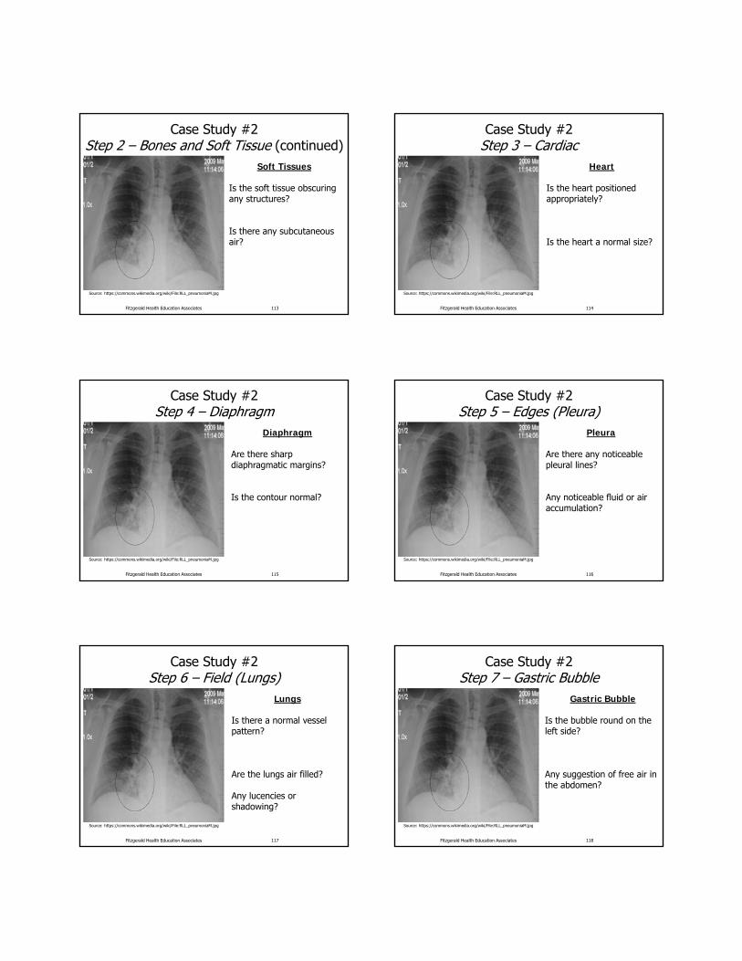
Case Study #2Step 2 – Bones and Soft Tissue (continued)
Soft Tissues
Is the soft tissue obscuring any structures?
Is there any subcutaneous air?
Source: https://commons.wikimedia.org/wiki/File:RLL_pneumoniaM.jpg
Fitzgerald Health Education Associates 113
Case Study #2Step 3 – Cardiac
Heart
Is the heart positioned appropriately?
Is the heart a normal size?
Source: https://commons.wikimedia.org/wiki/File:RLL_pneumoniaM.jpg
Fitzgerald Health Education Associates 114
Case Study #2Step 4 – Diaphragm
Diaphragm
Are there sharp diaphragmatic margins?
Is the contour normal?
Source: https://commons.wikimedia.org/wiki/File:RLL_pneumoniaM.jpg
Fitzgerald Health Education Associates 115
Case Study #2Step 5 – Edges (Pleura)
Pleura
Are there any noticeable pleural lines?
Any noticeable fluid or air accumulation?
Source: https://commons.wikimedia.org/wiki/File:RLL_pneumoniaM.jpg
Fitzgerald Health Education Associates 116
Case Study #2Step 6 – Field (Lungs)
Lungs
Is there a normal vessel pattern?
Are the lungs air filled?
Any lucencies or shadowing?
Source: https://commons.wikimedia.org/wiki/File:RLL_pneumoniaM.jpg
Fitzgerald Health Education Associates 117
Case Study #2Step 7 – Gastric Bubble
Gastric Bubble
Is the bubble round on the left side?
Any suggestion of free air in the abdomen?
Source: https://commons.wikimedia.org/wiki/File:RLL_pneumoniaM.jpg
Fitzgerald Health Education Associates 118
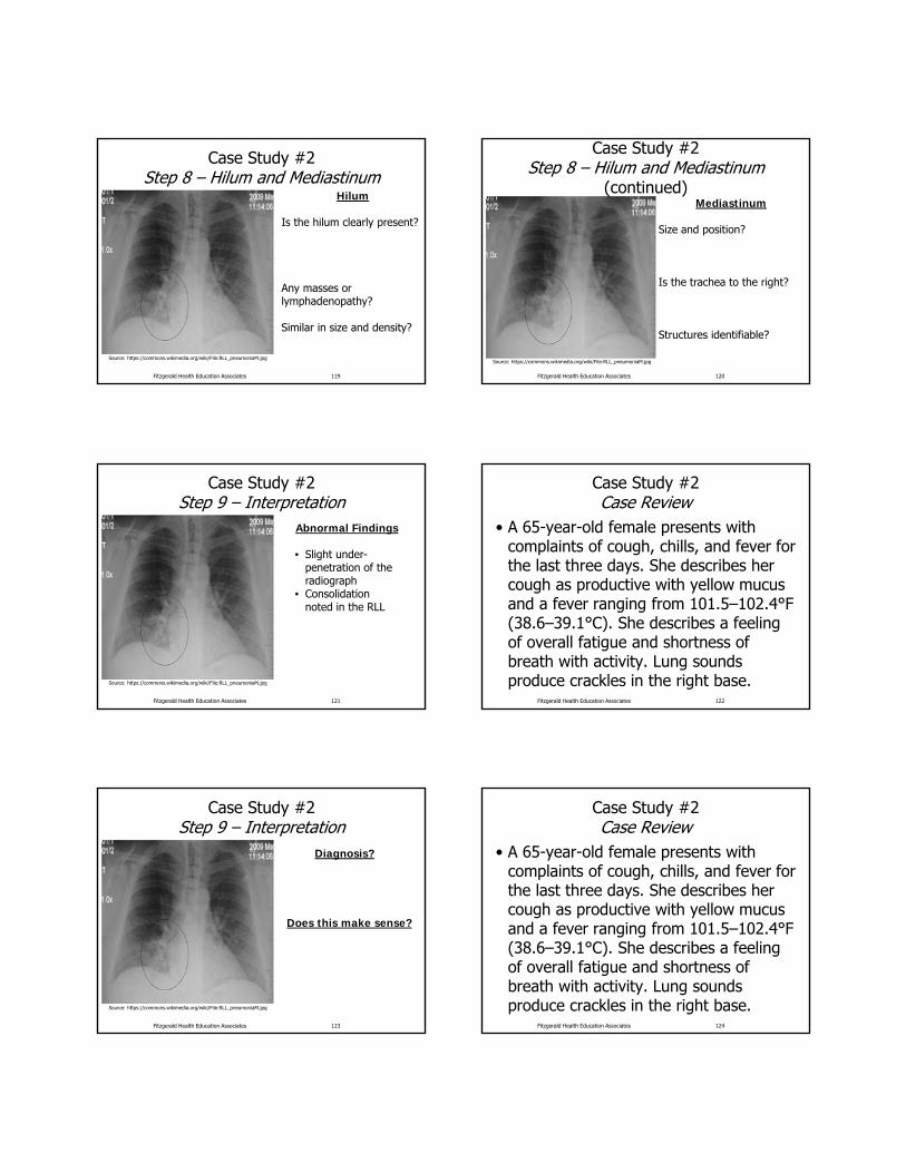
Case Study #2Step 8 – Hilum and Mediastinum
Hilum
Is the hilum clearly present?
Any masses or lymphadenopathy?
Similar in size and density?
Source: https://commons.wikimedia.org/wiki/File:RLL_pneumoniaM.jpg
Fitzgerald Health Education Associates 119
Case Study #2Step 8 – Hilum and Mediastinum
(continued)Mediastinum
Size and position?
Is the trachea to the right?
Structures identifiable?
Source: https://commons.wikimedia.org/wiki/File:RLL_pneumoniaM.jpg
Fitzgerald Health Education Associates 120
Case Study #2Step 9 – Interpretation
Abnormal Findings
• Slight under-penetration of the radiograph
• Consolidation noted in the RLL
Source: https://commons.wikimedia.org/wiki/File:RLL_pneumoniaM.jpg
Fitzgerald Health Education Associates 121
Case Study #2Case Review
• A 65-year-old female presents with complaints of cough, chills, and fever for the last three days. She describes her cough as productive with yellow mucus and a fever ranging from 101.5‒102.4°F (38.6‒39.1°C). She describes a feeling of overall fatigue and shortness of breath with activity. Lung sounds produce crackles in the right base.
Fitzgerald Health Education Associates 122
Case Study #2Step 9 – Interpretation
Diagnosis?
Does this make sense?
Source: https://commons.wikimedia.org/wiki/File:RLL_pneumoniaM.jpg
Fitzgerald Health Education Associates 123
Case Study #2Case Review
• A 65-year-old female presents with complaints of cough, chills, and fever for the last three days. She describes her cough as productive with yellow mucus and a fever ranging from 101.5‒102.4°F (38.6‒39.1°C). She describes a feeling of overall fatigue and shortness of breath with activity. Lung sounds produce crackles in the right base.
Fitzgerald Health Education Associates 124

Case Study #3
Fitzgerald Health Education Associates 125
Case Study #3Cough with Hemoptysis
• A 60-year-old male with a 40-pack-year smoking history presented with a complaint of cough with noted blood in his sputum and breathlessness. He states he has been short of breath for the last few months, but contributes that to his smoking. He also admits to a 10 lb (4.5 Kg) unprovoked weight loss over the last “few weeks.”
Fitzgerald Health Education Associates 126
Case Study #3Cough, Chills, and Fever
Source: https://en.wikipedia.org/wiki/File:Chest_Xray.jpg
Fitzgerald Health Education Associates 127
Case Study #3Step 1 – Adequacy and Airway
Source: https://en.wikipedia.org/wiki/File:Chest_Xray.jpg
Adequacy
Inspiration?
Penetration?
Rotation?
Fitzgerald Health Education Associates 128
Case Study #3Step 1 – Adequacy and Airway (continued)
Airway
Is the trachea midline?
Is the tracheal bifurcation at the level of T4‒T5?
Source: https://en.wikipedia.org/wiki/File:Chest_Xray.jpg
Fitzgerald Health Education Associates 129
Case Study #3Step 2 – Bones and Soft Tissue
Bones
Fractured ribs?
Clavicles?
Scapula, humerus?
Source: https://en.wikipedia.org/wiki/File:Chest_Xray.jpg
Fitzgerald Health Education Associates 130

Case Study #3Step 2 – Bones and Soft Tissue
(continued)Soft Tissues
Is the soft tissue obscuring any structures?
Is there any subcutaneous air?
Source: https://en.wikipedia.org/wiki/File:Chest_Xray.jpg
Fitzgerald Health Education Associates 131
Case Study #3Step 3 – Cardiac
Heart
Is the heart positioned appropriately?
Is the heart a normal size?
Source: https://en.wikipedia.org/wiki/File:Chest_Xray.jpg
Fitzgerald Health Education Associates 132
Case Study #3Step 4 – Diaphragm
Diaphragm
Are there sharp diaphragmatic margins?
Is the contour normal?
Source: https://en.wikipedia.org/wiki/File:Chest_Xray.jpg
Fitzgerald Health Education Associates 133
Case Study #3Step 5 – Edges (Pleura)
Pleura
Are there any noticeable pleural lines?
Any noticeable fluid or air accumulation?
Source: https://en.wikipedia.org/wiki/File:Chest_Xray.jpg
Fitzgerald Health Education Associates 134
Case Study #3Step 6 – Field (Lungs)
Lungs
Is there a normal vessel pattern?
Are the lungs air filled?
Any lucencies or shadowing?
Source: https://en.wikipedia.org/wiki/File:Chest_Xray.jpg
Fitzgerald Health Education Associates 135
Case Study #3Step 7 – Gastric Bubble
Gastric Bubble
Is the bubble round on the left side?
Any suggestion of free air in the abdomen?
Source: https://en.wikipedia.org/wiki/File:Chest_Xray.jpg
Fitzgerald Health Education Associates 136

Case Study #3Step 8 – Hilum and Mediastinum
Hilum
Is the hilum clearly present?
Any masses or lymphadenopathy?
Similar in size and density?
Source: https://en.wikipedia.org/wiki/File:Chest_Xray.jpg
Fitzgerald Health Education Associates 137
Case Study #3Step 8 – Hilum and Mediastinum
(continued)Mediastinum
Size and position?
Is the trachea to the right?
Structures identifiable?
Source: https://en.wikipedia.org/wiki/File:Chest_Xray.jpg
Fitzgerald Health Education Associates 138
Case Study #3Step 9 – Interpretation
Abnormal Findings
• Singular mass/lesion located in the left lung
• Left hilum is obstructed by the presence of a singular mass
Source: https://en.wikipedia.org/wiki/File:Chest_Xray.jpg
Fitzgerald Health Education Associates 139
Case Study #3Case Review
• A 60-year-old male with a 40-pack-year smoking history presented with a complaint of cough with noted blood in his sputum and breathlessness. He states he has been short of breath for the last few months, but contributes that to his smoking. He also admits to a 10 lb (4.5 Kg) unprovoked weight loss over the last “few weeks.”
Fitzgerald Health Education Associates 140
Case Study #3Step 9 – Interpretation
Diagnosis?
Does this make sense?
Source: https://en.wikipedia.org/wiki/File:Chest_Xray.jpg
Fitzgerald Health Education Associates 141
Case Study #3Case Review
• A 60-year-old male with a 40-pack-year smoking history presented with a complaint of cough with noted blood in his sputum and breathlessness. He states he has been short of breath for the last few months, but contributes that to his smoking. He also admits to a 10 lb (4.5 Kg) unprovoked weight loss over the last “few weeks.”
Fitzgerald Health Education Associates 142

Case Study #4
Fitzgerald Health Education Associates 143
Case Study #4Chest Pain After Being Hit with a Bat
• An 11-year-old male with a chief complaint of left sided chest pain presented to the ED after sustaining a blow to the chest with a bat during a baseball game. He states the pain is sharp in nature and worsens with inspiration. On physical examination there is noted chest tenderness to palpation in the left upper chest.
Fitzgerald Health Education Associates 144
Case Study #4Chest Pain After Being Hit with a Bat
(continued)
Source: https://commons.wikimedia.org/wiki/File:PfracturedribX.png
Fitzgerald Health Education Associates 145
Case Study #4Step 1 – Adequacy and Airway
Source: https://commons.wikimedia.org/wiki/File:PfracturedribX.png
Adequacy
Inspiration?
Penetration?
Rotation?
Fitzgerald Health Education Associates 146
Case Study #4Step 1 – Adequacy and Airway (continued)
Airway
Is the trachea midline?
Is the tracheal bifurcation at the level of T4‒T5?
Source: https://commons.wikimedia.org/wiki/File:PfracturedribX.png
Fitzgerald Health Education Associates 147
Case Study #4Step 2 – Bones and Soft Tissue
Bones
Fractured ribs?
Clavicles?
Scapula, humerus?
Source: https://commons.wikimedia.org/wiki/File:PfracturedribX.png
Fitzgerald Health Education Associates 148

Case Study #4Step 2 – Bones and Soft Tissue
(continued)Soft Tissues
Is the soft tissue obscuring any structures?
Is there any subcutaneous air?
Source: https://commons.wikimedia.org/wiki/File:PfracturedribX.png
Fitzgerald Health Education Associates 149
Case Study #4Step 3 – Cardiac
Heart
Is the heart positioned appropriately?
Is the heart a normal size?
Source: https://commons.wikimedia.org/wiki/File:PfracturedribX.png
Fitzgerald Health Education Associates 150
Case Study #4Step 4 – Diaphragm
Diaphragm
Are there sharp diaphragmatic margins?
Is the contour normal?
Source: https://commons.wikimedia.org/wiki/File:PfracturedribX.png
Fitzgerald Health Education Associates 151
Case Study #4Step 5 – Edges (Pleura)
Pleura
Are there any noticeable pleural lines?
Any noticeable fluid or air accumulation?
Source: https://commons.wikimedia.org/wiki/File:PfracturedribX.png
Fitzgerald Health Education Associates 152
Case Study #4Step 6 – Field (Lungs)
Lungs
Is there a normal vessel pattern?
Are the lungs air filled?
Any lucencies or shadowing?
Source: https://commons.wikimedia.org/wiki/File:PfracturedribX.png
Fitzgerald Health Education Associates 153
Case Study #4Step 7 – Gastric Bubble
Gastric Bubble
Is the bubble round on the left side?
Any suggestion of free air in the abdomen?
Source: https://commons.wikimedia.org/wiki/File:PfracturedribX.png
Fitzgerald Health Education Associates 154

Case Study #4Step 8 – Hilum and Mediastinum
Hilum
Is the hilum clearly present?
Any masses or lymphadenopathy?
Similar in size and density?
Source: https://commons.wikimedia.org/wiki/File:PfracturedribX.png
Fitzgerald Health Education Associates 155
Case Study #4Step 8 – Hilum and Mediastinum (continued)
Mediastinum
Size and position?
Is the trachea to the right?
Structures identifiable?
Source: https://commons.wikimedia.org/wiki/File:PfracturedribX.png
Fitzgerald Health Education Associates 156
Case Study #4Step 9 – Interpretation
Abnormal Findings
• Slightly displaced fracture of the left 3rd rib
Source: https://commons.wikimedia.org/wiki/File:PfracturedribX.png
Fitzgerald Health Education Associates 157
Case Study #4Case Review
• An 11-year-old male with a chief complaint of left sided chest pain presented to the ED after sustaining a blow to the chest with a bat during a baseball game. He states the pain is sharp in nature and worsens with inspiration. On physical examination there is noted chest tenderness to palpation in the left upper chest.
Fitzgerald Health Education Associates 158
Case Study #4Step 9 – Interpretation
Diagnosis?
Does this make sense?
Source: https://commons.wikimedia.org/wiki/File:PfracturedribX.png
Fitzgerald Health Education Associates 159
Case Study #4Case Review
• An 11-year-old male with a chief complaint of left sided chest pain presented to the ED after sustaining a blow to the chest with a bat during a baseball game. He states the pain is sharp in nature and worsens with inspiration. On physical examination there is noted chest tenderness to palpation in the left upper chest.
Fitzgerald Health Education Associates 160

Case Study #5
Fitzgerald Health Education Associates 161
Case Study #5Hospitalized Patient Sudden Onset SOB
• An 47-year-old male who was admitted to the TICU after sustaining multiple traumatic injuries requiring numerous blood product transfusions complains this morning of severe onset of shortness of breath. He denies chest pain, cough, and no fever is documented. On physical examination his lung sounds are decreased with diffuse crackles.
Fitzgerald Health Education Associates 162
Case Study #5Hospitalized Patient Sudden Onset SOB
(continued)
Source: https://commons.wikimedia.org/wiki/File:ARDS_X-Ray.jpg
Fitzgerald Health Education Associates 163
Case Study #5Step 1 – Adequacy and Airway
Source: https://commons.wikimedia.org/wiki/File:ARDS_X-Ray.jpg
AdequacyInspiration?
Penetration?
Rotation?
Fitzgerald Health Education Associates 164
Case Study #5Step 1 – Adequacy and Airway
(continued)Airway
Is the trachea midline?
Is the tracheal bifurcation at the level of T4‒T5?
Source: https://commons.wikimedia.org/wiki/File:ARDS_X-Ray.jpg
Fitzgerald Health Education Associates 165
Case Study #5Step 2 – Bones and Soft Tissue
Bones
Fractured ribs?
Clavicles?
Scapula, humerus?
Source: https://commons.wikimedia.org/wiki/File:ARDS_X-Ray.jpg
Fitzgerald Health Education Associates 166

Case Study #5Step 2 – Bones and Soft Tissue
(continued)Soft Tissues
Is the soft tissue obscuring any structures?
Is there any subcutaneous air?
Source: https://commons.wikimedia.org/wiki/File:ARDS_X-Ray.jpg
Fitzgerald Health Education Associates 167
Case Study #5Step 3 – Cardiac
Heart
Is the heart positioned appropriately?
Is the heart a normal size?
Source: https://commons.wikimedia.org/wiki/File:ARDS_X-Ray.jpg
Fitzgerald Health Education Associates 168
Case Study #5Step 4 – Diaphragm
Diaphragm
Are there sharp diaphragmatic margins?
Is the contour normal?
Source: https://commons.wikimedia.org/wiki/File:ARDS_X-Ray.jpg
Fitzgerald Health Education Associates 169
Case Study #5Step 5 – Edges (Pleura)
Pleura
Are there any noticeable pleural lines?
Any noticeable fluid or air accumulation?
Source: https://commons.wikimedia.org/wiki/File:ARDS_X-Ray.jpg
Fitzgerald Health Education Associates 170
Case Study #5Step 6 – Field (Lungs)
Lungs
Is there a normal vessel pattern?
Are the lungs air filled?
Any lucencies or shadowing?
Source: https://commons.wikimedia.org/wiki/File:ARDS_X-Ray.jpg
Fitzgerald Health Education Associates 171
Case Study #5Step 7 – Gastric Bubble
Gastric Bubble
Is the bubble round on the left side?
Any suggestion of free air in the abdomen?
Source: https://commons.wikimedia.org/wiki/File:ARDS_X-Ray.jpg
Fitzgerald Health Education Associates 172

Case Study #5Step 8 – Hilum and Mediastinum
Hilum
Is the hilum clearly present?
Any masses or lymphadenopathy?
Similar in size and density?Source: https://commons.wikimedia.org/wiki/File:ARDS_X-Ray.jpg
Fitzgerald Health Education Associates 173
Case Study #5Step 8 – Hilum and Mediastinum
(continued)Mediastinum
Size and position?
Is the trachea to the right?
Structures identifiable?
Source: https://commons.wikimedia.org/wiki/File:ARDS_X-Ray.jpg
Fitzgerald Health Education Associates 174
Case Study #5Step 9 – Interpretation
Abnormal Findings
• Costovertebral angles not clear
• Bilateral, diffuse patchy infiltrates
Source: https://commons.wikimedia.org/wiki/File:ARDS_X-Ray.jpg
Fitzgerald Health Education Associates 175
Case Study #5Case Review
• An 47-year-old male who was admitted to the TICU after sustaining multiple traumatic injuries requiring numerous blood product transfusions complains this morning of severe onset of shortness of breath. He denies chest pain, cough, and no fever is documented. On physical examination his lung sounds are decreased with diffuse crackles.
Fitzgerald Health Education Associates 176
Case Study #5Step 9 – Interpretation
Diagnosis?
Does this make sense?
Source: https://commons.wikimedia.org/wiki/File:ARDS_X-Ray.jpg
Fitzgerald Health Education Associates 177
Case Study #5Case Review
• An 47-year-old male who was admitted to the TICU after sustaining multiple traumatic injuries requiring numerous blood product transfusions complains this morning of severe onset of shortness of breath. He denies chest pain, cough, and no fever is documented. On physical examination his lung sounds are decreased with diffuse crackles.
Fitzgerald Health Education Associates 178

Case Study #6
Fitzgerald Health Education Associates 179
Case Study #6Cough
• An 25-year-old female presents with complaints of cough for two days and is demanding a chest x-ray for evaluation. The cough is non-productive, she denies SOB, and denies fever. There are no other symptoms reported by the patient, although she states she does have a history of well-controlled asthma. Her physical examination is unremarkable.
Fitzgerald Health Education Associates 180
Case Study #6Cough (continued)
Source: https://pixabay.com/en/diagnosis-xray-chest-lungs-ribs-1476620/
Fitzgerald Health Education Associates 181
Case Study #6Step 1 – Adequacy and Airway
Source: https://pixabay.com/en/diagnosis-xray-chest-lungs-ribs-1476620/
Adequacy
Inspiration?
Penetration?
Rotation?
Fitzgerald Health Education Associates 182
Case Study #6Step 1 – Adequacy and Airway
(continued)Airway
Is the trachea midline?
Is the tracheal bifurcation at the level of T4‒T5?
Source: https://pixabay.com/en/diagnosis-xray-chest-lungs-ribs-1476620/
Fitzgerald Health Education Associates 183
Case Study #6Step 2 – Bones and Soft Tissue
Bones
Fractured ribs?
Clavicles?
Scapula, humerus?
Source: https://pixabay.com/en/diagnosis-xray-chest-lungs-ribs-1476620/
Fitzgerald Health Education Associates 184

Case Study #6Step 2 – Bones and Soft Tissue
(continued)Soft Tissues
Is the soft tissue obscuring any structures?
Is there any subcutaneous air?
Source: https://pixabay.com/en/diagnosis-xray-chest-lungs-ribs-1476620/
Fitzgerald Health Education Associates 185
Case Study #6Step 3 – Cardiac
Heart
Is the heart positioned appropriately?
Is the heart a normal size?
Source: https://pixabay.com/en/diagnosis-xray-chest-lungs-ribs-1476620/
Fitzgerald Health Education Associates 186
Case Study #6Step 4 – Diaphragm
Diaphragm
Are there sharp diaphragmatic margins?
Is the contour normal?
Source: https://pixabay.com/en/diagnosis-xray-chest-lungs-ribs-1476620/
Fitzgerald Health Education Associates 187
Case Study #6Step 5 – Edges (Pleura)
Pleura
Are there any noticeable pleural lines?
Any noticeable fluid or air accumulation?
Source: https://pixabay.com/en/diagnosis-xray-chest-lungs-ribs-1476620/
Fitzgerald Health Education Associates 188
Case Study #6Step 6 – Field (Lungs)
Lungs
Is there a normal vessel pattern?
Are the lungs air filled?
Any lucencies or shadowing?
Source: https://pixabay.com/en/diagnosis-xray-chest-lungs-ribs-1476620/
Fitzgerald Health Education Associates 189
Case Study #6Step 7 – Gastric Bubble
Gastric Bubble
Is the bubble round on the left side?
Any suggestion of free air in the abdomen?
Source: https://pixabay.com/en/diagnosis-xray-chest-lungs-ribs-1476620/
Fitzgerald Health Education Associates 190
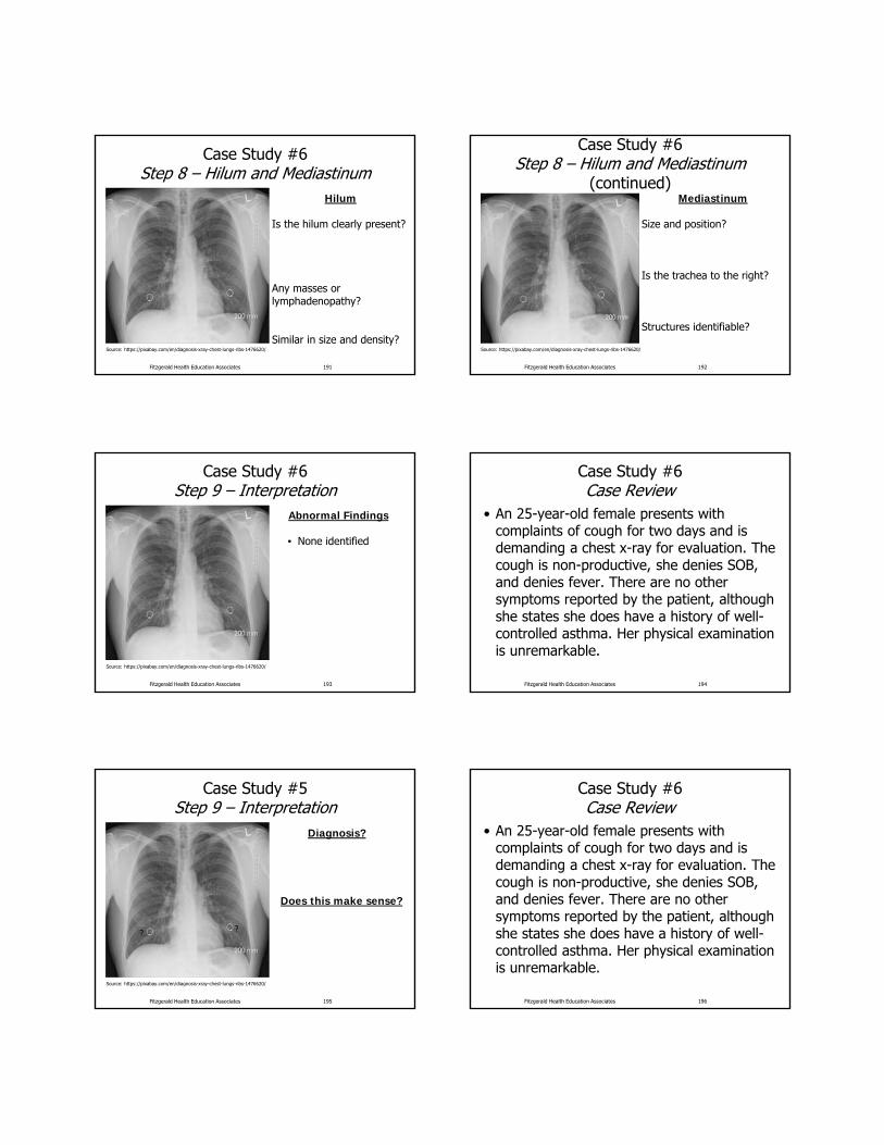
Case Study #6Step 8 – Hilum and Mediastinum
Hilum
Is the hilum clearly present?
Any masses or lymphadenopathy?
Similar in size and density?Source: https://pixabay.com/en/diagnosis-xray-chest-lungs-ribs-1476620/
Fitzgerald Health Education Associates 191
Case Study #6Step 8 – Hilum and Mediastinum
(continued)Mediastinum
Size and position?
Is the trachea to the right?
Structures identifiable?
Source: https://pixabay.com/en/diagnosis-xray-chest-lungs-ribs-1476620/
Fitzgerald Health Education Associates 192
Case Study #6Step 9 – Interpretation
Abnormal Findings
• None identified
Source: https://pixabay.com/en/diagnosis-xray-chest-lungs-ribs-1476620/
Fitzgerald Health Education Associates 193
Case Study #6Case Review
• An 25-year-old female presents with complaints of cough for two days and is demanding a chest x-ray for evaluation. The cough is non-productive, she denies SOB, and denies fever. There are no other symptoms reported by the patient, although she states she does have a history of well-controlled asthma. Her physical examination is unremarkable.
Fitzgerald Health Education Associates 194
Case Study #5Step 9 – Interpretation
Diagnosis?
Does this make sense?
Source: https://pixabay.com/en/diagnosis-xray-chest-lungs-ribs-1476620/
? ?
Fitzgerald Health Education Associates 195
Case Study #6Case Review
• An 25-year-old female presents with complaints of cough for two days and is demanding a chest x-ray for evaluation. The cough is non-productive, she denies SOB, and denies fever. There are no other symptoms reported by the patient, although she states she does have a history of well-controlled asthma. Her physical examination is unremarkable.
Fitzgerald Health Education Associates 196

Reference Slides
Other Common Abnormalities
Fitzgerald Health Education Associates 197
Atelectasis• Condition of volume loss in some
portion of lung• May involve sub-segment, segment,
lobe or entire lung• Increased density usually linear• Collapse or incomplete expansion of
the lung or part of the lung • Segmental and sub-segmental collapse
may show linear, curvilinear, wedge shaped opacities
Fitzgerald Health Education Associates 198
Atelectasis Causes
• Obstructive– Most common– Bronchus obstructed by mucous plug,
neoplasm, or foreign body
• Compressive– Normal lung compressed by tumor,
emphysematous bulla or heart enlargement
Fitzgerald Health Education Associates 199
Atelectasis Causes (continued)
• Cicatrization– Organizing scar tissue– Most often after healing granulomatous
disease (i.e., TB), pulmonary infarct or trauma
• Adhesive– Inactivation of surfactant (i.e., hyaline
membrane disease)
Fitzgerald Health Education Associates 200
Atelectasis Causes (continued)
• Passive– Normal compliance of the lung with
pneumothorax or pleural effusion– Airway remains patent
Fitzgerald Health Education Associates 201
Linear Atelectasis
Source: https://commons.wikimedia.org/wiki/File:Thorax_mit_bds_Unterlappen-Atelektase_mit_Voraufnahme.jpg
Fitzgerald Health Education Associates 202

Pulmonary Edema• Cardiogenic
– Increased hydrostatic pulmonary capillary pressure
• Non-cardiogenic– Altered capillary
membrane permeability or decreased plasma oncotic pressure
• NOT CARDIAC (pneumonic)– Near-drowning, Oxygen
therapy, Transfusion or Trauma, CNS disorder, ARDS, Aspiration, or Altitude sickness, Renal disorder or Resuscitation, Drugs, Inhaled toxins, Allergic Alveolitis, Contrast or Contusion
Fitzgerald Health Education Associates 203
Cardiogenic Pulmonary Edema
• Cephalization of the pulmonary vessels• Kerley A lines
– Thin linear opacities in mid and upper zones radiating to hila
• Kerley B lines – Linear opacities 1‒2 cm long and 1‒2 mm
thick perpendicular to pleural surface caused by interstitial fluid (septal lines)
Fitzgerald Health Education Associates 204
Cardiogenic Pulmonary Edema (continued)
• Peribronchial cuffing• “Bat wing" pattern
– Perihilar and medullary consolidation of both lungs
• Patchy shadowing with air bronchograms
• Heart enlargement • Pleural effusions
Fitzgerald Health Education Associates 205
Pulmonary EdemaCephalization of Vessels
Source: Anthony Angelow
Fitzgerald Health Education Associates 206
Bat Wing Pattern
Source: Anthony Angelow and Theresa Campo (used with permission)
Fitzgerald Health Education Associates 207
Pulmonary Edema
Source: Anthony Angelow
Fitzgerald Health Education Associates 208

Heart Failure
Source: Anthony Angelow
Fitzgerald Health Education Associates 209
Pleural Effusion
• Causes– CHF– Infection (parapneumonic)– Trauma– PE– Tumor– Autoimmune disease– Renal failure
Fitzgerald Health Education Associates 210
Pleural Effusion (continued)
Source: Anthony Angelow
Fitzgerald Health Education Associates 211
Pulmonary Nodules
Source: Anthony Angelow
Fitzgerald Health Education Associates 212
Pulmonary Nodules (continued)
Fitzgerald Health Education Associates 213
References
• Campo, T.M. (2017). Clinical Chest Radiography Podium Presentation at National Conference of Nurse Practitioners.
• Campo, T.M. (2016). Medical Imaging for Health Care Providers: Practical Radiograph Interpretation. Springer Publishing: New York, NY.
Fitzgerald Health Education Associates 214

References(continued)
• Collins and Stern. (2008). Chest Radiology: The Essentials. Wolters Kluwer/LWW: Philadelphia, PA.
• Daffner and Hartman (2014). Clinical Radiology: The Essentials 4th ed. Wolters Kluwer/LWW: Philadelphia, PA.
Fitzgerald Health Education Associates 215
References(continued)
• Hermann et al. (2012). Best practices in digital radiography: White paper. American Society of Radiologic Technologies accessed from http://www.asrt.org/docs/whitepapers/asrt12_bstpracdigradwhp_final.pdf
• Wallace, J.E. (2015). Radiographic exposure: Principles and practice. FA Davis: Philadelphia, PA.
Fitzgerald Health Education Associates 216
• Images/Illustrations: Unless otherwise noted, all images/illustrations are from open sources, such as the CDC or Wikipedia or property of FHEA or author.
• All websites listed active at the time of publication.
Fitzgerald Health Education Associates 217
Copyright Notice
Copyright by Fitzgerald Health Education AssociatesAll rights reserved. No part of this publication may be reproduced or transmitted
in any form or by any means, electronic or mechanical, including photocopy, recording or any information storage and retrieval system, without permission
from Fitzgerald Health Education Associates
Requests for permission to make copies of any part of the work should be mailed to:
Fitzgerald Health Education Associates85 Flagship Drive
North Andover, MA 01845-6184
Fitzgerald Health Education Associates 218
Statement of Liability
• The information in this program has been thoroughly researched and checked for accuracy. However, clinical practice and techniques are a dynamic process and new information becomes available daily. Prudent practice dictates that the clinician consult further sources prior to applying information obtained from this program, whether in printed, visual or verbal form.
• Fitzgerald Health Education Associates disclaims any liability, loss, injury or damage incurred as a consequence, directly or indirectly, of the use and application of any of the contents of this presentation.
Fitzgerald Health Education Associates 219
Fitzgerald Health Education Associates
85 Flagship Drive
North Andover, MA 01845-6154978.794.8366 Fax-978.794.2455
Website: fhea.com
Learning and Testing Center: fhea.com/npexpert
www.facebook.com/fitzgeraldhealth
@npcert
Fitzgerald Health Education Associates 220