Embed Size (px)
DESCRIPTION
Discipline : “ Preventive Dentistry” Programme of lectures: – 7 lectures + 7 lectures Textbooks for self-study are: - Norman O. Harris, Primary Preventive Dentistry, 6th Ed. (2004); 7 th ed.(2009) - Hardy Limeback , Comprehensive Preventive Dentistry, 2012 - PowerPoint PPT Presentation
Citation preview

Discipline : “ Preventive Dentistry” Programme of lectures: – 7 lectures + 7 lectures Textbooks for self-study are:
- Norman O. Harris, Primary Preventive Dentistry, 6th Ed. (2004); 7th ed.(2009) - Hardy Limeback , Comprehensive Preventive Dentistry, 2012
Exam : after 7th semester - in January 2015. Exam consists : 1
written question from the syllabus and test ( multiple choice), all
in one day.

Etiology and pathogenesis of dental caries.
Prof.d-r R.Kabaktchieva- 2014
The three general disease categories of focus in dentistry are :
- dental decay, - periodontal disease, - oral cancer.
Dentistry in the past has been treatment oriented,but we are witnessing an interest prevention.

It is obviously better to prevent the disease in the first place, than treat it once it has happened.
The goals of preventive dentistry are to avoid disease altogether.

Maintaining a disease-free state can result from primary prevention. When lifestyle changes are made early on, the risk for developing dental disease are minimized.
Lifestyle changes – less carbohydrates – better oral hygiene – improved nutrition – better education

Secondary prevention (reverse, arrest incipient caries) and early intervention (MID &“Preventive Resins”)
can be used to reverse the initiation of disease.
An outcome of good health can still be achieved, when incipient enamel lesions are reversed before cavities form.
MID- minimal intervention dentistry

Far too often though, dentists spend most of their time treating dental disease
in an endless cycle of repeat restorations, which leads…… to tooth loss.
!
The goal of primary prevention is never to have had
any kind of dental disease.
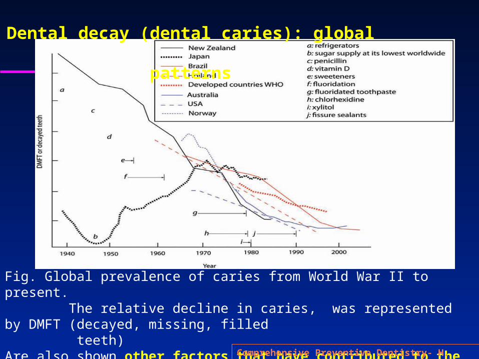
Fig. Global prevalence of caries from World War II to present. The relative decline in caries, was represented by DMFT (decayed, missing, filled teeth) Are also shown other factors that have contributed to the decline in caries worldwide (labeled a to j)
Comprehensive Preventive Dentistry- H. Limeback
Dental decay (dental caries): global patterns
Тhe prevalence of caries has changed over the decades.
In every developed country, there has been a steady decline in dental decay.
Experts believe, that it was primarily the introduction of fluoride therapies after the
1960s that had a huge impact on dental decay rates.
To know how to prevent , need to know disease, its etiology and pathogenesis
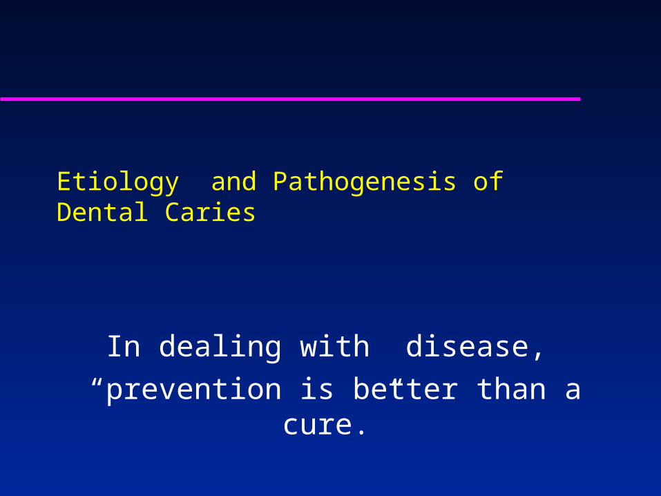
Etiology and Pathogenesis of Dental Caries
In dealing with disease, “prevention is better than a cure.”

Definition
Dental caries is a dietary carbohydrate-modified bacterial infectious disease with saliva as a critical regulator.
It is the most common chronic infectious disease of childhood

Contemporary definition : Tooth decay is localized progressive disease, whose character consists in the destruction of tooth
structures mainly under the influence of metabolic products of the oral microflora;
Each level of decomposition is clinically differentiated.

Kidd
Caries process takes place in the biofilm on the tooth
surface .
Carious lesion is the result of carious process developing
between the microbial biofilm and tooth structure
The metabolic activity of the microorganisms in
the biofilm is invisible to the clinician,
but carious lesion that is a result of this activity
is clinically apparent.

The classic Vennediagram of caries.
Must have a tooth, plaque bacteria,
fermentable carbohydrate, saliva,
and enough time in order for a
carious lesion to develop .
Caries results when all of the factors
that contribute to caries overlap.
(red color, center).
Several factors influencing each
component, ( see the diagram,) affect
the rate and severity of the caries.
Caries Factors
diagram

This is a convenient analogy to understand and is anoffshoot of the classic Venn diagram ( first introduced by Keyes (1962).

The role of microorganisms
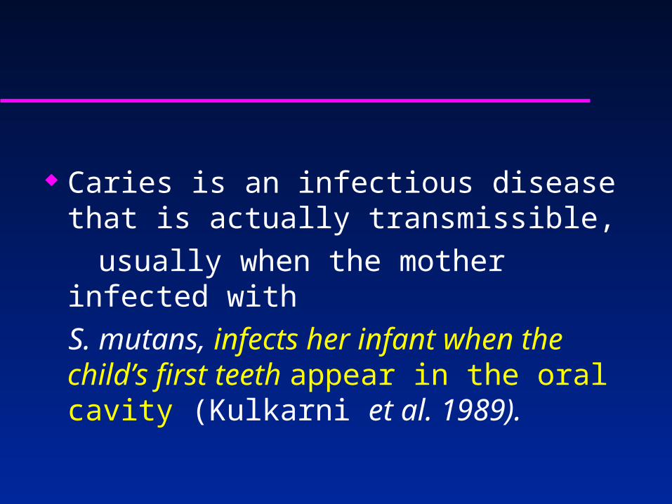
Caries is an infectious disease that is actually transmissible,
usually when the mother infected with S. mutans, infects her infant when the
child’s first teeth appear in the oral cavity (Kulkarni et al. 1989).

Dental caries does not occur in a sterile mouth. (no mouth can ever be made sterile) The conditions in the oral cavity are ideal for the growth of bacteria that metabolize sugar to acids. The oral cavity is generally a warm place, at body temperature (37°C) encouraging the growth of bacteria.
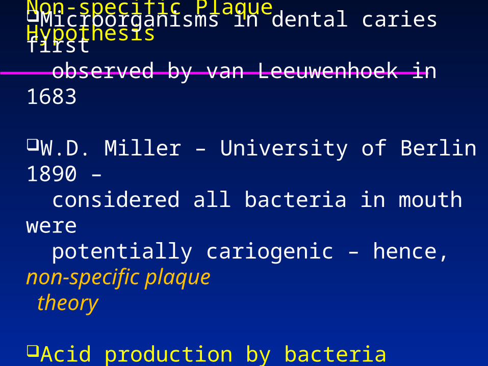
Non-specific Plaque Hypothesis
Microorganisms in dental caries first observed by van Leeuwenhoek in 1683
W.D. Miller – University of Berlin 1890 – considered all bacteria in mouth were potentially cariogenic – hence, non-specific plaque theory
Acid production by bacteria considered responsible for breakdown of tooth

Specific Plaque Hypothesis
1924 – Clarke isolated a streptococcus species from a cavity in a child
The bacteria underwent some changes as the culture aged – Clarke named it Streptococcus mutans for “mutation”

Mutans streptococci 1960 – Keyes “rediscovered” S. mutans He demonstrated that:
– specific microorganisms were responsible for caries– caries was transmissible
Later, the responsible bacteria were found to comprise seven distinct species – only mutans and sobrinus are associated with caries in humans
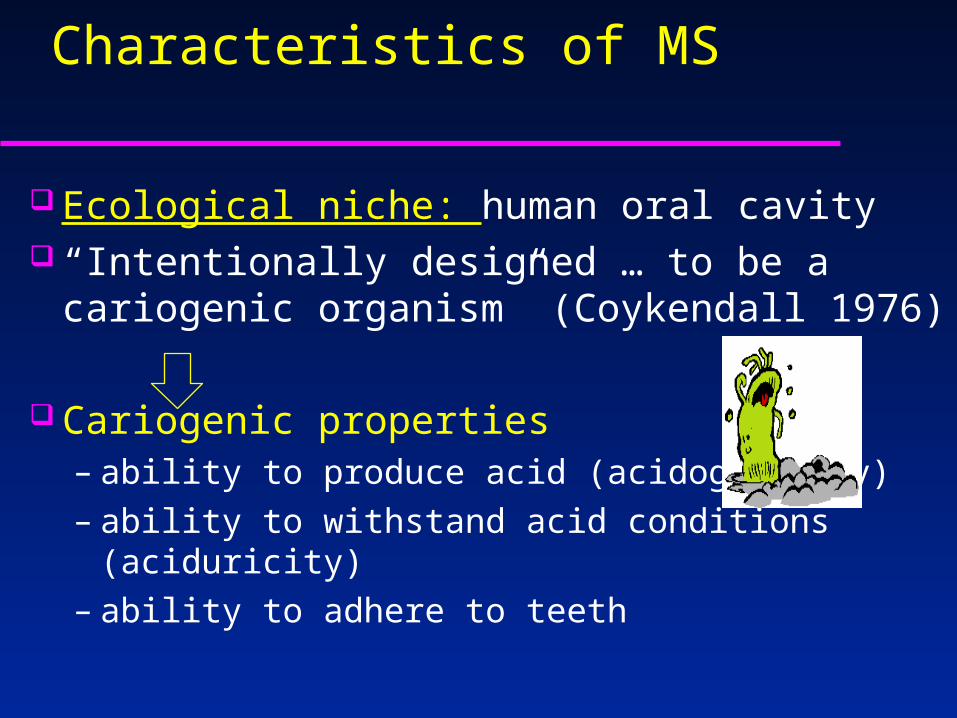
Characteristics of MS
Ecological niche: human oral cavity “Intentionally designed … to be a cariogenic
organism” (Coykendall 1976) Cariogenic properties
– ability to produce acid (acidogenicity)– ability to withstand acid conditions (aciduricity)– ability to adhere to teeth
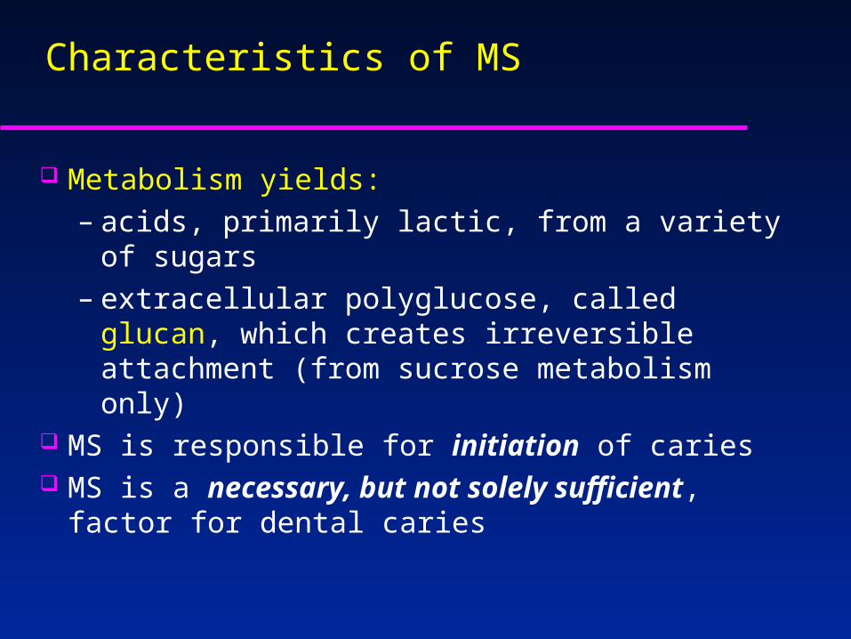
Characteristics of MS
Metabolism yields:– acids, primarily lactic, from a variety of sugars– extracellular polyglucose, called glucan, which
creates irreversible attachment (from sucrose metabolism only)
MS is responsible for initiation of caries MS is a necessary, but not solely sufficient, factor for
dental caries

Acquisition of MS by Infants
MS colonize oral cavity after eruption of teeth – require hard, non-desquamating surface;
Some believe in “window of infectivity “ that relievs on virgin tooth surfaces for initial colonization;
Second “window may open” when permanent dentition erupts

Acquisition of MS by Infants
MS is poor competitor for colonization – once stable biofilm is in place, ability for MS to colonize is reduced
Infants who acquire sanguis early have less MS
Birthmitis
1
sanguis
8 11 19 26 33 mos.
mutans
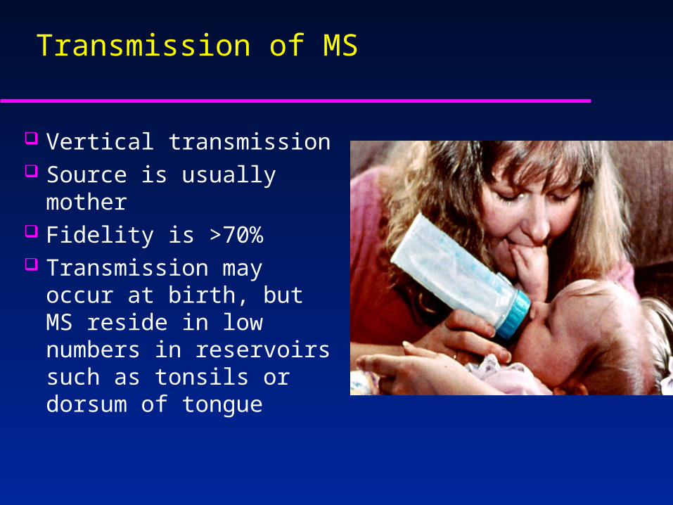
Transmission of MS
Vertical transmission Source is usually mother Fidelity is >70% Transmission may occur at
birth, but MS reside in low numbers in reservoirs such as tonsils or dorsum of tongue

Other Microorganisms
Lactobacilli sp.– found in large numbers in some children– considered opportunistic, not initiators– numbers in cavity increase after DEJ
invaded– lactobacilli are good indicators of total
carbohydrate intake
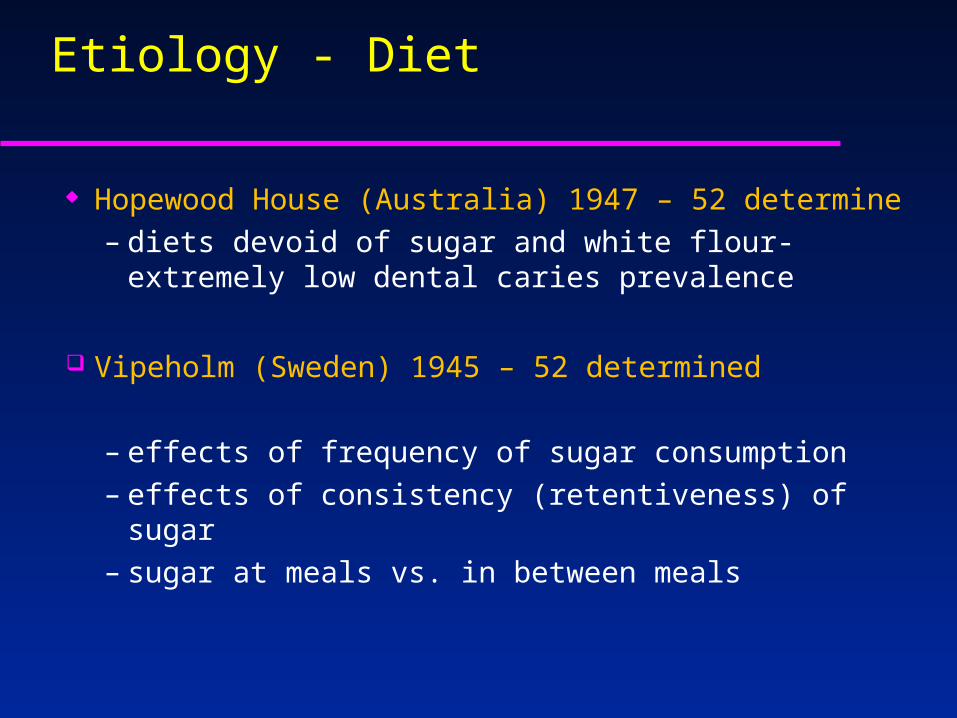
Etiology - Diet
Hopewood House (Australia) 1947 – 52 determine– diets devoid of sugar and white flour- extremely low
dental caries prevalence
Vipeholm (Sweden) 1945 – 52 determined
– effects of frequency of sugar consumption– effects of consistency (retentiveness) of sugar– sugar at meals vs. in between meals
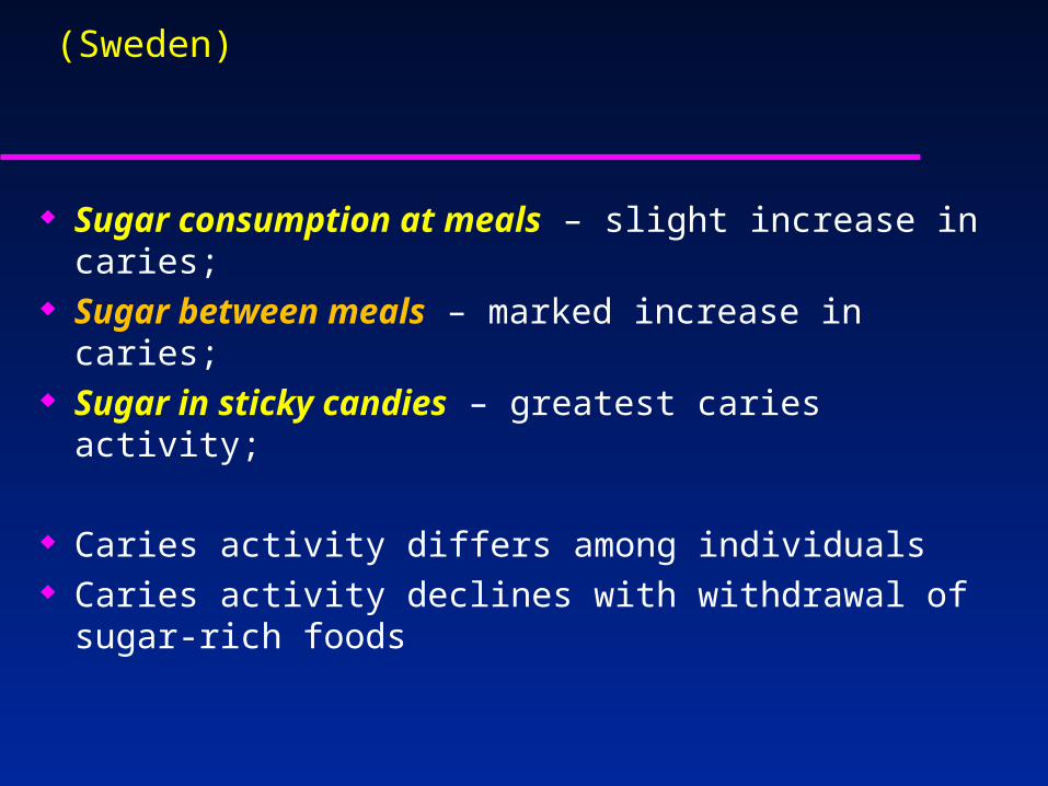
Lessons from Vipeholm (Sweden)
Sugar consumption at meals – slight increase in caries; Sugar between meals – marked increase in caries; Sugar in sticky candies – greatest caries activity;
Caries activity differs among individuals Caries activity declines with withdrawal of sugar-rich
foods

Not all sugars are cariogenic.
The role of dietary sugars

The more common dietary sugars are presented. The cariogenic potential of carbohydrates are presented too.
The sugars with the most
cariogenicity are sucrose
and glucose (red).
Other carbohydrates (maltose,
lactose, fructose, and starch)
are less cariogenic.
The sugar alcohols, such as
sorbitol and mannitol, are the
least cariogenic (yellow)
Xylitol has even been shown to
be anticariogenic (green).
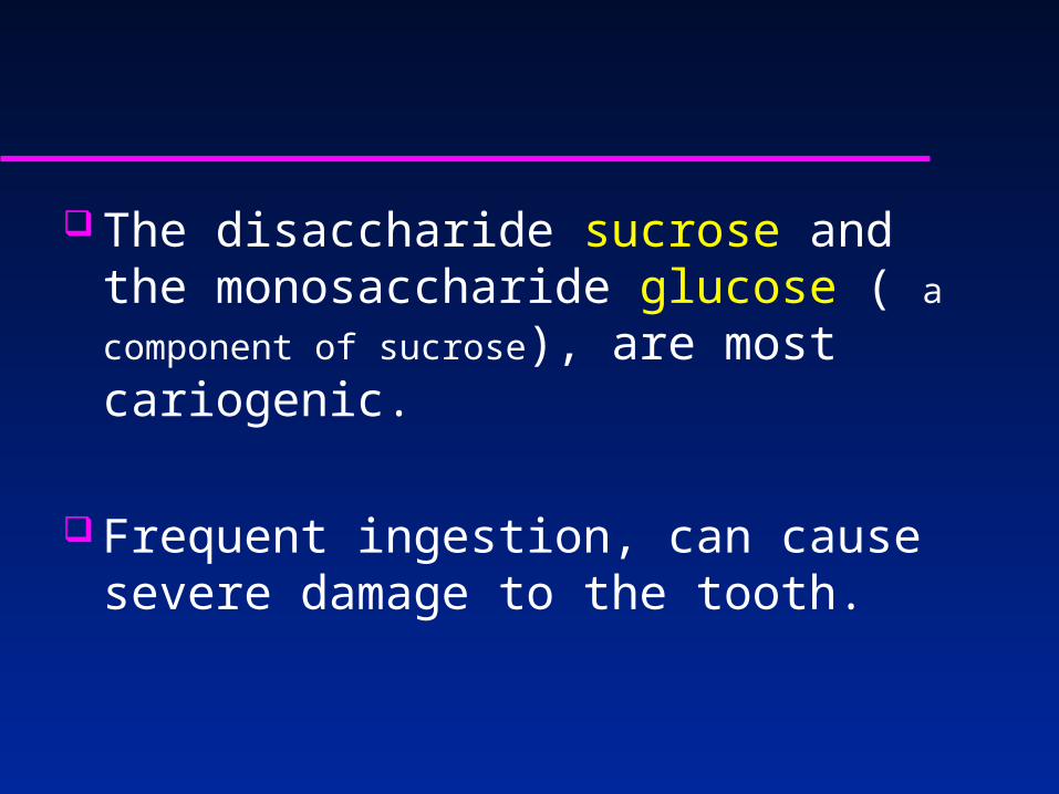
The disaccharide sucrose and the monosaccharide glucose ( a component of sucrose), are most cariogenic.
Frequent ingestion, can cause severe damage to the tooth.

There is no question that carbohydrates are the main etiological reason for the development of caries.
One of the strategies in prevention of caries is to limit access to the more cariogenic sugars and substitute them with the anti-cariogentic ones.
Not only does their conversion to acid result in enamel dissolution, but they also encourage the growth of more virulent cariogenic bacteria.
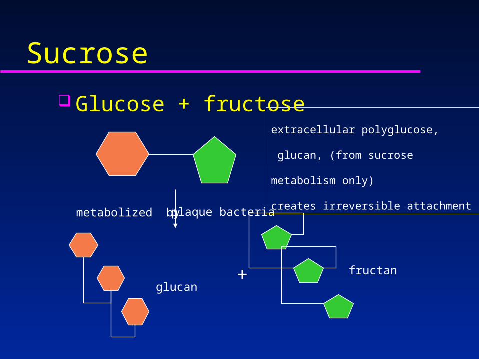
Sucrose Glucose + fructose
glucanfructan
metabolized by plaque bacteria
+
extracellular polyglucose,
glucan, (from sucrose metabolism only)
creates irreversible attachment

Glucan Water soluble Extracellular “glue” Enables adhesion to tooth
– reduced susceptibility to mechanical disruption
Inhibits diffusion properties of plaque– reduces buffering capacity of saliva– inhibits transport of acid away from tooth
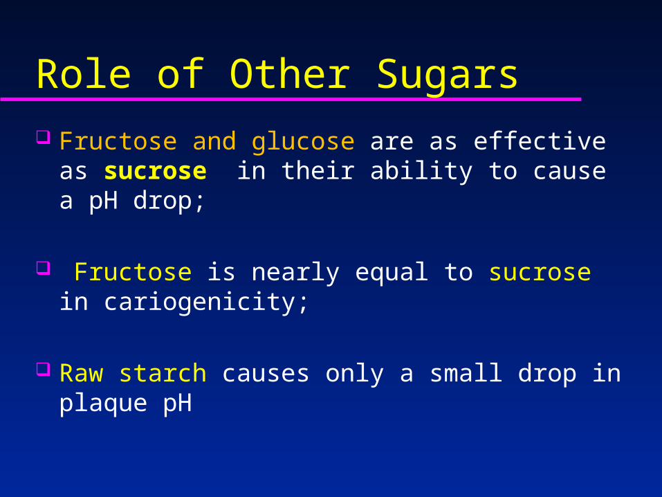
Role of Other Sugars Fructose and glucose are as effective as sucrose in
their ability to cause a pH drop;
Fructose is nearly equal to sucrose in cariogenicity;
Raw starch causes only a small drop in plaque pH
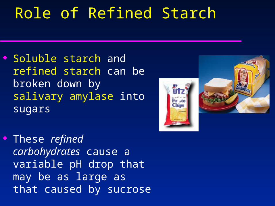
Role of Refined Starch
Soluble starch and refined starch can be broken down by salivary amylase into sugars
These refined carbohydrates
cause a variable pH drop that may be as large as that caused by sucrose

The supragingival bacteria are dominated with streptococci
and lactobacilli that can lower the plaque pH and induce decalcifications
white spot lesions.
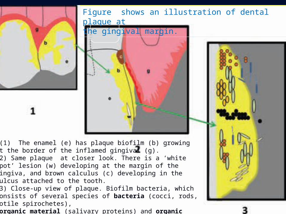
(1) The enamel (e) has plaque biofilm (b) growing at the border of the inflamed gingival (g). (2) Same plaque at closer look. There is a ‘white spot’ lesion (w) developing at the margin of the gingiva, and brown calculus (c) developing in the sulcus attached to the tooth. (3) Close-up view of plaque. Biofilm bacteria, whichconsists of several species of bacteria (cocci, rods, motile spirochetes), organic material (salivary proteins) and organic matter secreted by the bacteria (yellow-stained)
Figure shows an illustration of dental plaque atthe gingival margin.
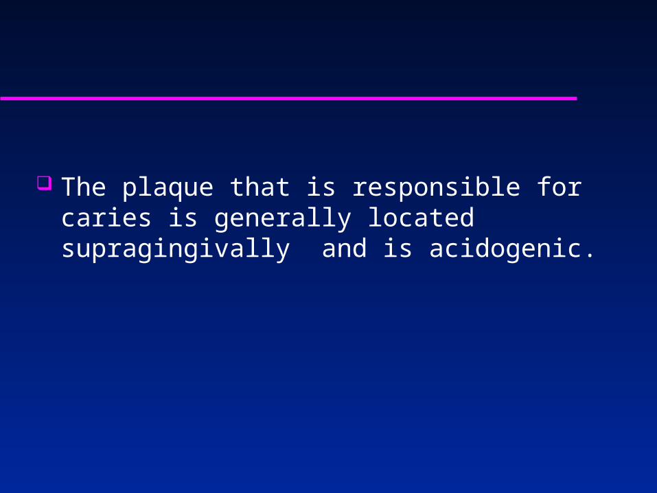
The plaque that is responsible for caries is generally located supragingivally and is acidogenic.

People who consume sugars frequently in their diet increase the levels of streptococci and lactobacilli
The two bacteria species thought to be responsible for caries.
These bacteria continue to thrive as the pH drops.
If the plaque is not removed, eventually, the enamel starts to decalcify and an incipient ‘white spot’ lesion ensues.

Figure .The enamel white spot lesion at the mesial contact zone of thefirst maxillary right molar .These white-spot lesions are sometimes filled by dentistsbut can be remineralized.

Marsh (1994) was able to show, that feeding of bacteria a meal of glucose can encourage the growth of cariogenic bacteria
when the pH is allowed to drop .
Repeated glucose rinses encourages SM and LB growth when plaque acid is not controlledFluoride at high concentrations inhibits SM , but not LB !!!
Xylitol had inhibitory properties for both cariogenic and periodontal bacteria.

The demineralization–remineralizationbalance in caries
The plaque thickness dominated by cariogenic bacteria, can effectively keep the saliva from reaching the enamel surface.
In addition, the more plaque there is, the more acid is produced.
These acids have a longer time to penetrate into the enamel under thick biofilm - This allows the tooth to demineralize!!!!
If the saliva reaches the acids they are washed away and neutralized by the salivary buffers - This allows the tooth to remineralize.
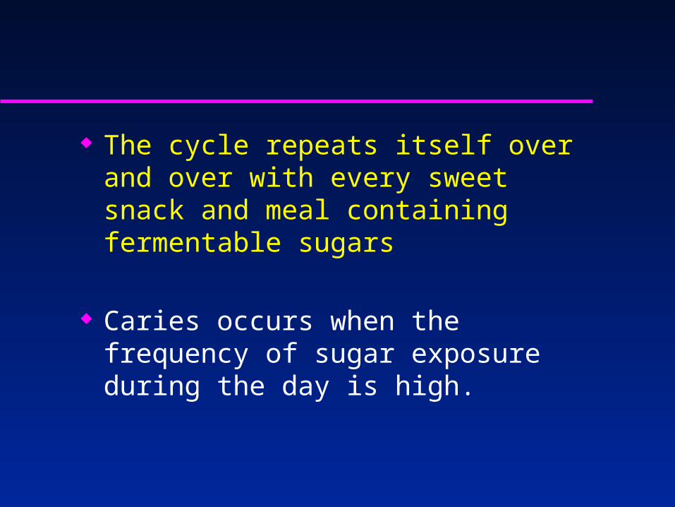
The cycle repeats itself over and over with every sweet snack and meal containing fermentable sugars
Caries occurs when the frequency of sugar exposure during the day is high.

The repeated cycle of ‘sugar attacks.’

The pH of dental plaque in response to glucose has been studied using the classic
Stephan curve The diagram illustrates the plaque pH response curves that have been obtained from patients with differentrisks for caries.
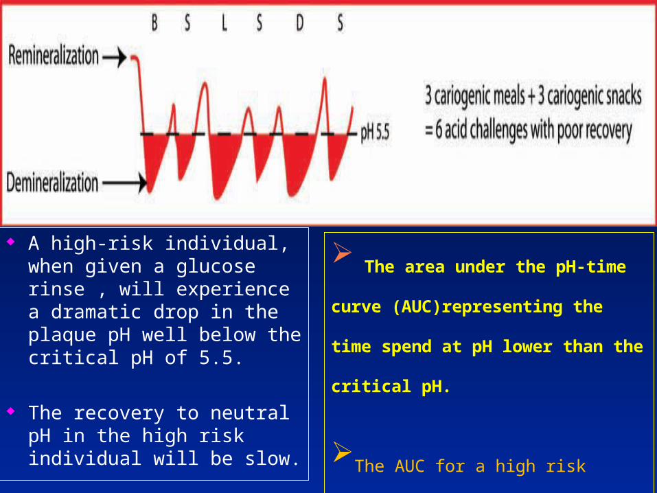
A high-risk individual, when given a glucose rinse , will experience a dramatic drop in the plaque pH well below the critical pH of 5.5.
The recovery to neutral pH in the high risk individual will be slow.
The area under the pH-time curve
(AUC)representing the time spend at pH
lower than the critical pH.
The AUC for a high risk
individual will be very large.
AUC is a better measure of total caries
risk.

The person with a high risk for caries snacks frequently during the day,
and the total AUCs clearly are excessive and will not allow remineralization to occur.
If that daily trend continues, the person will experience dental decay.

For a moderate risk individual (yellow), the initial pH drop may only be a little lower than the critical pH, and the AUC will be much less.
For a caries-resistant person
(green),the initial pH drop of that
person’s plaque may
not even reach the critical pH, and
the recovery will be very quick.
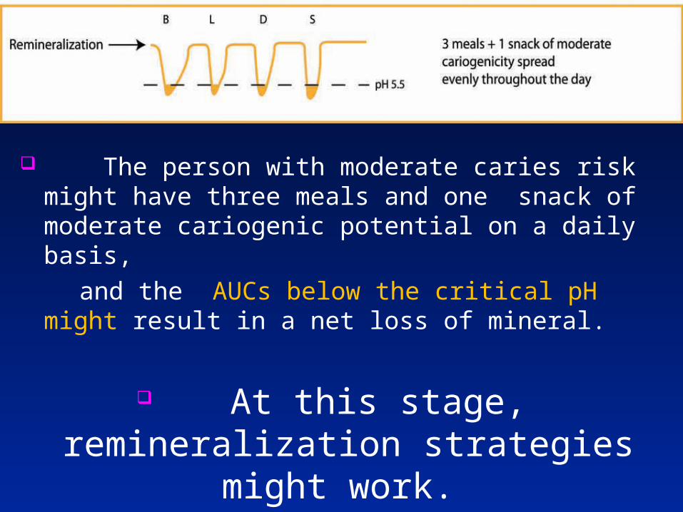
The person with moderate caries risk might have three meals and one snack of moderate cariogenic potential on a daily basis,
and the AUCs below the critical pH might result in a net loss of mineral.
At this stage, remineralization strategies might work.

Тhe person at low risk may not snack at all and has three meals of low cariogenicity spread apart during the day.
This allow remineralization to occur.

Researchers have determined that: it is not only the frequency of ingestion that is
important, but it is also the type of fermentable carbohydrate that is
ingested.
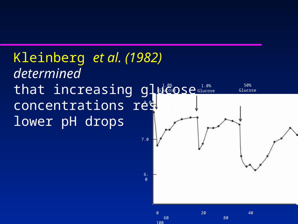
Kleinberg et al. (1982) determinedthat increasing glucose concentrations results in lower pH drops 1.0% Glucose 50% Glucose
7.0
8.0
6.0
0 20 40 60 80 100
1.0% Glucose

Etiology – Host Factors
Tooth factors– quality of enamel– presence/depth of pits and fissures– hypoplasia– fluoride exposure
Saliva– pH– flow rate– buffering capacity– antimicrobial components
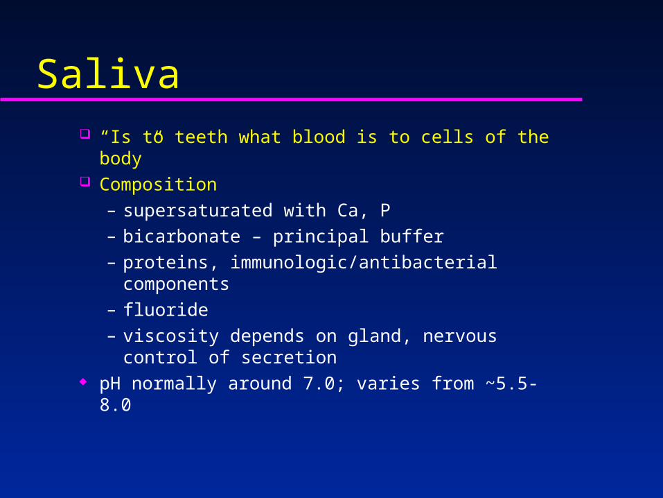
Saliva “Is to teeth what blood is to cells of the body” Composition
– supersaturated with Ca, P– bicarbonate – principal buffer– proteins, immunologic/antibacterial components– fluoride– viscosity depends on gland, nervous control of
secretion pH normally around 7.0; varies from ~5.5-8.0
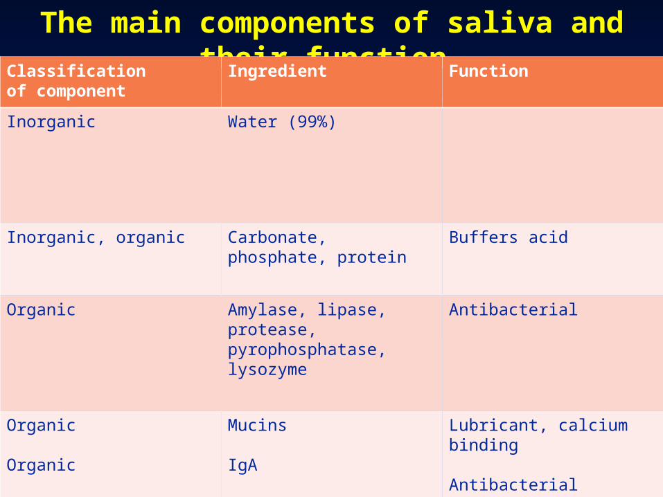
The main components of saliva and their function Classificationof component
Ingredient Function
Inorganic Water (99%)
Inorganic, organic Carbonate,phosphate, protein
Buffers acid
Organic Amylase, lipase,protease,pyrophosphatase,lysozyme
Antibacterial
Organic
Organic
Mucins
IgA
Lubricant, calcium binding
Antibacterial

The role of saliva Saliva contains antibacterial proteins,
electrolytes for remineralization and the essential nutrients for bacteria to grow.
The host provides the dietary carbohydrates that are easily converted to energy and acids by the bacteria that leads to dissolution of dental hard tissues.
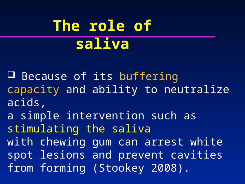
Because of its buffering capacity and ability to neutralize acids, a simple intervention such as stimulating the saliva with chewing gum can arrest white spot lesions and prevent cavities from forming (Stookey 2008).
The role of saliva
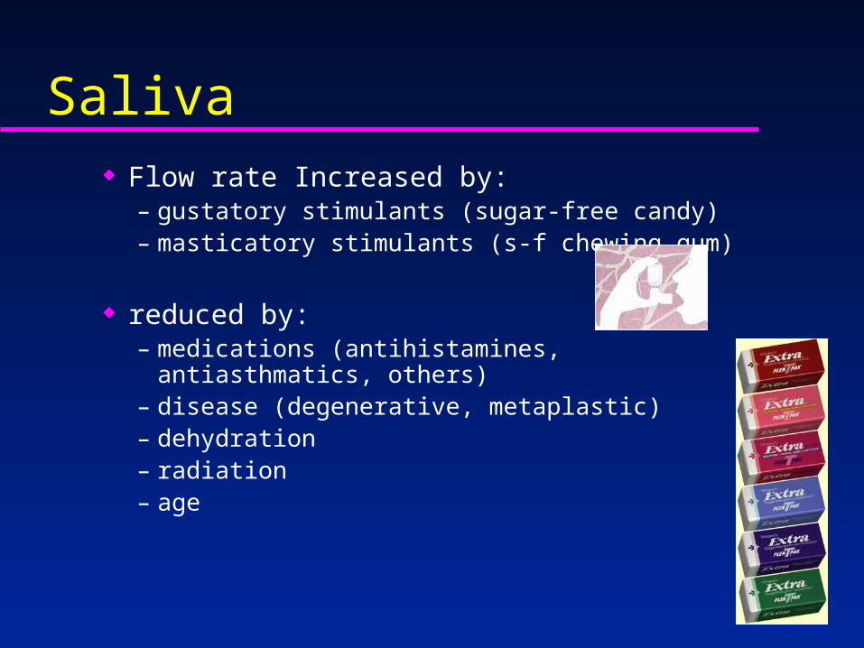
Saliva Flow rate Increased by:
– gustatory stimulants (sugar-free candy)– masticatory stimulants (s-f chewing gum)
reduced by:– medications (antihistamines, antiasthmatics, others)– disease (degenerative, metaplastic)– dehydration– radiation– age

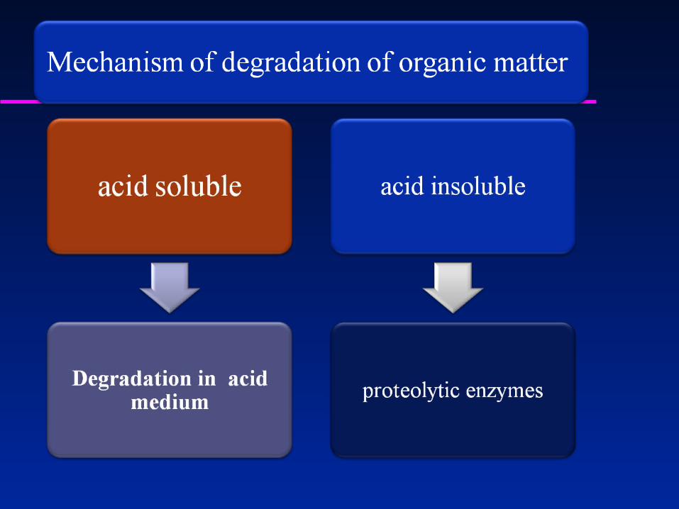
2. Pathogenesis of dental caries
Mechanism of development of dental caries
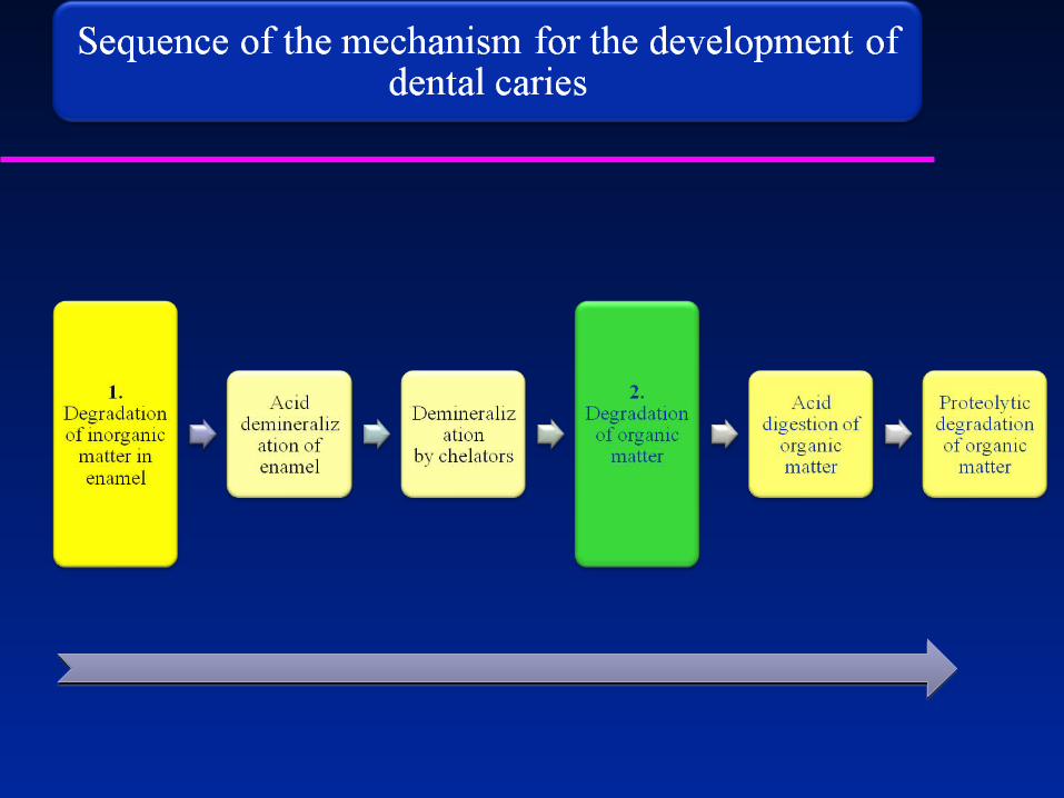

Mechanism of acid demineralization
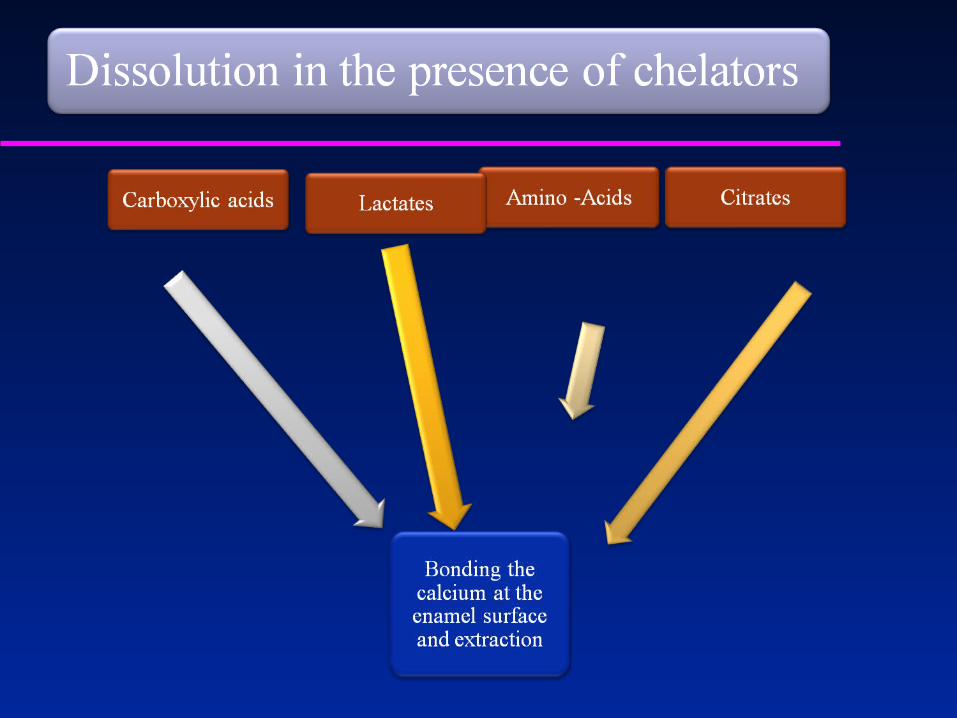
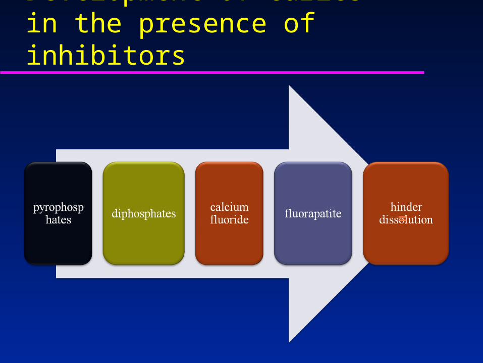
Development of caries in the presence of inhibitors
=
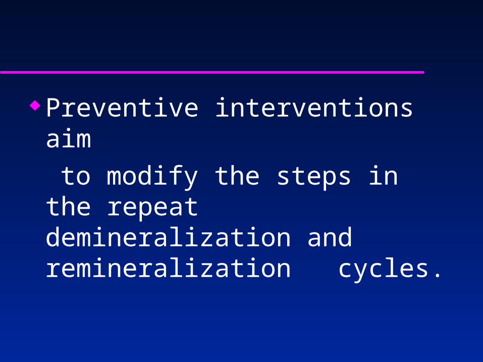
Preventive interventions aim to modify the steps in the repeat
demineralization and remineralization cycles.
1. Neutralize the plaque acids: This can be done by adding base
or adding buffers such as sodium bicarbonate (baking soda) to the saliva to boost its ability to neutralize acids.
2. Improve hygiene: With bacterial levels low, less acid is
produced. Plaque layers don’t have a chance to grow
thick; Saliva can penetrate better to the enamel surface through thin layers of plaque.

3. Introduce antimicrobials: Since caries is a disease caused by
bacteria, simply eliminating the bacteria or controlling their growth will reduce the caries incidence.
Chlorhexidine, xylitol, ozone, even experimental antibodies, have been
used to control bacterial growth.

4. Stimulate saliva: Saliva contains numerous components -
that fight tooth decay buffers, remineralizing minerals,
antimicrobial enzymes, antibodies.

5. Topical fluorides: Fluoride added to the remineralizing incipient lesion increases the enamel crystals’ resistance to dissolution by plaque acids.
6. Remineralizing strategies: Remineralization can be promoted with the use of calcium-
phosphate complexes such and ACP-CPP.

end