Embed Size (px)
Citation preview
Biochemical Journal 441 (1) : 487-498 (2012)
Directed evolution of a temperature, peroxide and
alkaline pH tolerant versatile peroxidase
Eva GARCIA-RUIZ*, David GONZALEZ-PEREZ*, Francisco J. RUIZ-DUEÑAS†, Angel T. MARTINEZ† and Miguel ALCALDE*,1
*Department of Biocatalysis, Institute of Catalysis, CSIC, Cantoblanco, 28049 Madrid, Spain and †Centro de Investigaciones Biológicas, CSIC, Ramiro de Maeztu 9, 28040 Madrid, Spain.
1To whom correspondence should be addressed: [email protected].
Page heading title: Directed evolution of versatile peroxidase.
Biochemical Journal 441 (1) : 487-498 (2012)
SYNOPSIS
The versatile peroxidases (VPs) secreted by white-rot fungi are fundamental for the natural decay of lignin. In this study, a fusion gene containing the VP from Pleurotus eryngii was subjected to six rounds of directed evolution, achieving a level of secretion in Saccharomyces cerevisiae (21 mg/L) as yet unseen for any ligninolytic peroxidase. The evolved variant for expression harboured 4 mutations and increased its total VP activity 129-fold. The signal leader processing by the STE13 protease at the Golgi compartment changed as consequence of over-expression, retaining the additional N-terminal sequence EAEA that enhanced secretion. The engineered N-terminal truncated variant displayed similar biochemical properties as the non-truncated counterpart in terms of kinetics, stability and spectroscopic features. Additional cycles of evolution raised the T50 8ºC and significantly increased the enzyme’s stability at alkaline pHs. In addition, the Km for H2O2 was enhanced up to 15-fold while the catalytic efficiency was maintained, and there was an improvement in peroxide stability (with half lives for H2O2 of 43 min at a H2O2/enzyme molar ratio of 4000:1). Overall, the directed evolution approach described provides a set of strategies for selecting VPs with improvements in secretion, activity and stability.
Key words: versatile peroxidase, directed evolution, enzyme promiscuity, Saccharomyces cerevisiae, α-factor prepro-leader.
Abbreviation used: ABTS, 2,2´-azino-bis(3-ethylbenzothiazoline-6-sulfonic acid); CIP, Coprinopsis cinerea peroxidase; DMP; 2,6 dimethoxyphenol; Eº, redox potential; HRP, horseradish peroxidase; HTP, high-throughput; IA, initial activity; IvAM, in vivo assembly of mutant libraries with different mutational spectra; IVOE, in vivo overlap extension; LiP, lignin peroxidase; MALDI-TOF, matrix assisted laser desorption and ionization-time of flight; MnP, manganese peroxidase; MW, molecular weight; pI, isoelectric point; RA, residual activity; RB5, reactive black 5; Rz, Reinheitszahl value; StEP, staggered extension process; T50, temperature at which the enzyme retains 50 % of its activity after a 10 min incubation; Ta, thermo-activity; TAI, total activity improvement; T1/2,H2O2, half lives in the presence of hydrogen peroxide; VA, veratryl alcohol; VP, versatile peroxidase.
Biochemical Journal 441 (1) : 487-498 (2012)
INTRODUCTION
The ligninolytic enzymatic consortium secreted by white-rot fungi represents a remarkable example of a highly efficient oxidative system [1]. For decades, the complex interactions and synergies between enzymes and diffusible electron carriers generated by the wood-decaying fungi during lignin combustion have constituted a hot topic of research [2]. Typically formed by different oxidoreductases (mostly laccases, peroxidases and H2O2-supplying oxidases), this lignin-degrading array of enzymes have potential applications in the production of 2nd generation biofuels, pulp biobleaching, the design of nanobiodevices (biosensors and biofuel cells), organic synthesis or bioremediation, to name a few [3-6]. Among these enzymes, the high-redox potential versatile peroxidase (VP) is arousing great interest because of its catalytic promiscuity, being mainly described in Pleurotus, Bjerkandera and some other basidiomycete species [7-13].
As a protein, VP is classified by its structural properties and sequence information in the Class II corresponding to fungal secreted haem-containing peroxidases. As an enzyme, it is presented as a high redox-potential substrate and Mn2+ oxidizing peroxidase (E.C. 1.11.1.6) [14]. VPs combine the general catalytic features of other haem-containing enzymes (in terms of substrate specificity and reaction mechanisms), like the high-redox potential ligninolytic peroxidases, lignin peroxidase (LiP) and manganese peroxidase (MnP), with those of peroxidases with a lower redox potential, such as horseradish peroxidase (HRP) and Coprinopsis cinerea peroxidase (CIP), [15]. The VPs substrate promiscuity is associated with a high-redox potential (Eº > +1.4 V) and the presence of different catalytic sites for the oxidation of low and high redox-potential compounds [14,16]. Comprehensive structure-function studies have helped reveal the mechanisms underlying the complex activity of these enzymes, which is governed by a haem domain located in the middle of a structure formed by twelve helixes and that is connected to the protein surface by two small channels. In a scaffold of ~300 amino acids, VPs contain three carboxylic residues implicated in the coordination of Mn2+ and one catalytic tryptophan involved in the oxidation of high-redox potential compounds through a long range electron transfer pathway. In addition, VP has an oxidation site for low-medium redox potential compounds (0.6-0.8 V) associated with the main haem access channel [17-19]. Thus, VP behaves as a generalist biocatalyst, readily oxidizing: veratryl alcohol, methoxybenzenes or non-phenolic lignin model compounds (like LiP); Mn2+ to Mn3+ (like MnP), the latter capable to act as diffusible oxidizer; and phenolic compounds (like HRP
and CIP). Furthermore, and unlike laccases that require the help of redox mediators [20], VPs can oxidise high-redox potential compounds alone, such as polycyclic aromatic hydrocarbons, azo-dyes, and many other complex recalcitrant molecules that LiP only oxidizes in the presence of veratryl alcohol [16].
Despite its broad catalytic promiscuity, the lack of suitable heterologous host to functionally express VPs has impeded their engineering by directed evolution. Their extreme structural complexity (four disulfide bridges, two structural Ca2+, the haem prosthetic group) often hampers proper folding and functional expression at reasonable levels. Indeed, when Emericella nidulans or Aspergillus niger have been tested as hosts, VP expression was similar to that achieve from the homologous VP in Pleurotus eryngii (~0.4 mg/L), [21]. Moreover, further optimization by engineering only slightly improved the yields of secretion up to 2 mg/L [22,23]. When Escherichia coli has been used (one of the favourite hosts for directed evolution along with Saccharomyces cerevisiae) inclusion bodies were generally formed, and when a soluble enzyme was obtained, its atypical properties were related to improper folding [24]. In E. coli, the in vitro refolding from inclusion bodies can be attempted [25], which
Biochemical Journal 441 (1) : 487-498 (2012)
although useful for structure-function studies [17-19], it is not appropriate for high-throughput screening or directed evolution experiments. Rather than facing the shortcomings found for a correct VP folding in bacteria (from different codon usage to the lack of posttranslational modifications and missing chaperones), we circumvented these bottlenecks by engineering strategies to perform laboratory evolution of VP in S. cerevisiae. Here, for the first time we report an evolved VP heterologously and functionally produced in a soluble, active and stable form. To extract further benefits of this system, additional rounds of molecular evolution were carried out to tailor a highly stable enzyme. The laboratory evolution approach based on the yeast eukaryotic machinery is discussed in detail, together with the properties of a set of novel VPs that include, among others, strong functional expression and improved stability against temperature, alkaline pH and inactivation by H2O2.
A comprehensive biochemical analysis of the VP variants is provided.
EXPERIMENTAL PROCEDURES
Laboratory evolution: General aspects
The original parental α-vpl2 fusion gene was constructed as described in the Supplemental Experimental Procedures. In each generation, PCR fragments were cleaned, concentrated and loaded onto a low melting point preparative agarose gel for purification using the Zymoclean gel DNA recovery kit (Zymo Research). PCR products were cloned under the control of GAL1p in the pJRoC30 expression shuttle vector, replacing the native gene in pJRoC30. The pJRoC30 plasmid was linearised with XhoI and BamHI to remove the native gene, and the linear plasmid was concentrated and purified as described above for the PCR fragments.
Evolution for secretion: 1st generation
A mutagenic library (1400 clones) was constructed by error prone PCR using the following primers for amplification: RMLN sense, 5’-CCTCTATACTTTAACGTCAAGG-3’, which binds to bp 160-181 of pJRoC30-αvpl2; and RMLC antisense, 5’-GGGAGGGCGTGAATGTAAGC-3’, which binds to bp 1532-1551 of pJRoC30-αvpl2. To promote in vivo ligation, overhangs of 40 and 66 bp homologous to the linear vector were designed. The reaction mixture was prepared in a final volume of 50 μL containing 90 nM RMLN, 90 nM RMLC, 0.1 ng/μL pJRoC30-αvpl2, 0.3 mM dNTPs (0.075 mM each), 3 %
dimethylsulfoxide (DMSO), 1.5 mM MgCl2 and 0.05 U/μL Taq polymerase (Sigma). Different concentrations of MnCl2 were tested to estimate the appropriate mutation rate before adopting 0.01 mM as the final concentration. Error prone-PCR was carried out on a gradient thermocycler (Mycycler, BioRad, US) using the following programme: 95ºC for 2 min (1 cycle); 94ºC for 0.45 min, 53ºC for 0.45 min, 74ºC for 3 min (28 cycles); and 74ºC for 10 min (1 cycle).The PCR product (400 ng) was mixed with the linearised vector (100 ng) and transformed into competent cells using the yeast transformation kit (Sigma). Transformed cells were plated in SC drop-out plates and incubated for 3 days at 30ºC. Colonies containing the whole autonomously replicating vector were picked and screened, and then subjected to additional screenings as described in the Supplemental Experimental Procedures. From the first to the fourth round of evolution the libraries were explored for improvements in total activity. From the 4th round onwards, libraries were explored for improvements in thermostability. Here and in other parts of the study (see Supplemental Material), VP activity was measured by oxidation of 2 mM ABTS
(2,2´-azino-bis(3-ethylbenzothiazoline-6-sulfonic acid), ABTS + 36,000 M-1 cm-1)
Biochemical Journal 441 (1) : 487-498 (2012)
in 100 mM sodium tartrate buffer, pH 3.5 (the optimum ABTS oxidation value for the VPL2 parent) in the presence of 0.1 mM H2O2.
Evolution for secretion: 2nd generation
The best secretion variants from the first round (11H10, 15G9, 4B5 and 4B1; Fig. 1) were subjected to Taq/MnCl2 amplification and further recombined by in vivo DNA shuffling (~1800 clones). The Taq/MnCl2 amplifications were prepared as described above for the first round. Mutated PCR products were mixed in equimolar amounts and transformed along with the linearised vector into yeast (4:1 ratio PCR products:vector).
Evolution for secretion: 3rd generation
The best secretion variants of the second round (16E12, 19C2, 20D1 and 13G1; Fig. 1) were subjected to Taq/MnCl2 amplification and recombined by in vivo DNA shuffling (~1100 clones) as described for the second generation.
Evolution for secretion and thermostability: 4th generation
The best secretion variants of the third round (10C3, 6B1, 13E4, 6E7 and 11F3; Fig. 1) were subjected to mutagenic StEP + in vivo DNA shuffling (~2000 clones). StEP was performed as reported elsewhere [26] with some modifications. In order to favor random mutagenesis during StEP, Taq DNA-polymerase was employed for the PCR reaction along with a low template concentration to promote the introduction of point mutations during
amplification. The PCR reactions were performed in a final volume of 50 L containing: 90 nM RMLN, 90 nM RMLC, 0.3 mM dNTPs, 3 % DMSO, 0.05 U/μL of Taq polymerase (Sigma), 1.5 mM MgCl2 and 0.1 ng/ μL of the 10C3, 6B1, 13E4, 6E7 and 11F3 DNA-template mixture. The thermal cycling programme for StEP was: 95ºC for 5 min (1 cycle), 94ºC for 30 s and 55ºC for 20 s (90 cycles). Purified PCR products were further recombined by in vivo DNA-shuffling as described for earlier generations. A thermostability assay was incorporated to screen this and future generations (see Supplemental Experimental Procedures for details). The temperature established to screen this generation was 60ºC.
Evolution for thermostability: 5th generation
A library of ~1400 clones was built by in vivo assembly of mutant libraries constructed with different mutational spectra (IvAM), [27]. The 24E10 thermostable mutant was used as the parental type (Fig. 1), and Taq/MnCl2 (Sigma) and Mutazyme libraries (Stratagene) were mixed in equimolar amounts, and they were transformed into competent S. cerevisiae cells along with the linearised vector as described above (4:1 ratio library:vector). The library was explored by thermostability screening as described in the Supplemental Experimental Procedures. The temperature established to screen this generation was 80ºC.
Evolution for thermostability: 6th generation
The best thermostable variants of the former round (3H9 and 15B4; Fig. 1) were subjected to Taq/MnCl2 amplification and recombined by in vivo DNA shuffling (~1400 clones) as described for the third generation. The library was screened as described for the previous generation with the screening temperature established as 90ºC.
Biochemical Journal 441 (1) : 487-498 (2012)
Engineering truncated variants
In order to remove the extra N-terminal sequence, the best secretion mutant (R4, 4th generation) was subjected to deletion mutagenesis by In Vivo Overlap Extension (IVOE) [28] (see details in Supplemental Experimental Procedures).
High-throughput screening
Activity and thermostability screening assays were performed as indicated the Supplemental Experimental Procedures. Selected mutants were produced, purified and characterized as described in the Supplemental Experimental Procedures.
RESULTS AND DISCUSSION
Directed evolution strategy
Our starting point in this study was the cDNA of the high-redox potential VP from the white-rot fungus P. eryngii (allelic variant vpl2). This gene encodes a mature protein of 331 amino acids, with a 30 amino acid signal leader that directs its secretion. Initially, the native VP signal leader was replaced by the α-factor prepro-leader from S. cerevisiae, a commonly used signal peptide for heterologous protein expression in yeast (29). The fusion gene constructed (α-vpl2) was cloned into the corresponding shuttle vector and it was shown to be functionally expressed in S. cerevisiae (120 ABTS-Units/L of culture flask). The media composition (haem supply, ethanol content, CaCl2) and the culture conditions (temperature, pH, oxygen uptake, shaking) were optimized for microcultures of S. cerevisiae in 96-well plates (see Supplemental Experimental Procedures). Several specific substrates for different VP catalytic sites were tested to design a suitable screening assay for VP evolution: 2,2´-azino-bis(3-ethylbenzothiazoline-6-sulfonic acid) (ABTS), Reactive Black 5 (RB5), veratryl alcohol (VA), Mn2+, 2,6 dimethoxyphenol (DMP). In terms of reliability, signal stability and interference with the supernatant from the culture broth, ABTS appeared to be the most appropriate. It is worth noting that this molecule can be very efficiently oxidized by VP at the catalytic tryptophan, Trp164, and less efficiently in the haem channel [16]. The ABTS-colorimetric assay was adjusted and validated (limit of sensitivity ~5 nU/mL, coefficient of variance <13 %) to screen for total activity (i.e. the product of functional expression and specific activity for ABTS), and later in the evolution process this colorimetric assay was adapted to a HTP-protocol to screen for thermostability [30].
Around 9,000 clones were explored in six rounds of molecular evolution to improve functional expression and thermostability (Fig. 1, Table S1). Following a general premise, the whole α-vpl2 fusion gene was targeted for random mutagenesis and DNA recombination to improve secretion and activity. Once the secretion levels had been considerably improved, mutants were screened at high temperatures in an attempt to transform the VP into a more stable enzyme. Most of the tools employed for library construction were based on S. cerevisiae physiology, encouraged by our previous results when using the in vivo gap repair mechanism of this yeast coupled to its high frequency of homologous DNA recombination [31-33]. In vivo cloning and/or repair of mutagenic products was tackled by engineering specific overhangs with homologous regions of 30 to 60 bp that annealed to the linearised vector without altering the ORF. In vivo DNA shuffling, in vivo assembly of mutant libraries with different mutational spectra (IvAM), or deletion mutagenesis by in vivo overlap extension (IVOE) to construct truncated variants were the
Biochemical Journal 441 (1) : 487-498 (2012)
preferred DNA recombination tools [27,28,34]. Accordingly, error prone PCR was combined with the aforementioned in vivo strategies, which was particularly helpful to overcome the characteristic trade-off between activity and stability when evolving towards thermal stability [35,36]. In order to further enhance the complexity of the library (i.e. the frequency of crossover events between mutant templates), in vitro and in vivo recombination tools were mixed in the same cycle of evolution (e.g., by combining mutagenic StEP with in vivo DNA shuffling). Up to three re-screenings were incorporated in the evolution protocol to rule out the presence of false positives.
Directed evolution for functional expression
The first four cycles of evolution aimed to enhance the secretion of the
VP in S. cerevisiae (Fig. 1, Table S1). The total activity improvement (TAI) over the parental VPL2 obtained from large scale fermentations was ~129-fold (the TAI value represents the enhancement of both ABTS specific activity and secretion, Table S2). The breakdown of the TAI value reflected a 51.6-fold improvement in functional expression and a 2.5-fold increase in ABTS oxidation activity with respect to the parental enzyme. The secretion of the most strongly expressed variant (R4 mutant) was 21.6 mg/L, to the best of our knowledge the highest functional expression reported to date for a high-redox potential peroxidase (from ligninolytic fungi).
During evolution, several mutations were introduced in the α-factor prepro-leader (up to eight mutations that included two synonymous ones, four in the pre-leader and four in the pro-leader), although consecutive cycles of DNA recombination ruled out all of them (Fig. 1, Table S1). Hence, the native signal sequence of the α-factor appeared to drive efficient secretion of the mature R4 mutant in yeast and it did not need further adjustment to successfully export the VP polypeptide. The mutation rate coupled with DNA recombination was highly tuned to generate c.a. one mutation per round of evolution on average (i.e. the R4 expression mutant harboured 4 mutations after 4 cycles of evolution, Fig. 1). Therefore, the accumulation of neutral or deleterious mutations was almost completely avoided, which was advantageous in terms of identifying the roles of specific mutations. The four mutations E37K, V160A, T184M and Q202L harboured in the R4 mutant provided an amino-acidic backbone responsible for the enhancement in functional expression and ABTS oxidation activity. Three of these four mutations (E37K, V160A, T184M) were introduced independently in different first generation mutants (the 11H10, 15G9 and 4B1 variants that had TAI values ranging from 9 to 3.6-fold over parental type), whereas Q202L came from the best mutant of second generation, the 16E12 variant. The DNA recombination approach enabled us to recreate crossover events in such a way that these mutations, discovered early in the evolution pathway, represented a common and well conserved scaffold on which new thermostable variants could be constructed from the 4th round onwards.
The extra N-terminal sequence
The α-factor prepro-leader fusion proteins are processed in yeast by: i) a signal peptidase acting on the pre-leader during the translocation of the nascent secretory protein at the endoplasmic-reticulum membrane; and ii) the action of STE13 and KEX2 proteases in the Golgi compartment to remove the pro-leader (Fig. S1A), [29,37,38]. It has been reported that when high levels of fusion protein secretion are achieved with the α-factor prepro-leader, alternative processing occurs such that extracellular proteins contain a dipeptide spacer sequence linked to the N-terminus. This effect is attributed to
Biochemical Journal 441 (1) : 487-498 (2012)
the fact that the yeast produced insufficient STE13 protease to process the high levels of heterologous proteins expressed from these synthetic genes. When we sequenced the N-terminal end of the R4 mutant over-expressed by S. cerevisiae, an extra acidic N-terminal sequence (EAEA) led the original mature protein, confirming that STE13 failed to adequately process the pro-leader (Fig. S1B).
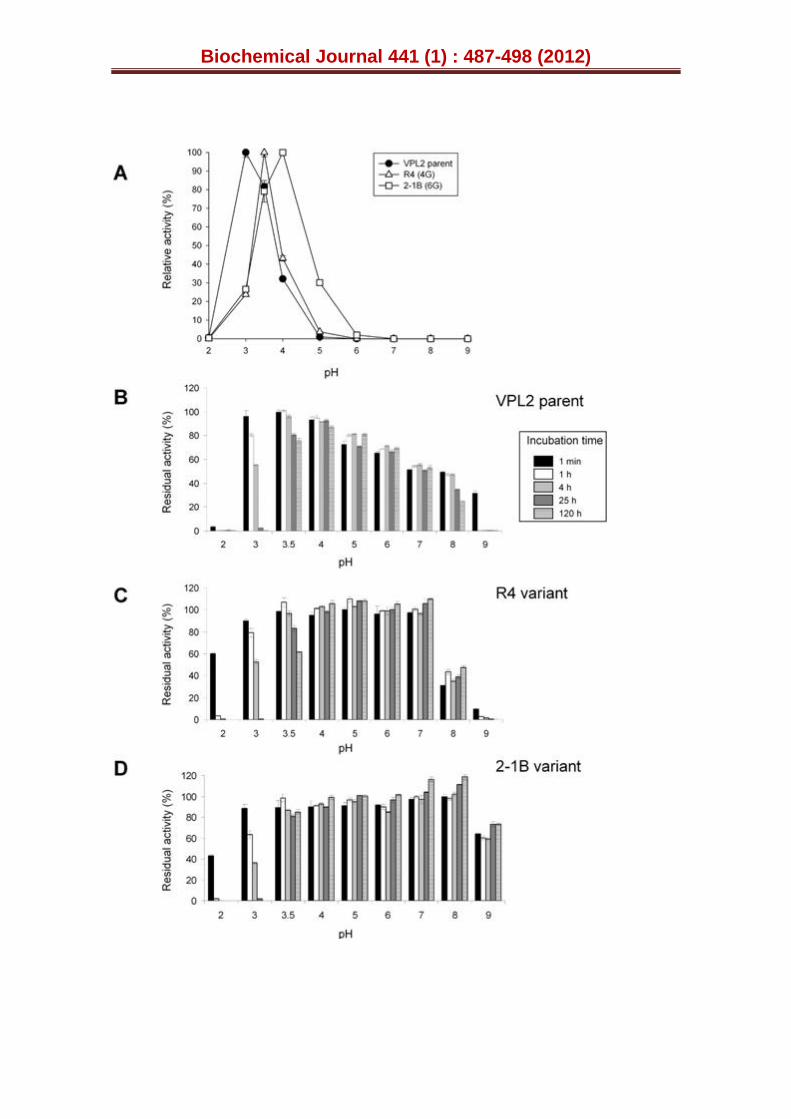
We decided to delete the portion of our synthetic gene encoding the spacer dipeptide (Fig. S1C), engineering a truncated version of the R4 mutant by IVOE deletion mutagenesis [28]. This strategy took advantage of the eukaryotic S. cerevisiae machinery by designing specific overhangs between fragments with homologous regions that tolerated the splicing of the truncated products between each other and with the linearised vector, giving rise to an autonomously replicating plasmid (see Experimental Procedures, Fig. S2). The new variant was produced, purified and the N-terminus sequenced to verify that it was correctly processed and secreted without the STE13 cleavage site (i.e. only through KEX2 protease activity, Fig. S1C). However, the secretion levels were reduced ~60 % upon deletion of the EAEA sequence, Table S3. Since the removal of the charged spacer peptide linked the fusion directly to the Lys-Arg processing site, making it a poor substrate for the KEX2 protease, an acidic environment in the proximity of the KEX2 cleavage site would appear to be important for secretion. Similar effects have been observed with other fusions, such as the α-factor leader-interferon-α1, in which the intracellular accumulation and secretion of unprocessed and partially processed forms occurred [39]. The truncated R4 variant behaved similarly to its extra N-terminal sequence counterpart in terms of spectroscopic characteristics, kinetic parameters and stability (including pH activity profiles and temperature or pH stability), indicating that the N-terminal extension had little or no impact on the biochemical properties of the mutant VPs (Tables 1 and S3; Figs. 3 and S3). Therefore, the R4 mutant containing the N-terminal sequence was subjected to further engineering.
Directed evolution for thermal stability
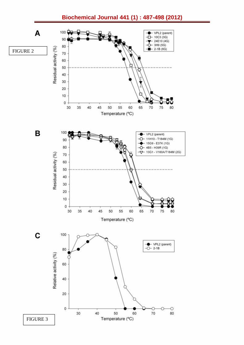
After considerably augmenting the levels of secretion by evolution, our next goal was to tailor a more thermostable VP. Accordingly, further rounds of evolution were carried out and screened with a HTP thermostability assay, based on the ratio of residual activity/initial activity (RA/IA) in combination with the estimated T50 values (see Supplemental Experimental Procedures for details, [30]). The selection pressure was progressively increased from 60ºC (4th cycle) to 80ºC and 90ºC in the 5th and 6th generations, respectively. This approach was possible because the improvements accomplished in each generation were sufficient to retain >30 % of residual activity making them suitable to evolve stability. The T50 (i.e. the temperature at which the enzyme retains 50 % of its activity after a 10 min incubation) shifted ~2ºC per round of evolution for thermostability (Fig. 2A), attaining a final T50 of 66ºC in the most thermostable mutant, the 2-1B variant (with a global improvement in T50 of 8ºC over the parental VPL2). Interestingly, an improvement of ~2ºC in T50 was detected in the 3rd generation 10C3 mutant when compared with original parental VPL2, despite the fact that the thermal stability was not targeted during the first three rounds of evolution, only secretion (Fig. 1, Fig. 2A). Hence, some of the mutations discovered during the in vitro evolution for expression also displayed improved stability.
Intrigued by this result, we evaluated the T50 values in several variants of the first and second generation that harboured beneficial mutations finally inherited by the 10C3 mutant. The improvements in stability could be
Biochemical Journal 441 (1) : 487-498 (2012)
precisely attributed to each mutation under study as at this primary stage of evolution, most of the mutations appeared individually in the mutants or in combination with other mutations that were either silent or located at the signal leader, and that did not affect stability (Fig. 1, Table S1). Accordingly, the T50 of the 11H10 mutant containing the T184M mutation and the 15G9 mutant containing the E37K mutation improved 2–4ºC over VPL2, which corroborated the stabilizing effect of T184M and E37K mutations (Fig. 1, Fig. 2B). The T50 remained constant when the 13G1 mutant from the 2nd generation was compared with 11H10, indicating that the V160A mutation did not affect thermal stability. It is also notable that the H39R mutation (found for the first time in the 4B5 mutant from 1st generation) was lost during the recombination events that took place in the 4th round, although it was rediscovered in the final cycle for thermal stability (in the 2-1B mutant: Fig. 1). The T50 of the 4B5
mutant was 4ºC higher than that of the parental type, which confirmed that the H39R mutation also had a strong influence on protein stability during secretion (Fig. 2B).
Enhanced thermostability often coincides with improved thermo-activity (Ta) and/or a widening of the Ta range [40]. The 2-1B mutant had the same Ta (defined as the optimal temperature for activity) as the parental VPL2 at 40ºC, although the range of temperature at which the enzyme is highly active was broader after artificial selection (2-1B retaining over 80 % of relative activity in the range from 30ºC to 50ºC as opposed to the range from 30ºC to 45ºC for the parental type: Fig. 2C). Thus, the relative activity at 50ºC of the 2-1B mutant was double that of the parental type at the same temperature. Generally, when thermostability is improved there is an inherent trade-off between stability and activity, with the concomitant detrimental effect on the specific activity. This is basically due to the fact that stabilizing mutations are not necessarily beneficial for activity but rather, they may confer robustness to the protein structure that compromises flexibility in some cases and consequently, turnover rates. Indeed, the TAI was reduced from 129-fold (10C3 mutant, 3G) to 87-fold (2-1B mutant, 6G) from the 4th to 6th round of evolution (Fig. 1). However, as might be expected this reduction did not drastically affect the kinetics but rather, expression above all. With all the substrates tested (except for Mn2+ where several stabilizing mutations were located around the Mn2+ binding site, see below), the catalytic efficiencies were either improved (at the haem channel site, with ~4-fold improvements for ABTS or DMP) or conserved (at the catalytic Trp164) when compared with the R4 mutant (Table 1).
In general terms, the secretion backbone generated for functional expression helped tolerate the introduction of a new set of stabilizing mutations during evolution towards thermal stability without compromising the kinetics. This was particularly noticeable in the 4th generation when screening for total activity and thermostability was combined in the same cycle (Fig. 1). For the first time, all the 4th generation VPL2 variants selected bought together the E37K, V160A, T184M and Q202L mutations to produce the most active mutant R4. The related 24E10 mutant also selected harboured an additional G330R mutation that while impeding large gains in total ABTS oxidation activity it increased its thermostability by >2ºC (Fig 1). The DNA recombination method used in this cycle (mutagenic StEP in combination with in vivo DNA shuffling) favoured the joining of the four mutations beneficial for total activity, which buffered the effect of incorporating the stabilizing mutation G330R in the same template. Therefore, a more thermostable mutant with similar activity to the parental types was created.
Characterization of evolved VPs
Biochemical Journal 441 (1) : 487-498 (2012)
The parental and evolved R4 and 2-1B VPs were purified and their
averaged molecular weights measured by MALDI-TOF and found to marginally differ from their expected masses (Table 2). The other biochemical features (pI, 4.6; ~9% glycosylation) also remained unchanged in the mutant VPs (Fig S4, Table 2). Unlike other proteins heterologously expressed in S. cerevisiae that are hyperglycosylated upon secretion, the low sugar content of the parental VPL2 and the mutants probably reflects the short residence in the Golgi compartment that is associated with the ease of exocytosis by yeast. The spectroscopic characteristics of VP variants expressed by S. cerevisiae were similar to those of VP from P. eryngii or the in vitro refolded VP from E. coli inclusion bodies (Table 2, Fig. S4D). Thus, the absorption spectrum showed a maximum in the Soret2 region and two charge transference bands (CT2 and CT1) that are characteristic of Fe3+ in a high spin state. The pH activity profile
was shifted in the course of evolution and while the parental VPL2 displayed an optimum pH for activity at 3.0, this value rose to 3.5 and 4.0 for the R4 and 2-1B variants, respectively (Fig. 3A). Together with this unexpected change, the stability at alkaline pHs was notably improved in the thermostable 2-1B (Fig. 3B-D). When maintained for 120 h at pH 9.0, the 2-1B variant conserved ~60 % of its residual activity as it was very stable in the pH range 3.5-9. By contrast, the parental VPL2 (from yeast) was very unstable at pH 9.0 loosing nearly all its residual activity after 1 hour at this pH. Similar alkaline instability has been reported for the wild-type VP produced by P. eryngii as well as for the Aspergillus-expressed recombinant enzyme, being related to the lost of structural calcium ions (as shown by enzyme stability in Ca2+-containing buffer) [23].
Kinetic constants were assessed using an array of substrates that bind to the different catalytic sites of VP (Table 1). The substrates ABTS and DMP are both oxidized with high efficiency at the catalytic Trp164 and less efficiently at the haem channel site. Substrate RB5 is exclusively oxidized at Trp164 while the substrate Mn2+ is only oxidized at the Mn2+ site [16]. The R4 mutant had an improved affinity for ABTS and DMP at the haem channel with a 10-fold lower Km and almost 2-fold higher kcat for ABTS that enhanced its kcat/Km 16-fold compared to VPL2. This improvement prevented the measurement of ABTS oxidation at the catalytic Trp164 since the kinetics of ABTS oxidation at the haem channel masked the plots for its oxidation at this site. However, the kcat/Km values for RB5 at the catalytic Trp164 were readily measured and they were similar to those of the parental VPL2 (~1600 mM-1 s-
1), indicating that the improvement in activity was mainly concentrated at the haem channel site. Similar behaviour was detected for the thermostable 2-1B mutant with RB5 as the substrate, yet the kcat/Km for ABTS was much improved at the haem channel site (over 61-fold better than the parental type). By contrast, and even though the kcat for Mn2+ was slightly improved over the course of evolution, the Mn2+ binding site was negatively affected by the mutations (see the structural analysis of the mutations below), with a 2.6 and 94-fold enhancement of the Km for Mn2+ for R4 and 2-1B, respectively. We chose ABTS for the screening as this molecule can be oxidized at both the haem channel site and the catalytic tryptophan, thereby avoiding the tendency to lose performance at the different catalytic sites. Indeed, activity at both sites was fairly well conserved or even substantially improved during evolution. The drop in Mn2+ activity underscores the fact that all the properties not addressed in the screen could drift. For future applications in which Mn2+ activity might be required, the kinetics of the enzyme with this specific substrate can be enhanced by directed evolution or semi-rational approaches.
Stability of the evolved VPs against peroxide
Biochemical Journal 441 (1) : 487-498 (2012)
Peroxidases are inhibited by excess of H2O2 and the Pleurotus VP is not
an exception [41]. Although this inhibition is caused by relatively large molar excesses of H2O2 (with respect to enzyme concentration) it results in substantial activity losses when the enzyme is in the absence of a reducing substrate. The explanation for such inhibition is found in the peroxidase catalytic cycle, which includes a highly reactive two-electron oxidized specie (compound I, a porphyrin radical Fe4+=O complex) that under turnover conditions is reduced back to the resting state (via compound II, Fe4+=O haem). In the absence of reducing substrate, compound I reacts with H2O2 resulting in compound III (a Fe3+ superoxide complex) formation and then in irreversible inactivation due to the haem/protein oxidative degradation, as reported for ligninolytic and other peroxidases [42, 43]. Comparing peroxide stability between different peroxidases is not straightforward because the
process is strongly dependent on pH; nevertheless wild VP susceptibility would be in the same order as other ligninolytic peroxidases (60% inactivation after 1 h incubation with 250 H2O2 equivalents at pH 4.5, and 90% inactivation at pH 3.0) [41]. Inactivation by their oxidizing substrate is an important problem to be overcome for biotechnological application of peroxidases, since some loss of activity is produced by H2O2 even under steady-state operation conditions, and in this context has been referred to as a suicide inactivation of the enzyme (43-45). While this problem has yet to be fully resolved, it has been examined by controlling the addition of H2O2 by sensors, by co-immobilization with glucose oxidase to generate H2O2 in situ coupled to the oxidation of glucose, as well as through site directed mutagenesis and directed evolution (with low- and high-redox potential peroxidases but not VPs) (46-53).
In the course of evolution, we detected a dramatic shift in the Km for H2O2. The peroxide kcat was inferred from ABTS oxidation at the haem channel site (taking into account the stoichiometry of the reaction: 1 molecule of H2O2 is reduced by oxidizing 2 substrate molecules, Table 1, Fig. S5). Indeed, the R4 and 2-1B mutants reduced their affinity for hydrogen peroxide by 4- and 15-fold, respectively, without the catalytic efficiency being modified. This means that the evolved VP can operate at higher H2O2 concentrations with a significant improvement in turnover rates (kcat, mole of peroxide molecules reduced per mole VP per second) from 135 s-1 for VPL2 to 1720 s-1 for 2-1B. In contrast, the 2 to 7-fold increases in Km for H2O2 attained by engineering hydrogen peroxide stability in bacterial dye-decolorizing peroxidases by site directed mutagenesis of Met residues resulted in reductions in kcat and subsequent catalytic efficiency kcat/Km (53). Thus, R4 and 2-1B worked efficiently in the presence of high concentrations of H2O2, showing specific activities with ABTS as high as 3530 U/mg and 11300 U/mg respectively, when estimated using saturating H2O2 conditions (i.e. at concentrations of 2.0 mM and 7.6 mM for R4 and 2-1B, respectively, representing 10-fold the H2O2
Km of each variant). Under these conditions, the total ABTS-oxidation activity of the R4 mutant secreted into the culture broth were ~57,000 U/L, to date the highest value reported for any peroxidase as far as we are aware.
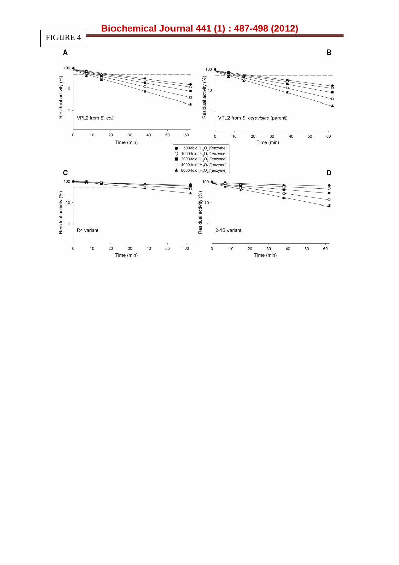
The half lives in the presence of H2O2 (t1/2 H2O2) were assessed at several H2O2/enzyme molar ratios (Fig. 4). Both the R4 and 2-1B mutants significantly improved their peroxide stability relative to VPL2. The t1/2 H2O2 of R4 and 2-1B at a H2O2/enzyme molar ratio of 4000:1 was enhanced from 10 min for VPL2 to 53 and 19 min, respectively (Fig 4). In contrast, the Km for H2O2 for 2-1B was 4- and 16-fold higher than R4 and VPL2 (Table 1).This apparent catalytic inconsistency is probably a consequence of the differing experimental conditions used to measure Km for H2O2 (steady state conditions in the presence of both peroxidase oxidizing and reducing substrates) and t1/2 H2O2 (no reducing substrate present). The enhanced Km for H2O2 detected in
Biochemical Journal 441 (1) : 487-498 (2012)
R4 and 2-1B suggests that the kinetic constants of the transient-state has been altered by directed evolution of VP, the estimation of which could provide additional information on the mechanisms underlying peroxide stability. These changes may affect different stages of the VP catalytic cycle (i.e. from the resting state to compound I, or its derivative compound II). In fact, any subtle modification in the balance between these intermediates could vary the overall catalytic cycle, affecting the formation of compound III, which is the main precursor for peroxidase inactivation by H2O2, as described above. Possibly, the improve oxidative stability is a side effect of laboratory evolution suggesting a potential use of our mutants to further select for VP variants with improved stability against H2O2.
Structural analysis of mutations
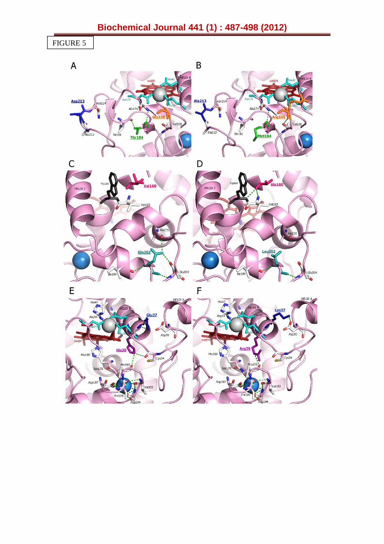
The mutations introduced by directed evolution were mapped using the crystal structure of VP isolated from P. eryngii (PDB 3FJW, Fig. S6). In this structure the Thr184, Glu37 and Val160 residues mutated in R4 are partially exposed to the solvent and distributed in different regions of the protein (Fig. S6). In contrast, the mutated Leu202 occupies an internal position in the VP molecule. Upon inspection of the VP structure, the Thr184 mutation may interrupt a H-bond with the neighbouring Ile181, relieving possible structural tension in this region (Fig. 5A, B). This change might also cause reorientation of the H-bonded Ala174 and adjoining Asp175 that constitutes one of the three carboxylic residues of the Mn2+ binding site, possibly explaining the increased Km for Mn2+ by R4 (Table 1). The Q202L mutation may interrupt two H-bonds with Ala235 and Glu304. This residue is located deep in the protein, at the end of the last β-strand in the enzyme, which is followed by a long C-terminal tail (Fig. S6). The possible changes in the hydrogen-bonding structure provoked by the Q202L mutation in this area might enhance the structural flexibility of the protein, improving its secretion (Fig. 5C, D). The E37K mutation increased the T50 by 4ºC (Fig 2B). The side-chain carboxyl group of Glu37 is H-bonded to the backbone amide proton of Glu36 (both residues located in the helix B, Fig. 5E, F). However, inspection of the protein model suggests that the resulting Lys37 may establish a salt bridge with the adjacent Asp30 of helix A. This new interaction could pack the two neighbouring A and B helixes more tightly, which may improve stability [54]. Moreover, Glu36 is one of the three acidic residues forming the Mn2+ binding site and it seems plausible that helix B could shift slightly due to this mutation, varying the relative position of Glu36 with respect to the other two acidic residues. This shift could ultimately affect the geometry of the coordination sphere of the Mn2+ ion and consequently, the affinity for this substrate [18] (Table 1). Interestingly, helix B also harbours the residues involved in H2O2 binding (His47 and Arg43). Any subtle change in the position of this helix relative to the haem co-factor could affect peroxide binding and indeed, the Km for H2O2 in the R4 mutant was 15-fold higher than that of the parental VPL2 (Table 1, Fig. S5). Finally, the V160A mutation seems to render two new H-bonds (with Trp164 and Val163, Fig. 5C, D), without affecting either the kinetics at the catalytic Trp164 (Table 1) or stability (Fig. 2B). The change in this position could be related with more correct polypeptide maturation in S. cerevisiae.
The remaining mutations discovered during the evolution to improve thermal stability were also located at the surface of the protein (G330R, D213A and H39R). The G330R mutation is situated at the C-terminal tail of VP, the most mobile region in the protein (Fig. S6). According to our model, this mutation seems to produce a new H-bond with Val328 (Fig. 5A, B), which
Biochemical Journal 441 (1) : 487-498 (2012)
could stabilize this area (the T50 was enhanced by 2.2ºC upon mutation, Fig. 1). The D213A mutation is very exposed to the solvent in the most external part of the haem channel (Fig. 5A, B). The change of a negative residue for a neutral one at this position would help VP to accommodate negatively-charged substrates, like ABTS, to be oxidized at the haem edge. It is likely that a change in the polarity of this region might suppress unfolding at higher temperatures. The H39R mutation was discovered at different points of the evolution route and indeed, the secretion mutant 4B5 and the final 2-1B thermostable variant incorporated this mutation in the 1st and last cycle of evolution, respectively. As discussed previously, this mutation considerably enhances the stability of the enzyme, improving secretion and thermal stability (Figs. 1, 2B). In native VP, the His39 is hydrogen-bond to Pro190 acting as a bridge between helix B and the loop containing the proximal calcium ion (one
of the two structural Ca2+ conserved in all Class-II peroxidases [10]). In fact, this Proline residue is contiguous to Thr189, one of the five residues involved in the coordination of the proximal calcium ion in VP (together with Ser170, Asp187, Val192 and Asp194). Differences in the strength of the hydrogen-bonds between His39 and Pro190 in native VP and between Arg39 and Pro190 in 2-1B (Fig. 5E, F) could be related to the improvement in thermal (and alkaline) resistance of the latter variant by modifying the stability of the proximal Ca2+ binding site. This effect is supported by the fact that the first generation 4B5 variant (containing only the H39R beneficial mutation) also showed improved stability (Figs. 1, 2B).The suggested changes in the proximal Ca2+ loop after mutation are in good agreement with the reported role for the protein stability of the two structural Ca2+, as for LiP and VP among other class-II peroxidases [13, 55, 56]. The H39R mutation may also break down the H-bond with Cys34 (Fig. 5E, F), which in turn could affect the distance between helix B and the haem domain, modifying the binding of peroxide (Fig. S5). It is also worth noting that this mutation may affect the oxidation of Mn2+ in the 2-1B variant (exhibiting a Km value 100-fold higher than that of the parental VPL2), enhancing the effect previously described for the E37K mutation since both Arg39 and Lys37 are contiguous to Glu40, which along with Glu36 and Asp175 contributes to the Mn2+ binding site (Table 1). A summary of the different characteristics (such as location, distance to the catalytic sites and interactions affected) of the seven residues that were mutated during VP directed evolution is provided in Table S4.
Conclusions
VPs are a typical example of generalist enzymes with phylogenetic sequence comparisons indicating they are related to MnP and LiP peroxidases. In principle, VP has adapted through natural selection to combine many catalytic phenotypes from its related peroxidases. Indeed, modern VPs likely possess a broader substrate specificity than their ancestral progenitors (14,16). Given their intrinsic enzymatic promiscuity it is likely VPs have the capacity to evolve novel catalytic properties, making them a potentially powerful model enzyme to engineer by molecular evolution towards specialised functions. S. cerevisiae is considered by many to be the preferred eukaryotic model organism for molecular and cell biology, with this versatility making it a highly useful expression host in directed protein evolution studies (28, 46, 57). Indeed, in this study we have demonstrated the usefulness of S. cerevisiae for evolving VP variants via laboratory evolution strategies to select for more stable and active forms, possibly serving as a suitable platform to tailor VPs with targeted catalytic attributes [58]. Recently, new laccases have been engineered by artificial evolution, strengthening the array of ligninolytic enzymes available for different potential applications [31,59]. These evolutionary models could be employed as a biomolecular tool-box in order to
Biochemical Journal 441 (1) : 487-498 (2012)
address both traditional problems and new challenges in synthetic biology that have so far hindered the practical use of VPs and other high-redox potential oxidoreductases: from inactivation by hydrogen peroxide to the construction of artificial operons of evolved laccases and VPs for the directed evolution of whole ligninolytic cell factories in yeast.
AUTHOR CONTRIBUTION
Eva Garcia-Ruiz carried out all the laboratory evolution experiments. David Gonzalez-Perez helped in the biochemical characterization of evolved variants. Francisco J. Ruiz-Dueñas and Angel T. Martinez contributed to the structure-function analysis of the mutations and helped with the revision of the manuscript. Miguel Alcalde conceived the project, supervised its development and wrote the manuscript.
ACKNOWLEDGEMENTS
We thank Rita Getzlaff from the Helmholtz Centre for Infection Research (HZI, Braunschweig, Germany) for sequence analyses. We thank Dr Francisco J. Plou from the ICP (CSIC, Spain) for assistance with the HPLC purification.
FUNDING
This study is based upon work funded by EU Projects [NMP2-CT-2006-026456-Biorenew, FP7-KBBE-2010-4-26537-Peroxicats and COST Action CM0701] and National projects [BIO2010-19697 and BIO2008-01533]. We also thank NeuronBiopharma for financial support through Research Contracts [020401070029, Profit Program and 020401070004, Idea Program]. E.G.R. was supported by a Biorenew grant, D.G.P. by a Peroxicats contract and F.J.R.D. by the MICINN Ramón y Cajal Program.
REFERENCES
1. Martínez, A. T., Ruiz-Dueñas, F. J., Martínez, M. J., del Río, J. C., and Gutiérrez, A. (2009) Enzymatic delignification of plant cell-wall: From nature to mill. Curr. Opin. Biotechnol. 20, 348-357
2. Ruiz-Dueñas, F. J. and Martínez, A. T. (2009) Microbial degradation of lignin: How a bulky recalcitrant polymer is efficiently recycled in nature
and how we can take advantage of this. Microbial Biotechnol. 2, 164-177
3. Kamm, B. and Kamm, M. (2007) Biorefineries - Multi product processes. In Ulber, R. and Sell, D., editors. White Biotechnology, Springer, Berlin
4. Orts, W. J., Imam, S., Glenn, G. M., Inglesby, M., Wong, D., Guttman, M., and Samac, D. A. (2004) Envisioning biorefineries based on utilization of lignocellulosic straws and bagasses. Abs. Papers Amer. Chem. Soc. 227, U310
5. Alcalde, M., Ferrer, M., Plou, F. J., and Ballesteros, A. (2006) Environmental biocatalysis: from remediation with enzymes to novel green processes. Trends Biotechnol. 24, 281-287
6. Xu, F. (2005) Applications of oxidoreductases: Recent progress. Industr. Biotechnol. 1, 38-50
Biochemical Journal 441 (1) : 487-498 (2012)
7. Camarero, S., Sarkar, S., Ruiz-Dueñas, F. J., Martínez, M. J., and
Martínez, A. T. (1999) Description of a versatile peroxidase involved in natural degradation of lignin that has both Mn-peroxidase and lignin-peroxidase substrate binding sites. J. Biol. Chem. 274, 10324-10330
8. Martínez, M. J., Ruiz-Dueñas, F. J., Guillén, F., and Martínez, A. T. (1996) Purification and catalytic properties of two manganese-peroxidase isoenzymes from Pleurotus eryngii. Eur. J. Biochem. 237, 424-432
9. Ruiz-Dueñas, F. J., Martínez, M. J., and Martínez, A. T. (1999) Molecular characterization of a novel peroxidase isolated from the ligninolytic fungus Pleurotus eryngii. Mol. Microbiol. 31, 223-236
10. Martínez, A. T. (2002) Molecular biology and structure-function of lignin-degrading heme peroxidases. Enzyme Microb. Technol. 30, 425-444
11. Rainio, A., Maijala, P., Hildén, K., and Hatakka, A. (2008) A novel peroxidase from the white-rot fungus Cerrena unicolor. Proc.8th Intern.Peroxidase Symp., Tampere, 20-23
12. Moreria, P. R., HDuez, C H., HDehareng, DH., HAntunes, A.,H HAlmeida-Vara, E H., HFrere, J. MH., HMalcata, F. X H. HDuarte, J.C H. (2005) HMolecular characterisation of a versatile peroxidase from a Bjerkandera strainH. J. Biotechnol. 118, 339-352
13. Verdín, J., Pogni, R., Baeza, A., Baratto, M. C., Basosi, R. and Vázquez-
Duhalt, R. (2006) Mechanism of versatile peroxidase inactivation by
Ca2+ depletion. Biophys. Chem. 121, 163-170
14. Hofrichter, M., Ullrich, R., Pecyna, M. J., Liers, C., and Lundell, T. (2010) New and classic families of secreted fungal heme peroxidases. Appl. Microbiol. Biotechnol. 87, 871-897
15. Martínez, A. T. (2007) High redox potential peroxidases. In Polaina, J. and MacCabe, A. P., editors. Industrial Enzymes: Structure, Function and Applications, Springer, Berlin
16. Ruiz-Dueñas, F. J., Morales, M., García, E., Miki, Y., Martínez, M. J., and Martínez, A. T. (2009) Substrate oxidation sites in versatile peroxidase and other basidiomycete peroxidases. J. Exp. Bot. 60, 441-452
17. Ruiz-Dueñas, F. J., Morales, M., Mate, M. J., Romero, A., Martínez, M. J., Smith, A. T., and Martínez, A. T. (2008) Site-directed mutagenesis of the catalytic tryptophan environment in Pleurotus eryngii versatile peroxidase. Biochemistry 47, 1685-1695
18. Ruiz-Dueñas, F. J., Morales, M., Pérez-Boada, M., Choinowski, T., Martínez, M. J., Piontek, K., and Martínez, A. T. (2007) Manganese oxidation site in Pleurotus eryngii versatile peroxidase: A site-directed mutagenesis, kinetic and crystallographic study. Biochemistry 46, 66-77
19. Ruiz-Dueñas, F. J., Pogni, R., Morales, M., Giansanti, S., Mate, M. J., Romero, A., Martínez, M. J., Basosi, R., and Martínez, A. T. (2009)
Biochemical Journal 441 (1) : 487-498 (2012)
Protein radicals in fungal versatile peroxidase: Catalytic tryptophan radical in both Compound I and Compound II and studies on W164Y, W164H and W164S variants. J. Biol. Chem. 284, 7986-7994
20. Cañas, A. and Camarero, S. (2010) Laccases and their natural mediators: biotechnological tools for sustainable eco-friendly processes. Biotechnol. Adv. 28, 694-705
21. Ruiz-Dueñas, F. J., Martínez, M. J., and Martínez, A. T. (1999) Heterologous expression of Pleurotus eryngii peroxidase confirms its ability to oxidize Mn2+ and different aromatic substrates. Appl. Environ. Microbiol. 65, 4705-4707
22. Eibes, G. M., Lu-Chau, T. A., Ruiz-Duenas, F. J., Feijoo, G., Martinez, M. J., Martinez, A. T., and Lema, J. M. (2009) Effect of culture temperature on the heterologous expression of Pleurotus eryngii versatile peroxidase in Aspergillus hosts. Bioproc.Biosyst. Eng. 32, 129-134
23. Lú-Chau, T. A., Ruiz-Dueñas, F. J., Camarero, S., Feijoo, G., Martínez, M. J., Lema, J. M., and Martínez, A. T. (2004) Effect of pH on the stability of Pleurotus eryngii versatile peroxidase during heterologous production in Emericella nidulans. Bioproc.Biosyst. Eng. 26, 287-293
24. Mohorcic, M., Bencina, M., Friedrich, J., and Jerala, R. (2009) Expression of soluble versatile peroxidase of Bjerkandera adusta in Escherichia coli. Bioresource Technol. 100, 851-858
25. Pérez-Boada, M., Doyle, W. A., Ruiz-Dueñas, F. J., Martínez, M. J., Martínez, A. T., and Smith, A. T. (2002) Expression of Pleurotus eryngii versatile peroxidase in Escherichia coli and optimisation of in vitro folding. Enzyme Microb. Technol. 30, 518-524
26. Zhao, H. M., Giver, L., Shao, Z. X., Affholter, J. A., and Arnold, F. H. (1998) Molecular evolution by staggered extension process (StEP) in vitro recombination. Nat. Biotech. 16, 258-261
27. Zumárraga, M., Camarero, S., Shleev, S., Martinez-Arias, A., Ballesteros, A., Plou, F. J., and Alcalde, M. (2008) Altering the laccase functionality by in vivo assembly of mutant libraries with different mutational spectra. Proteins 71, 250-260
28. Alcalde, M. (2010) Mutagenesis protocols in Saccharomyces cerevisiae by in vivo overlap extension. In Bramman, J., editor. In vitro Mutagenesis Protocols, Humana Press, Totowa, New Jersey, US
29. Brake, A. J. (1990) Alpha-factor leader-directed secretion of heterologous proteins from yeast. Methods in Enzymology 185, 408-421
30. García-Ruiz, E., Maté, D., Ballesteros, A., Martínez, A. T., and Alcalde, M. (2010) Evolving thermostability in mutant libraries of ligninolytic oxidoreductases expressed in yeast. Microb. Cell Fact. 9, 17.
31. Mate, D., Garcia-Burgos, C., Garcia-Ruiz, E., Ballesteros, A. O., Camarero, S., and Alcalde, M. (2010) Laboratory evolution of high-redox potential laccases. Chem. Biol.17, 1030-1041
Biochemical Journal 441 (1) : 487-498 (2012)
32. Zumárraga, M., Bulter, T., Shleev, S., Polaina, J., Martinez-Arias, A.,
Plou, F. J., Ballesteros, A., and Alcalde, M. (2007) In vitro evolution of a fungal laccase in high concentrations of organic cosolvents. Chem. Biol. 14, 1052-1064
33. Bulter, T., Alcalde, M., Sieber, V., Meinhold, P., Schlachtbauer, C., and Arnold, F. H. (2003) Functional expression of a fungal laccase in Saccharomyces cerevisiae by directed evolution. Appl. Environ. Microbiol. 69, 987-995
34. Alcalde, M., Zumárraga, M., Polaina, J., Ballesteros, A., and Plou, F. J. (2006) Combinatorial saturation mutagenesis by in vivo overlap extension for the engineering of fungal laccases. Comb. Chem.High T.
Scr. 9, 719-727
35. Bloom, J. D. and Arnold, F. H. (2009) In the light of directed evolution: pathways of adaptive protein evolution. Proc.Natl. Acad. Sci. USA 106, 9995-10000
36. Romero, P. A. and Arnold, F. H. (2009) Exploring protein fitness landscapes by directed evolution. Nat. Rev. Mol. Cell Bio. 10, 866-876
37. Brake, A. J., Merryweather, J. P., Coit, D. G., Heberlein, U. A., Masiarz, F. R., Mullenbach, G. T., Urdea, M. S., Valenzuela, P., and Barr, P. J. (1984) Alfa-factor-directed synthesis and secretion of mature foreign proteins in Saccharomyces cerevisiae. Proc.Natl. Acad. Sci. USA 81, 4642-4646
38. Romanos, M. A., Scorer, C. A., and Clare, J. J. (1992) Foreign gene expression in yeast: a review. Yeast 8, 423-488
39. Zsebo, K. M., Lu, H. S., Fieschko, J. C., Goldstein, L., Davis, J., Duker, K., Suggs, S. V., Lai, P. H., and Bitter, G. A. (1986) Protein secretion from Saccharomyces cerevisiae directed by the prepro-α-factor leader region. J. Biol. Chem.261, 5858-5865
40. Cirino, P. C. and Georgescu, R. (2003) Screening for thermostability. In Arnold, F. H. and Georgiou, G., editors. Directed enzyme evolution. Screening and selection methods, Humana Press, Totowa, New Jersey,
US
41. Böckle, B., Martínez, M. J., Guillén, F. and Martínez, A. T. (1999)
Mechanism of peroxidase inactivation in liquid cultures of the
ligninolytic fungus Pleurotus pulmonarius. Appl. Environ. Microbiol. 65,
923-928
42. Wariishi, H. and Gold, M. H. (1989) Lignin peroxidase compound III:
Formation, inactivation, and conversion to the native enzyme. FEBS
Lett. 243, 165-168
43. Valderrama, B., Ayala, M., and Vázquez-Duhalt, R. (2002) Suicide inactivation of peroxidases and the challenge of engineering more robust enzymes. Chem. Biol. 9, 555-565
Biochemical Journal 441 (1) : 487-498 (2012)
44. Timofeevski, S. L., Reading, N. S., and Aust, S. D. (1998) Mechanisms
for protection against inactivation of manganese peroxidase by hydrogen peroxide. Arch. Biochem. Biophys. 356, 287-295
45. Hernández-Ruiz, J., Arnao, M. B., Hiner, A. N. P., García-Canovas, F., and Acosta, M. (2001) Catalase-like activity of horseradish peroxidase: relationship to enzyme inactivation by H2O2. Biochem. J. 354, 107-114
46. Cherry, J. R., Lamsa, M. H., Schneider, P., Vind, J., Svendsen, A., Jones, A., and Pedersen, A. H. (1999) Directed evolution of a fungal peroxidase. Nat. Biotechnol. 17, 379-384
47. Morawski, B., Quan, S., and Arnold, F. H. (2001) Functional expression
and stabilization of horseradish peroxidase by directed evolution in Saccharomyces cerevisiae. Biotechnol. Bioeng. 76, 99-107
48. Miyazaki-Imamura, C., Oohira, K., Kitagawa, R., Nakano, H., Yamane, T., and Takahashi, H. (2003) Improvement of H2O2 stability of manganese peroxidase by combinatorial mutagenesis and high-throughput screening using in vitro expression with protein disulfide isomerase. Protein Eng. 16, 423-428
49. Ryu, K., Hwang, S. Y., Kim, K. H., Kang, J. H., and Lee, E. K. (2008) Functionality improvement of fungal lignin peroxidase by DNA shuffling for 2,4-dichlorophenol degradability and H2O2 stability. J. Biotechnol. 133, 110-115
50. Ryu, K., Kang, J. H., Wang, L. S., and Lee, E. K. (2008) Expression in yeast of secreted lignin peroxidase with improved 2,4-dichlorophenol degradability by DNA shuffling. J. Biotechnol. 135, 241-246
51. Miyazaki, C. and Takahashi, H. (2001) Engineering of the H2O2-binding pocket region of a recombinant manganese peroxidase to be resistant to H2O2. FEBS Lett. 509, 111-114
52. Ryan, B. J. and O'Fagain, C. (2007) Effects of single mutations on the stability of horseradish peroxidase to hydrogen peroxide. Biochimie 89, 1029-1032
53. Ogola, H. J. O., Hashimoto, N., Miyabe, S., Ashida, H., Ishikawa, T., Shibata, H., and Sawa, Y. (2010) Enhancement of hydrogen peroxide stability of a novel Anabaena sp. DyP-type peroxidase by site-directed mutagenesis of methionine residues. Appl. Microbiol. Biotechnol. 87, 1727-1736
54. Musafia, B., Buchner, V. and Arad, D. (1995) Complex salt bridges in
proteins: Statistical analysis of structure and function. J. Mol. Biol.
254:761-770
55. Nie, G. J. and Aust, S. D. (1997) Effect of calcium on the reversible
thermal inactivation of lignin peroxidase. Arch. Biochem. Biophys.
337:225-231
56. George, S. J., Kvaratskhelia, M., Dilworth, M. J. and Thorneley, R. N. F.
(1999) Reversible alkaline inactivation of lignin peroxidase involves the
Biochemical Journal 441 (1) : 487-498 (2012)
release of both the distal and proximal site calcium ions and
bishistidine co-ordination of the haem. Biochem. J. 344, 237-244
57. Okkels, J. S. (2004) In vivo gene shuffling in yeast: a fast and easy method for directed evolution of enzymes. In Svendsen, A., editor. Enzyme functionality: design, engineering, and screening, Marcel Dekker, Inc., New York
58. García, E., Martínez, F. J., Ruiz-Dueñas, F. J., Martínez, A. T., and Alcalde, M. (2010) High redox potential peroxidases designed by directed evolution. Patent: PCT/ES2010/070316.
59. Camarero, S., Cañas, A., Martínez, M. J., Martínez, A. T., Ballesteros,
A., Plou, F. J., Record, E., Asther, M., and Alcalde, M. (2009) High redox potential laccases engineered by directed evolution. Patent PCT/ES2009/070519
Biochemical Journal 441 (1) : 487-498 (2012)
FIGURE LEGENDS
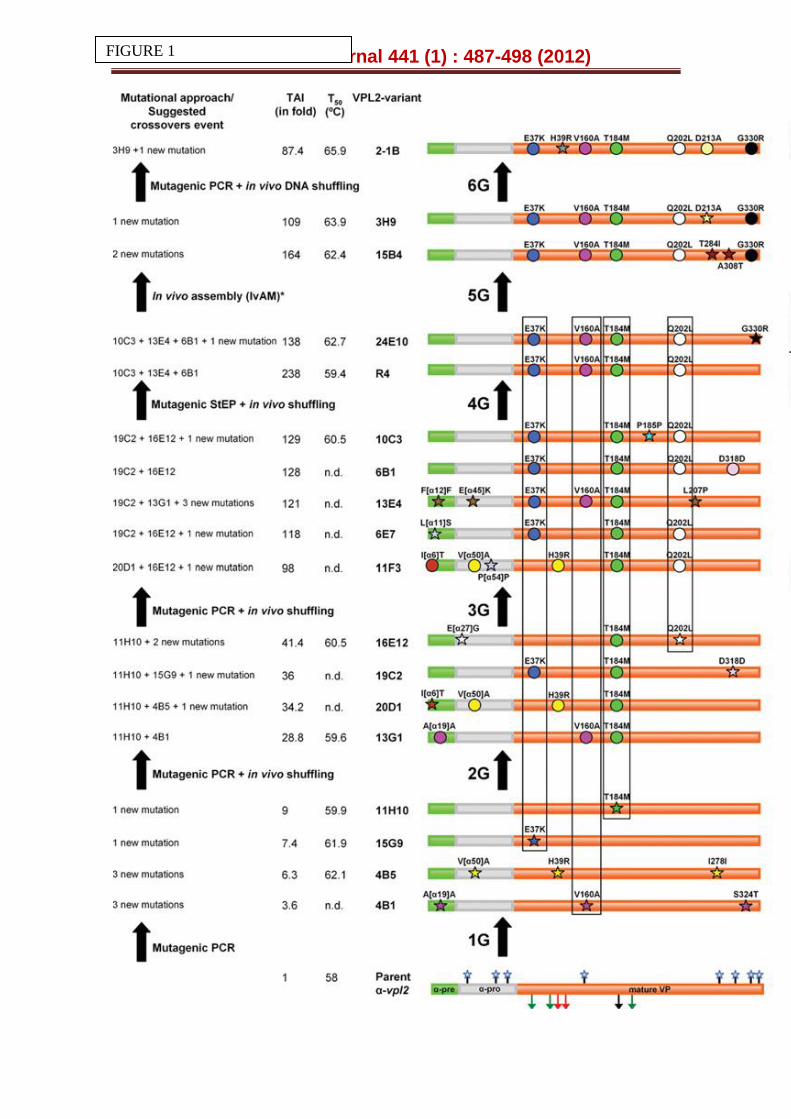
Figure 1. Laboratory evolution of the α-vpl2 fusion gene. The properties targeted for evolution were secretion (from 1st to 4th generation) and thermal stability (from 4th to 6th generation). In the 4th generation, secretion and thermal stability were combined during the screening. The α-factor pre-leader is represented in green, the α-factor pro-leader in grey, and the mature VP in orange. In the parent α-vpl2 the glycosylation sites are represented as blue stars, the Mn2+ binding site (E36, E40 and D175) as green arrows, the
catalytic Trp164 as black arrow and the H2O2 binding site (from distal side, R43 and H47) as red arrows. New mutations are depicted as stars and accumulated mutations as circles. The amino acid backbone for secretion is highlighted in boxes. TAI (total activity improvement): this value indicates the improvement in VP activity detected in S. cerevisiae microcultures for each mutant when compared with the parental α-vpl2. The breakdown of the TAI into specific activity and expression for the best secretion variant is represented in Table S2. The suggested crossover events and the T50 values are included (n.d. not determined). See also Table S1.
Figure 2. Thermostability of evolved VPs. (A) T50 profiles of mutants from the 3rd to 6th generations. (B) T50 profiles of mutants from the 1st and 2nd generations. (C) Thermo-activity (optimum temperature for activity) of parental VPL2 and the 2-1B mutant. Each point is from three independent experiments and includes the standard deviation.
Figure 3. Activity and stability vs pH. (A) pH activity profile for the parental VPL2, R4 and 2-1B mutants. Activities were measured in 100 mM Britton and Robinson (B&R) buffer at different pHs with 2 mM ABTS and 0.1 mM H2O2. VP activity was normalized to the optimum activity value and each point comes from three independent experiments, including the standard deviation. The pH stability of the parental VPL2 (B), the R4 (C) and the 2-1B mutants (D). Enzyme samples were incubated in 100 mM B&R buffer at different pH values, and the residual activity was measured in 100 mM sodium tartrate buffer pH 3.5 containing 2 mM ABTS and 0.1 mM H2O2. Each point comes from three independent experiments and includes the standard deviation (See also Table
2 and Fig. S4).
Figure 4. Inactivation of parental and VP mutants at different H2O2:enzyme ratios. (A) VPL2 from E. coli after in vitro refolding; (B) Functional expression in S. cerevisiae of the parental VPL2, (C) the R4 and (D) 2-1B mutant. The purified VPs (0.06 μM) were incubated at room temperature in 20 mM B&R buffer containing different hydrogen peroxide concentrations. The following [H2O2]/[VP] molar ratios were assayed: black circles, 500-fold; white circles, 1000-fold; black squares, 2000-fold; white squares, 4000-fold; black triangles, 6000-fold. The residual activity was measured with 2 mM ABTS and 0.1 mM H2O2 in 100 mM tartrate buffer [pH 3.5]. The residual activity refers to the corresponding VP variant incubated in the absence of H2O2, taking into account the final concentration of H2O2 for each assay. Each point is from three independent experiments and includes the standard deviation. The dashed line indicates 50 % of the residual activity (See also Figure S5).
Biochemical Journal 441 (1) : 487-498 (2012)
Figure 5. Structural examination of the selected mutations in VPL2. The haem group is represented in red, the catalytic Trp164 in black and the Mn2+ binding site formed by acidic residues Glu36, Glu40 and Asp175 in blue. Grey sphere, Mn2+ ; blue sphere, Ca2+. (A, B) Mutations G330R (in orange and underlined), D213A (in blue and underlined) and T184M (in green and underlined). (C, D) Mutations V160A (in red and underlined) and Q202L (in green and underlined). (E, F) Mutations H39R (in violet and underlined) and E37K (in blue and underlined). The residues involved in the coordination of the proximal Ca2+ (Ser170, Asp187, Thr189, Val192, Asp194) are highlighted. Based on PDB entry 3FJW (parental VPL2) and model with the mutations introduced by directed evolution.
Biochemical Journal 441 (1) : 487-498 (2012)
FIGURE 1
Biochemical Journal 441 (1) : 487-498 (2012)
FIGURE 2
FIGURE 3
Biochemical Journal 441 (1) : 487-498 (2012)
Biochemical Journal 441 (1) : 487-498 (2012)
FIGURE 4
Biochemical Journal 441 (1) : 487-498 (2012)
FIGURE 5
Biochemical Journal 441 (1) : 487-498 (2012)
SUPPLEMENTARY FIGURE LEGENDS
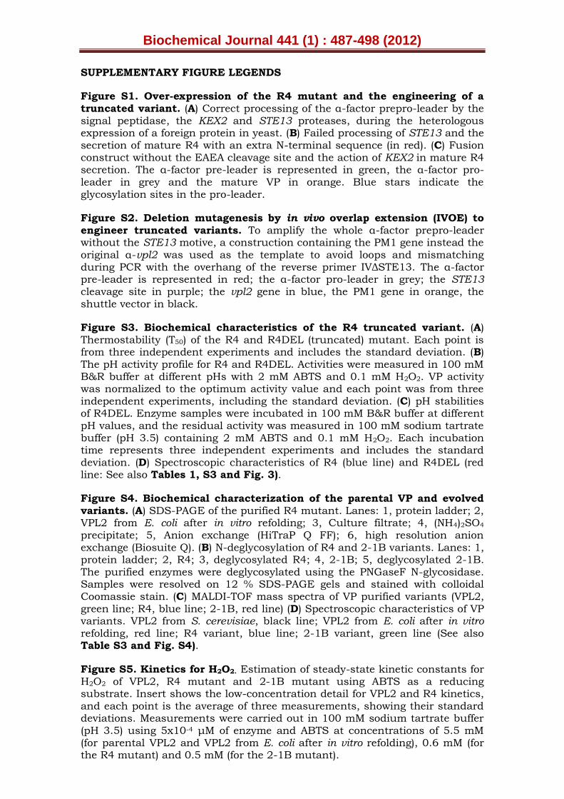
Figure S1. Over-expression of the R4 mutant and the engineering of a truncated variant. (A) Correct processing of the α-factor prepro-leader by the signal peptidase, the KEX2 and STE13 proteases, during the heterologous expression of a foreign protein in yeast. (B) Failed processing of STE13 and the secretion of mature R4 with an extra N-terminal sequence (in red). (C) Fusion construct without the EAEA cleavage site and the action of KEX2 in mature R4 secretion. The α-factor pre-leader is represented in green, the α-factor pro-leader in grey and the mature VP in orange. Blue stars indicate the glycosylation sites in the pro-leader.
Figure S2. Deletion mutagenesis by in vivo overlap extension (IVOE) to
engineer truncated variants. To amplify the whole α-factor prepro-leader without the STE13 motive, a construction containing the PM1 gene instead the original α-vpl2 was used as the template to avoid loops and mismatching during PCR with the overhang of the reverse primer IVΔSTE13. The α-factor pre-leader is represented in red; the α-factor pro-leader in grey; the STE13 cleavage site in purple; the vpl2 gene in blue, the PM1 gene in orange, the shuttle vector in black.
Figure S3. Biochemical characteristics of the R4 truncated variant. (A) Thermostability (T50) of the R4 and R4DEL (truncated) mutant. Each point is from three independent experiments and includes the standard deviation. (B) The pH activity profile for R4 and R4DEL. Activities were measured in 100 mM B&R buffer at different pHs with 2 mM ABTS and 0.1 mM H2O2. VP activity was normalized to the optimum activity value and each point was from three independent experiments, including the standard deviation. (C) pH stabilities of R4DEL. Enzyme samples were incubated in 100 mM B&R buffer at different pH values, and the residual activity was measured in 100 mM sodium tartrate buffer (pH 3.5) containing 2 mM ABTS and 0.1 mM H2O2. Each incubation time represents three independent experiments and includes the standard deviation. (D) Spectroscopic characteristics of R4 (blue line) and R4DEL (red line: See also Tables 1, S3 and Fig. 3).
Figure S4. Biochemical characterization of the parental VP and evolved variants. (A) SDS-PAGE of the purified R4 mutant. Lanes: 1, protein ladder; 2, VPL2 from E. coli after in vitro refolding; 3, Culture filtrate; 4, (NH4)2SO4
precipitate; 5, Anion exchange (HiTraP Q FF); 6, high resolution anion
exchange (Biosuite Q). (B) N-deglycosylation of R4 and 2-1B variants. Lanes: 1, protein ladder; 2, R4; 3, deglycosylated R4; 4, 2-1B; 5, deglycosylated 2-1B. The purified enzymes were deglycosylated using the PNGaseF N-glycosidase. Samples were resolved on 12 % SDS-PAGE gels and stained with colloidal Coomassie stain. (C) MALDI-TOF mass spectra of VP purified variants (VPL2, green line; R4, blue line; 2-1B, red line) (D) Spectroscopic characteristics of VP variants. VPL2 from S. cerevisiae, black line; VPL2 from E. coli after in vitro refolding, red line; R4 variant, blue line; 2-1B variant, green line (See also Table S3 and Fig. S4).
Figure S5. Kinetics for H2O2. Estimation of steady-state kinetic constants for H2O2 of VPL2, R4 mutant and 2-1B mutant using ABTS as a reducing substrate. Insert shows the low-concentration detail for VPL2 and R4 kinetics, and each point is the average of three measurements, showing their standard deviations. Measurements were carried out in 100 mM sodium tartrate buffer (pH 3.5) using 5x10⁻⁴ μM of enzyme and ABTS at concentrations of 5.5 mM (for parental VPL2 and VPL2 from E. coli after in vitro refolding), 0.6 mM (for the R4 mutant) and 0.5 mM (for the 2-1B mutant).
Biochemical Journal 441 (1) : 487-498 (2012)
Figure S6. Scheme of the wild-type VP and 2-1B variant molecular structure. (A) Wild-type VPL2 isolated from P. eryngii (PDB entry 3FJW) indicating the 7 residues that were seen to be modified during directed evolution. (B) Model for the 2-1B evolved variant including the 7 mutations selected. The secondary protein structure (dominated by helixes), the position of the haem cofactor (CPK sticks) and the solvent access surfaces of both proteins (in partially-transparent electrostatic-potential colours) are shown.
SUPPLEMENTAL EXPERIMENTAL PROCEDURES
Reagents and Enzymes
ABTS, DMP, VA, RB5, haemoglobin from bovine blood, Taq polymerase for
random mutagenesis, StEP and the S. cerevisiae transformation kit were purchased from Sigma-Aldrich (Madrid, Spain). The E. coli XL2-blue competent cells and the Genemorph II random mutagenesis kit were obtained from Stratagene (La Jolla, CA, USA). The protease deficient S. cerevisiae strain BJ5465 was from LGCPromochem (Barcelona, Spain), the uracil independent and ampicillin resistance shuttle vector pJRoC30 was obtained from the California Institute of Technology (Caltech, CA, USA), while the pGAPZαA vector containing α-factor prepro-leader and the Taq polymerase used to construct the α-vpl2 fusion were from Invitrogen, USA. The zymoprep yeast plasmid miniprep kit, zymoclean gel DNA recovery kit, and the DNA clean and concentrator TM-5 kit were all from Zymo Research (Orange, CA). The NucleoSpin Plasmid kit was purchased from Macherey-Nagel (Germany) and the restriction enzymes BamHI, XhoI, EcoRI and NotI were from New England Biolabs (Hertfordshire, UK). VPL2 from E. coli was prepared by in vitro refolding from inclusion bodies as reported elsewhere [25]. All chemicals were reagent-grade purity.
Culture Media
Minimal medium contained 100 mL 6.7% sterile yeast nitrogen base, 100 mL 19.2 g/L sterile yeast synthetic drop-out medium supplement without uracil, 100 mL sterile 20% raffinose, 1 mL 25 g/L chloramphenicol and 700 mL sddH2O. YP medium contained 10 g yeast extract, 20 g peptone and ddH2O to 650 mL. Flask expression medium contained 720 mL YP, 67 mL 1 M KH2PO4 buffer (pH 6.0), 111 mL 20% galactose, 25 g/L ethanol, 500 mg/L bovine haemoglobin, 1 mM CaCl2, 1 mL 25 g/L chloramphenicol and ddH2O to 1000 mL. Microplate expression medium contained 720 mL YP, 67 mL 1 M KH2PO4 buffer (pH 6.0), 111 mL 20% galactose, 100 mg/L bovine haemoglobin, 1 ml 25 g/L chloramphenicol and ddH2O to 1000 mL. YPD solution contained 10 g yeast extract, 20 g peptone, 100 mL 20% sterile glucose, 1 mL 25 g/L chloramphenicol and ddH2O to 1000 mL. SC drop-out plates contained 100 mL 6.7 % sterile yeast nitrogen base, 100 mL 19.2 g/L sterile yeast synthetic drop-out medium supplement without uracil, 20 g bacto agar, 100 mL 20% sterile glucose, 1 mL 25 g/L chloramphenicol and ddH2O to 1000 mL.
Construction of pJRoC30-α-vpl2
The pGAPZ-αA vector was used as a template to fuse the native vpl2 with the α-factor prepro-leader. Firstly, the cDNA from vpl2 (996 bp) excluding the native signal leader was amplified using the following primers: NtEcoRI-direct (5´-CGGAATTCGCAACTTGCGACGACGGACGC-3´) and CtNotl-reverse (AAGGAAAAAAGCGGCCGCTTACGATCCAGGGACGGGAGG-3´). The target
Biochemical Journal 441 (1) : 487-498 (2012)
sequences for EcoRI and Notl are underlined. PCR reactions were performed in
a final volume of 50 L containing 400 nM NecoRI-direct, 400 nM CtNotl-reverse, 0.25 mM dNTPs, 0.05 U/μL of Taq polymerase (Invitrogen), 4 mM MgCl2 and 0.5 ng/ μL cDNA vpl2. The thermal cycling programme was: 94ºC for 5 min, 55ºC for 5 min, 72ºC for 5 min (1 cycle); 95ºC for 0.35 min, 50ºC for 2 min, 72ºC for 4 min (25 cycles); and 72ºC for 10 min (1 cycle). The amplified α-vpl2 was purified by low melting point gel extraction and recovered with zymoclean gel DNA recovery kit. The vpl2 product and the pGAPZαA vector were both digested with EcoRI and Notl, and ligated, giving rise to pGAPZ-α-vpl2. This construct was used to transform E. coli XL2-blue cells and the product was prepared in large amounts. The pGAPZα-vpl2 was used to amplify the α-vpl2 fusion gene with the following primers: NtpJRBamHI-direct (5´-CGCGGATCCATGAGATTTCCTTCAATTTTTACTGC-3´), which included the BamHI target (underlined); and CtNotL-reverse sequences. PCR reactions were
performed in a final volume of 50 L containing 400 nM NtpJRBamHI-direct, 400 nM CtNotl-reverse, 0.25 mM dNTPs, 0.05 U/μL of Taq polymerase (Invitrogen), 4 mM MgCl2 and 0.4ng/ μL pGAPZ-α-vpl2. The amplified α-vpl2 fusion was purified and cleaned as described above and then cloned into the pJRoC30 episomal shuttle vector. Both α-vpl2 and pJRoC30 were linearised with BamHI and NotI, and ligated to generate pJRoC30-α-vpl2.
Truncated variant
The extra N-terminal sequence was removed by deletion mutagenesis (R4DEL variant) using In Vivo Overlap Extension (IVOE, as summarized in Fig. S2: [28]).
R4DEL mutant: The primers for PCR 1 were RMLN and IVΔSTE13 R (5’ GCAGCATTTGCGGTGGTGCGTCCGTCGTCGCAAGTTGCTCTTTTCTCGAGAGATACCCCTTC 3´ that binds to bp 5´- 441-465 -3´ of pJRoC30-αPM1). The primers for PCR 2 were IVΔEcoRI F (5’ GCAACTTGCGACGACGGACGC 3’ that binds to bp 5´-483-503-3´ of pJRoC30-α-vpl2) and RMLC. The products from PCR 1 and PCR 2 have overhangs with homologous regions of 38 bp between each other, and of 40 bp and 66 bp with the linearised vector for in vivo cloning. The linearised plasmid (100 ng) was mixed with products from PCR1 and PCR 2 (400 ng each) and transformed into competent S. cerevisiae cells. Individual clones were picked and cultured in 96-well plates (GreinerBio-One,
Germany) containing 50 L of minimal medium per well and subjected to the screening procedure described below. Positive clones were re-screened (see below), the in vivo repaired plasmid was recovered and the truncated fusion gene was confirmed by DNA sequencing.
High-throughput screening assays
Due to the low levels of secretion, longer inductions times in expression media were used in the first round of evolution (up to 72 h). From the 2nd cycle onwards, expression was sufficiently strong that the time required for protein induction was reduced to 24 h. Moreover, in the first two cycles the screening was carried out in end-point mode after incubating the supernatants for several hours in the presence of ABTS. As a consequence of the high levels of secretion achieved, from the third generation on the supernatants were diluted 1:10 before screening.
HTP-Screening assays for secretion (total activity)
Individual clones were picked and cultured in 96-well plates (GreinerBio-One,
Germany) containing 50 L of minimal medium per well. In each plate, column
Biochemical Journal 441 (1) : 487-498 (2012)
number 6 was inoculated with the parental type and one well (H1-control) was inoculated with non-transformed yeast cells as a negative control. The plates were sealed to prevent evaporation and incubated at 30ºC, 225 rpm and 80 % relative humidity in a humid shaker (Minitron-INFORS, Biogen, Spain). After
48 h, 160 L of expression medium was added to each well and the plates were incubated for 24 h. The plates (master plates) were centrifuged for 5 min
at 4ºC (Eppendorf 5810R centrifuge, Germany) and 3000 x g, and 20 L of supernatant was transferred from the master plate onto a replica plate using a robot (Liquid Handler Quadra 96-320, Tomtec, Hamden, CT, USA). The replica
plate was filled with 180 L of 100 mM sodium tartrate buffer (pH 3.5) containing 2 mM ABTS and 0.1 mM H2O2. The plates were stirred briefly and
the absorption at 418 nm (ABTS+=36,000 M-1cm-1) was recorded in the plate
reader (SPECTRAMax Plus 384, Molecular Devices, Sunnyvale, CA). The plates
were incubated at RT until the colour developed and the absorption was again measured. Relative activities were calculated from the difference between the absorption after incubation and that of the initial measurement normalized against the parental type and used as a reference in the corresponding plate (the reference parental types were as follows: 1G, α-VPL2; 2G, 11H10; 3G, 16E12; 4G, 10C3; 5G, 24E10; 6G, 3H9).
HTP-Screening assay for thermostability
From the 4th generation onwards a thermostability assay was incorporated, as
described elsewhere [30] with minor modifications. Accordingly, 20 L of supernatant was transferred to the replica plate from the master plate using
the robot. Subsequently, 180 L of stability buffer (10 mM sodium tartrate buffer (pH 5.0)) was added to each replica and they were stirred briefly. The
replica plate was duplicated with the help of the robot by transferring 50 L of the mixtures to a thermocycler plate (Multiply PCR plate without skirt,
neutral, Sarstedt, Germany) and 20 L to the initial activity plate. Thermocycler plates were sealed with thermoresistant film (Deltalab, Spain) and incubated at the corresponding temperature in a thermocycler (MyCycler, Biorad, USA). The incubation took place over 10 min so that the activity assessed was reduced to 2/3rds of the initial activity. Afterwards, the thermocycler plates were placed on ice for 10 min and further incubated for 5
min at room temperature. Subsequently, 20 L of the supernatants were transferred from both the thermocycler and the initial activity plates to new plates to estimate the initial and residual activities in ABTS buffer. The plates were briefly stirred and the ABTS oxidation activity was measured as described in the previous section. The plates were incubated at RT until a green colour developed and the absorption was measured again. The same experiment was performed for both the initial activity plate and the residual activity plate. The relative activity was calculated from the difference between the absorption after incubation and that of the initial measurement normalized against the parental type in the corresponding plate. Thermostability values were taken as the ratio between the residual and initial activity.
Re-screening
To rule out false positives, two consecutive re-screenings were carried out according to an earlier protocol with some modifications [30]. A third rescreening was incorporated to calculate the T50 of the mutants selected from the thermostability assay.
Biochemical Journal 441 (1) : 487-498 (2012)
First re-screening: Aliquots (5 L) of the best clones were removed from the
master plates to inoculate minimal media (50 L) in new 96-well plates. Columns 1 and 12 (rows A and H) were not used to prevent the appearance of false positives. After a 24 h incubation at 30ºC and 225 rpm, an aliquot (5 μL) was transferred to the adjacent wells and further incubated for 24 h. Finally, expression medium (160 μL) was added and the plates were incubated for 24 h. Thus, each mutant was grown in 4 wells. The parental types were subjected to the same procedure (lane D, wells 7-11) and the plates were assessed using the same protocol as that used for the screenings described above.
Second re-screening: An aliquot from the wells with the best clones from the first rescreening was inoculated in YPD (3 mL), incubated at 30ºC and 225 rpm for 24 h, and the plasmids from these cultures were recovered (Zymoprep
yeast plasmid miniprep kit, Zymo Research). As the product of the zymoprep was very impure and the DNA concentration extracted was very low, the shuttle vectors were transformed into super-competent E. coli cells (XL2-Blue, Stratagene) and plated onto LB-amp plates. Single colonies were picked and used to inoculate LB-amp media (5 mL) and they were grown overnight at 37ºC and 225 rpm. The plasmids were then extracted (NucleoSpin® Plasmid kit, Macherey-Nagel, Germany) and S. cerevisiae was transformed with plasmids from the best mutants and the parental type. Five colonies for each mutant were picked and rescreened as described above.
Third re-screening for thermostability (T50): Fresh transformants of selected mutants and of the parental types were cultured (10 mL) in a 100 mL flask for VP production. The supernatants were assayed for thermostability to accurately estimate their T50 using 96/384 well gradient thermocyclers. Appropriate dilutions of supernatants were prepared with the help of the robot such that aliquots (20 μL) produced a linear response in kinetic mode, and for each point in the gradient scale aliquots (50 μL) from both selected mutants and the parental types were used. A temperature gradient profile ranging from 30 to 80ºC was established and after a 10 min incubation, samples were chilled on ice for 10 min and further incubated at RT for 5 min. Finally, aliquots (20 μL) were removed and subjected to the same ABTS-based colourimetric assay described above for the screening. Thermostability values were deduced from the ratio between the residual activities incubated at different temperatures and the initial activity at room temperature.
Production and purification of VP variants
Production of VPs in S. cerevisiae
A single colony from the S. cerevisiae clone containing the parental or mutant vp gene was picked from a SC drop-out plate, inoculated in minimal medium (10 ml) and incubated for 48 h at 30ºC and 225 rpm (Micromagmix shaker, Ovan, Spain). An aliquot of cells was removed and used to inoculate minimal medium (50 ml) in a 500 ml flask (optical density, OD600=0.25). The cells completed two growth phases were (6 to 8 h) and then, expression medium (450 ml) was inoculated with the preculture (50 ml) in a 2 litre baffled flask (OD600=0.1). After incubating for 48 h at 30ºC and 225 rpm (maximal VP activity; OD600=28-30), the cells were recovered by centrifugation for 15 min at 6000 rpm (4ºC: Avantin J-E Centrifuge, Beckman Coulter, Fullerton, CA) and
Biochemical Journal 441 (1) : 487-498 (2012)
the supernatant was double-filtered (through both a glass filter and then a
nitrocellulose membrane of 0.45 m pore size).
Purification
VPs were purified by fast protein liquid chromatography (FPLC: LCC-500CI, Amersham Bioscience, Barcelona, Spain) and high performance liquid chromatography (HPLC: Waters 600E System with a PDA detector Varian, USA). The crude extract was first submitted to a fractional precipitation with ammonium sulphate (50 %, first cut) and after removing the pellet, the supernatant was again precipitated with ammonium sulphate (65 %, second cut). The final pellet was recovered in 20 mM piperazine buffer (buffer P, pH 5.5), and the sample was filtered and loaded onto the FPLC coupled with a strong anionic exchange column (HiTraP QFF, Amersham Bioscience) pre-equilibrated with buffer P. The proteins were eluted with a linear gradient from 0 to 1 M of NaCl in two phases at a flow rate of 1 mL/min: from 0 to 25 % over 50 min and from 25 to 100 % over 10 min. Fractions with VP activity were pooled, concentrated, dialyzed against buffer P and further purified by HPLC-PDA coupled with a 10 μm high resolution anion exchange Biosuite Q (Waters, MA, USA) pre-equilibrated with buffer P. The proteins were eluted on a linear gradient from 0 to 1 M of NaCl at a flow rate of 1 mL/min in two phases: from 0 to 6 % in 65 min and from 6 to 100 % in 10 min.
For R4DEL, the non retained fraction (with VP activity) was dialyzed against 20 mM bis-tris buffer (buffer BT, pH 6.5) and purified by HPLC-PDA coupled with a 10 μm high resolution anion exchange Biosuite Q (Waters, MA, USA) pre-equilibrated with buffer BT. The proteins were eluted with a linear gradient from 0 to 1 M of NaCl at a flow rate of 1 mL/min in three phases: from 0 to 7 % in 25 min, from 7 to 9 % in 65 min and from 9 to 100% in 10 minutes. The fractions with VP activity were pooled, dialyzed against buffer BT and purified by HPLC-PDA in the same conditions described above until Reinheitszahl values (Rz) ~4 were attained. The fractions with VP activity were pooled, dialyzed against 10 mM sodium tartrate buffer [pH 5.0], concentrated and stored at –20ºC. Throughout the purification protocol the fractions were analyzed by SDS-PAGE on 12 % gels and the proteins were stained with colloidal Coomassie (Protoblue Safe, National Diagnostics, GA, USA). The concentrations of all crude protein extracts were determined using the Bio-Rad protein reagent and bovine serum albumin as a standard. Purified VP concentrations were determined by measuring the absorbance at 407 nm (εVP = 150,000 M-1 cm-1).
MALDI-TOF analysis
The MALDI experiments were performed on an Autoflex III MALDI-TOF-TOF instrument with a smartbeam laser (Bruker Daltonics, Bremen, Germany). The spectra were acquired using a laser power just above the ionization threshold, and the samples were analysed in the positive ion detection and delayed extraction linear mode. Typically, 1000 laser shots were summed into a single mass spectrum. External calibration was performed, using the BSA from Bruker, covering the range from 15000 to 70000 Da. The 2,5-Dihydroxy-acetophenone (2,5-DHAP) matrix solution was prepared by dissolving 7.6 mg (50 μmol) in 375 μl ethanol followed by the addition of 125 μl of 80 mM diammonium hydrogen citrate aqueous solution. To prepare the samples, 2.0 μL of purified enzyme was diluted with 2.0 μL of 2% trifluoroacetic acid aqueous solution and 2.0 μl of the matrix solution. An aliquot of this mixture (1.0 μL) was spotted onto the stainless steel target and allowed to dry at room temperature.
Biochemical Journal 441 (1) : 487-498 (2012)
pI determination
Purified VPs (8 μg each) were subjected to bi-dimensional electrophoresis at the Proteomic and Genomic Services from CIB (CSIC, Spain).
N-terminal analysis
Purified VPs were subjected to SDS-PAGE and the protein bands were blotted onto polyvinylidene difluoride (PVDF) membranes. The PVDF membranes were stained with Coomassie Brilliant Blue R-250, after which the enzyme bands were cut out and processed for N-terminal amino acid sequencing on a precise sequencer at the Core facilities of the Helmholtz Centre for Infection Research (HZI: Braunschweig, Germany).
Determination of thermostability (T50)
The thermostability of the different VP samples was estimated by assessing their T50 values using 96/384 well gradient thermocyclers. Appropriate VP dilutions were prepared with the robot such that the aliquots (20 μL) gave rise to a linear response in the kinetic mode. The samples (50 μL) were then used for each point in the gradient scale and a temperature gradient profile ranging from 30 to 80ºC was established as follows (in ºC): 30.0, 31.4, 34.8, 39.3, 45.3, 49.9, 53, 55, 56.8, 59.9, 64.3, 70.3, 75, 78.1 and 80. After a 10 min incubation, samples were chilled on ice for 10 min and further incubated at RT for 5 min. Afterwards, aliquots (20 μL) were subjected to the ABTS-based colorimetric assay described above for screening. The thermostability values were deduced from the ratio between the residual activities incubated at different temperatures and the initial activity at RT.
Kinetics parameters
As reported elsewhere [17], steady-state enzyme kinetics were determined using the following extinction coefficients: ABTS product, ε418 = 36000 M-1 cm-
1; DMP product, ε469 = 27500 M-1 cm-1 (in reference to the substrate concentration); RB5, ε598 = 50000 M-1 cm-1; Mn3+ - tartrate, ε238 = 6500 M-1 cm-1.
DNA sequencing
Plasmids containing VP variants were sequenced with an ABI 3730 DNA Analyzer-Applied Biosystems Automatic Sequencer from Secugen (CIB, Madrid). The primers were designed with Fast-PCR software (University of Helsinki, Finland) as follows: RMLN; 3R-direct (5´-GTTCCATCATCGCGTTCG-3´); 5F-reverse (5´-GGATTCCTTTCTTCTTGG-3´) and RMLC.
Protein modelling
The structural model of wild VPL2 (purified from P. eryngii culture) at a resolution of 2.8 Ǻ (PDB entry 3FJW) was used to map the mutations, and the 1.33 Ǻ crystal structure of non-glycosylated VP from E. coli (PDB entry 2BOQ) was used for comparison (only 0.36 Ǻ rms-distance between 319 Cα). Mutations selected upon VP evolution were analyzed by DeepView/Swiss-Pdb Viewer (Glaxo SmithKline) and the PyMol Viewer (DeLano Scientific LLC), and evolved VP variants were modelled using the Swiss-Model protein automated modelling server (http://swissmodel.expasy.org/).
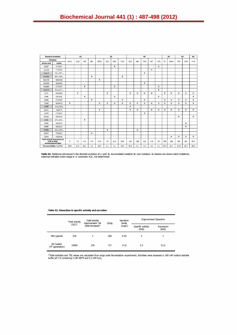
Biochemical Journal 441 (1) : 487-498 (2012)
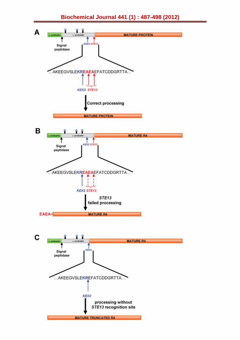
Biochemical Journal 441 (1) : 487-498 (2012)
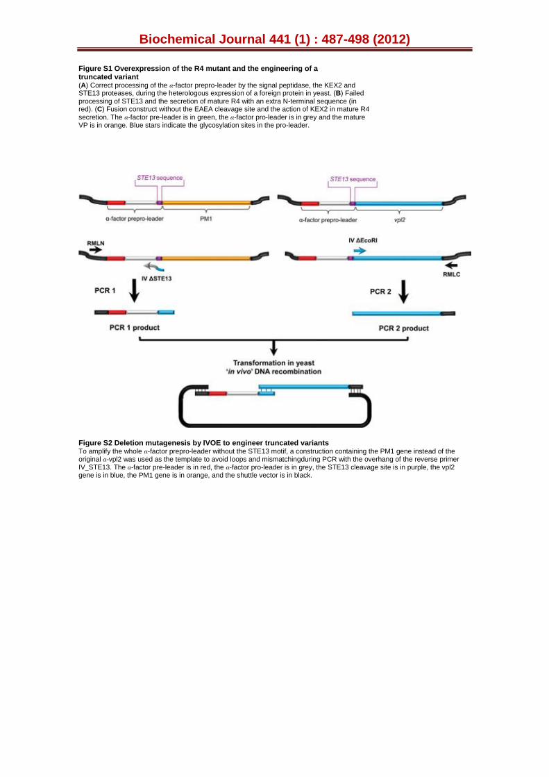
Biochemical Journal 441 (1) : 487-498 (2012)
Figure S1 Overexpression of the R4 mutant and the engineering of a truncated variant (A) Correct processing of the α-factor prepro-leader by the signal peptidase, the KEX2 and STE13 proteases, during the heterologous expression of a foreign protein in yeast. (B) Failed processing of STE13 and the secretion of mature R4 with an extra N-terminal sequence (in red). (C) Fusion construct without the EAEA cleavage site and the action of KEX2 in mature R4 secretion. The α-factor pre-leader is in green, the α-factor pro-leader is in grey and the mature VP is in orange. Blue stars indicate the glycosylation sites in the pro-leader.
Figure S2 Deletion mutagenesis by IVOE to engineer truncated variants To amplify the whole α-factor prepro-leader without the STE13 motif, a construction containing the PM1 gene instead of the original α-vpl2 was used as the template to avoid loops and mismatchingduring PCR with the overhang of the reverse primer IV_STE13. The α-factor pre-leader is in red, the α-factor pro-leader is in grey, the STE13 cleavage site is in purple, the vpl2 gene is in blue, the PM1 gene is in orange, and the shuttle vector is in black.
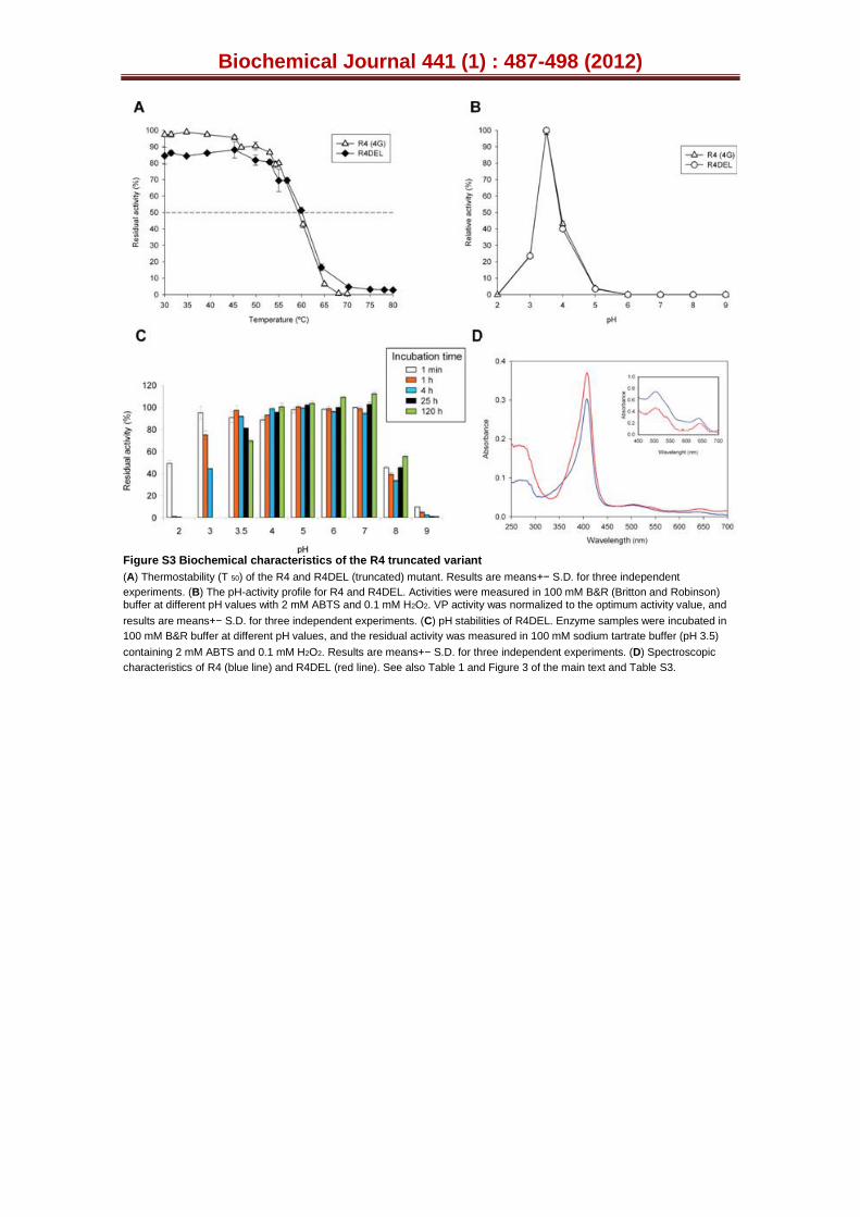
Biochemical Journal 441 (1) : 487-498 (2012)
Figure S3 Biochemical characteristics of the R4 truncated variant
(A) Thermostability (T 50) of the R4 and R4DEL (truncated) mutant. Results are means+− S.D. for three independent
experiments. (B) The pH-activity profile for R4 and R4DEL. Activities were measured in 100 mM B&R (Britton and Robinson) buffer at different pH values with 2 mM ABTS and 0.1 mM H2O2. VP activity was normalized to the optimum activity value, and
results are means+− S.D. for three independent experiments. (C) pH stabilities of R4DEL. Enzyme samples were incubated in
100 mM B&R buffer at different pH values, and the residual activity was measured in 100 mM sodium tartrate buffer (pH 3.5)
containing 2 mM ABTS and 0.1 mM H2O2. Results are means+− S.D. for three independent experiments. (D) Spectroscopic
characteristics of R4 (blue line) and R4DEL (red line). See also Table 1 and Figure 3 of the main text and Table S3.
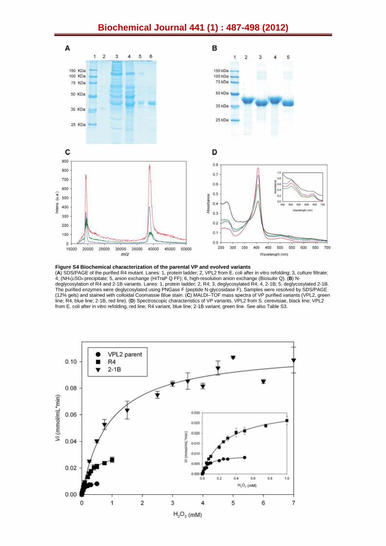
Biochemical Journal 441 (1) : 487-498 (2012)
Figure S4 Biochemical characterization of the parental VP and evolved variants (A) SDS/PAGE of the purified R4 mutant. Lanes: 1, protein ladder; 2, VPL2 from E. coli after in vitro refolding; 3, culture filtrate; 4, (NH4)2SO4 precipitate; 5, anion exchange (HiTraP Q FF); 6, high-resolution anion exchange (Biosuite Q). (B) N-deglycosylation of R4 and 2-1B variants. Lanes: 1, protein ladder; 2, R4; 3, deglycosylated R4; 4, 2-1B; 5, deglycosylated 2-1B. The purified enzymes were deglycosylated using PNGase F (peptide N-glycosidase F). Samples were resolved by SDS/PAGE (12% gels) and stained with colloidal Coomassie Blue stain. (C) MALDI–TOF mass spectra of VP purified variants (VPL2, green line; R4, blue line; 2-1B, red line). (D) Spectroscopic characteristics of VP variants. VPL2 from S. cerevisiae, black line; VPL2 from E. coli after in vitro refolding, red line; R4 variant, blue line; 2-1B variant, green line. See also Table S3.
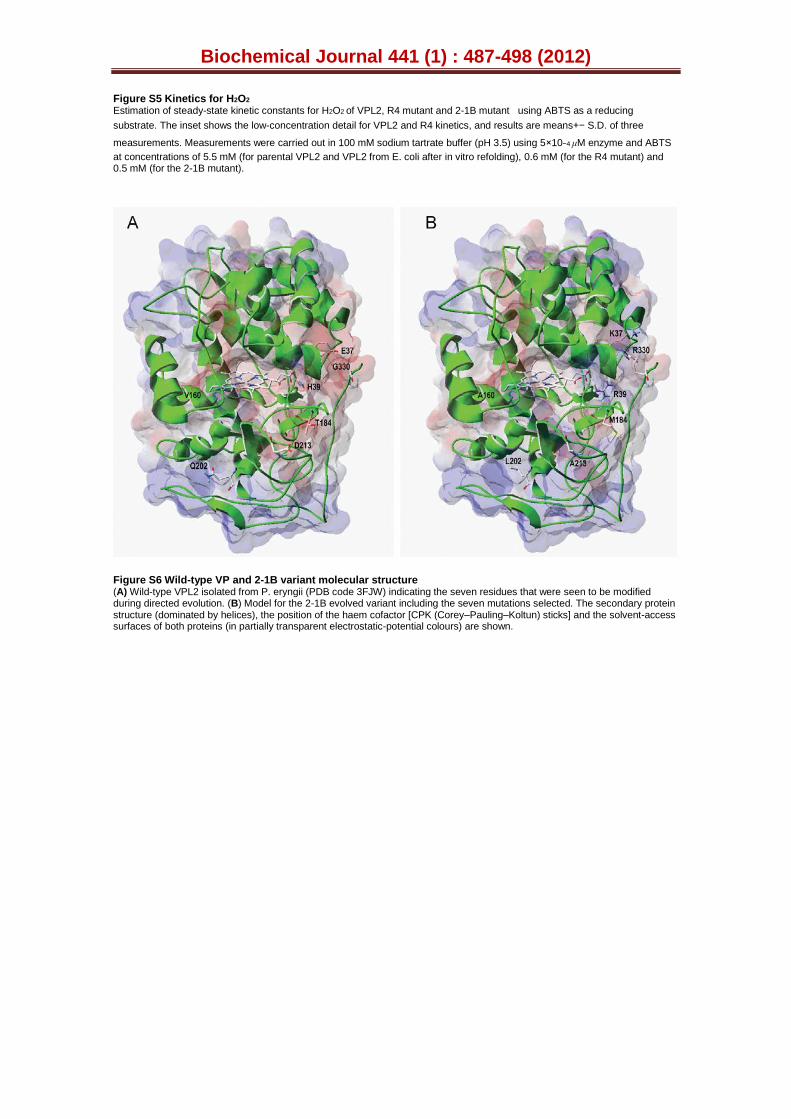
Biochemical Journal 441 (1) : 487-498 (2012)
Figure S5 Kinetics for H2O2
Estimation of steady-state kinetic constants for H2O2 of VPL2, R4 mutant and 2-1B mutant using ABTS as a reducing
substrate. The inset shows the low-concentration detail for VPL2 and R4 kinetics, and results are means+− S.D. of three
measurements. Measurements were carried out in 100 mM sodium tartrate buffer (pH 3.5) using 5×10⁻⁴ μM enzyme and ABTS
at concentrations of 5.5 mM (for parental VPL2 and VPL2 from E. coli after in vitro refolding), 0.6 mM (for the R4 mutant) and 0.5 mM (for the 2-1B mutant).
Figure S6 Wild-type VP and 2-1B variant molecular structure (A) Wild-type VPL2 isolated from P. eryngii (PDB code 3FJW) indicating the seven residues that were seen to be modified during directed evolution. (B) Model for the 2-1B evolved variant including the seven mutations selected. The secondary protein structure (dominated by helices), the position of the haem cofactor [CPK (Corey–Pauling–Koltun) sticks] and the solvent-access surfaces of both proteins (in partially transparent electrostatic-potential colours) are shown.

Biochemical Journal 441 (1) : 487-498 (2012)