Embed Size (px)
Citation preview

Direct Observation of Coupling between Structural Fluctuation andUltrafast Hydration Dynamics of Fluorescent Probes in AnionicMicellesSusobhan Choudhury,† Prasanna Kumar Mondal,† V. K. Sharma,‡ S. Mitra,‡ V. Garcia Sakai,§
R. Mukhopadhyay,‡ and Samir Kumar Pal*,†
†Department of Chemical, Biological & Macromolecular Sciences, S. N. Bose National Centre for Basic Sciences, Block JD, Sector III,Salt Lake, Kolkata 700 098, India‡Solid State Physics Division, Bhabha Atomic Research Centre, Mumbai 400085, India§Science and Technology Facilities Council, Rutherford Appleton Laboratory, Didcot, OX11 0QX, U.K.
ABSTRACT: The coupling of structural fluctuation and the dynamicsof associated water molecules of biological macromolecules is vital forvarious biological activities. Although a number of molecular dynamics(MD) studies on proteins/DNA predicted the importance of suchcoupling, experimental evidence of variation of hydration dynamics withcontrolled structural fluctuation even in model macromolecule is sparseand raised controversies in the contemporary literature. Here, we haveinvestigated dynamics of hydration at the surfaces of two similar anionicmicelles sodium dodecyl sulfate (SDS) and sodium dodecylbenzene-sulfonate (SDBS) as model macromolecules using coumarin 500 (C500)as spectroscopic probe with femtosecond to picosecond time resolutionup to 20 ns time window. The constituting surfactants SDS and SDBSare structurally similar except one benzene moiety in the SDBS may offer additional rigidity to the SDBS micelles through π-stacking and added bulkiness. The structural integrity of the micelles in the aqueous medium is confirmed in dynamic lightscattering (DLS) studies. A variety of studies including polarization gated fluorescence spectroscopy and quasielastic neutronscattering (QENS) have been used to confirm differential structural fluctuation of SDS and SDBS micelles. We have alsoemployed femtosecond-resolved Forster resonance energy transfer (FRET) in order to study binding of a cationic organic ligandethidium bromide (EtBr) salt at the micellar surfaces. The distance distribution of the donor (C500)−acceptor (EtBr) in themicellar media reveals the manifestation of the structural flexibility of the micelles. Our studies on dynamical coupling of thestructural flexibility with surface hydration in the nanoscopic micellar media may find the relevance in the “master−slave” typewater dynamics in biologically relevant macromolecules.
■ INTRODUCTION
Exchange of dynamical information between biological macro-molecules and water molecules in their vicinity (hydrationwater) is found to be key for the biological function of water asthe “matrix of life”.1−4 The ubiquitous dynamical role ofhydration water in various biological processes including chargetransfer,5−7 productive enzyme substrate complex formation,8,9
and protein folding10,11 is well documented in the early andrecent literature. Master role of water molecules at surface12
and interior13 of biologically relevant macromolecules in thecontrol of macromolecular dynamics has also been proposed.Whereas the influence of hydration water on the dynamics andfunction of several biologically relevant macromolecules isevidenced14 and believed to be slave to those of the solvent, yetto date, how dynamical coupling between the hydration waterand biologically relevant macromolecules occurs remainsunclear.15−18 This is because of experimental difficulties indirectly accessing hydration water dynamics.15,19 The usefulnessof femtosecond resolved electronic spectroscopy in the
exploration of the dynamics of hydration in various biologicalmacromolecules is evidenced and reviewed.14,20
In the present work we have used two anionic micellessodium dodecyl sulfate (SDS; C12H25SO4Na) and sodiumdodecylbenzenesulfonate (SDBS; C12H25C6H4SO3Na) asmodel macromolecule. SDBS monomer has the same alkylmoiety as SDS with an extra phenyl ring linked to a sulfonategroup. Although SDBS and SDS micelles are very similar froma structural point of view, their dynamical behavior differssignificantly. Dynamic light scattering (DLS) studies confirmstructural integrity of the micelles in our experimentalcondition. Recent molecular dynamics21 studies clearly showthat the dynamics of SDBS micelles are more restrictedcompared to SDS micelles. The structure factor indicates that
Special Issue: Biman Bagchi Festschrift
Received: November 29, 2014Revised: April 8, 2015Published: April 15, 2015
Article
pubs.acs.org/JPCB
© 2015 American Chemical Society 10849 DOI: 10.1021/jp511899qJ. Phys. Chem. B 2015, 119, 10849−10857
Dow
nloa
ded
by S
.N. B
OSE
NA
TL
CT
R B
ASI
C S
CIE
NC
ES
on A
ugus
t 28,
201
5 | h
ttp://
pubs
.acs
.org
P
ublic
atio
n D
ate
(Web
): A
pril
24, 2
015
| doi
: 10.
1021
/jp51
1899
q

the alkyl chains are more flexible in SDS compared withSDBS.22 The restricted structural flexibility of the SDBSmicelles is believed to be due to the π-stacking of two benzenemoieties of two adjacent surfactants,23 though other possibil-ities including increase in the molar mass of SDBS cannot becompletely ruled out. Results of our QENS study revealrestricted internal motion in the case of SDBS micellecompared to that of the SDS micelle.We have employed temperature dependent polarization
gated fluorescence anisotropy of a well-known fluorescenceprobe coumarin 500 (C500)24 in the micelles revealingmicroviscosity of the immediate probe environments andassociated energetics. The strategy of investigating waterdynamics in similar micellar systems using time-resolveddynamics Stokes shift is well documented in the literature.25
Femtosecond resolved Stokes shifts of a well-known solvationprobe C500 in the micelles have been followed for theinvestigation of hydration dynamics at the micellar surface. Acoupling of the structural flexibility of the micelles with theirdynamics of hydration is evident from our studies. We have alsoemployed femtosecond resolved Forster resonance energytransfer (FRET) for the investigation of nonspecific binding ofa cationic dye ethidium bromide (EtBr) salt to the micellarsurface. A strong spectral overlap of the emission of C500 withthe absorption spectrum of EtBr reveals the possibility ofenergy transfer from C500 (donor) to the acceptor EtBrthrough dipole−dipole coupling.26 The distribution of thedonor−acceptor distances in the micelles clearly reveals acorrelation of the structural flexibility of the micelles in theirmolecular recognition by a small organic ligand (EtBr). Overall,our present study is an attempt to explore the coupling ofstructural flexibility with the dynamics of hydration ofnanoscopic micellar systems using femtosecond to picosecondresolved electronic spectroscopy.
■ MATERIALS AND METHODSMaterials. Sodium dodecyl sulfate (SDS; C12H25SO4Na)
and s od i um dod e c y l b e n z e n e s u l f o n a t e ( SDBS ;C12H25C6H4SO3Na) were products of Sigma (purity≥99.0%). For QENS studies, micellar solution was preparedseparately in D2O (99.9% atom D purity) for both thesurfactants. Coumarin 500 (C500) and ethidium bromide(EtBr) are from Exciton and Molecular Probes, respectively.The water is from Millipore system.Optical Studies. Steady-state absorption and emission
spectra of the corresponding systems are measured withShimadzu UV-2450 spectrophotometer and Jobin YvonFluorolog fluorimeter, respectively. Femtosecond-resolvedfluorescence is measured using a femtosecond upconversionsetup (FOG 100, CDP) with full width at half-maximum(fwhm) of 185 fs. The samples were excited at 400 nm, andmore details of the system can be found elsewhere.27 Time-resolved emission spectrum (TRES) and solvent correlationfunction, C(t), anisotropy r(t), were constructed followingearlier published work.7,27,28 FRET distance between donor−acceptor (r) was calculated from the equation r6 = [R0
6(1 −E)]/E, where E is the energy transfer efficiency between donorand acceptor and following the procedure published else-where.29 Distance distribution function P(r) was calculatedusing nonlinear least-squares fitting procedure using SCIEN-TIST software to the following function p(r) = {1/[σ-(2π)1/2)]}exp{−1/2[(r − r)/σ]2}, where r is the mean of theGaussian with a standard deviation of σ, and r is the donor−
acceptor distance. Detailed theory and calculations of distancedistribution can be found elsewhere.29−31
Dynamic Light Scattering (DLS). Hydrodynamic diame-ter (dh) of the micelles was calculated using the equation dh =(kBT)/(3πηD), in which kB, η, and D are Boltzmann constant,viscosity, and translational diffusion coefficient, respectively, atabsolute temperature T. Details of DLS setup and experimentcan be found elsewhere.32 Following modified Stokes−Einstein−Debye equation,32 τr = (ηmVm f C)/(kBT) micro-viscosity (ηm) can be calculated, where τr is rotational timeconstant of the probe in the micelles and kB and Vm areBoltzmann constant and molecular volume of the probe (198Å3) at absolute temperature T. C represents solute−solventcoupling constant, whereas f is the shape factor. Here we haveconsidered the values of C and f to be unity.
Quasielastic Neutron Scattering (QENS). A 0.3 Mmicellar solution was prepared separately in D2O (99.9%atom D purity) for both surfactants. Experiments were carriedout using the IRIS time-of-flight spectrometer at the ISISpulsed neutron facility, U.K. IRIS is an inelastic neutronscattering instrument that works on inverted geometry with anarray of pyrolytic graphite analyzer crystals situated nearbackscattering geometry. PG (002) analyzer provides an elasticresolution of 17.5 μeV at a final energy of 1.84 meV over wavevector transfer (Q) range from 0.44 to 1.83 Å−1 with an energywindow from 0.35 to 1.2 meV (in offset configuration). Inorder to achieve no more than 10% scattering, the samples wereplaced in a cylindrical aluminum can with an internal spacing of1 mm. QENS measurements were carried out on a micellarsolution in D2O at temperatures of 300 K. Since we areinterested in the dynamics of the micelles, we subtract thescattering contribution from D2O after measuring the QENSspectra for pure D2O. Data reduction involving backgroundsubtraction and detector efficiency corrections were performedby using the ISIS data analysis package, MODES.33
■ RESULTS AND DISCUSSIONSteady-State Optical Studies. Figure 1a shows steady
state absorption and emission spectra of the probe (C500) insolvents of different degrees of polarity. The emission peak incyclohexane at 417 nm moves to 510 nm in water. Theabsorption spectra of the probe in the two solvents do not showmuch change (data not shown) revealing significantly largertransition dipole moment upon excitation.34 The emissionmaxima of the probe C500 in the SDS and SDBS micelles arefound to be 505 and 508 nm, respectively, indicating thelocation of the probe at the micellar interface. Similar spectralwidth of the emission profiles of the probe C500 in the micellarmedia and bulk water rules out significant heterogeneity in thelocation of the probe in the micelles. A slight but reproducibleshift of emission spectrum of the probe C500 in SDBScompared to that in SDS is indicative of more polarenvironment in the vicinity of the probe in SDBS micelle.Relatively higher steady state fluorescence anisotropy value ofthe probe C500 in SDBS micelle (∼0.05) compared to that inSDS micelle (∼0.03) is evident from Figure 1b, which suggestsa more restricted environment of the probe in the formermedium. The persistency of the steady state anisotropy valuesin a range of emission wavelengths (470−530 nm) in themicelles is also indicative of less heterogeneity in the probe’smicroenvironments in the micelles. Dynamics light scattering(DLS) experiments on the micelles (Figure 1c) reveals similarand almost monodispersed distribution of the SDS and SDBS
The Journal of Physical Chemistry B Article
DOI: 10.1021/jp511899qJ. Phys. Chem. B 2015, 119, 10849−10857
10850
Dow
nloa
ded
by S
.N. B
OSE
NA
TL
CT
R B
ASI
C S
CIE
NC
ES
on A
ugus
t 28,
201
5 | h
ttp://
pubs
.acs
.org
P
ublic
atio
n D
ate
(Web
): A
pril
24, 2
015
| doi
: 10.
1021
/jp51
1899
q

micelles with hydrodynamic diameters of 4.1 and 4.8 nm,respectively, consistent with earlier reported values.35,36
Quasielastic Neutron Scattering (QENS). Neutronscattering data from a micellar system have dominantcontribution from hydrogen atoms present due to their highincoherent cross-section compared to other elements. Themeasured scattering intensity can therefore be written as37
σω
σ ω= ∂∂Ω ∂
∝Ikk
S Q( , )2
i
finc inc
(1)
where Sinc(Q,ω) is the dynamical scattering function describingsingle particle motion. Here Q (=ki − kf) is the wave vectortransfer, ki and kf are the incident and scattered neutron wavevectors, respectively, and ℏω is the energy transfer. Tominimize the scattering contribution from the solvent,deuterated water was used. In order to obtain the contributionfrom the micelles alone, data from the pure solvent weresubtracted from that of the micellar solution. Figure 2 showsQENS spectra measured for SDS and SDBS micelles(contribution of D2O subtracted) at Q = 1.0 Å−1. Observedquasielastic broadening for SDS micelles is found to be
significantly larger in SDS compared to SDBS, suggestingslower dynamics in SDBS compared to SDS micelle.Varieties of motions are plausible in a micellar system, such
as global motion (which includes translational and rotation ofthe micelles), internal motions of different CH2 units, and fasttorsional motion within the micelle.38 It is these dynamicalprocesses that are expected to contribute within the time scalesof the neutron scattering technique used (10−9−10−12 s).Therefore, QENS spectra shown in Figure 2 consist of all thesemotions, and to find out the contribution from differentdynamical processes, we need to model the total dynamicalscattering function. Assuming that these dynamical processesare independent of each other, the scattering law, Smicelles(Q,ω)can be expressed as a convolution of the global and internalmotions of the micelles.22 It may be noted that “global” motionof the micelle involves both translational and rotational motionof the whole micelle. It has been shown that the total scatteringlaw comprising both translational and rotational motion can bedescribed by a single Lorentzian function for dispersedspherical nanometer sized macromolecules.39 Therefore, thescattering law for global motion can be written as37
ω ωπ ω
= Γ =Γ
Γ +S Q L( , ) ( , )
1G G G
G
G2 2
(2)
Here ΓG is the half width at half maxima (hwhm) of theLorentzian function corresponding to global motion and isproportional to the global diffusivity, DG, of the micelles. Perezet al.39 have also showed numerically that the DG, obtained withsingle Lorentzian description, is higher compared to the self-diffusion coefficient, Ds.Apart from the global motion the data also have contribution
from internal motion of the surfactant monomers. Thisscattering law consists of an elastic component and aquasielastic component. The scattering law for the internalmotion, Sin(Q,ω) can be expressed as22
ω δ ω ω= + − ΓS Q A Q A Q L( , ) ( ) ( ) (1 ( )) ( , )in in in (3)
The quasielastic component is approximated as a singleLorentzian function, Lin(Γin,ω) with hwhm, Γin. Elasticincoherent structure factor (EISF),22 defined as the fractionof the elastic scattering out of total scattering, is useful tounderstand the geometry of a dynamical motion and can beidentified as A(Q) in eq 3. As mentioned above, the scatteringlaw for micelles is the convolution product of the global andinternal motions, which can be written as22
Figure 1. (a) Steady state emission spectra of fluorescence probe C500in various solvent and in SDS and SDBS micelles. Steady stateanisotropy of the probe in SDS and SDBS micelles are shown in (b)and (c) respectively. (d) Typical DLS spectra of both micelles showingtheir corresponding hydrodynamic diameter.
Figure 2. Typical QENS spectra for 0.3 M SDS and SDBS micellarsolution at Q = 1.0 Å−1. The instrument resolution is shown by dashedline. All the spectra are normalized to peak intensities.
The Journal of Physical Chemistry B Article
DOI: 10.1021/jp511899qJ. Phys. Chem. B 2015, 119, 10849−10857
10851
Dow
nloa
ded
by S
.N. B
OSE
NA
TL
CT
R B
ASI
C S
CIE
NC
ES
on A
ugus
t 28,
201
5 | h
ttp://
pubs
.acs
.org
P
ublic
atio
n D
ate
(Web
): A
pril
24, 2
015
| doi
: 10.
1021
/jp51
1899
q

ω ω ω= Γ + − ΓS Q A Q L A Q L( , ) [ ( ) ( , ) (1 ( )) ( , )]micelles G G tot tot
(4)
Here, Ltot (Γtot,ω) represents the total contribution from globaland internal motions and Γtot = ΓG + Γin. The program DAVE40
developed at the NIST Center for Neutron Research is used toanalyze the QENS data. The parameters A(Q), ΓG, and Γtot areobtained by least-squares fit with the measured data afterconvoluting the above scattering law (eq 4) with theinstrumental resolution function. It is found that the modelfunction (eq 4) could fit the observed QENS spectra very wellat all the Q values. Typical fitted spectra for SDS and SDBSmicelles are shown in Figure 3 at some typical Q values. Thevariation of the hwhm, ΓG, of the first Lorentzian for SDBS isshown in Figure 4a along with that of SDS micelles. Larger ΓGfor SDS suggests that the global motion is faster in SDS thanSDBS micelles. It is clear that the global diffusion follows Fick’slaw (ΓG = DGQ
2, DG being the global diffusion coefficient) forboth micellar systems as shown by solid lines in Figure 4a. DG =(1.9 ± 0.3) × 10−6 is obtained for SDBS, whereas for SDSmicelle, it is found to be (3.4 ± 0.4) × 10−6 cm2/s.22 Self-diffusion constant for pure translational motion of the micelles,Ds, can be calculated using Stokes−Einstein relation, Ds = kBT/(6πηR). Here R is the hydrodynamic radius of micelles, η, theviscosity of D2O, and T, the temperature of the solution. ForSDS micelles having radius R = 20.5 Å, ηD2O = 0.9 cP 41 at T =300 K, the value of Ds obtained as 1.2 × 10⁻⁶ cm2/s which isbetween the value reported by Hayter and Penfold42 and thepresent work. Although several studies including MDsimulation studies21 on SDS and SDBS estimated the similarsizes of SDS and SDBS micelle, global diffusivities obtainedfrom QENS study are found to be significantly different in SDSand SDBS micelles.
Figure 3. Typical fitted S(Q,ω) for 0.3 M SDS and SDBS micelles at different Q values, assuming the scattering function given in eq 4.
Figure 4. (a) Variation of half width at half maxima (hwhm), ΓG,corresponds to global motion, with Q2 for SDBS and SDS micelles. (b)Variation of the hwhm, Γin, which corresponds to the internal motionof the monomer for SDS and SDBS micelle with Q2.
The Journal of Physical Chemistry B Article
DOI: 10.1021/jp511899qJ. Phys. Chem. B 2015, 119, 10849−10857
10852
Dow
nloa
ded
by S
.N. B
OSE
NA
TL
CT
R B
ASI
C S
CIE
NC
ES
on A
ugus
t 28,
201
5 | h
ttp://
pubs
.acs
.org
P
ublic
atio
n D
ate
(Web
): A
pril
24, 2
015
| doi
: 10.
1021
/jp51
1899
q
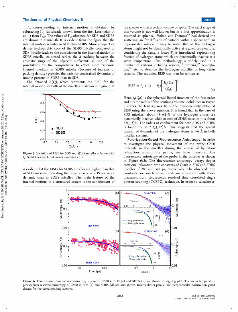
Γin corresponding to internal motion is obtained bysubtracting ΓG (as already known from the first Lorentzian ineq 4) from Γtot. The values of Γin obtained for SDS and SDBSare shown in Figure 4b. It is evident from the figure that theinternal motion is faster in SDS than SDBS. More compact ordenser hydrophobic core of the SDBS micelle compared toSDS micelle leads to the constriction in the internal motion inSDBS micelle. As stated earlier, the π stacking between thearomatic rings of the adjacent surfactants is one of thepossibilities for the compactness. In effect, more “viscous”(dense) medium in SDBS micelle (because of increase inpacking density) provides the basis for constrained dynamics ofmobile protons in SDBS than in SDS.The obtained A(Q), which represents the EISF for the
internal motion for both of the micelles, is shown in Figure 5. It
is evident that the EISFs for SDBS micelles are higher than thatof SDS micelles, indicating that alkyl chains in SDS are moredynamic than in SDBS micelles. The main feature of theinternal motions in a structured system is the confinement of
the species within a certain volume of space. The exact shape ofthis volume is not well-known but in a first approximation isassumed as spherical. Volino and Dianoux43 had derived thescattering law for diffusion of particles within a sphere with animpermeable surface. It may be noted that all the hydrogenatoms might not be dynamically active at a given temperature,considering the same, a factor Px is introduced, representingfraction of hydrogen atoms which are dynamically inactive at agiven temperature. This methodology is widely used in anumber of systems including vesicles,44 proteins,39 hemoglo-bin,45 etc. to describe the hydrogen mobility in long chainsystems. The modified EISF can then be written as
= + −⎡⎣⎢
⎤⎦⎥P P
j Qa
QaEISF (1 )
3 ( )
( )x x1
2
(5)
Here, j1(Qa) is the spherical Bessel function of the first orderand a is the radius of the confining volume. Solid lines in Figure5 shows the least-squares fit of the experimentally obtainedEISF using the above equation. It is found that in the case ofSDS micelles, about 48(±2)% of the hydrogen atoms aredynamically inactive, while in case of SDBS micelles it is about62(±2)%. The radius of confinement for both SDS and SDBSis found to be 2.9(±0.2)Å. This suggests that the spatialdomain of dynamics of the hydrogen atoms is ∼6 Å in bothmicellar systems.
Polarization-Gated Fluorescence Anisotropy. In orderto investigate the physical movement of the probe C500molecule in the micelles during the course of hydrationrelaxation around the probe, we have measured thefluorescence anisotropy of the probe in the micelles as shownin Figure 6a,b. The fluorescence anisotropy decays depictrotational relaxation time constants of C500 in SDS and SDBSmicelles of 283 and 342 ps, respectively. The observed timeconstants are much slower and are consistent with thosemeasured from picoseconds resolved time correlated singlephoton counting (TCSPC) technique. In order to calculate π-
Figure 5. Variation of EISF for SDS and SDBS micellar solution withQ. Solid lines are fitted curves assuming eq 5.
Figure 6. Femtosecond fluorescence anisotropy decays of C500 in SDS (a) and SDBS (b) are shown in log−log plot. The room temperaturepicoseconds resolved anisotropy of C500 in SDS (c) and SDBS (d) are also shown. Inserts shows parallel and perpendicular polarization gateddecays for the corresponding systems.
The Journal of Physical Chemistry B Article
DOI: 10.1021/jp511899qJ. Phys. Chem. B 2015, 119, 10849−10857
10853
Dow
nloa
ded
by S
.N. B
OSE
NA
TL
CT
R B
ASI
C S
CIE
NC
ES
on A
ugus
t 28,
201
5 | h
ttp://
pubs
.acs
.org
P
ublic
atio
n D
ate
(Web
): A
pril
24, 2
015
| doi
: 10.
1021
/jp51
1899
q

stacking energy in the case of SDBS micelle, we have measuredtemperature dependent anisotropy of both the micelles withTCSPC. Figure 6c,d shows the anisotropy decay of the probe inSDS and SDBS micelle at 293 K temperature, respectively. Thecorresponding parallel and perpendicular polarization gateddecays are shown in the respective insets. Anisotropy at 278and 343 K of both the SDS and SDBS micelle are shown in theinset of Figure 7a,b, respectively. As observed from Figure 7a,b
rotational time constant (τrot) becomes faster upon increasingtemperature for both systems. We have estimated micro-viscosities at different temperature for the correspondingsystems, given the hydrodynamics diameter of the probe is7.6 Å as reported earlier32 and plotted with 1/T (K−1) as shownin Figure 7c. The plots are distinct and out of the experimentaluncertainty (5%). Linear fit of both plots provide activationenergy 22.8(±1.1) kJ/mol and 24.1(±1.2) kJ/mol for SDS andSDBS, respectively, using the equation46 η = η0 exp[Eη/(RT)],where Eη is the energy barrier for viscous flow. The relativelyhigher activation energy in the SDBS micelles compared to that
in SDS micelle is probably the manifestation of less flexibility ofthe alkyl chains due to π-stacking (1.3 kJ/mol) in the formermedium.47
Femtosecond-Resolved Dynamics of Hydration. Tocorrelate the fluctuations with hydration dynamics of themicelles, we have performed femtosecond solvation dynamicsof both micelles. The femtosecond resolved fluorescencetransients of C500 in SDS and SDBS micelles at threecharacteristic wavelengths (440, 480, and 560 nm) across theemission wavelengths are shown in Figure 8a and Figure 8b,
respectively. An ultrafast decay component in the blue end iseventually converted into a rise component of similar timeconstant in the red end for both the micelles. The observationis consistent with the solvation of the probe C500 in themedia.24 The constructed time dependent Stokes shifts(TDSSs) of the C500 emission at different times are shownin Figure 9a and Figure 9b, with a spectral shift of 1260 and1080 cm−1 for SDS and SDBS micelles, respectively, in a 1 nstime window. The hydration correlation functions [C(t)] for
Figure 7. Plot of rotational time constant (τrot) against temperature forSDS (a) and SDBS (b) micelle. Inserts are showing anisotropy atinitial and final temperature for the respective systems. (c) Arrheniusplots of microviscosity for SDS and SDBS micelles are shown (with5% error bar).
Figure 8. Femtosecond resolved fluorescence transients of C500 inthree representative detection wavelengths (440, 480, and 560 nm) forSDS (a) and SDBS (b) micelles are shown. The circles areexperimental data, and the solid lines are best multiexponential fit(see text).
The Journal of Physical Chemistry B Article
DOI: 10.1021/jp511899qJ. Phys. Chem. B 2015, 119, 10849−10857
10854
Dow
nloa
ded
by S
.N. B
OSE
NA
TL
CT
R B
ASI
C S
CIE
NC
ES
on A
ugus
t 28,
201
5 | h
ttp://
pubs
.acs
.org
P
ublic
atio
n D
ate
(Web
): A
pril
24, 2
015
| doi
: 10.
1021
/jp51
1899
q

the SDS and SDBS micelles up to 150 ps as shown in Figure 9ccan be fitted with biexponential decay functions. C(t) up to 1 nsfor SDS and SDBS as shown in inset is nonexponential becauseof contribution of fluctuation of the micellar headgroup. Up to150 ps for SDS micelles the decay time constants are 1.48 ps(59%) and 27 ps (41%), and for SDBS micelles the obtainedtime constants are 1.75 ps (48%) and 39 ps (52%). Our resultis consistent with earlier femtosecond resolved study48 on the
dynamics of hydration in TX-100 micelle reported hydrationtime constants of 2.9 ps (45%) and 58 ps (55%).49 It has to benoted that the correlation functions for both micelles shownonexponential behavior after 150 ps up to a time window of 1ns. The nonexponential behavior of biologically relevantmacromolecules including DNA has been well documented ata time window from femtosecond until microsecond.50 Earlierit has been concluded that the faster response in the C(t) is aconsequence of the movement of counterions away from theiraverage position through the electric field at the probe. On theother hand relatively slower components are from various kindsof motions of the host system itself.50,51 Overall, a variety ofdynamical events are likely to be included in the solvationresponse and the relaxation dynamics must be treated as acollective response of the whole system. The faster dynamics ofhydration in SDS than that in SDBS micelle is clearly evidentfrom the study. The structural flexibility of the SDS and SDBSmicelles is also evident from polarization gated fluorescenceanisotropy studies (Figure 6), revealing higher rigidity in thelatter case. Thus, our observation of faster hydration dynamicsin structurally flexible SDS micelle may find a correlationbetween the dynamics of hydration water and internal motionof the micelles.
Femtosecond-Resolved Forster Resonance EnergyTransfer (FRET). In Figure 10 we have shown the molecularcomplexation of a cationic dye EtBr with the anionic micellesSDS and SDBS using FRET techniques.52 Figure 10a shows theoverlap (overlap integral value of 3.37 × 10¹⁴ M−1 cm−1 nm4)between the emission spectrum of C500 (donor) and theabsorption spectrum of EtBr (acceptor) in the SDS micelle.The overlap for the SDBS micelles remains almost unchanged(overlap integral value of 3.1 × 10¹⁴ M−1 cm−1 nm4; data notshown in the figure). Femtosecond resolved fluorescencetransients of C500 at 500 nm in the micelles before and afterthe complexation with the acceptor EtBr is shown in the Figure10b and Figure 10c. A significant faster component of 409 fs(32%) in the case of SDS micelles upon complexation withEtBr is clearly evident. In the case of SDBS micelle the fastercomponent is found to be 475 fs (10%). The estimated Forsterdistance (R0) values of the donor−acceptor pair for the SDSand SDBS micelles are 34.61 and 34.14 Å, respectively. Thecalculated donor−acceptor distances in the two micelles arefound to be 33.64 and 45.0 Å, respectively. The higher donor−acceptor distance in the case of SDBS compared to that in SDSmicelle may not be due to the distant location of the donorC500 from the micellar surface, as the fluorescence spectrum ofthe donor probe shows similar characteristics revealinginsignificant change in the polarity around the probe. In thecase of deep insertion of the probe from the surface, lowerpolarity around the probe is unavoidable. Thus, the observationcan be rationalized in terms of different position of the acceptorEtBr at the surface of the SDBS micelle compared to that of theSDS. The intrinsic micellar fluctuation, as evidenced inmicroviscosity (described above), is also substantiated in ourFRET studies on the molecular recognition of EtBr by the twomicellar systems. The distribution of donor−acceptor distancesin the micelles is also shown in Figure 10d revealing relativelyless broadening in the case of SDBS (half width of 2.5 Å)compared to that in SDS (half width of 3.4 Å). The observationof high width in the donor−acceptor distance distribution incase of SDS micelle compared to SDBS micelle can berationalized in terms of dynamical fluctuation of the hostmicelles.31,53 The calculated half width values, revealing
Figure 9. Time dependent emission spectra (TRES) of fluorescentprobe C500 in SDS (a) and SDBS (b) micelles are shown. The dottedlines are the steady state fluorescence spectra of the correspondingsystems. Δυ is the spectral shift for the micellar system in 1 ns timewindow. (c) Hydration correlation functions for the SDS and SDBSare shown up to 150 ps. The solid lines are the best biexponential fit toC(t). Inset shows the correlation function in long time range.
The Journal of Physical Chemistry B Article
DOI: 10.1021/jp511899qJ. Phys. Chem. B 2015, 119, 10849−10857
10855
Dow
nloa
ded
by S
.N. B
OSE
NA
TL
CT
R B
ASI
C S
CIE
NC
ES
on A
ugus
t 28,
201
5 | h
ttp://
pubs
.acs
.org
P
ublic
atio
n D
ate
(Web
): A
pril
24, 2
015
| doi
: 10.
1021
/jp51
1899
q

heterogeneity or fluctuation in the observed distances betweenthe donor and the acceptor, thus correlated with the dynamicsof hydration.
■ CONCLUSION
Here we employed femtosecond resolved electronic spectros-copy for the exploration of hydration dynamics of a well-knownsolvation probe C500 in two structurally similar anionicmicelles SDS and SDBS. Dynamic light scattering (DLS)reveals the structural integrity of the micelles. We have usedpolarization gated fluorescence anisotropy for the estimation ofstructural flexibility of the micelles. We have found SDBS ismore compact than SDS. The extracted dynamics of hydrationin the micelles reveal slower water motion at the less flexibleSDBS micelle compared to that in SDS micelle. Thecomparison of the dynamics of hydration across two micellesof different compactness reveals a gradient of coupling betweenhydration water and internal micellar motions, which isstronger in relatively flexible SDS and weaker in compactSDBS micelles. Slower internal motion in SDBS compared toSDS micelle is also confirmed from QENS studies. We havealso used femtosecond resolved FRET in order to investigatethe molecular recognition of the micelles by a small cationicligand EtBr. Relatively broad distribution of EtBr at the micellarsurfaces with respect to the probe C500 in the case of SDSmicelle compared to that in SDBS micelle is evident. Theobservation is correlated with the internal flexibility of themicelles. Thus, our present study can be considered as anexploration of a coherent picture of structure, dynamics, andfunction of molecular recognition at physiologically relevantimportant nanoscopic micellar environments.
■ AUTHOR INFORMATION
Corresponding Author*E-mail:[email protected].
NotesThe authors declare no competing financial interest.
■ ACKNOWLEDGMENTS
S.C. thanks CSIR, India, for the research fellowships. Financialgrants SB/S1/PC-011/2013 and DST/TM/SERI/2k11/103from DST (India) and Grant 2013/37P/73/BRNS from DAE(India) are gratefully acknowledged.
■ REFERENCES(1) Pal, S. K.; Zewail, A. H. Dynamics of Water in BiologicalRecognition. Chem. Rev. 2004, 104, 2099−2124.(2) Ball, P. Water as an Active Constituent in Cell Biology. Chem.Rev. 2008, 108, 74−108.(3) Wang, L.; Yu, X.; Hu, P.; Broyde, S.; Zhang, Y. A Water-Mediatedand Substrate-Assisted Catalytic Mechanism for Sulfolobus SolfataricusDNA Polymerase IV. J. Am. Chem. Soc. 2007, 129, 4731−4737.(4) Zong, C.; Papoian, G. A.; Ulander, J.; Wolynes, P. G. Role ofTopology, Nonadditivity, and Water-Mediated Interactions inPredicting the Structures of alpha/beta Proteins. J. Am. Chem. Soc.2006, 128, 5168−5176.(5) Pechstedt, K.; Whittle, T.; Baumberg, J.; Melvin, T. Photo-luminescence of Colloidal Cdse/Zns Quantum Dots: The CriticalEffect of Water Molecules. J. Phys. Chem. C 2010, 114, 12069−12077.(6) Migliore, A.; Corni, S.; Di Felice, R.; Molinari, E. Water-MediatedElectron Transfer between Protein Redox Centers. J. Phys. Chem. B2007, 111, 3774−3781.(7) Choudhury, S.; Batabyal, S.; Mondol, T.; Sao, D.; Lemmens, P.;Pal, S. K. Ultrafast Dynamics of Solvation and Charge Transfer in aDNA-Based Biomaterial. Chem.Asian J. 2014, 9, 1395−1402.(8) Rupley, J. A.; Careri, G. Protein Hydration and Function. Adv.Protein Chem. 1991, 41, 37−172.(9) Kornblatt, J.; Kornblatt, M. Water as It Applies to the Function ofEnzymes. Int. Rev. Cytol. 2002, 215, 49−73.(10) Levy, Y.; Onuchic, J. N. Water Mediation in Protein Folding andMolecular Recognition. Annu. Rev. Biophys. Biomol. Struct. 2006, 35,389−415.(11) Kim, S. J.; Born, B.; Havenith, M.; Gruebele, M. Real-TimeDetection of Protein−Water Dynamics upon Protein Folding byTerahertz Absorption Spectroscopy. Angew. Chem., Int. Ed. 2008, 47,6486−6489.(12) Zhou, F.; Schulten, K. Molecular Dynamics Study of aMembrane−Water Interface. J. Phys. Chem. 1995, 99, 2194−2207.
Figure 10. Spectral overlap of C500 emission and EtBr absorption in SDS micelles is shown in (a). (b) and (c) show fluorescence transient of C500in the micelles at 500 nm before and after complexation with cationic dye EtBr. (d) shows distribution of donor−acceptor distances in the twomicelles.
The Journal of Physical Chemistry B Article
DOI: 10.1021/jp511899qJ. Phys. Chem. B 2015, 119, 10849−10857
10856
Dow
nloa
ded
by S
.N. B
OSE
NA
TL
CT
R B
ASI
C S
CIE
NC
ES
on A
ugus
t 28,
201
5 | h
ttp://
pubs
.acs
.org
P
ublic
atio
n D
ate
(Web
): A
pril
24, 2
015
| doi
: 10.
1021
/jp51
1899
q

(13) Helms, V. Protein Dynamics Tightly Connected to theDynamics of Surrounding and Internal Water Molecules. ChemPhy-sChem 2007, 8, 23−33.(14) Bhattacharyya, K. Nature of Biological Water: A FemtosecondStudy. Chem. Commun. 2008, 2848−2857.(15) Gallat, F.-X.; Laganowsky, A.; Wood, K.; Gabel, F.; Van Eijck,L.; Wuttke, J.; Moulin, M.; Hartlein, M.; Eisenberg, D.; Colletier, J.-P.Dynamical Coupling of Intrinsically Disordered Proteins and TheirHydration Water: Comparison with Folded Soluble and MembraneProteins. Biophys. J. 2012, 103, 129−136.(16) Qvist, J.; Ortega, G.; Tadeo, X.; Millet, O.; Halle, B. HydrationDynamics of a Halophilic Protein in Folded and Unfolded States. J.Phys. Chem. B 2012, 116, 3436−3444.(17) Conti Nibali, V.; D’Angelo, G.; Paciaroni, A.; Tobias, D. J.;Tarek, M. On the Coupling between the Collective Dynamics ofProteins and Their Hydration Water. J. Phys. Chem. Lett. 2014, 5,1181−1186.(18) Heyden, M.; Tobias, D. J. Spatial Dependence of Protein-WaterCollective Hydrogen-Bond Dynamics. Phys. Rev. Lett. 2013, 111,218101.(19) Tarek, M.; Tobias, D. J. Single-Particle and Collective Dynamicsof Protein Hydration Water: A Molecular Dynamics Study. Phys. Rev.Lett. 2002, 89, 275501.(20) Pal, S. K.; Zhao, L.; Zewail, A. H. Water at DNA Surfaces:Ultrafast Dynamics in Minor Groove Recognition. Proc. Natl. Acad. Sci.U.S.A. 2003, 100, 8113−8118.(21) Palazzesi, F.; Calvaresi, M.; Zerbetto, F. A Molecular DynamicsInvestigation of Structure and Dynamics of Sds and Sdbs Micelles. SoftMatter 2011, 7, 9148−9156.(22) Sharma, V.; Mitra, S.; Johnson, M.; Mukhopadhyay, R.Dynamics in Anionic Micelles: Effect of Phenyl Ring. J. Phys. Chem.B 2013, 117, 6250−6255.(23) Islam, M.; Rojas, E.; Bergey, D.; Johnson, A.; Yodh, A. HighWeight Fraction Surfactant Solubilization of Single-Wall CarbonNanotubes in Water. Nano Lett. 2003, 3, 269−273.(24) Rakshit, S.; Saha, R.; Verma, P. K.; Pal, S. K. Role of SolvationDynamics in Excited State Proton Transfer of 1-Naphthol inNanoscopic Water Clusters Formed in a Hydrophobic Solvent.Photochem. Photobiol. 2012, 88, 851−859.(25) Nandi, N.; Bhattacharyya, K.; Bagchi, B. Dielectric Relaxationand Solvation Dynamics of Water in Complex Chemical and BiologicalSystems. Chem. Rev. 2000, 100, 2013−2046.(26) Majumder, P.; Sarkar, R.; Shaw, A. K.; Chakraborty, A.; Pal, S.K. Ultrafast Dynamics in a Nanocage of Enzymes: Solvation andFluorescence Resonance Energy Transfer in Reverse Micelles. J.Colloid Interface Sci. 2005, 290, 462−474.(27) Banerjee, D.; Makhal, A.; Pal, S. K. Sequence DependentFemtosecond-Resolved Hydration Dynamics in the Minor Groove ofDNA and HistoneDNA Complexes. J. Fluoresc. 2009, 19, 1111−1118.(28) Horng, M. L.; Gardecki, J. A.; Papazyan, A.; Maroncelli, M.Subpicosecond Measurements of Polar Solvation Dynamics: Coumar-in 153 Revisited. J. Phys. Chem. 1995, 99, 17311−17337.(29) Batabyal, S.; Mondol, T.; Choudhury, S.; Mazumder, A.; Pal, S.K. Ultrafast Interfacial Solvation Dynamics in Specific Protein DNARecognition. Biochimie 2013, 95, 2168−2176.(30) Lakowicz, J. R.; Geddes, C. D. Topics in FluorescenceSpectroscopy; Springer: New York, 1991; Vol. 1.(31) Chaudhuri, S.; Batabyal, S.; Polley, N.; Pal, S. K. Vitamin B2 inNanoscopic Environments under Visible Light: PhotosensitizedAntioxidant or Phototoxic Drug? J. Phys. Chem. A 2014, 118, 3934−3943.(32) Banerjee, D.; Verma, P. K.; Pal, S. K. Temperature-DependentFemtosecond-Resolved Hydration Dynamics of Water in AqueousGuanidinium Hydrochloride Solution. Photochem. Photobiol. Sci. 2009,8, 1441−1447.(33) Howells, W. S.; Garcia Sakai, V.; Demmel, F.; Telling, M. T. F.;Fernandez-Alonso, F. The MODES User Guide, version 3; RutherfordAppleton Laboratory: Didcot, U.K., 2010.
(34) Lewis, J.; Maroncelli, M. On the (Uninteresting) Dependence ofthe Absorption and Emission Transition Moments of Coumarin 153on Solvent. Chem. Phys. Lett. 1998, 282, 197−203.(35) Bruce, C. D.; Berkowitz, M. L.; Perera, L.; Forbes, M. D.Molecular Dynamics Simulation of Sodium Dodecyl Sulfate Micelle inWater: Micellar Structural Characteristics and Counterion Distribu-tion. J. Phys. Chem. B 2002, 106, 3788−3793.(36) Cheng, D. C.; Gulari, E. Micellization and IntermicellarInteractions in Aqueous Sodium Dodecyl Benzene SulfonateSolutions. J. Colloid Interface Sci. 1982, 90, 410−423.(37) Bee, M. Quasielastic Neutron Scattering; Adam Hilger: Bristol,U.K., 1988.(38) Sharma, V. K.; Mitra, S.; Garcia Sakai, V.; Hassan, P. A.; PeterEmbs, J.; Mukhopadhyay, R. The Dynamical Landscape in CTABMicelles. Soft Matter 2012, 8, 7151−7160.(39) Perez, J.; Zanotti, J.-M.; Durand, D. Evolution of the InternalDynamics of Two Globular Proteins from Dry Powder to Solution.Biophys. J. 1999, 77, 454−469.(40) Azuah, R. T.; Kneller, L. R.; Qiu, Y.; Tregenna-Piggott, P. L.;Brown, C. M.; Copley, J. R.; Dimeo, R. M. DAVE: A ComprehensiveSoftware Suite for the Reduction, Visualization, and Analysis of LowEnergy Neutron Spectroscopic Data. J. Res. Natl. Inst. Stand. Technol.2009, 114, 341−358.(41) Castelletto, V.; Hamley, I. W.; Yang, Z.; Haeussler, W. NeutronSpin-Echo Investigation of the Dynamics of Block CopolymerMicelles. J. Chem. Phys. 2003, 119, 8158−8161.(42) Hayter, J. B.; Penfold, J. J. Chem. Soc., Faraday Trans. 1 1981, 77,1851−1863.(43) Volino, F.; Dianoux, A. Neutron Incoherent Scattering Law forDiffusion in a Potential of Spherical Symmetry. J. Mol. Phys. 1980, 41,271−279.(44) Gerelli, Y.; Sakai, V. G.; Ollivier, J.; Deriu, A. Conformationaland Segmental Dynamics in Lipid-Based Vesicles. Soft Matter 2011, 7,3929−3935.(45) Caronna, C.; Natali, F.; Cupane, A. Incoherent Elastic andQuasi-Elastic Neutron Scattering Investigation of HemoglobinDynamics. Biophys. Chem. 2005, 116, 219−225.(46) Mitra, R. K.; Verma, P. K.; Pal, S. K. Exploration of theDynamical Evolution and the Associated Energetics of WaterNanoclusters Formed in a Hydrophobic Solvent. J. Phys. Chem. B2009, 113, 4744−4750.(47) Huber, R. G.; Margreiter, M. A.; Fuchs, J. E.; von Grafenstein,S.; Tautermann, C. S.; Liedl, K. R.; Fox, T. Heteroaromatic π-StackingEnergy Landscapes. J. Chem. Inf. Model. 2014, 54, 1371−1379.(48) Pal, S. K.; Peon, J.; Zewail, A. H. Biological Water at the ProteinSurface: Dynamical Solvation Probed Directly with FemtosecondResolution. Proc. Natl. Acad. Sci. U.S.A. 2002, 99, 1763−1768.(49) Pal, S. K.; Sukul, D.; Mandal, D.; Sen, S.; Bhattacharyya, K.Solvation Dynamics of Dcm in Micelles. Chem. Phys. Lett. 2000, 327,91−96.(50) Andreatta, D.; Perez Lustres, J. L.; Kovalenko, S. A.; Ernsting, N.P.; Murphy, C. J.; Coleman, R. S.; Berg, M. A. Power-Law SolvationDynamics in DNA over Six Decades in Time. J. Am. Chem. Soc. 2005,127, 7270−7271.(51) Brauns, E. B.; Madaras, M. L.; Coleman, R. S.; Murphy, C. J.;Berg, M. A. Complex Local Dynamics in DNA on the Picosecond andNanosecond Time Scales. Phys. Rev. Lett. 2002, 88, 158101.(52) Pal, S. K.; Mandal, D.; Bhattacharyya, K. PhotophysicalProcesses of Ethidium Bromide in Micelles and Reverse Micelles. J.Phys. Chem. B 1998, 102, 11017−11023.(53) Nag, S.; Sarkar, B.; Chandrakesan, M.; Abhyanakar, R.;Bhowmik, D.; Kombrabail, M.; Dandekar, S.; Lerner, E.; Haas, E.;Maiti, S. A Folding Transition Underlies the Emergence of MembraneAffinity in Amyloid-B. Phys. Chem. Chem. Phys. 2013, 15, 19129−19133.
The Journal of Physical Chemistry B Article
DOI: 10.1021/jp511899qJ. Phys. Chem. B 2015, 119, 10849−10857
10857
Dow
nloa
ded
by S
.N. B
OSE
NA
TL
CT
R B
ASI
C S
CIE
NC
ES
on A
ugus
t 28,
201
5 | h
ttp://
pubs
.acs
.org
P
ublic
atio
n D
ate
(Web
): A
pril
24, 2
015
| doi
: 10.
1021
/jp51
1899
q