Embed Size (px)
Citation preview

JOURNAL OF CLINICAL MICROBIOLOGY, Aug. 1983, p. 310-3170095-1137/83/080310-08$02.00/0Copyright C 1983, American Society for Microbiology
Vol. 18, No. 2
Direct Isolation in Cell Culture of Human Rotaviruses andTheir Characterization into Four Serotypes
RICHARD G. WYATT,* HARVEY D. JAMES, JR., ANNIE L. PITTMAN, YASUTAKA HOSHINO,HARRY B. GREENBERG, ANTHONY R. KALICA, JORGE FLORES, AND ALBERT Z. KAPIKIAN
Laboratory of Infectious Diseases, National Institute ofAllergy and Infectious Diseases, Bethesda, Maryland20205
Received 28 February 1983/Accepted 28 April 1983
Of 73 rotavirus-positive fecal specimens tested, 39 yielded a human rotavirusthat could be cultivated serially in MA104 or primary African green monkeykidney cells or both; 18 were serotyped. Four distinct serotypes were identified byplaque reduction or tube neutralization assay or both, and three of these serotypeswere the same .as those established previously by plaque reduction, using humanrotaviruses cultivated by genetic reassortment with a cultivable bovine rotavirus.Ten human rotavirus strains received from Japan were found to be similar, if notidentical, to our candidate prototype strains representing these four humanrotavirus serotypes.
Previous inability to cultivate human rotavi-ruses hindered the serotypic characterization ofthese viruses by neutralization assay. The fluo-rescent-focus neutralization assay was used suc-cessfully, however, since the virus was notrequired to undergo efficient replication or toproduce a cytopathic effect (CPE) (3, 23). Subse-quently, after serial passage in gnotobiotic pig-lets, a single strain of human rotavirus, Wa, wassuccessfully propagated in primary Africangreen monkey kidney (AGMK) cells (28). Thisstrain was characterized by both plaque reduc-tion neutralization assay and fluorescent-focusneutralization assay and was found to be distinctfrom selected animal rotavirus strains (28, 29).More recently, Japanese investigators demon-strated successful isolation of multiple humanrotavirus strains in roller tubes of MA104 cellcultures, using trypsin (21, 25). Successful culti-vation of human rotaviruses has also been re-ported in primary cynomolgus monkey kidneycells (10). These newly cultivated strains havebeen characterized by neutralization of plaquesor CPE and were found to represent three to fivedistinct serotypes (20, 24). An enzyme immuno-assay for distinguishing three serotypes of hu-man rotavirus from clinical specimens has re-cently been described, in which antiseraabsorbed with heterologous rotaviruses wereused (22).Human rotaviruses were also cultivated or
"rescued" by the technique of genetic reassort-ment, in which a temperature-sensitive mutantof bovine rotavirus was reassorted with a non-cultivable human rotavirus under the selective
pressure of temperature and high-titered hyper-immune antiserum, so that the resultant virusesexhibited the growth characteristics of the bo-vine parent and the neutralization phenotype ofthe human parent (7, 9). Human rotavirusesrescued in this manner and characterized byplaque reduction neutralization assay fell intothree distinct serotypes (26). Serotypes could becorrectly predicted by using a single dilution ofhigh-titered hyperimmune antiserum in a simpli-fied CPE assay (9). The use of the geneticreassortment technique also permitted the as-signment of function to selected genes (6, 13,14). Of particular interest is the observationthat, depending on the rotavirus strain, gene 8 or9 codes for the primary neutralization determi-nant (6, 14). Thus, in this description of serotyp-ic characterization, it is likely that antibody tothe product of gene 8 or 9 is primarily responsi-ble for neutralization, although recently a neu-tralization function has also been assigned to theproducts of genes 4, 6, and 10 (1, 8a, 13, 16).
In this study, 18 of 39 human rotavirus strains,which were cultivated directly in cell culture,were classified into four distinct serotypes bythe use of plaque reduction assay or tube neu-tralization read by enzyme immunoassay orboth. These strains were further compared withother human rotavirus strains isolated in Japan(17, 20, 24).
MATERIALS AND METHODS
Cell cultures. MA104 cell cultures were obtainedoriginally from E. H. Bohl, Ohio Agricultural Re-search and Development Center, Wooster, and were
310
on June 16, 2018 by guesthttp://jcm
.asm.org/
Dow
nloaded from

FOUR HUMAN ROTAVIRUS SEROTYPES 311
prepared either at MA Bioproducts, Walkersville,Md., or in our laboratory. An effort was made not toexceed passage 72. Cultures of primary AGMK werepurchased from MA Bioproducts.
Viruses. Rotaviruses for primary isolation were inthe form of diarrheal or normal feces, either as approx-imate 10%o suspensions or as rectal swab fluids. Seven-ty-three such fecal samples containing rotavirus werekindly supplied by the following investigators: H. W.Kim and colleagues, Children's Hospital NationalMedical Center, Washington, D.C.; I. Perez and col-leagues, Hospital de Nifios, Caracas, Venezuela; J.Banatvala and colleagues, St. Thomas Hospital, Lon-don, England; R. Bishop and colleagues, University ofMelbourne, Melbourne, Australia; J. G. Kapsenberg,Rijkinstituut Voor de Volksgezondheid, Bilthoven,The Netherlands; R. Black and colleagues, Universityof Maryland, Baltimore, Md.; R. Yolken, Johns Hop-kins University, Baltimore, Md.; and 0. Sobeslavsky,who was coordinating a collaborative study of diarrhe-al diseases for the World Health Organization. Select-ed rotavirus strains were previously passaged one tothree times in gnotobiotic calves or piglets beforeinoculation into cell cultures as a part of collaborativestudies with C. A. Mebus and A. Torres-Medina at theUniversity of Nebraska, Lincoln, and with E. H.Bohl, L. J. Saif, and K. W. Theil at the Ohio Agricul-tural Research and Development Center, Wooster.The isolation and characterization of human/bovinerotavirus reassortants were previously described (9).Human rotavirus strains were received from Japanfrom S. Urasawa, T. Konno, Y. Inaba, and theircolleagues in the form of cell culture suspensions ofcultivable viruses.
Cultivation method. The method of Sato et al. wasused for cultivation of human rotaviruses with onlyminor modifications (21). The first nine viruses inocu-lated into cell culture were partially purified as de-scribed before (21), but subsequently 10%o suspensionsor rectal swab fluids were used directly for inocula-tion. Before inoculation, virus suspensions were treat-ed with 10 pLg of trypsin (type IX; Sigma Chemical Co.,St. Louis, Mo.) per ml for 1 h at 37°C. After anadsorption period of 1 h at 37°C, tubes were washedonce and fed with Eagle minimum essential mediumcontaining 0.5 Fg of trypsin per ml. DEAE-dextranwas added to the medium at a concentration of 100,ug/ml for use on MA104 cells (18), but was omitted foruse on AGMK cells because of toxicity for the cells.Eagle minimum essential medium was prepared ac-cording to the description of Sato et al., except for theomission of sodium glutamate (21). Cells were incubat-ed for 3 to 10 days on a roller apparatus at 37°C andwere observed for CPE. After that time, cells werefrozen and thawed once and then tested by indirectenzyme immunoassay for viral antigens (15).Hyperimmune antisera. Antisera were prepared as
previously described (7, 9, 26). Hyperimmune antiseraagainst selected Japanese rotavirus strains were kindlysupplied by S. Urasawa and T. Konno.Plaque reduction assay. Rotaviruses were plaqued in
six-well dishes of MA104 cells as previously described(27). For neutralization assay, the hyperimmune anti-sera were inactivated at 56°C for 30 min. Fourfolddilutions were then prepared and mixed with a dilutionof trypsin-treated virus calculated to yield approxi-mately 30 PFU per well (27). Antibody titers were
estimated based on 60%o reduction of plaque count.Viruses that exhibited a reciprocal 20-fold or greaterdifference in serum antibody titer were considereddistinct serotypes (26).
Modified tube neutralization assay read by enzymeimmunoassay. Approximately 10 to 100 50%o tissueculture infective doses of trypsin-treated virus wereincubated with 10-fold dilutions of inactivated hyper-immune antisera before inoculation into one tube eachof MA104 roller tube cultures under the same condi-tions described above. After 2 or more days of incuba-tion, the tubes were harvested and examined by indi-rect enzyme immunoassay for rotavirus antigens (15)(Fig. 1). The antibody titer was considered to be thehighest dilution of antiserum which completely neu-tralized the virus as measured by indirect enzymeimmunoassay. A 20-fold reciprocal difference in anti-body titers was considered the basis for differentiatingserotypes (26).Subgrouping analysis. Rotavirus fecal suspensions
and cell culture isolates were analyzed for subgroupantigen by the use of monoclonal antibodies as previ-ously described (8), except that indirect enzyme im-munoassay was used instead of radioimmunoassay insome cases. Goat anti-mouse immunoglobulin G con-jugated with alkaline phosphatase (Kirkegaard andPerry, Inc., Gaithersburg, Md.) was used in the reac-tion, and human rotaviruses DS-1 and Wa, subgroup 1and 2 viruses, respectively, were used as positivecontrols.RNA analysis by polyacrylamide gel electrophoresis.
RNA was extracted from rotavirus preparations andexamined on polyacrylamide gels as previously de-scribed (14).
RESULTSCultivation of rotaviruses. Thirty-five strains
of human rotavirus were successfully cultivatedfrom 73 rotavirus-positive fecal specimens, us-ing MA104 cell cultures. These strains werepassaged serially 5 to 14 times. Eighteen of the35 fecal samples which yielded rotavirus oninoculation in MA104 cells were also inoculatedinto primary AGMK cells, and 16 of the 18 grew.Five strains which did not grow in MA104 cellswere inoculated into AGMK cells, and fourgrew. Strains were grown in primary AGMKcells for 3 to 14 passages. Thus, a total of 39human rotavirus strains were cultivated in cellculture. Growth of rotavirus was determined byindirect enzyme immunoassay, since CPE wasvariable. Most strains grew well by passage 2 or3, and cell culture fluids were strongly positiveby enzyme immunoassay. Five (14%) of the 35strains passaged in MA104 cells, however, werenot positive in enzyme immunoassay until pas-sage 4 to 6. These same five strains were laterinoculated into primary AGMK cells, and fourgrew during passage 1 and during five subse-quent serial passages; one strain grew duringtwo serial passages but failed to grow thereafter.Most strains which could not be serially propa-gated in cell culture were negative in enzyme
VOL. 18, 1983
on June 16, 2018 by guesthttp://jcm
.asm.org/
Dow
nloaded from

312 WYATT ET AL. J. CLIN. MICROBIOL.
Es 88 8 8 88 0Ivv I I I" vv, Iv- I v vI ooC;C
A\ A\
88888VVV VV
vvv
-4 -
v v
8888 88888 888_t4 _e4 _- _. _ 4 - -4 V4 "4 - -
VVV VV V I VV- _-4 T- -
A\ A\ A\ A\
o o o -'.
N -
00 8 00 0 0 in)oo oo 0 00 00oov v vvv v v nIIi vv
en 0%
eIn
00 I-'T 00" I
-IIOvO I I"
aDI es
0 0 0000 0 0 .)0 a) )0 0
%O_-4a -4 'I'D %N00 00 00 NN NN NN0 0 0 % 00N %0%0o0%sofi oi as---4_ 1_t-4
00 00Iv I
o oZ +1 Z
0% 0% 0%
co coc < < O,, X
>C
t-o W) 00
IV1 S^ z 6
V" 43 CT
%) Dmt rm %0
. . r_8n Cl CO
(tO 0
I. S.%
.8 <=F,
en~~~~~~~~~U
CU
C)
=0 a)3
CU.2
tv( a) 0
U)
en C Uc
>00
^ E
CU
o e
r. 00 0e
oe cd 0~~~~c
Qqtcd _R~~~c
o oo E
oar) . oQ u7 ce
*= b * 0
C CU
.to . Q
JFS E >)
C0c *cOgCN .5I ,
888 8888o o880vvvv v vvvv _; _tAA4 A\
/\ A\ A\
88888 8OOOV V V VV I oo^~C
A\ A\
.0la
0.
r_
.e
a)
CoCL
0
10cx
a)ci*F
0
N
0
.0
0I=
l)a)
'aI
Z
a)
._
u
C)cua.
uz
._
~0
U,o
a-
c
0
CU
2
(A
0
a
CZ
2C.
0
0.
0
F-0
00 00 00
0° 0N IN ON ONceo o
0000 00 00 00A\ A\ A\ A\ A\
T4 T-4 rl - 4 4
Nt tNt t t0a ON 0% as ON0%
.! -< < <: <: <ce CO) ut va ut3: 3) 3: 3: 3) 3
0>1 c
0
uC
U,*a)
on June 16, 2018 by guesthttp://jcm
.asm.org/
Dow
nloaded from
FOUR HUMAN ROTAVIRUS SEROTYPES 313
immunoassay by passage 2. Twenty-three of the73 fecal samples were tested in the form of rectalswabs, and none of these specimens yielded a
cultivable rotavirus. The 39 isolates were recov-
ered from 50 fecal suspensions tested in thesame manner as the rectal swabs, thus yieldingan isolation rate of 78% from such specimens.Twenty-six of 39 cultivated rotavirus strains
were derived from fecal specimens obtainedfrom infants and young children who had anacute diarrheal episode for which medical atten-tion was sought. The remaining 13 cultivatedstrains were derived from fecal samples ob-tained from 20 neonates who were asymptomat-ic (4, 19).
In addition, 10 tissue culture-adapted humanrotavirus strains (K8, KU, S2, YO, KUN, MO,Ito, Nemoto, Hosokawa, and Hochi) receivedfrom Japan were passaged further in MA104cells (17, 20, 24).
Plaquing of rotavirus isolates. Thirty-five iso-lates were studied for plaque formation inMA104 cells. Only 11 (31%) produced easilyrecognized plaques, whereas 12 (34%) producedvery small and faint plaques. Twelve isolates(34%) did not produce any plaques. Thus, not allstrains could be tested for serotype by plaquereduction assay as previously described for hu-man/bovine reassortants, which usually produceclear plaques at a titer of >106 PFU/ml. Titers ofisolates recovered directly in MA104 cellsranged from 102 to 106 PFU/ml, and CPE in theroller tube cultures was variable.
Determination of serotype. Twenty-nine of the
39 strains, including Wa as a reference strain,which were cultivated directly in cell culturewere examined by plaque reduction or modifiedtube neutralization assay or both with hyperim-mune antisera to the three previously describedhuman rotavirus serotypes (Table 1). Serotypewas established for 18 of the 29; results with theremaining 11 were inconclusive. Eight of the 18were classified by plaque reduction assay asbelonging to one of the three distinct serotypesdescribed previously (Table 1) (26). Four addi-tional human rotaviruses, designated St. Thom-as (ST) no. 1, 2, 3, and 4, in the form ofhuman/bovine reassortants (9), were also exam-ined by plaque reduction assay and found to bedistinct from these three serotypes (only data forST no. 3 and 4 are shown in Table 1). With theuse of hyperimmune serum developed againstthe ST no. 4 strain isolated directly in MA104cells, it was then established that this strain wasin fact a fourth serotype (Table 2). Althoughthese four strains were also available as directisolates, they did not produce distinct enoughplaques to permit characterization by the plaquereduction assay.A modified roller tube neutralization assay
was used to study 22 of the 39 direct humanrotavirus isolates, including the previously culti-vated Wa strain. Fourteen of the 22 could beassigned to one of the three serotypes (Wa, DS-1, and M) previously reported (Table 1). Thetube neutralization assay also clearly establishedthe ST no. 3, 4, and 6 isolates as representing a
serotype distinct from the three previously es-
TABLE 2. Composite of two plaque reduction assays which demonstrate the presence of a fourth rotavirusserotype
Reciprocal of titer against hyperimmune antiserum to given serotypeRotavirus S WALK Rhesus
Wa DS_la 57/14a (MMU1800) ST no. 4
Wa a81,920b.c d <80b84c
DS-1 - a81,920c - <80c
DS-la -,:81,920b -<80b
WALK 57/14a 75,035b <80b
Rhesus (MMU18006) ---81,920c <80c
ST no. 4a <80b,c 2,229 <80b <80c 7,322b
3,895c
a Human/bovine reassortant (9).b Values were obtained in test no. 1; antiserum to DS-1 is different from that used in tests shown in Table 1.
Homologous values are underlined.c Values were obtained in test no. 2. Homologous values are underlined.d -, Not tested.
VOL. 18, 1983
on June 16, 2018 by guesthttp://jcm
.asm.org/
Dow
nloaded from
314 WYATT ET AL.
ST. THOMAS # 4 VIRUS +INDICATED HYPERIMMUNE GUIN3EA PIG ANTISERA
ST.THOMAS 4VIRUS ALONE
anti-Wa ant*-14 anti-ST#*4'1--1'00 1:100 ,- i:oS10 01:10 1@ 1:1000 Q
1:10,000 1:10,1000 1:10,000anti-DS-1 anti-NCDV _ anti-ED
1:100 1:100 1:100 _
1:100 1:100@ 1:1000
1:10,000 1:10,000*._
io,. 0
I ".-|
r L
undiluted Iundiluted *
undl,uted u
Positive virus (D strain) controlfor enzyme immunoassay _ _
X 0-1 1 0-2 '-!-
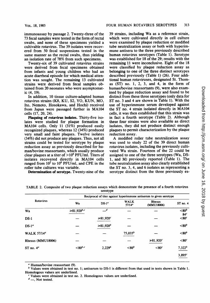
FIG. 1. Serotypic characterizaton of human rotavirus (ST no. 4) by neutralization in MA104 tube culture withviral replication detected by enzyme immunoassay: microtiter plate wells in which tube harvests from a modifiedneutralization test were assayed by indirect enzyme immunoassay. Hyperimmune guinea pig antisera were
diluted 1:100, 1:1,000, and 1:10,000 and mixed with a constant amount of ST, no. 4 virus before inoculation oftube cultures. Back titrations of virus (10-1, 10-2, and 10-') were included in the test. In this test, humanrotavirus strain ST no. 4 was neutralized only by the anti-ST no. 4 antiserum to a dilution of 1:1,000 and not byantisera against Wa, DS-1, WALK 57/14 (indicated by 14), Nebraska calf diarrhea virus (NCDV), or epizooticdiarrhea of infant mice virus (EDIM). Microtiter plate variation was controlled by the titration of humanrotavirus, strain D, in 10-fold dilutions. A Kodak Wratten no. 49 (blue) filter was used to photograph themicrotiter plate to enhance the contrast and density for black and white reproduction.
tablished serotypes (Table 1; Fig. 1). Sevendirect isolates were studied by both plaque re-duction assay and tube neutralization assay, andboth tests yielded the same results in each case.The assignment of strains to a serotype wasclear-cut, with reciprocal 20-fold antibody dif-ferences being demonstrated in each instanceexcept for strain Pz, which had a low-level one-way relationship with ST no. 4.RNA analysis. Ten of the newly isolated
strains were examined by RNA polyacrylamidegel electrophoresis, and 8 of the 10 were sero-typed. Each appeared to be a single strain basedon an appropriate number and pattern of RNAsegments.Comparison with Japanese rotaviruses. Ten,
human rotavirus strains from Japan were co _pared by plaque reduction assay with the fourserotypes encountered in these studies (Table3). Hyperimmune antisera against Wa (serotype1), DS-1 (serotype 2), WALK 57/14 (serotype 3),and ST no. 4 (serotype 4) were utilized. StrainsK8 and KU were similar if not identical to strainWa, whereas strains S2 and KUN were similar if
not identical to strain DS-1. Strains YO, MO,Ito, and Nemoto were serotyped as being similarif not identical to strain WALK 57/14, althoughthere was a low level of cross-reactivity of Itowith serotype 4 reference antiserum. StrainsHosokawa and Hochi appeared to be similar toST no. 4 in this one-way comparison. Two-waycomparisons were made only for strains K8,KU, S2, KUN, YO, and MO, and in each case atwo-way relationship of identity was furtherestablished (data not shown). Thus, each of 10strains from Japan belonged to one of fourserotypes established in our studies. The sub-group of each of these 10 strains was deter-mined, and the results are shown in Table 3.
DISCUSSIONPlaque reduction assay and a modified tube
neutralizaton assay were used to define theserotype of a series of newly isolated humanrotaviruses, and these strains were comparedwith other strains previously isolated in Japan(17, 20, 24). Other investigators have used thefluorescent-focus neutralization assay or CPE
J. CLIN. MICROBIOL.
on June 16, 2018 by guesthttp://jcm
.asm.org/
Dow
nloaded from

FOUR HUMAN ROTAVIRUS SEROTYPES 315
TABLE 3. Neutralization specificity of human rotavirus strains from Japan as determined with the use ofantisera against strains of U.S. origin
Reciprocal of titer by 60%o plaque reduction assay againstantiserum from guinea pigs hyperimmunized with given
Sub- rotavirusRotavirus Investigators (reference) group
no. Wa DS-1 Reassortant STno4(:81,920)a (31,273)a (W81,9274 (7,4414)
K8 Urasawa et al. (24) 2 :81,920 <80 <80 <80KU Urasawa et al. (24) 2 >81,920 <80 <80 <80
S2 Urasawa et al. (24) 1 <80 7,227 <80 <80KUN Kutsuzawa et al. (17) 1 150 9,347 <80 <80
YO Urasawa et al. (24) 2 144 <80 72,043 <80MO Kutsuzawa et al. (17) 2 <80 <80 35,%7 <80Ito Sato et al. (20) 2 1,827 320 20,058 304Nemoto Sato et al. (20) 2 552 179 42,870 125
Hosokawa Sato et al. (20) 2 714 <80 <80 813bHochi Sato et al. (20) 2 415 <80 <80 862b
a Reciprocal of homologous titer from previous assay.b Homologous titer of anti-ST no. 4 serum in same test = 1:3,460.
assay to determine serotype (3, 22). All of these was observed among these four strains (20).assays measure antibody which neutralizes rota- Each strain was also reported to be distinct fromvirus and thus characterize the antigenic deter- strain Wa (20), and our findings confirm thatminant(s) of the rotavirus which most likely is observation. We require a 20-fold difference ininvolved in protective immunity (5, 30). homologous and heterologous antibody titers to
In this study, we identified four serotypes of establish distinct serotypes and tentatively clas-human rotavirus isolated from specimens ob- sify these same four strains as belonging to twotained from Australia, England, Hong Kong, the serotypes, 3 and 4, using our reference antisera.United States, and Venezuela. Ten human rota- A new fourth serotype emerged from our studiesviruses isolated previously in Japan were also when a series of neonatal specimens from En-tested and found to belong to one of the four gland was examined; the identification in thisserotypes. Our findings are in agreement with study of a similar fourth serotype from Japanthose of Urasawa et al., which established the must be confirmed by the development of high-existence of three distinct serotypes among titered hyperimmune antisera against the strainsstrains KU, K8, YO, and S2 (24). The four and comparisons in a two-way fashion.Japanese strains described by Sato et al. were The numbering system used in this report isreported to represent four different serotypes, consistent with that used by Thouless and col-since at least an eightfold difference between leagues (Table 4) (2, 22). Of interest, strainshomologous and heterologous antibody titers belonging to the third of these serotypes have
TABLE 4. Comparison of human rotaviruses by serotypeDesignation of rotavirus strains which are similar if not identical by
Investigators (reference) Country neutralization assaysla 2a 3a 4
Beards et al. (3) England I II IIIUrasawa et al. (24) Japan K8 * S2 YO
KUKutsuzawa et al. (17) Japan KUN MOSato et al. (20) Japan Ito Hosokawa
Nemoto HochiWyatt et al. (26); this USA Wa DS-1 pb ST no. 4
studya Designation originally established and currently used by Thouless and colleagues (22).b Also includes rhesus rotavirus MMU18006 and reassortant rotavirus WALK 57/14.
VOL. 18, 1983
on June 16, 2018 by guesthttp://jcm
.asm.org/
Dow
nloaded from

316 WYATT ET AL.
been recovered from simian, canine, and felinespecies (11, 26). Further comparisons betweenanimal and human rotavirus strains may lead toadditional examples of shared serotypes, as oc-
curs with the reoviruses (12). It was also ofinterest that 5 of the 13 strains isolated fromasymptomatic newborns were either serotype 3(two cases) or serotype 4 (three cases).Each of the rotaviruses isolated in these stud-
ies was determined to be either subgroup 1 or 2.Subgroup specificity represents a function of thegene 6 product of rotaviruses and is not associat-ed with neutralization specificity in a major way(1, 6, 14, 16). However, determination of sub-group in the serial cultivation of rotavirusesoffers a simple way of monitoring for contamina-tion by rotavirus of a different subgroup. Inaddition, it appears that subgroup 1 viruses ofhuman origin belong to serotype 2.
Determination of serotype is often difficultbecause of poor growth of direct isolates andtheir failure to produce discrete plaques. Humanrotaviruses cultivated or rescued by the use ofgenetic reassortment plaque well, usually at atiter of 106 PFU/ml. Rescue of human rotavirus-es is more sensitive than the method of directisolation used in this study, since eight rotavi-ruses which were previously rescued from rectalswab preparations could not be grown directlyfrom the same swab preparation in MA104 orAGMK cells. It is also of interest that rotavirus-es grown either directly or by reassortment wereof the same serotype. Some other methods fordetermination of serotype which do not requireplaquing depend on the development of CPE,which is often variable. Thus, the use of fluores-cence or enzyme immunoassay has been usefulto measure the presence of rotavirus antigens inneutralization assays (22, 23). The use of en-
zyme immunoassay in the current study to mea-sure rotavirus replication in roller tube culturesoffers the advantage of a sensitive cultivationsystem, and yet it is not dependent on thedevelopment of CPE. The technique is, howev-er, tedious and time-consuming and requireslarge numbers of roller tube cultures. An en-
zyme immunoassay has recently been describedwhich is potentially useful to determine serotypeon clinical specimens by the use of absorbedantisera (22). This and other simplified solid-phase systems utilizing serotype-specific mono-clonal antibodies, probes which will detect spe-cific genes, e.g., gene 8 or 9, or antibodiesdirected against specific gene products willhopefully allow the rapid determination of sero-type required for larger-scale epidemiologicalstudies.
ACKNOWLEDGMENTS
We thank R. M. Chanock for helpful and critical appraisalof the manuscript; we also thank R. Jones, R. Marquina, M.
Sereno, and J. Valdesuso for valuable technical assistance andT. Popkin for expert photography.
LITERATURE CITED
1. Bastardo, J. W., J. L. McKimm-Breschkin, S. Sonza,L. D. Mercer, and I. H. Holmes. 1981. Preparation andcharacterization of antisera to electrophoretically purifiedSA-11 virus polypeptides. Infect. Immun. 34:641-647.
2. Beards, G. M. 1982. Polymorphism of genomic RNAswithin rotavirus serotypes and subgroups. Arch. Virol.74:65-70.
3. Beards, G. M., J. N. PUlford, M. E. Thouless, and T. H.Flewett. 1980. Rotavirus serotypes by serum neutralisa-tion. J. Med. Virol. 5:231-237.
4. Chrystie, I. L., B. Totterdell, M. J. Baker, J. W. Scopes,and J. E. Banatvala. 1975. Rotavirus infections in a mater-nity unit. Lancet 1:79.
5. Gaul, S. K., T. F. Simpson, G. N. Woode, and R. W.Fulton. 1982. Antigenic relationships among some animalrotaviruses: virus neutralizaton in vitro and cross-protec-tion in piglets. J. Clin. Microbiol. 16:495-503.
6. Greenberg, H. B., J. Flores, A. Kalica, R. Wyatt, and R.Jones. 1983. Gene coding assignments for growth restric-tion and neutralization and subgroup specificities of the Wand DS-1 strains of human rotavirus. J. Gen. Virol.64:313-324.
7. Greenberg, H. B., A. R. Kalica, R. G. Wyatt, R. W.Jones, A. Z. Kapikian, and R. M. Chanock. 1981. Rescueof non-cultivatable human rotavirus by gene reassortmentduring mixed infection with ts mutants of a cultivatablebovine rotavirus. Proc. Natl. Acad. Sci. U.S.A. 78:420-424.
8. Greenberg, H. B., V. McAuliffe, J. Valdesuso, R. Wyatt, J.Flores, A. Kalica, Y. Hoshino, and N. Singh. 1983. Sero-logical analysis of the subgroup protein of rotavirus, usingmonoclonal antibodies. Infect. Immun. 39:91-99.
8a.Greenberg, H. B., J. Valdesuso, K. van Wyke, K.Midthun, M. Walsh, V. McAuliffe, R. G. Wyatt, A. R.Kalica, J. Flores, and Y. Hoshino. 1983. Production andpreliminary characterization of monoclonal antibodiesdirected at two surface proteins of rhesus rotavirus. J.Virol. 47:267-275.
9. Greenberg, H. B., R. G. Wyatt, A. Z. Kapikian, A. R.Kalica, J. Flores, and R. Jones. 1982. Rescue and serotyp-ic characterization of noncultivatable human rotavirus bygene reassortment. Infect. Immun. 37:104-109.
10. Hasegawa, A., S. Matsuno, S. Inouye, R. Kono, Y. Tsuru-kubo, A. Mukoyama, and Y. Salto. 1982. Isolation ofhuman rotaviruses in primary cultures of monkey kidneycells. J. Clin. Microbiol. 16:387-390.
11. Hoshino, Y., R. G. Wyatt, F. W. Scott, and M. J. Appel.1982. Isolation and characterization of a canine rotavirus.Arch. Virol. 72:113-125.
12. Hrdy, D. B., L. Rosen, and B. N. Fields. 1979. Polymor-phism of the migration of double-stranded RNA genomesegments of reovirus isolates from humans, cattle, andmice. J. Virol. 31:104-111.
13. Kalica, A. R., J. Flores, and H. B. Greenberg. 1983.Identification of the rotaviral gene that codes for hemag-glutination and protease-enhanced plaque formation. Vi-rology 125:194-205.
14. Kalica, A. R., H. B. Greenberg, R. G. Wyatt, J. Flores,M. M. Sereno, A. Z. Kapikian, and R. M. Chanock. 1981.Genes of human (strain Wa) and bovine (strain UK)rotaviruses that code for neutralization and subgroupantigens. Virology 112:385-390.
15. Kapikian, A. Z., R. H. Yolken, H. B. Greenberg, R. G.Wyatt, A. R. Kalica, R. M. Chanock, and H. W. Kim.1979. Gastroenteritis viruses, p. 927-995. In E. H. Len-nette and N. J. Schmidt (ed.), Diagnostic procedures forviral rickettsial and chlamydial infections. American Pub-lic Health Association, Washington, D.C.
16. Killen, H. M., and N. J. Dimmock. 1982. Identification ofa neutralization-specific antigen of a calf rotavirus. J.Gen. Virol. 62:297-311.
J. CLIN. MICROBIOL.
on June 16, 2018 by guesthttp://jcm
.asm.org/
Dow
nloaded from

FOUR HUMAN ROTAVIRUS SEROTYPES 317
17. Kutsuzawa, T., T. Konno, H. Suzuki, A. Z. Kapikian, T.Ebina, and N. Ishida. 1982. Isolation of human rotavirussubgroups 1 and 2 in cell culture. J. Clin. Microbiol.16:727-730.
18. Matsuno, S., S. Inouye, and R. Kono. 1977. Plaque assayof neonatal calf diarrhea virus and the neutralizing anti-body in human sera. J. Clin. Microbiol. 5:1-4.
19. Rodgers, S. M., R. F. Bhbop, C. BhIch, B. McLean, andI. H. Holmes. 1981. Molecular epidemiology of humanrotaviruses in Melbourne, Australia, from 1973 to 1979, asdetermined by electrophoresis of genome ribonucleicacid. J. Clin. Microbiol 13:272-278.
20. Sato, K., Y. Inaba, Y. Mhwa, S. Tokuhisa, and M.Matumoto. 1982. Antigenic relationships between rotavi-ruses from different species as studied by neutralizationand immunofluorescence. Arch. Virol. 73:45-50.
21. Sato, K., Y. Inaba, T. Shinozakl, R. FuNi, and M. Matsu-moto. 1981. Isolation of human rotavirus in cell culture.Arch. Virol. 69:155-160.
22. Thouess, M. E, G. M. Beards, and T. H. Flewett. 1982.Serotyping and subgrouping of rotavirus strains by theELISA test. Arch. Virol. 73:219-230.
23. Tboules, M. E., A. S. Bryden, T. H. Flewett, G. N.Woode, J. C. Bridger, D. R. Snodgras, and J. A. Herring.1977. Serological relationships between rotaviruses fromdifferent species as studied by complement fixation andneutralization. Arch. Virol. 53:287-294.
24. Urasawa, S., T. Urasawa, and K. Taniguchi. 1982. Threehuman rotavirus serotypes demonstrated by plaque neu-
tralization of isolated strains. Infect. Immun. 38:781-784.25. Urasawa, T., S. Urswa, and K. Taniguchi. 1981. Sequen-
tial passages of human rotavirus in MA104 cells. Microbi-ol. Immunol. 25:1025-1035.
26. Wyatt, R. G., H. B. Greenberg, W. D. James, A. L. PItt-man, A. R. Klica, J. Fores, R. M. Chanock, and A. Z.Kapilkan. 1982. Definition of human rotavirus serotypesby plaque reduction assay. Infect. Immun. 37:110-115.
27. Wyatt, R. G., and W. D. James. 1982. Methods of gastro-enteritis virus culture in vivo and in vitro, p. 13-35. InD. A, J. Tyrrell and A. Z. Kapikian (ed.), Virus infectionsof the gastrointestinal tract. Marcel Dekker, Inc., NewYork.
28. Wyatt, R. G., W. D. James, E. H. Bobi, K. W. Thell,L. H. Saif, A. R. Kalca, H. B. Greenberg, A. Z. Kapi-klan, and R. M. Chanock. 1980. Human rotavirus type 2:cultivation in vitro. Science 207:189-191.
29. Wyatt, R. G., A. Z. Kapiklan, H. B. Greenberg, A. R.Kalica, and R. M. Chanock. 1982. Prospects for develop-ment of a vaccine against rotavirus diarrhea, p. 505-522.In T. Holme, J. Holmgren, M. H. Merson, and R. MolIby(ed.), Proceedings of the Conference on Acute EntericInfections in Children. Elsevier/North-Holland Biomedi-cal Press, Amsterdam.
30. Wyatt, R. G., C. A. Mebus, R. H. Yoken, A. R. Kalica,H. D. James, Jr., A. Z. Kapyikan, and R. M. Cbanock.1979. Rotaviral immunity in gnotobiotic calves: heterolo-gous resistance to human virus induced by bovine virus.Science 203:548-550.
VOL. 18, 1983
on June 16, 2018 by guesthttp://jcm
.asm.org/
Dow
nloaded from