Embed Size (px)
Citation preview
of June 12, 2018.This information is current as
βHomeostatic Function of IL-1Osteoclast Precursors Identifies a
+Direct Inhibition of Human RANK
Ivashkiv and Jong Dae JiSong, Jeongwon Sohn, Kyung-Hyun Park-Min, Lionel B.Jin-Hyun Woo, Sung Jae Choi, Young Ho Lee, Gwan Gyu Bitnara Lee, Tae-Hwan Kim, Jae-Bum Jun, Dae-Hyun Yoo,
http://www.jimmunol.org/content/185/10/5926doi: 10.4049/jimmunol.1001591October 2010;
2010; 185:5926-5934; Prepublished online 8J Immunol
MaterialSupplementary
1.DC1http://www.jimmunol.org/content/suppl/2010/10/08/jimmunol.100159
Referenceshttp://www.jimmunol.org/content/185/10/5926.full#ref-list-1
, 18 of which you can access for free at: cites 47 articlesThis article
average*
4 weeks from acceptance to publicationFast Publication! •
Every submission reviewed by practicing scientistsNo Triage! •
from submission to initial decisionRapid Reviews! 30 days* •
Submit online. ?The JIWhy
Subscriptionhttp://jimmunol.org/subscription
is online at: The Journal of ImmunologyInformation about subscribing to
Permissionshttp://www.aai.org/About/Publications/JI/copyright.htmlSubmit copyright permission requests at:
Email Alertshttp://jimmunol.org/alertsReceive free email-alerts when new articles cite this article. Sign up at:
Print ISSN: 0022-1767 Online ISSN: 1550-6606. Immunologists, Inc. All rights reserved.Copyright © 2010 by The American Association of1451 Rockville Pike, Suite 650, Rockville, MD 20852The American Association of Immunologists, Inc.,
is published twice each month byThe Journal of Immunology
by guest on June 12, 2018http://w
ww
.jimm
unol.org/D
ownloaded from
by guest on June 12, 2018
http://ww
w.jim
munol.org/
Dow
nloaded from

The Journal of Immunology
Direct Inhibition of Human RANK+ Osteoclast PrecursorsIdentifies a Homeostatic Function of IL-1b
Bitnara Lee,*,1 Tae-Hwan Kim,*,1 Jae-Bum Jun,* Dae-Hyun Yoo,* Jin-Hyun Woo,†
Sung Jae Choi,† Young Ho Lee,† Gwan Gyu Song,† Jeongwon Sohn,‡ Kyung-Hyun Park-Min,x
Lionel B. Ivashkiv,x and Jong Dae Ji†
IL-1b is a key mediator of bone resorption in inflammatory settings, such as rheumatoid arthritis (RA). IL-1b promotes
osteoclastogenesis by inducing RANKL expression on stromal cells and synergizing with RANKL to promote later stages of
osteoclast differentiation. Because IL-1Rs share a cytosolic Toll–IL-1R domain and common intracellular signaling molecules with
TLRs that can directly inhibit early steps of human osteoclast differentiation, we tested whether IL-1b also has suppressive
properties on osteoclastogenesis in primary human peripheral blood monocytes and RA synovial macrophages. Early addition of
IL-1b, prior to or together with RANKL, strongly inhibited human osteoclastogenesis as assessed by generation of TRAP+
multinucleated cells. IL-1b acted directly on human osteoclast precursors (OCPs) to strongly suppress expression of RANK, of
the costimulatory triggering receptor expressed on myeloid cells 2 receptor, and of the B cell linker adaptor important for
transmitting RANK-induced signals. Thus, IL-1b rendered early-stage human OCPs refractory to RANK stimulation. Similar
inhibitory effects of IL-1b were observed using RA synovial macrophages. One mechanism of RANK inhibition was IL-1b–
induced proteolytic shedding of the M-CSF receptor c-Fms that is required for RANK expression. These results identify a ho-
meostatic function of IL-1b in suppressing early OCPs that contrasts with its well-established role in promoting later stages of
osteoclast differentiation. Thus, the rate of IL-1–driven bone destruction in inflammatory diseases, such as RA, can be restrained
by its direct inhibitory effects on early OCPs to limit the extent of inflammatory osteolysis. The Journal of Immunology, 2010,
185: 5926–5934.
Bone resorption and osteolysis are a prominent feature anda cause of substantial morbidity in several inflammatorydiseases, including rheumatoid arthritis (RA), perio-
dontitis, and peri-prosthetic loosening (1–3). Osteoclasts are theprimary bone-resorbing cells and are essential for bone destructionin these inflammatory diseases. Osteoclasts are multinucleatedgiant cells that are differentiated from hematopoietic cells ofmyeloid lineage. RANKL and M-CSF are essential molecules fordifferentiation of osteoclasts from their precursors, and theseosteoclastogenic molecules are abundantly expressed in inflam-matory conditions, such as RA and periodontitis (4, 5). M-CSFbinds to the surface receptor c-Fms (also termed colony-stimulatingfactor 1 receptor), which is responsible for early differentiation
of osteoclasts and acts as a potent stimulator of RANK expression(6). RANKL binds to RANK on the cell surface of osteoclastprecursors (OCPs) and induces the full differentiation of osteo-clasts and their bone resorbing activity. Osteoprotegerin is anotherreceptor for RANKL and a potent inhibitor of osteoclastogenesisthat acts as a decoy receptor for RANKL. Other inflammatorymolecules also positively or negatively contribute to bone de-struction by regulating the differentiation of osteoclasts. There-fore, the extent of bone destruction is determined by the balancebetween stimulatory and inhibitory factors of osteoclastogenesisin inflammatory conditions.In RA, several inflammatory molecules, such as TNF-a, IL-1b,
IL-6, IL-17, and PGs play a vital role in osteoclastogenesis andbone resorption. These molecules promote osteoclastogenesis in-directly by increasing expression of RANKL and M-CSF by stro-mal cells and T cells, and also by acting directly on OCPs tosynergize with RANKL in driving osteoclastogenesis (1, 2).Among these molecules, TNF-a is the most important osteoclas-togenic molecule in pathologic conditions, such as RA. TNF-aincreases osteoclastogenesis through several different mechanisms(7). TNF-a increases the pool size of marrow OCPs, enhances theRANKL-induced osteoclastogenic actions, and increases expres-sion of RANKL in synovial cells, T cells, and osteoblast/stromalcells.IL-1 is a multifunctional cytokine that has predominately pro-
inflammatory properties but can also engage feedback inhibitorymechanisms (e.g., induction of glucocorticoid production) thatrestrain and balance its proinflammatory function (8). This cyto-kine was initially described as an osteoclast-activating factor dueto its potent bone-resorbing activity (9). Like TNF-a, IL-1b alsoplays an essential role in the pathogenesis of bone destruction inRA. Although IL-1 alone does not induce osteoclastogenesis, it
*Hospital for Rheumatic Diseases, Hanyang University; †Division of Rheumatologyand ‡Department of Biochemistry, College of Medicine, Korea University, Seoul,South Korea; and xArthritis and Tissue Degeneration Program, Hospital for SpecialSurgery, New York, NY 10021
1B.L. and T.-H.K. contributed equally to this work.
Received for publication May 14, 2010. Accepted for publication September 8, 2010.
This work was supported by Grant A084224 from the Korea Healthcare TechnologyR&D Project, Ministry for Health, Welfare and Family Affairs, Republic of Korea (toJ.D.J.) and by grants from the National Institutes of Health (to L.B.I.) and theArthritis Foundation (to K.-H.P.-M.).
Address correspondence and reprint requests to Dr. Jong Dae Ji, Division of Rheu-matology, College of Medicine, Korea University, 126-1, Anam-Dong 5-Ga,Sungbuk-Ku, Seoul 136-705, South Korea. E-mail address: [email protected]
The online version of this article contains supplemental material.
Abbreviations used in this paper: BLNK, B cell linker; COX-2, cyclooxygenase-2;IL-1Ra, IL-1R antagonist; MMP, matrix metalloproteinase; OCP, osteoclast precur-sor; Pam3Cys, Pam3CysSer(Lys)4; PKC, protein kinase C; RA, rheumatoid arthritis;TRAP, tartrate-resistant acid phosphatase; TREM-2, triggering receptor expressed onmyeloid cells 2.
Copyright� 2010 by TheAmericanAssociation of Immunologists, Inc. 0022-1767/10/$16.00
www.jimmunol.org/cgi/doi/10.4049/jimmunol.1001591
by guest on June 12, 2018http://w
ww
.jimm
unol.org/D
ownloaded from
augments RANKL-induced osteoclast differentiation and pro-motes osteoclast activation and survival (10). IL-1 also mediatesTNF-induced bone resorption (11). The IL-1 gene family hasseveral members, such as IL-1a, IL-1b, and the IL-1R antagonist(IL-1Ra) (8). IL-1a and IL-1b are agonists, and IL-1Ra is a spe-cific receptor antagonist. There are two members of the IL-1Rgene family. The type I receptor IL-1RI transduces signals,whereas IL-1RII does not transduce signals and instead works asa decoy receptor. IL-1 exerts its biological effects by forming acomplex with the IL-1RI and IL-1R accessory protein. IL-1 usesthe adaptor molecule MyD88 to activate signaling pathwaysleading to the activation of NF-kB and MAPKs and downstreamtranscription factors that drive inflammatory gene expression (12).While inflammatory molecules, such as TLR ligands, drive bone
destruction, these molecules also engage potent homeostatic mecha-nisms to limit damage associated with inflammation, and these mech-anisms may limit the extent of bone resorption. Direct stimulation ofvariousTLRsonOCPs inhibitsRANKL-inducedosteoclastogenesisin mouse cells (13–16), and we also found that TLR ligands inhibithuman osteoclast differentiation by acting directly on OCPs (17).Generally, TLR ligands and other inflammatory molecules inhibitosteoclast differentiation at early stages of osteoclastogenesis, suchas generation of OCPs, and lose their inhibitory properties at laterstages, at which point they augment RANKL-induced osteoclastdifferentiation. In chronic inflammatory conditions, TLR ligandsand other inflammatory molecules predominately work as pro-osteoclastogenic molecules, despite their direct inhibitory effecton the osteoclastogenesis.TLRs and IL-1R share a cytosolic domain, termedToll–IL-1R, and
common intracellular signaling molecules, such as MyD88, IL-1R–associated kinase, and TNFR-associated factor 6 (12), and the knownstimulatory effects of IL-1b on the osteoclast differentiation aresimilar to the effects of TLR ligands. These findings suggest the
possibility that IL-1b also may regulate osteoclast differentiation byacting directly on OCPs, similarly to TLR ligands. Because little isknown about the direct effect on human osteoclastogenesis by IL-1b,we examined the effects of IL-1b on osteoclastogenesis in primaryhuman peripheral blood monocytes and RA synovial macrophages.We found that IL-1b induces shedding and thereby inactivation ofc-Fms that drives RANK expression and makes early human OCPsrefractory to RANK stimulation by downregulating expression ofRANK, its costimulatory receptor, triggering receptor expressed onmyeloid cells 2 (TREM-2), and downstream signaling molecules,such as B cell linker (BLNK). These findings identify a homeostaticfunction for a predominately inflammatory cytokine and suggesta new mechanism that can restrain osteoclastogenesis in inflam-matory settings.
Materials and MethodsMaterials
Recombinant human IL-1b was from R&D Systems (Minneapolis, MN),and human M-CSF and soluble RANKL were from PeproTech (RockyHill, NJ). SB203580, U0126, PD98059, GF 109203X, and TAPI-1 werepurchased from Calbiochem (San Diego, CA). SB202190 was purchasedfrom Sigma–Aldrich (St. Louis, MO). p38 Ab (catalog no. sc-535) wasfrom Santa Cruz Biotechnology (Santa Cruz, CA), and STAT3 Ab (clone84/Stat3) was from BD Transduction Laboratories (Franklin Lakes, NJ).M-CSFR Ab (catalog no. 3152) was from Cell Signaling Technology(Beverly, MA), and RANK Ab (clone 9A725) was from Alexis Bio-chemicals (San Diego, CA).
Cell isolation and culture
PBMCs were obtained from normal blood donors or blood leukocytepreparations purchased from the New York Blood Center (New York, NY)by density gradient centrifugation with Ficoll (Invitrogen, Carlsbad, CA),using a protocol approved by the Hospital for Rheumatic Disease (Han-yang University, Seoul, Korea) and by the Hospital for Special Surgery(New York, NY) institutional review boards. Monocytes were obtained
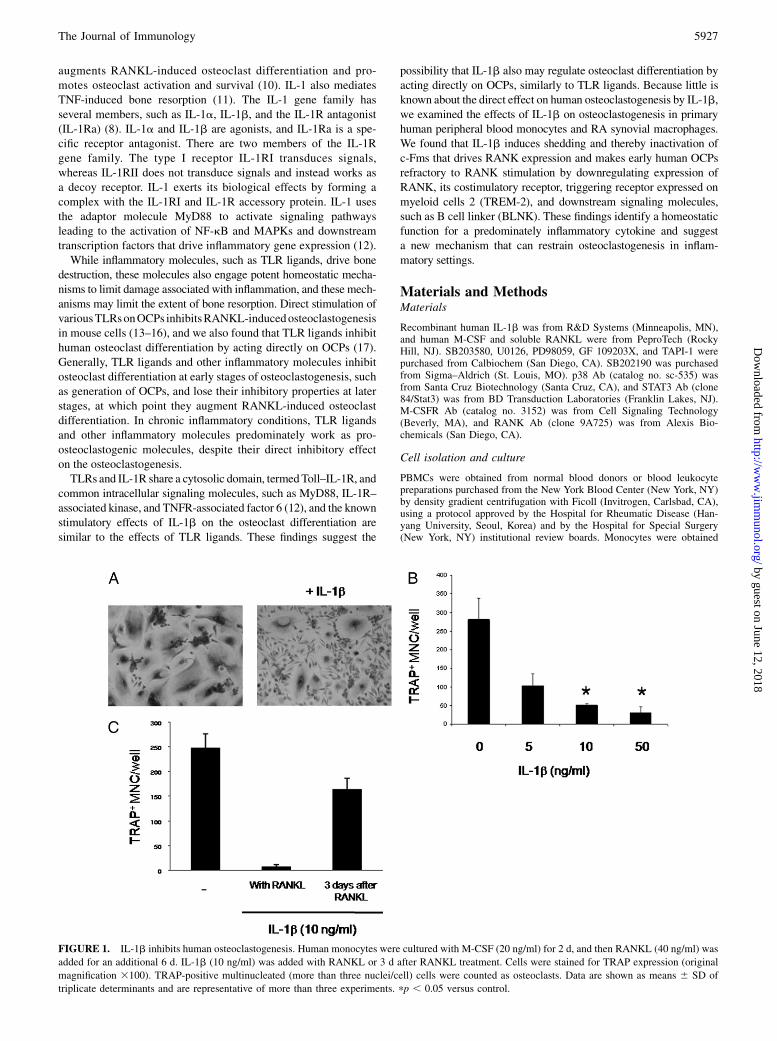
FIGURE 1. IL-1b inhibits human osteoclastogenesis. Human monocytes were cultured with M-CSF (20 ng/ml) for 2 d, and then RANKL (40 ng/ml) was
added for an additional 6 d. IL-1b (10 ng/ml) was added with RANKL or 3 d after RANKL treatment. Cells were stained for TRAP expression (original
magnification 3100). TRAP-positive multinucleated (more than three nuclei/cell) cells were counted as osteoclasts. Data are shown as means 6 SD of
triplicate determinants and are representative of more than three experiments. pp , 0.05 versus control.
The Journal of Immunology 5927
by guest on June 12, 2018http://w
ww
.jimm
unol.org/D
ownloaded from
from PBMCs, using anti-CD14 magnetic beads, as recommended by themanufacturer (Miltenyi Biotec, Auburn, CA). Monocytes were cultured for1 d in a-MEM medium (Invitrogen) supplemented with 10% FBS(Hyclone, Logan, UT) with either M-CSF (20 ng/ml) in the presence orabsence of IL-1b. Monocyte-derived preosteoclasts obtained after 2 d ofculture with M-CSF were used unless otherwise noted in the figure leg-ends, and the purity of monocytes/macrophages was .95%, as verified byflow cytometric analysis. RA synovial macrophages were obtained fromRA synovial fluid mononuclear cells using anti-CD14 magnetic beads, asrecommended by the manufacturer (Miltenyi Biotec). Thirteen patientswere included in this study, and all patients fulfilled the revised AmericanCollege of Rheumatology classification criteria for the diagnosis of RA.Except for one patient, the remaining 12 RA patients were treated withlow-dose corticosteroids. Five patients were treated with methotrexatealone, two patients with methotrexate and sulfasalazine, two patients withmethotrexate and leflunomide, one patient with methotrexate and tacroli-mus, one patient with sulfasalazine, one patient with hydroxychloroquine,and one patient without antirheumatic drugs due to pregnancy. Twelvepatients had active disease (erythrocyte sedimentation rate, 46–128 mm/h),and one patient had inactive disease (erythrocyte sedimentation rate, 9mm/h). This study protocol was approved by the institutional review boardof the Hospital for Rheumatic Disease, Hanyang University.
Osteoclast differentiation
Human CD14+ cells were incubated with 20 ng/ml M-CSF for 2 d togenerate OCPs. OCPs were further incubated with 20 ng/ml M-CSF and40 ng/ml human soluble RANKL for an additional 6 d in a-MEM
supplemented with 10% FBS. Cytokines were replenished every 3 d. Onday 8, cells were fixed and stained for tartrate-resistant acid phosphatase(TRAP) using the Acid Phosphatase Leukocyte diagnostic kit (Sigma, SanDiego, CA) as recommended by the manufacturer. Multinucleated (morethan three nuclei) TRAP-positive osteoclasts were counted in triplicatewells.
Gene expression analysis
For real-time PCR, DNA-free RNAwas obtained using the RNeasyMini Kit(Qiagen, Hilden, Germany) or TRIzol reagent (Invitrogen Life Technolo-gies, Carlsbad, CA) according to the manufacturer’s instructions, and 1 mgtotal RNAwas reverse transcribed using a First Strand cDNA Synthesis kit(Fermentas, Hanover, MD). Real-time PCR was performed in triplicateusing the iCycler iQ thermal cycler and detection system (Bio-Rad Lab-oratories, Hercules, CA) following the manufacturer’s protocols. Expres-sion of the tested gene was normalized relative to levels of GAPDH.Primary transcripts were measured using primers that amplified eitherexon–intron junctions or intronic sequences.
Immunoblotting
Whole-cell extracts were prepared by lysis in a buffer containing 20 mMHEPES (pH 7.0), 300 mM NaCl, 10 mM KCl, 1 mM MgCl2, 0.1% TritonX-100, 0.5 mM DTT, 20% glycerol, and 13 proteinase inhibitor mixture(Roche, Basel, Switzerland). The protein concentration of extracts wasquantified using a Bradford assay (Bio-Rad, Hercules, CA). For immu-noblotting, 10 mg cell lysates was fractionated on 7.5% polyacrylamide
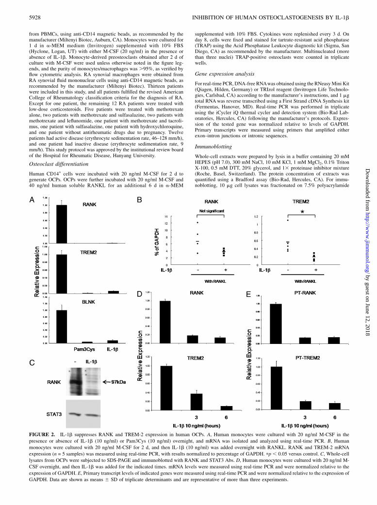
FIGURE 2. IL-1b suppresses RANK and TREM-2 expression in human OCPs. A, Human monocytes were cultured with 20 ng/ml M-CSF in the
presence or absence of IL-1b (10 ng/ml) or Pam3Cys (10 ng/ml) overnight, and mRNA was isolated and analyzed using real-time PCR. B, Human
monocytes were cultured with 20 ng/ml M-CSF for 2 d, and then IL-1b (10 ng/ml) was added overnight with RANKL. RANK and TREM-2 mRNA
expression (n = 5 samples) was measured using real-time PCR, with results normalized to percentage of GAPDH. pp , 0.05 versus control. C, Whole-cell
lysates from OCPs were subjected to SDS-PAGE and immunoblotted with RANK and STAT3 Abs. D, Human monocytes were cultured with 20 ng/ml M-
CSF overnight, and then IL-1b was added for the indicated times. mRNA levels were measured using real-time PCR and were normalized relative to the
expression of GAPDH. E, Primary transcript levels of indicated genes were measured using real-time PCR and were normalized relative to the expression of
GAPDH. Data are shown as means 6 SD of triplicate determinants and are representative of more than three experiments.
5928 INHIBITION OF HUMAN OSTEOCLASTOGENESIS BY IL-1b
by guest on June 12, 2018http://w
ww
.jimm
unol.org/D
ownloaded from

gels using SDS-PAGE, transferred to polyvinylidene fluoride membranes(Millipore, Billerica, MA), incubated with specific Abs, and ECL was usedfor detection.
Flow cytometry
Staining for cell surface expression ofM-CSF receptor was performed usingmonoclonal anti-human M-CSF receptor (clone 61708; R&D Systems). AFACScan flow cytometer with CELLQuest software (Becton Dickinson,Franklin Lakes, NJ) was used.
Statistical analysis
Results are expressed as means 6 SD. A Student t test was applied toevaluate group differences; a p value of ,0.05 was considered significant.
ResultsIL-1b inhibits RANKL-induced human osteoclastogenesis
We examined the effects of IL-1b on human osteoclastogenesis usinga standard, validated system of human osteoclast differentiation (18).Treatment with M-CSF for 2 d induces the differentiation of pri-mary human monocytes into RANKL-responsive OCPs via inducingRANK expression. OCPs are subsequently differentiated into matureosteoclasts in the presence of RANKL. Control cells cultured withM-CSF and RANKL efficiently differentiated into multinucleated(more than three nuclei/cell) TRAP-positive giant cells (Fig. 1A).
When added together with RANKL, treatment with IL-1b stronglyinhibited RANKL-induced osteoclastogenesis (Fig. 1A). Osteoclas-togenesis was inhibited by IL-1b in a dose-dependent fashion (Fig.1B). IL-1b significantly inhibited osteoclastogenesis when addedprior to addition of RANKL, although IL-1b–induced inhibition ofosteoclastogenesis was less effective than when added together withRANKL (Supplemental Fig. 1). Also, when IL-1b was added severaldays after stimulation with RANKL, the IL-1b–induced inhibitionof human osteoclastogenesis was significantly diminished (Fig. 1C).These results show a direct, but time-dependent, inhibitory effect ofIL-1b on human osteoclast differentiation.
IL-1b suppresses RANK, TREM-2, and BLNK expression inhuman OCPs
To begin to investigate mechanisms by which IL-1b inhibitsosteoclastogenesis, we examined the effects of IL-1b on RANK,TREM-2, and BLNK expression. Previously, we found that TLRligands inhibit human osteoclast differentiation by suppressingRANK and TREM-2 expression (17). We compared the effects ofIL-1b on RANK, TREM-2, and BLNK expression with the ef-fects of Pam3CysSer(Lys)4 (Pam3Cys; TLR2 ligand). TREM-2 isa DAP12-associated receptor that provides an essential ITAM-mediated costimulatory signal for RANKL-induced osteoclasto-
FIGURE 3. IL-1Ra reverses IL-1b–induced inhibition of human osteoclastogenesis and RANK gene expression. A, Human monocytes were cultured
with M-CSF (20 ng/ml) for 2 d, and then RANKL (40 ng/ml) was added for an additional 6 d. IL-1b (10 ng/ml) and/or IL-1Ra (200 ng/ml) were added with
RANKL. Cells were stained for TRAP expression (original magnification 3100). TRAP-positive multinucleated (more than three nuclei/cell) cells were
counted as osteoclasts. Data are shown as means 6 SD of triplicate determinants and are representative of more than three experiments. pp , 0.05 versus
control. B, Human monocytes were cultured with 20 ng/ml M-CSF in the presence or absence of IL-1b (10 ng/ml) and/or IL-1Ra (200 ng/ml) overnight,
and mRNA was isolated and analyzed using real-time PCR and was normalized relative to the expression of GAPDH. Data are shown as means 6 SD of
triplicate determinants and are representative of more than three experiments.
The Journal of Immunology 5929
by guest on June 12, 2018http://w
ww
.jimm
unol.org/D
ownloaded from
genesis in human cells (19–21). BLNK is an adaptor important fortransducing RANK signals (22). Treatment of human OCPs withIL-1b resulted in a significant decrease of RANK, TREM-2, andBLNK mRNA, similar to treatment with Pam3Cys (Fig. 2A).Thus, pre-exposure to IL-1b very effectively suppresses expres-sion of key molecules important for osteoclastogenesis, consistentwith significant suppression of osteoclast differentiation whenprecursor cells were preincubated with IL-1b prior to additionof RANKL (Supplemental Fig. 1). Because IL-1b suppressedosteoclastogenesis when added together with RANKL (Fig. 1), wealso tested the effects of simultaneous IL-1b and RANKL addi-tion on RANK and TREM-2 expression. IL-1b significantlyinhibited TREM-2 expression when added together with RANKL(Fig. 2B); inhibition of RANK was observed (Fig. 2B) but wasless effective than in the absence of RANKL (Fig. 2A). Theseresults suggest that downregulation of RANK contributes to thesuppression of osteoclastogenesis when IL-1b and RANKL areadded together, but that additional inhibitory mechanisms areengaged. Parallel to downregulation of RANK mRNA expression,expression of RANK protein was also suppressed (Fig. 2C). Also,RANK and TREM-2 mRNA decreased in a time-dependent man-
ner after IL-1b treatment (Fig. 2D). Inhibition of RANK, TREM-2,and BLNK expression was maintained for at least 2 d of IL-1btreatment (data not shown). These results suggest that IL-1binduces the unresponsiveness of OCPs to stimulation by RANKLvia suppression of RANK, TREM-2, and BLNK expression inhuman OCPs.We used primary transcript analysis (23) to determine the effects
of IL-1b on transcription of the RANK and TREM-2 genes. Theaddition of IL-1b had a striking inhibitory effect on RANK andTREM-2 transcription (Fig. 2E).Take et al. (24) found that PGE2 inhibits osteoclast differ-
entiation in human peripheral blood CD14+ cells. IL-1b in-creases PGE2 production via the induction and activation ofcyclooxygenase-2 (COX-2). We wished to test whether the in-hibitory effect of IL-1b on human osteoclastogenesis is mediatedvia the production of PGE2 using NS398, a COX-2 inhibitor.However, addition of NS398 inhibited RANK expression andhuman osteoclastogenesis even in the absence of IL-1b (Supple-mental Fig. 2), and thus the potential reversal of IL-1b–mediatedinhibition of RANK expression and osteoclastogenesis by in-hibition of PG production could not be addressed.
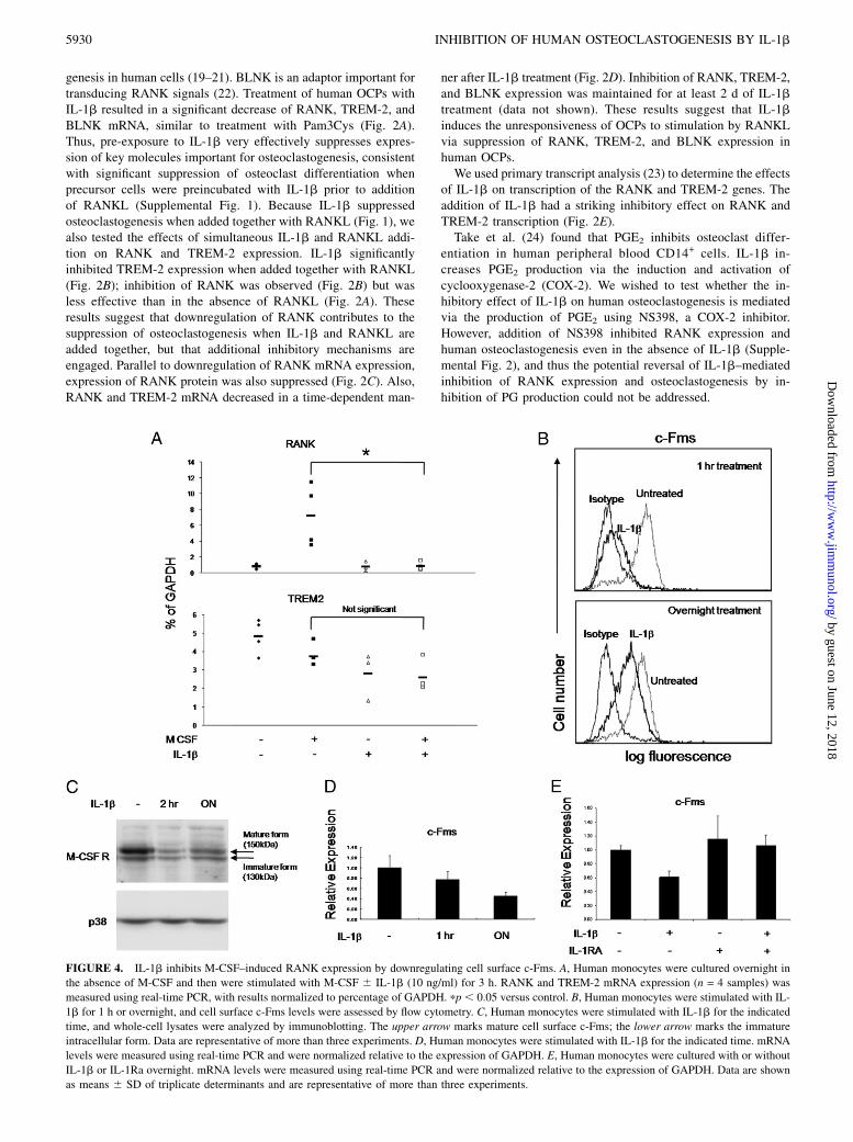
FIGURE 4. IL-1b inhibits M-CSF–induced RANK expression by downregulating cell surface c-Fms. A, Human monocytes were cultured overnight in
the absence of M-CSF and then were stimulated with M-CSF 6 IL-1b (10 ng/ml) for 3 h. RANK and TREM-2 mRNA expression (n = 4 samples) was
measured using real-time PCR, with results normalized to percentage of GAPDH. pp, 0.05 versus control. B, Human monocytes were stimulated with IL-
1b for 1 h or overnight, and cell surface c-Fms levels were assessed by flow cytometry. C, Human monocytes were stimulated with IL-1b for the indicated
time, and whole-cell lysates were analyzed by immunoblotting. The upper arrow marks mature cell surface c-Fms; the lower arrow marks the immature
intracellular form. Data are representative of more than three experiments. D, Human monocytes were stimulated with IL-1b for the indicated time. mRNA
levels were measured using real-time PCR and were normalized relative to the expression of GAPDH. E, Human monocytes were cultured with or without
IL-1b or IL-1Ra overnight. mRNA levels were measured using real-time PCR and were normalized relative to the expression of GAPDH. Data are shown
as means 6 SD of triplicate determinants and are representative of more than three experiments.
5930 INHIBITION OF HUMAN OSTEOCLASTOGENESIS BY IL-1b
by guest on June 12, 2018http://w
ww
.jimm
unol.org/D
ownloaded from

We used IL-1Ra to block specifically IL-1 signaling and toconfirm that the inhibitory effects observed were mediated by IL-1b. IL-1Ra significantly reversed the inhibition of human osteo-clastogenesis by IL-1b (Fig. 3A). Also, the inhibition of RANKgene expression was completely reversed by IL-1Ra (Fig. 3B).These results demonstrated that human osteoclastogenesis is spe-cifically suppressed by IL-1b.
IL-1b downregulates c-Fms and inhibits M-CSF–inducedRANK expression
RANK expression is dependent on M-CSF, which binds to andsignals via c-Fms (6). We tested whether IL-1b inhibits RANKexpression by inhibiting the c-Fms–mediated induction. Isolatedmonocytes were cultured overnight in the absence of M-CSF tomaintain low basal RANK expression, and then these cells weretreated with M-CSF to induce the expression of RANK mRNA.M-CSF treatment rapidly induced expression of RANK mRNA,and this induction was significantly blocked by IL-1b treatment(Fig. 4A). In contrast with RANK expression, TREM-2 expressionwas not dependent on M-CSF (Fig. 4A).To investigate the mechanism underlying inhibition of M-CSF–
induced RANK expression, we examined the effect of IL-1b onsurface expression of c-Fms. Cell surface expression of c-Fms wasnearly completely downregulated by a 1-h treatment with IL-1b(Fig. 4B, upper panel) and remained partially suppressed afterovernight incubation with IL-1b (Fig. 4B, lower panel). M-CSFreceptor expression was examined using immunoblotting. Con-sistent with the results of flow cytometry, the mature form of theM-CSF receptor that is expressed on the cell surface (Fig. 4C,upper band) was rapidly downregulated by IL-1b, and thisdownregulation was partially maintained after overnight incu-bation with IL-1b. In contrast, the immature intracellular form ofc-Fms (Fig. 4C, lower band) was minimally affected by IL-1treatment. We then tested whether rapid downregulation of surfacec-Fms expression is caused by inhibiting expression of c-FmsmRNA. In contrast with strong and rapid downregulation of pro-tein expression, expression of c-Fms mRNA was minimally af-fected after 1-h treatment with IL-1b (Fig. 4D) but evidenceda slow, time-dependent decrease after addition of IL-1b (Fig. 4D).
These results suggest that inhibition of c-Fms gene expression isnot the major mechanism of the early phase of downregulation ofc-Fms expression observed after 1 h of IL-1b treatment, althoughdecreased c-Fms mRNA levels could contribute to the decreasedtotal cellular pool of c-Fms protein observed at the overnight timepoint. Similar to the results obtained with RANK mRNA, IL-1Racompletely reversed the inhibition of c-Fms gene expressionby IL-1b (Fig. 4E).Ectodomain shedding contributes to rapid downregulation of
cell surface c-Fms expression by cell surface a disintegrin andmetalloproteases (25–27). To examine the involvement of metal-loproteinases in the downregulation of M-CSF receptor, cells werepretreated with the metalloproteinase inhibitor TAPI-1 for 1 hbefore treatment with IL-1b. TAPI-1 significantly reversed thenearly complete downregulation of c-Fms expression induced bya 1-h treatment with IL-1b (Fig. 5A, upper panel). Inhibition ofmetalloproteinases with TAPI-1 completely reversed the down-regulation of c-Fms cell surface expression induced by overnighttreatment of IL-1b (Fig. 5A, lower panel). These results suggestthat metalloproteinase-mediated proteolysis is involved in thedownregulation of c-Fms observed after IL-1b treatment.Ectodomain shedding can be induced by many inflammatory
stimuli via protein kinase C (PKC) or via ERK or p38 MAPKs, butpathways that mediate IL-1–induced shedding have not been wellstudied (28–30). Previous studies demonstrated that PKC andERK are involved in downregulation of M-CSF receptor expres-sion (25, 31). Therefore, kinase inhibitors were used to examinethe involvement of PKC, p38, and ERK in IL-1b–induceddownregulation of the M-CSF receptor. The p38 inhibitorSB203580 partially reversed IL-1b–induced inhibition of c-Fmsexpression when used alone (Fig. 5B, upper left panel). IL-1b–induced inhibition of c-Fms expression was also reversed bySB202190, another selective inhibitor of p38 MAPK (data notshown). The MEK–ERK inhibitor U0126 also partially reverseddownregulation of c-Fms expression when used alone (Fig. 5B,upper right panel). Downregulation of c-Fms expression was alsoreversed by PD98059, a specific MEK inhibitor that is structur-ally unrelated to U0126 (data not shown). Combined inhibitionof ERKs and p38 completely abrogated IL-1b–induced down-
FIGURE 5. IL-1b–induced downregulation of cell surface c-Fms is dependent on MMPs and ERK and p38 MAPKs. A, Human monocytes were cultured
overnight in the absence of M-CSF. These cells were stimulated with IL-1b for 1 h or overnight in the presence or absence of the MMP inhibitor TAPI-1. B,
Cells were cultured as in A, these cells were stimulated with IL-1b for 1 h, and GF109203X (1 mM), U0126 (40 mM), SB203580 (10 mM), or SP600125 (10
mM) were added 1 h prior to adding IL-1b. Cell surface c-Fms levels were assessed by flow cytometry. Data are representative of more than three
experiments.
The Journal of Immunology 5931
by guest on June 12, 2018http://w
ww
.jimm
unol.org/D
ownloaded from
regulation of cell surface c-Fms expression (Fig. 5B, lower leftpanel). In contrast, inhibition of PKC had no detectable effect onIL-1b–induced downregulation of c-Fms (Fig. 5B, lower rightpanel).
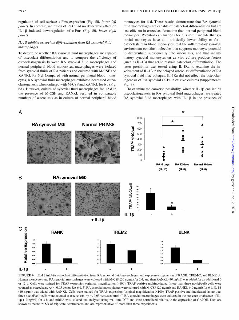
IL-1b inhibits osteoclast differentiation from RA synovial fluidmacrophages
To determine whether RA synovial fluid macrophages are capableof osteoclast differentiation and to compare the efficiency ofosteoclastogenesis between RA synovial fluid macrophages andnormal peripheral blood monocytes, macrophages were isolatedfrom synovial fluids of RA patients and cultured with M-CSF andRANKL for 6 d. Compared with normal peripheral blood mono-cytes, RA synovial fluid macrophages exhibited decreased osteo-clastogenesis when cultured with M-CSF and RANKL for 6 d (Fig.6A). However, culture of synovial fluid macrophages for 12 d inthe presence of M-CSF and RANKL resulted in comparablenumbers of osteoclasts as in culture of normal peripheral blood
monocytes for 6 d. These results demonstrate that RA synovialfluid macrophages are capable of osteoclast differentiation but areless efficient in osteoclast formation than normal peripheral bloodmonocytes. Potential explanations for this result include that sy-novial monocytes have an intrinsically lower ability to formosteoclasts than blood monocytes, that the inflammatory synovialenvironment contains molecules that suppress monocyte potentialto differentiate subsequently into osteoclasts, and that inflam-matory synovial monocytes on ex vivo culture produce factors(such as IL-1b) that act to restrain osteoclast differentiation. Thelatter possibility was tested using IL-1Ra to examine the in-volvement of IL-1b in the delayed osteoclast differentiation of RAsynovial fluid macrophages. IL-1Ra did not affect the osteoclas-togenesis of RA synovial OCPs in ex vivo cultures (SupplementalFig. 3).To examine the converse possibility, whether IL-1b can inhibit
osteoclastogenesis in RA synovial fluid macrophages, we treatedRA synovial fluid macrophages with IL-1b in the presence of
FIGURE 6. IL-1b inhibits osteoclast differentiation from RA synovial fluid macrophages and suppresses expression of RANK, TREM-2, and BLNK. A,
Human monocytes and RA synovial macrophages were cultured with M-CSF (20 ng/ml) for 2 d, and then RANKL (40 ng/ml) was added for an additional 6
or 12 d. Cells were stained for TRAP expression (original magnification 3100). TRAP-positive multinucleated (more than three nuclei/cell) cells were
counted as osteoclasts. pp, 0.05 versus RA 6 d. B, RA synovial macrophages were cultured with M-CSF (20 ng/ml) and RANKL (40 ng/ml) for 6 d. ΙL-1b(10 ng/ml) was added with RANKL. Cells were stained for TRAP expression (original magnification 3100). TRAP-positive multinucleated (more than
three nuclei/cell) cells were counted as osteoclasts. pp , 0.05 versus control. C, RA synovial macrophages were cultured in the presence or absence of IL-
1b (10 ng/ml) for 3 h, and mRNA was isolated and analyzed using real-time PCR and were normalized relative to the expression of GAPDH. Data are
shown as means 6 SD of triplicate determinants and are representative of more than three experiments.
5932 INHIBITION OF HUMAN OSTEOCLASTOGENESIS BY IL-1b
by guest on June 12, 2018http://w
ww
.jimm
unol.org/D
ownloaded from
M-CSF and RANKL. Similar to normal peripheral blood mono-cytes, addition of IL-1b strongly inhibited RANKL-inducedosteoclastogenesis in RA synovial fluid macrophages (Fig. 6B).To investigate the mechanisms by which IL-1b inhibits osteo-clastogenesis in RA synovial fluid macrophages, we tested theeffects of IL-1b on RANK, TREM-2, and BLNK expression.Addition of IL-1b to RA synovial fluid macrophages resulted ina decrease of RANK, TREM-2, and BLNK mRNA expression(Fig. 6C). Collectively, these results suggest that inflammatorymolecules, such as IL-1b, can restrain osteoclastogenesis in in-flammatory diseases, and this inhibition works as a homeostaticmechanism to prevent excessive bone destruction in inflammatorydiseases, such as RA.
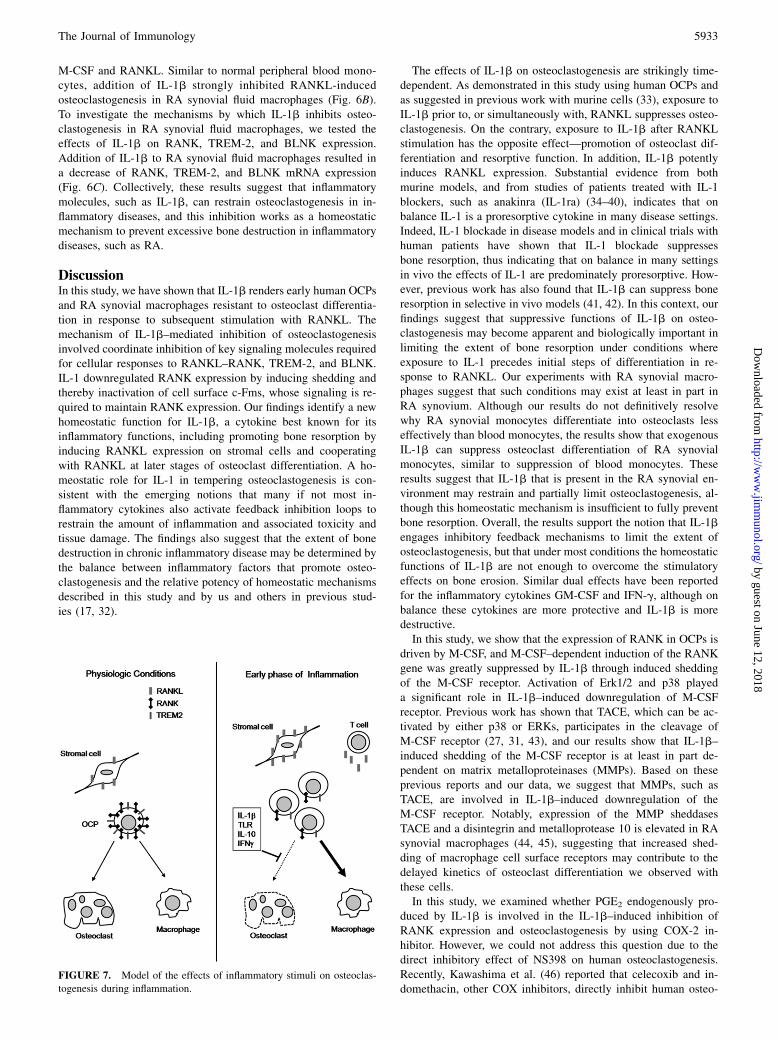
DiscussionIn this study, we have shown that IL-1b renders early human OCPsand RA synovial macrophages resistant to osteoclast differentia-tion in response to subsequent stimulation with RANKL. Themechanism of IL-1b–mediated inhibition of osteoclastogenesisinvolved coordinate inhibition of key signaling molecules requiredfor cellular responses to RANKL–RANK, TREM-2, and BLNK.IL-1 downregulated RANK expression by inducing shedding andthereby inactivation of cell surface c-Fms, whose signaling is re-quired to maintain RANK expression. Our findings identify a newhomeostatic function for IL-1b, a cytokine best known for itsinflammatory functions, including promoting bone resorption byinducing RANKL expression on stromal cells and cooperatingwith RANKL at later stages of osteoclast differentiation. A ho-meostatic role for IL-1 in tempering osteoclastogenesis is con-sistent with the emerging notions that many if not most in-flammatory cytokines also activate feedback inhibition loops torestrain the amount of inflammation and associated toxicity andtissue damage. The findings also suggest that the extent of bonedestruction in chronic inflammatory disease may be determined bythe balance between inflammatory factors that promote osteo-clastogenesis and the relative potency of homeostatic mechanismsdescribed in this study and by us and others in previous stud-ies (17, 32).
The effects of IL-1b on osteoclastogenesis are strikingly time-dependent. As demonstrated in this study using human OCPs andas suggested in previous work with murine cells (33), exposure toIL-1b prior to, or simultaneously with, RANKL suppresses osteo-clastogenesis. On the contrary, exposure to IL-1b after RANKLstimulation has the opposite effect—promotion of osteoclast dif-ferentiation and resorptive function. In addition, IL-1b potentlyinduces RANKL expression. Substantial evidence from bothmurine models, and from studies of patients treated with IL-1blockers, such as anakinra (IL-1ra) (34–40), indicates that onbalance IL-1 is a proresorptive cytokine in many disease settings.Indeed, IL-1 blockade in disease models and in clinical trials withhuman patients have shown that IL-1 blockade suppressesbone resorption, thus indicating that on balance in many settingsin vivo the effects of IL-1 are predominately proresorptive. How-ever, previous work has also found that IL-1b can suppress boneresorption in selective in vivo models (41, 42). In this context, ourfindings suggest that suppressive functions of IL-1b on osteo-clastogenesis may become apparent and biologically important inlimiting the extent of bone resorption under conditions whereexposure to IL-1 precedes initial steps of differentiation in re-sponse to RANKL. Our experiments with RA synovial macro-phages suggest that such conditions may exist at least in part inRA synovium. Although our results do not definitively resolvewhy RA synovial monocytes differentiate into osteoclasts lesseffectively than blood monocytes, the results show that exogenousIL-1b can suppress osteoclast differentiation of RA synovialmonocytes, similar to suppression of blood monocytes. Theseresults suggest that IL-1b that is present in the RA synovial en-vironment may restrain and partially limit osteoclastogenesis, al-though this homeostatic mechanism is insufficient to fully preventbone resorption. Overall, the results support the notion that IL-1bengages inhibitory feedback mechanisms to limit the extent ofosteoclastogenesis, but that under most conditions the homeostaticfunctions of IL-1b are not enough to overcome the stimulatoryeffects on bone erosion. Similar dual effects have been reportedfor the inflammatory cytokines GM-CSF and IFN-g, although onbalance these cytokines are more protective and IL-1b is moredestructive.In this study, we show that the expression of RANK in OCPs is
driven by M-CSF, and M-CSF–dependent induction of the RANKgene was greatly suppressed by IL-1b through induced sheddingof the M-CSF receptor. Activation of Erk1/2 and p38 playeda significant role in IL-1b–induced downregulation of M-CSFreceptor. Previous work has shown that TACE, which can be ac-tivated by either p38 or ERKs, participates in the cleavage ofM-CSF receptor (27, 31, 43), and our results show that IL-1b–induced shedding of the M-CSF receptor is at least in part de-pendent on matrix metalloproteinases (MMPs). Based on theseprevious reports and our data, we suggest that MMPs, such asTACE, are involved in IL-1b–induced downregulation of theM-CSF receptor. Notably, expression of the MMP sheddasesTACE and a disintegrin and metalloprotease 10 is elevated in RAsynovial macrophages (44, 45), suggesting that increased shed-ding of macrophage cell surface receptors may contribute to thedelayed kinetics of osteoclast differentiation we observed withthese cells.In this study, we examined whether PGE2 endogenously pro-
duced by IL-1b is involved in the IL-1b–induced inhibition ofRANK expression and osteoclastogenesis by using COX-2 in-hibitor. However, we could not address this question due to thedirect inhibitory effect of NS398 on human osteoclastogenesis.Recently, Kawashima et al. (46) reported that celecoxib and in-domethacin, other COX inhibitors, directly inhibit human osteo-
FIGURE 7. Model of the effects of inflammatory stimuli on osteoclas-
togenesis during inflammation.
The Journal of Immunology 5933
by guest on June 12, 2018http://w
ww
.jimm
unol.org/D
ownloaded from
clast differentiation. Therefore, additional studies are needed totest whether PGE2 is involved in the IL-1b–induced inhibition ofhuman osteoclastogenesis.Our results and previous work on the effects of inflammatory
factors on osteoclastogenesis support a model (depicted sche-matically in Fig. 7) where during early phases of inflammation orearly after entry of OCPs into inflamed tissues, inflammatorymolecules, such as IL-1b and TLR ligands, downregulate ex-pression of M-CSF receptors and RANK, TREM-2, and BLNK.This downregulation counterbalances the augmentation of RANKLexpression in inflammatory conditions by making cells less re-sponsive to RANKL and thus restrains the extent of osteoclasto-genesis. However, as inflammation progresses, such inhibition canbe overcome by high RANKL expression, expansion of OCPpools (47), and changes in inflammatory factor expression suchthat the cell microenvironment less effectively suppresses RANKexpression (e.g., decreased expression of TLR ligands as infectionis cleared). Under conditions of high and sustained RANKL ex-pression, IL-1b will directly promote osteoclast differentiationand function in cooperation with RANKL, and thus the overalleffect of IL-1b is to promote osteoclast formation and bonedestruction.
DisclosuresThe authors have no financial conflicts of interest.
References1. Takayanagi, H. 2007. Osteoimmunology: shared mechanisms and crosstalk be-
tween the immune and bone systems. Nat. Rev. Immunol. 7: 292–304.2. Teitelbaum, S. L. 2006. Osteoclasts; culprits in inflammatory osteolysis. Arthritis
Res. Ther. 8: 201.3. Walsh, N. C., T. N. Crotti, S. R. Goldring, and E. M. Gravallese. 2005. Rheumatic
diseases: the effects of inflammation on bone. Immunol. Rev. 208: 228–251.4. Bartold, P. M., R. I. Marshall, and D. R. Haynes. 2005. Periodontitis and
rheumatoid arthritis: a review. J. Periodontol. 76(11, Suppl): 2066–2074.5. McInnes, I. B., and G. Schett. 2007. Cytokines in the pathogenesis of rheumatoid
arthritis. Nat. Rev. Immunol. 7: 429–442.6. Arai, F., T. Miyamoto, O. Ohneda, T. Inada, T. Sudo, K. Brasel, T. Miyata,
D. M. Anderson, and T. Suda. 1999. Commitment and differentiation of osteo-clast precursor cells by the sequential expression of c-Fms and receptor activatorof nuclear factor kappaB (RANK) receptors. J. Exp. Med. 190: 1741–1754.
7. Boyce, B. F., P. Li, Z. Yao, Q. Zhang, I. R. Badell, E.M. Schwarz, R. J. O’Keefe, andL.Xing. 2005. TNF-alpha andpathologic bone resorption.Keio J.Med. 54: 127–131.
8. Dinarello, C. A. 1994. The interleukin-1 family: 10 years of discovery. FASEB J.8: 1314–1325.
9. Gowen, M., D. D. Wood, E. J. Ihrie, M. K. McGuire, and R. G. Russell. 1983. Aninterleukin 1 like factor stimulates bone resorption in vitro. Nature 306: 378–380.
10. Nakamura, I., and E. Jimi. 2006. Regulation of osteoclast differentiation andfunction by interleukin-1. Vitam. Horm. 74: 357–370.
11. Wei, S., H. Kitaura, P. Zhou, F. P. Ross, and S. L. Teitelbaum. 2005. IL-1mediates TNF-induced osteoclastogenesis. J. Clin. Invest. 115: 282–290.
12. Dunne, A., and L. A. O’Neill. 2003. The interleukin-1 receptor/Toll-like receptorsuperfamily: signal transduction during inflammation and host defense. Sci.STKE 2003: re3.
13. Ha, H., J. H. Lee, H. N. Kim, H. B. Kwak, H. M. Kim, S. E. Lee, J. H. Rhee,H. H. Kim, and Z. H. Lee. 2008. Stimulation by TLR5 modulates osteoclastdifferentiation through STAT1/IFN-beta. J. Immunol. 180: 1382–1389.
14. Ishii, J., R. Kitazawa, K. Mori, K. P. McHugh, E. Morii, T. Kondo, and S. Kitazawa.2008. Lipopolysaccharide suppresses RANK gene expression in macrophages bydown-regulating PU.1 and MITF. J. Cell. Biochem. 105: 896–904.
15. Takami, M., N. Kim, J. Rho, and Y. Choi. 2002. Stimulation by toll-likereceptors inhibits osteoclast differentiation. J. Immunol. 169: 1516–1523.
16. Zou, W., and Z. Bar-Shavit. 2002. Dual modulation of osteoclast differentiationby lipopolysaccharide. J. Bone Miner. Res. 17: 1211–1218.
17. Ji, J. D., K. H. Park-Min, Z. Shen, R. J. Fajardo, S. R. Goldring, K. P. McHugh, andL. B. Ivashkiv. 2009. Inhibition of RANK expression and osteoclastogenesis byTLRs and IFN-gamma in human osteoclast precursors. J. Immunol. 183: 7223–7233.
18. Sørensen, M. G., K. Henriksen, S. Schaller, D. B. Henriksen, F. C. Nielsen,M.H.Dziegiel, andM.A. Karsdal. 2007. Characterization of osteoclasts derived fromCD14+ monocytes isolated from peripheral blood. J. Bone Miner. Metab. 25: 36–45.
19. Cella, M., C. Buonsanti, C. Strader, T. Kondo, A. Salmaggi, and M. Colonna.2003. Impaired differentiation of osteoclasts in TREM-2-deficient individuals. J.Exp. Med. 198: 645–651.
20. Colonna, M., I. Turnbull, and J. Klesney-Tait. 2007. The enigmatic function ofTREM-2 in osteoclastogenesis. Adv. Exp. Med. Biol. 602: 97–105.
21. Park-Min, K. H., J. D. Ji, T. Antoniv, A. C. Reid, R. B. Silver, M. B. Humphrey,M. Nakamura, and L. B. Ivashkiv. 2009. IL-10 suppresses calcium-mediated
costimulation of receptor activator NF-kappa B signaling during human osteoclastdifferentiation by inhibiting TREM-2 expression. J. Immunol. 183: 2444–2455.
22. Shinohara, M., T. Koga, K. Okamoto, S. Sakaguchi, K. Arai, H. Yasuda, T. Takai,T. Kodama, T. Morio, R. S. Geha, et al. 2008. Tyrosine kinases Btk and Tec regulateosteoclast differentiation by linking RANK and ITAM signals. Cell 132: 794–806.
23. Murray, P. J. 2005. The primary mechanism of the IL-10-regulated antiin-flammatory response is to selectively inhibit transcription. Proc. Natl. Acad. Sci.USA 102: 8686–8691.
24. Take, I., Y. Kobayashi, Y. Yamamoto, H. Tsuboi, T. Ochi, S. Uematsu,N. Okafuji, S. Kurihara, N. Udagawa, and N. Takahashi. 2005. Prostaglandin E2strongly inhibits human osteoclast formation. Endocrinology 146: 5204–5214.
25. Baccarini, M., P. Dello Sbarba, D. Buscher, A. Bartocci, and E. R. Stanley. 1992.IFN-gamma/lipopolysaccharide activation of macrophages is associated withprotein kinase C-dependent down-modulation of the colony-stimulating factor-1 receptor. J. Immunol. 149: 2656–2661.
26. Rettenmier, C. W., M. F. Roussel, R. A. Ashmun, P. Ralph, K. Price, andC. J. Sherr. 1987. Synthesis of membrane-bound colony-stimulating factor 1(CSF-1) and downmodulation of CSF-1 receptors in NIH 3T3 cells transformedby cotransfection of the human CSF-1 and c-fms (CSF-1 receptor) genes. Mol.Cell. Biol. 7: 2378–2387.
27. Rovida, E., A. Paccagnini, M. Del Rosso, J. Peschon, and P. Dello Sbarba. 2001.TNF-alpha-converting enzyme cleaves the macrophage colony-stimulating fac-tor receptor in macrophages undergoing activation. J. Immunol. 166: 1583–1589.
28. Garton, K. J., P. J. Gough, and E. W. Raines. 2006. Emerging roles for ecto-domain shedding in the regulation of inflammatory responses. J. Leukoc. Biol.79: 1105–1116.
29. Takenobu, H., A. Yamazaki, M. Hirata, T. Umata, and E. Mekada. 2003. Thestress- and inflammatory cytokine-induced ectodomain shedding of heparin-binding epidermal growth factor-like growth factor is mediated by p38MAPK, distinct from the 12-O-tetradecanoylphorbol-13-acetate- and lysophos-phatidic acid-induced signaling cascades. J. Biol. Chem. 278: 17255–17262.
30. Xu, K. P., D. Zoukhri, J. D. Zieske, D. A. Dartt, C. Sergheraert, E. Loing, andF. S. Yu. 2001. A role for MAP kinase in regulating ectodomain shedding ofAPLP2 in corneal epithelial cells. Am. J. Physiol. Cell Physiol. 281: C603–C614.
31. Glenn, G., and P. van der Geer. 2008. Toll-like receptors stimulate regulatedintramembrane proteolysis of the CSF-1 receptor through Erk activation. FEBSLett. 582: 911–915.
32. Lorenzo, J., M. Horowitz, and Y. Choi. 2008. Osteoimmunology: interactions ofthe bone and immune system. Endocr. Rev. 29: 403–440.
33. Kim, J. H., H. M. Jin, K. Kim, I. Song, B. U. Youn, K. Matsuo, and N. Kim.2009. The mechanism of osteoclast differentiation induced by IL-1. J. Immunol.183: 1862–1870.
34. Aksentijevich, I., S. L. Masters, P. J. Ferguson, P. Dancey, J. Frenkel, A. vanRoyen-Kerkhoff, R. Laxer, U. Tedgard, E. W. Cowen, T. H. Pham, et al. 2009.An autoinflammatory disease with deficiency of the interleukin-1-receptor an-tagonist. N. Engl. J. Med. 360: 2426–2437.
35. Dinarello, C. A. 2010. Anti-inflammatory agents: present and future. Cell 140:935–950.
36. Gabay, C., C. Lamacchia, and G. Palmer. 2010. IL-1 pathways in inflammationand human diseases. Nat. Rev. Rheumatol. 6: 232–241.
37. Horai, R., S. Saijo, H. Tanioka, S. Nakae, K. Sudo, A. Okahara, T. Ikuse,M. Asano, and Y. Iwakura. 2000. Development of chronic inflammatory ar-thropathy resembling rheumatoid arthritis in interleukin 1 receptor antagonist-deficient mice. J. Exp. Med. 191: 313–320.
38. Jiang, Y., H. K. Genant, I. Watt, M. Cobby, B. Bresnihan, R. Aitchison, andD. McCabe. 2000. A multicenter, double-blind, dose-ranging, randomized,placebo-controlled study of recombinant human interleukin-1 receptor antago-nist in patients with rheumatoid arthritis: radiologic progression and correlationof Genant and Larsen scores. Arthritis Rheum. 43: 1001–1009.
39. Reddy, S., S. Jia, R. Geoffrey, R. Lorier, M. Suchi, U. Broeckel, M. J. Hessner,and J. Verbsky. 2009. An autoinflammatory disease due to homozygous deletionof the IL1RN locus. N. Engl. J. Med. 360: 2438–2444.
40. Sims, J. E., and D. E. Smith. 2010. The IL-1 family: regulators of immunity. Nat.Rev. Immunol. 10: 89–102.
41. Bajayo, A., I. Goshen, S. Feldman, V. Csernus, K. Iverfeldt, E. Shohami,R. Yirmiya, and I. Bab. 2005. Central IL-1 receptor signaling regulates bonegrowth and mass. Proc. Natl. Acad. Sci. USA 102: 12956–12961.
42. Vargas, S. J., A. Naprta, M. Glaccum, S. K. Lee, J. Kalinowski, andJ. A. Lorenzo. 1996. Interleukin-6 expression and histomorphometry of bonesfrom mice deficient in receptors for interleukin-1 or tumor necrosis factor.J. Bone Miner. Res. 11: 1736–1744.
43. Xu, P., and R. Derynck. 2010. Direct activation of TACE-mediated ectodomainshedding by p38 MAP kinase regulates EGF receptor-dependent cell pro-liferation. Mol. Cell 37: 551–566.
44. Ohta, S., M. Harigai, M. Tanaka, Y. Kawaguchi, T. Sugiura, K. Takagi, C. Fukasawa,M. Hara, and N. Kamatani. 2001. Tumor necrosis factor-alpha (TNF-alpha) con-verting enzyme contributes to production of TNF-alpha in synovial tissues frompatients with rheumatoid arthritis. J. Rheumatol. 28: 1756–1763.
45. van der Voort, R., A. W. van Lieshout, L. W. Toonen, A. W. Sloetjes, W. B. vanden Berg, C. G. Figdor, T. R. Radstake, and G. J. Adema. 2005. ElevatedCXCL16 expression by synovial macrophages recruits memory T cells intorheumatoid joints. Arthritis Rheum. 52: 1381–1391.
46. Kawashima, M., Y. Fujikawa, I. Itonaga, C. Takita, and H. Tsumura. 2009. Theeffect of selective cyclooxygenase-2 inhibitor on human osteoclast precursors toinfluence osteoclastogenesis in vitro. Mod. Rheumatol. 19: 192–198.
47. Schett, G. 2007. Erosive arthritis. Arthritis Res. Ther. 9(Suppl 1): S2.
5934 INHIBITION OF HUMAN OSTEOCLASTOGENESIS BY IL-1b
by guest on June 12, 2018http://w
ww
.jimm
unol.org/D
ownloaded from