Embed Size (px)
Citation preview
Full Terms & Conditions of access and use can be found athttps://www.tandfonline.com/action/journalInformation?journalCode=pcgn20
Cognitive Neuropsychology
ISSN: 0264-3294 (Print) 1464-0627 (Online) Journal homepage: https://www.tandfonline.com/loi/pcgn20
Direct electrical stimulation of the left frontalaslant tract disrupts sentence planning withoutaffecting articulation
Benjamin L. Chernoff, Max H. Sims, Susan O. Smith, Webster H. Pilcher &Bradford Z. Mahon
To cite this article: Benjamin L. Chernoff, Max H. Sims, Susan O. Smith, Webster H.Pilcher & Bradford Z. Mahon (2019): Direct electrical stimulation of the left frontal aslant tractdisrupts sentence planning without affecting articulation, Cognitive Neuropsychology, DOI:10.1080/02643294.2019.1619544
To link to this article: https://doi.org/10.1080/02643294.2019.1619544
View supplementary material
Published online: 18 Jun 2019.
Submit your article to this journal
View Crossmark data
Direct electrical stimulation of the left frontal aslant tract disrupts sentenceplanning without affecting articulationBenjamin L. Chernoffa, Max H. Simsb, Susan O. Smithc, Webster H. Pilcherc and Bradford Z. Mahon a,b,c
aDepartment of Psychology, Carnegie Mellon University, Pittsburgh, PA, USA; bDepartment of Neurology, University of Rochester, Rochester, NY,USA; cDepartment of Neurosurgery, University of Rochester Medical Center, Rochester, NY, USA
ABSTRACTSentence production involves mapping from deep structures that specify meaning and thematicroles to surface structures that specify the order and sequencing of production ready elements.We propose that the frontal aslant tract is a key pathway for sequencing complex actions withdeep hierarchical structure. In the domain of language, and primarily with respect to the leftFAT, we refer to this as the ‘Syntagmatic Constraints On Positional Elements’ (SCOPE) hypothesis.One prediction made by the SCOPE hypothesis is that disruption of the frontal aslant tractshould disrupt sentence production at grammatical phrase boundaries, with no disruption ofarticulatory processes. We test this prediction in a patient undergoing direct electricalstimulation mapping of the frontal aslant tract during an awake craniotomy to remove a leftfrontal brain tumor. We found that stimulation of the left FAT prolonged inter-word durations atthe start of grammatical phrases, while inter-word durations internal to noun phrases wereunaffected, and there was no effect on intra-word articulatory duration. These results provideinitial support for the SCOPE hypothesis, and motivate novel directions for future research toexplore the functions of this recently discovered component of the language system.
ARTICLE HISTORYReceived 9 June 2018Revised 22 February 2019Accepted 26 April 2019
KEYWORDSFrontal aslant tract; sentenceproduction; diffusion MRI;direct electrical stimulation;aphasia
Introduction
Sentence production requires planning at multiplelevels of processing. Planning is needed to grammati-cally structure phrases, position lexical elements andgrammatical morphemes, and retrieve and ultimatelyproduce phonological and articulatory information.Understanding the neural circuitry that supports sen-tence production is critical for understanding how thebrain processes language. Strong constraints on cogni-tive models are provided by careful studies of how thesystem can fail, either in the healthy system in the formof slips of the tongue or under conditions of neurologi-cal injury. The Frontal Aslant Tract is a recently discov-ered white matter pathway that connects, in onebranch, the inferior frontal gyrus to the pre-supplemen-tary motor area, and in another branch, the inferiorfrontal gyrus with the anterior cingulate cortex. Withinthe inferior frontal gyrus it is believed to project princi-pally to pars opercularis, but there is also evidence thatit may project to pars orbitalis (Szmuda et al., 2017). Theanatomy of the Frontal Aslant Tract was formalized inhuman Diffusion Tensor Imaging (DTI) tractography
studies by Catani et al. (2012, 2013) and previouslydescribed by others (Aron, Behrens, Smith, Frank, & Pol-drack, 2007; Ford, McGregor, Case, Crosson, & White,2010; Lawes et al., 2008). Post-mortem dissection ofhuman brains (Bozkurt et al., 2016; Goryainov et al.,2017; Koutsarnakis et al., 2017) and tractographystudies in nonhuman primates (Thiebaut de Schotten,Dell’Acqua, Valabregue, & Catani, 2012) have also pro-vided important information about the anatomy ofthe frontal aslant tract.
Lesions to the left frontal aslant tract can result inhalting and dysfluent speech that is otherwise seman-tically, morpho-syntactically, and phonologicallycorrect; furthermore, the dysfluency in spontaneoussentence production does not manifest during sen-tence repetition (Chernoff et al., 2018). A broader litera-ture has implicated the FAT in verbal fluency, in primaryprogressive aphasia (Catani et al., 2013; Mandelli et al.,2014), autism (Chenausky, Kernbach, Norton, &Schlaug, 2017), and post-stroke language difficulties(Li et al., 2017). There is also evidence for a role of theFAT in speech initiation. Direct electrical stimulation
© 2019 Informa UK Limited, trading as Taylor & Francis Group
CONTACT Bradford Z. Mahon [email protected] data for this article can be accessed https://doi.org/10.1080/02643294.2019.1619544.
COGNITIVE NEUROPSYCHOLOGYhttps://doi.org/10.1080/02643294.2019.1619544
intra-operatively of left frontal aslant tract has beenshown to be able to induce speech arrest (Vassal,Boutet, Lemaire, & Nuti, 2014). Further, when examin-ing post-operative outcome after damage to thefrontal aslant tract as compared to the fronto-striataltract (which is located more medially), damage to theformer was more likely to cause speech initiationimpairments, while damage to the fronto-striatal tractcaused non-speech motor initiation impairments(Fujii et al., 2015; Kinoshita et al., 2014). Damage tothe frontal aslant tract has also been related to areduction in functional connectivity between its corti-cal endpoints (Chernoff et al., 2018), suggesting thatfunctional connectivity of those structures criticallydepends on the FAT and not other pathways.
The frontal aslant tract, in addition to the corticosp-inal and corticobulbar (Cai et al., 2014; Chang, Erick-son, Ambrose, Hasegawa-Johnson, & Ludlow, 2008;Dick, Bernal, & Tremblay, 2014) has been associatedwith stuttering, both developmentally (Kronfeld-Duenias, Amir, Ezrati-Vinacour, Civier, & Ben-Shachar,2016a, 2016b) and intra-operatively (Kemerdereet al., 2016). While the right frontal aslant tract hasnot been as systematically studied, Dick, Garic, Gra-ziano, and Tremblay (2018) have suggested that theright FAT may play a critical role in executive function,especially inhibitory control. That proposal resonateswith the putative role of the right inferior frontalgyrus in inhibition (Aron, Fletcher, Bullmore, Sahakian,& Robbins, 2003; Chikazoe, Konishi, Asari, Jimura, &Miyashita, 2007; Hampshire, Chamberlain, Monti,Duncan, & Owen, 2010; Verbruggen & Logan, 2008),and the pre-supplementary motor area in selectionof movements (Amador & Fried, 2004) and resolvingconflicting motor plans (Nachev, Wydell, O’Neill,Husain, & Kennard, 2007).
Here we propose that the left frontal aslant tractserves as an interface between distinct levels of sen-tence planning—specifically, between planning syn-tagmatic relations and positional planning ofmorphophonological elements—we refer to this asthe “Syntagmatic Constraints On Positional Elements”(SCOPE) hypothesis. The key aspect of this proposal isthat the frontal aslant tract interfaces grammaticalspecifications of sentence structure with (alreadyaccessed) lexical representations, by hypothesis, inanticipation of planning articulatory phrases. This gen-erates the prediction that disruption of the frontalaslant tract should specifically disrupt sentence
production at phrasal boundaries, with no impairmentfor articulatory duration. The purpose of the currentinvestigation was to test that hypothesis by measuringsentence fluency, in a patient who was anticipated tohave direct electrical stimulation of the left frontalaslant tract during surgery for resection of a tumour.
Prior documented impairments for speech fluencyin patients with damage to the FAT have beenbased on neuropsychological tests of speech pro-duction—such as the BDAE “Cookie Theft” (Chernoffet al., 2018) test, the “Cinderella test” (Catani et al.,2013), and indices of stuttering severity, such as theStuttering Severity Instrument (SSI-III; Riley, 1994).While powerful, those tests capture gross fluencymeasures (such as mean length of utterance), andare less constrained than may be preferable for evalu-ating our proposal about the functions supported bythe frontal aslant tract. We thus designed a task thatcould be used intra-operatively while the frontalaslant tract was stimulated, and which would allowus to examine different components of sentence pro-duction. The patient was presented with a 2 × 2arrangement of coloured geometric shapes on eachtrial, with one of the four shapes cued via a thickblack outline. The patient’s task was to generate a sen-tence describing the spatial relation of the cued shapein relation to the shape that was either immediatelyabove or below it—for instance, “The red square isabove the yellow circle”. If stimulation of the frontalaslant tract disrupts sentence production by disrupt-ing lexical retrieval, then we predict that inter-worddurations (i.e., pauses between words) will be longerprior to all content words in the sentence. By contrast,if the frontal aslant tract supports integration of gram-matical information with positionally specifiedelements, then stimulation of that tract should leadto prolonged inter-word durations at the boundariesof grammatical phrases, but not within grammaticalphrases. Finally, if the frontal aslant tract supportsarticulatory processes, then stimulation of that tractshould prolong or disrupt articulation of all words inthe sentence.
Methods
Participants
Patient AI was, at the time of testing, a 46 year-oldman with a left frontal Oligodendroglioma (Figure
2 B. L. CHERNOFF ET AL.
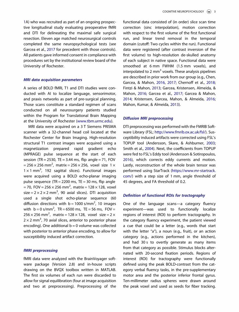
1A) who was recruited as part of an ongoing prospec-tive longitudinal study evaluating preoperative fMRIand DTI for delineating the maximal safe surgicalresection. Eleven age matched neurosurgical controlscompleted the same neuropsychological tests (seeGarcea et al., 2017 for precedent with those controls).All patients gave informed consent in compliance withprocedures set by the institutional review board of theUniversity of Rochester.
MRI data acquisition parameters
A series of BOLD fMRI, T1 and DTI studies were con-ducted with AI to localize language, sensorimotor,and praxis networks as part of pre-surgical planning.Those scans constitute a standard regimen of scansconducted on all neurosurgery patients studiedwithin the Program for Translational Brain Mappingat the University of Rochester (www.tbm.urmc.edu).
MRI data were acquired on a 3 T Siemens PRISMAscanner with a 32-channel head coil located at theRochester Center for Brain Imaging. High-resolutionstructural T1 contrast images were acquired using amagnetization prepared rapid gradient echo(MPRAGE) pulse sequence at the start of eachsession (TR = 2530, TE = 3.44 ms, flip angle = 71, FOV= 256 × 256 mm2, matrix = 256 × 256, voxel size 1 ×1 × 1 mm3, 192 sagittal slices). Functional imageswere acquired using a BOLD echo-planar imagingpulse sequence (TR = 2200 ms, TE = 30 ms, flip angle= 70, FOV = 256 × 256 mm2, matrix = 128 × 128, voxelsize = 2 × 2 × 2 mm3, 90 axial slices). DTI acquisitionused a single shot echo-planar sequence (60diffusion directions with b = 1000 s/mm2, 10 imageswith b = 0 s/mm2, TR = 6500 ms, TE = 56 ms, FOV =256 × 256 mm2, matrix = 128 × 128, voxel size = 2 ×2 × 2 mm3, 70 axial slices, anterior to posterior phaseencoding). One additional b = 0 volume was collectedwith posterior to anterior phase encoding, to allow forsusceptibility induced artifact correction.
fMRI preprocessing
fMRI data were analyzed with the BrainVoyager soft-ware package (Version 2.8) and in-house scriptsdrawing on the BVQX toolbox written in MATLAB.The first six volumes of each run were discarded toallow for signal equilibration (four at image acquisitionand two at preprocessing). Preprocessing of the
functional data consisted of (in order) slice scan timecorrection (sinc interpolation), motion correctionwith respect to the first volume of the first functionalrun, and linear trend removal in the temporaldomain (cutoff: Two cycles within the run). Functionaldata were registered (after contrast inversion of thefirst volume) to high-resolution de-skulled anatomyof each subject in native space. Functional data weresmoothed at 6 mm FWHM (1.5 mm voxels), andinterpolated to 2 mm3 voxels. These analysis pipelinesare described in prior work from our group (e.g., Chen,Garcea, & Mahon, 2016, 2017; Chernoff et al., 2018;Fintzi & Mahon, 2013; Garcea, Kristensen, Almeida, &Mahon, 2016; Garcea et al., 2017; Garcea & Mahon,2014; Kristensen, Garcea, Mahon, & Almeida, 2016;Mahon, Kumar, & Almeida, 2013).
Diffusion MRI preprocessing
DTI preprocessing was performed with the FMRIB Soft-ware Library (FSL; http://www.fmrib.ox.ac.uk/fsl/). Sus-ceptibility induced artifacts were corrected using FSL’sTOPUP tool (Andersson, Skare, & Ashburner, 2003;Smith et al., 2004). Next, the coefficients from TOPUPwere fed to FSL’s Eddy tool (Andersson & Sotiropoulos,2016), which corrects eddy currents and motion.Lastly, reconstruction of the whole brain tensor wasperformed using StarTrack (https://www.mr-startrack.com/) with a step size of 1 mm, angle threshold of45 degrees, and FA threshold of 0.2.
Definition of functional ROIs for tractography
One of the language scans—a category fluencyexperiment—was used to functionally localizeregions of interest (ROI) to perform tractography. Inthe category fluency experiment, the patient vieweda cue that could be a letter (e.g., words that startwith the letter “a”), a noun (e.g., fruit), or an actioncategory (e.g., actions performed in the kitchen),and had 30 s to overtly generate as many itemsfrom that category as possible. Stimulus blocks alter-nated with 20-second fixation periods. Regions ofinterest (ROI) for tractography were functionallydefined using the peak BOLD-contrast from the cat-egory verbal fluency tasks, in the pre-supplementarymotor area and the posterior inferior frontal gyrus.Ten-millimeter radius spheres were drawn aroundthe peak voxel and used as seeds for fiber tracking.
COGNITIVE NEUROPSYCHOLOGY 3
An exclusion mask was drawn around consecutivesagittal slices in the right hemisphere, four to eightmillimetrs from the midline, in order to exclude cross-ing callosal fibers. Deterministic tractography wasperformed using TrackVis (Wang, Benner, Sorensen,& Wedeen, 2007).
Neuropsychological tests
Patient AI was evaluated pre-operatively to broadlyassess language, praxis, visual processing, memory,and attention. All testing was video and/or audiorecorded for offline analysis (for a detailed
descriptions of the testing, see Garcea, Dombovy, &Mahon, 2013; Stasenko, Garcea, Dombovy, & Mahon,2014). We conduct a large array of tests as part of astandard battery for a broader longitudinal study inthe Program for Translational Brain Mapping, but inthis case we were particularly interested in languagefunction. The purpose of testing patients morebroadly is to understand the limits of each patient’simpairments, which is critical for deriving inferencesabout cognitive organization based on the underlingcognitive processes that are disrupted in any givenpatient (i.e., the “sufficiency” condition, as in Cara-mazza, 1984).
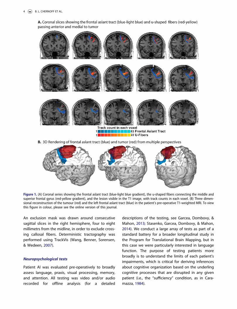
Figure 1. (A) Coronal series showing the frontal aslant tract (blue-light blue gradient), the u-shaped fibers connecting the middle andsuperior frontal gyrus (red-yellow gradient), and the lesion visible in the T1 image, with track counts in each voxel. (B) Three dimen-sional reconstruction of the tumour (red) and the left frontal aslant tract (blue) in the patient’s pre-operative T1-weighted MRI. To viewthis figure in colour, please see the online version of this journal.
4 B. L. CHERNOFF ET AL.
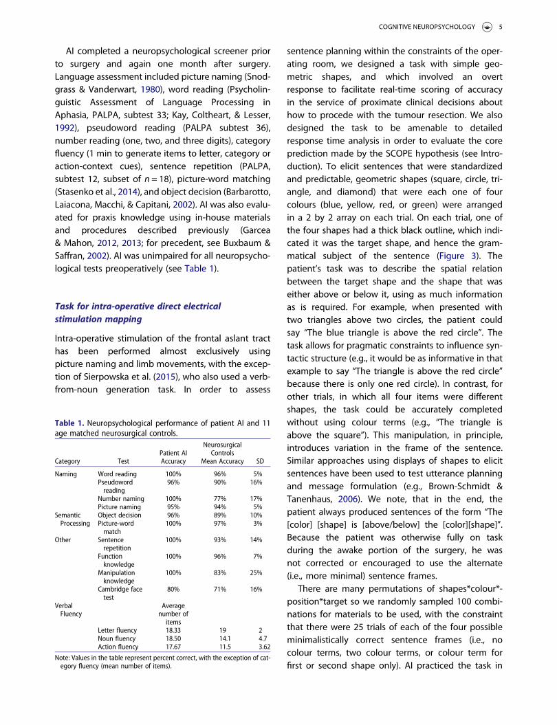
AI completed a neuropsychological screener priorto surgery and again one month after surgery.Language assessment included picture naming (Snod-grass & Vanderwart, 1980), word reading (Psycholin-guistic Assessment of Language Processing inAphasia, PALPA, subtest 33; Kay, Coltheart, & Lesser,1992), pseudoword reading (PALPA subtest 36),number reading (one, two, and three digits), categoryfluency (1 min to generate items to letter, category oraction-context cues), sentence repetition (PALPA,subtest 12, subset of n = 18), picture-word matching(Stasenko et al., 2014), and object decision (Barbarotto,Laiacona, Macchi, & Capitani, 2002). AI was also evalu-ated for praxis knowledge using in-house materialsand procedures described previously (Garcea& Mahon, 2012, 2013; for precedent, see Buxbaum &Saffran, 2002). AI was unimpaired for all neuropsycho-logical tests preoperatively (see Table 1).
Task for intra-operative direct electricalstimulation mapping
Intra-operative stimulation of the frontal aslant tracthas been performed almost exclusively usingpicture naming and limb movements, with the excep-tion of Sierpowska et al. (2015), who also used a verb-from-noun generation task. In order to assess
sentence planning within the constraints of the oper-ating room, we designed a task with simple geo-metric shapes, and which involved an overtresponse to facilitate real-time scoring of accuracyin the service of proximate clinical decisions abouthow to procede with the tumour resection. We alsodesigned the task to be amenable to detailedresponse time analysis in order to evaluate the coreprediction made by the SCOPE hypothesis (see Intro-duction). To elicit sentences that were standardizedand predictable, geometric shapes (square, circle, tri-angle, and diamond) that were each one of fourcolours (blue, yellow, red, or green) were arrangedin a 2 by 2 array on each trial. On each trial, one ofthe four shapes had a thick black outline, which indi-cated it was the target shape, and hence the gram-matical subject of the sentence (Figure 3). Thepatient’s task was to describe the spatial relationbetween the target shape and the shape that waseither above or below it, using as much informationas is required. For example, when presented withtwo triangles above two circles, the patient couldsay “The blue triangle is above the red circle”. Thetask allows for pragmatic constraints to influence syn-tactic structure (e.g., it would be as informative in thatexample to say “The triangle is above the red circle”because there is only one red circle). In contrast, forother trials, in which all four items were differentshapes, the task could be accurately completedwithout using colour terms (e.g., “The triangle isabove the square”). This manipulation, in principle,introduces variation in the frame of the sentence.Similar approaches using displays of shapes to elicitsentences have been used to test utterance planningand message formulation (e.g., Brown-Schmidt &Tanenhaus, 2006). We note, that in the end, thepatient always produced sentences of the form “The[color] [shape] is [above/below] the [color][shape]”.Because the patient was otherwise fully on taskduring the awake portion of the surgery, he wasnot corrected or encouraged to use the alternate(i.e., more minimal) sentence frames.
There are many permutations of shapes*colour*-position*target so we randomly sampled 100 combi-nations for materials to be used, with the constraintthat there were 25 trials of each of the four possibleminimalistically correct sentence frames (i.e., nocolour terms, two colour terms, or colour term forfirst or second shape only). AI practiced the task in
Table 1. Neuropsychological performance of patient AI and 11age matched neurosurgical controls.
Patient AINeurosurgical
ControlsCategory Test Accuracy Mean Accuracy SD
Naming Word reading 100% 96% 5%Pseudowordreading
96% 90% 16%
Number naming 100% 77% 17%Picture naming 95% 94% 5%
SemanticProcessing
Object decision 96% 89% 10%Picture-wordmatch
100% 97% 3%
Other Sentencerepetition
100% 93% 14%
Functionknowledge
100% 96% 7%
Manipulationknowledge
100% 83% 25%
Cambridge facetest
80% 71% 16%
VerbalFluency
Averagenumber ofitems
Letter fluency 18.33 19 2Noun fluency 18.50 14.1 4.7Action fluency 17.67 11.5 3.62
Note: Values in the table represent percent correct, with the exception of cat-egory fluency (mean number of items).
COGNITIVE NEUROPSYCHOLOGY 5
the lab prior to surgery, laying down on his right side,to simulate the ergonomics of the operating room.During those practice sessions, he understood thetask and performed all trials with no errors, hesita-tions, or paraphasias. During the surgery, an auditorycue (a click) initiated each trial; this auditory cue sig-nalled to the surgeon the start of a new trial. At thediscretion of the attending surgeon (WHP) directcurrent stimulation was then applied coincident
with trial onset, with the duration of stimulationlasting ∼4 s.
Intra-operative testing
Intra-operative experiments were performed usingStrongView, which has been developed within theProgram for Translational Brain Mapping at the Uni-versity of Rochester. StrongView includes a PC with a
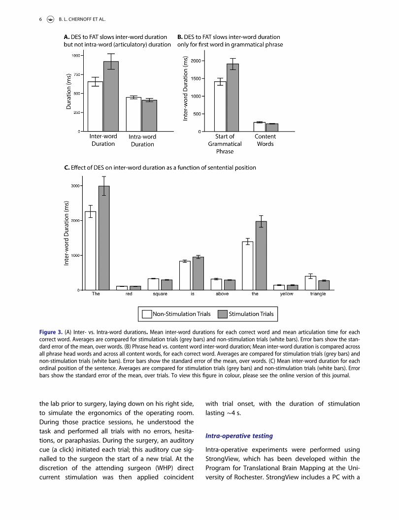
Figure 3. (A) Inter- vs. Intra-word durations. Mean inter-word durations for each correct word and mean articulation time for eachcorrect word. Averages are compared for stimulation trials (grey bars) and non-stimulation trials (white bars). Error bars show the stan-dard error of the mean, over words. (B) Phrase head vs. content word inter-word duration; Mean inter-word duration is compared acrossall phrase head words and across all content words, for each correct word. Averages are compared for stimulation trials (grey bars) andnon-stimulation trials (white bars). Error bars show the standard error of the mean, over words. (C) Mean inter-word duration for eachordinal position of the sentence. Averages are compared for stimulation trials (grey bars) and non-stimulation trials (white bars). Errorbars show the standard error of the mean, over trials. To view this figure in colour, please see the online version of this journal.
6 B. L. CHERNOFF ET AL.
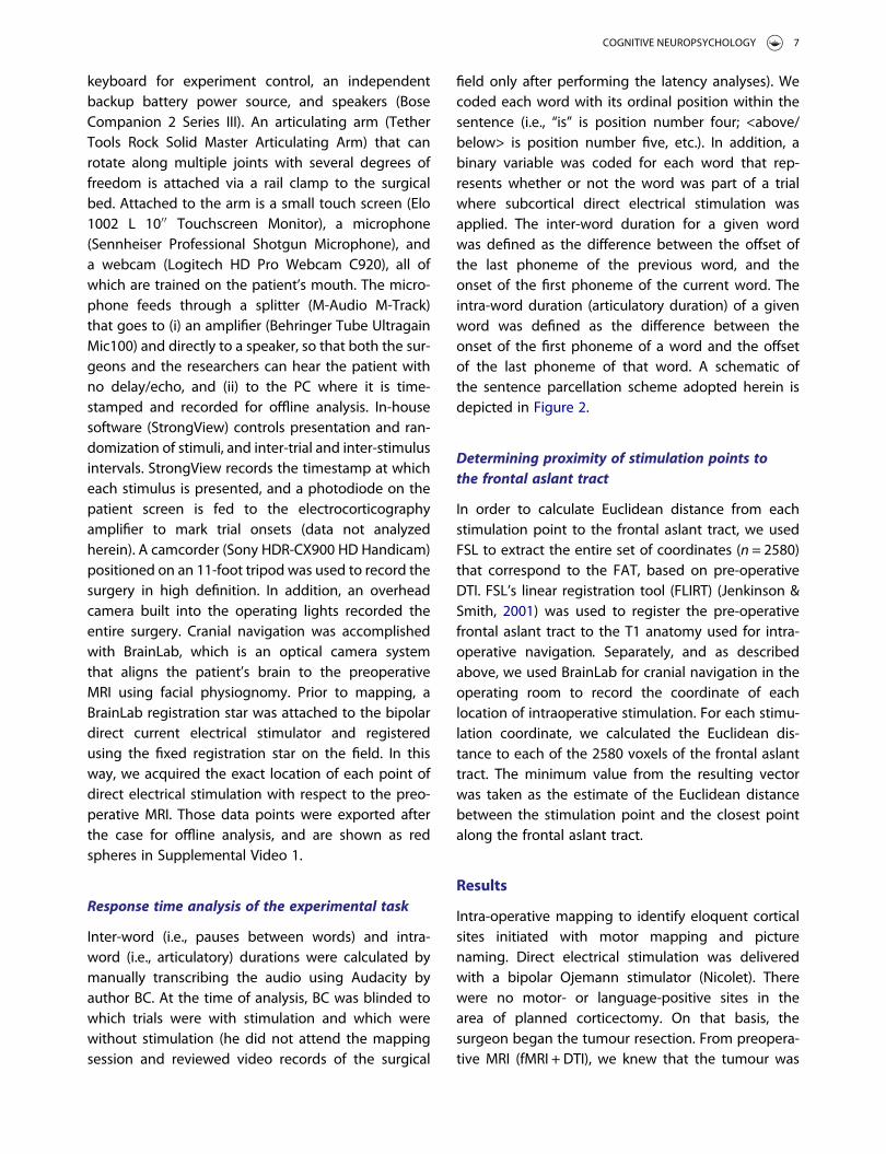
keyboard for experiment control, an independentbackup battery power source, and speakers (BoseCompanion 2 Series III). An articulating arm (TetherTools Rock Solid Master Articulating Arm) that canrotate along multiple joints with several degrees offreedom is attached via a rail clamp to the surgicalbed. Attached to the arm is a small touch screen (Elo1002 L 10′′ Touchscreen Monitor), a microphone(Sennheiser Professional Shotgun Microphone), anda webcam (Logitech HD Pro Webcam C920), all ofwhich are trained on the patient’s mouth. The micro-phone feeds through a splitter (M-Audio M-Track)that goes to (i) an amplifier (Behringer Tube UltragainMic100) and directly to a speaker, so that both the sur-geons and the researchers can hear the patient withno delay/echo, and (ii) to the PC where it is time-stamped and recorded for offline analysis. In-housesoftware (StrongView) controls presentation and ran-domization of stimuli, and inter-trial and inter-stimulusintervals. StrongView records the timestamp at whicheach stimulus is presented, and a photodiode on thepatient screen is fed to the electrocorticographyamplifier to mark trial onsets (data not analyzedherein). A camcorder (Sony HDR-CX900 HD Handicam)positioned on an 11-foot tripod was used to record thesurgery in high definition. In addition, an overheadcamera built into the operating lights recorded theentire surgery. Cranial navigation was accomplishedwith BrainLab, which is an optical camera systemthat aligns the patient’s brain to the preoperativeMRI using facial physiognomy. Prior to mapping, aBrainLab registration star was attached to the bipolardirect current electrical stimulator and registeredusing the fixed registration star on the field. In thisway, we acquired the exact location of each point ofdirect electrical stimulation with respect to the preo-perative MRI. Those data points were exported afterthe case for offline analysis, and are shown as redspheres in Supplemental Video 1.
Response time analysis of the experimental task
Inter-word (i.e., pauses between words) and intra-word (i.e., articulatory) durations were calculated bymanually transcribing the audio using Audacity byauthor BC. At the time of analysis, BC was blinded towhich trials were with stimulation and which werewithout stimulation (he did not attend the mappingsession and reviewed video records of the surgical
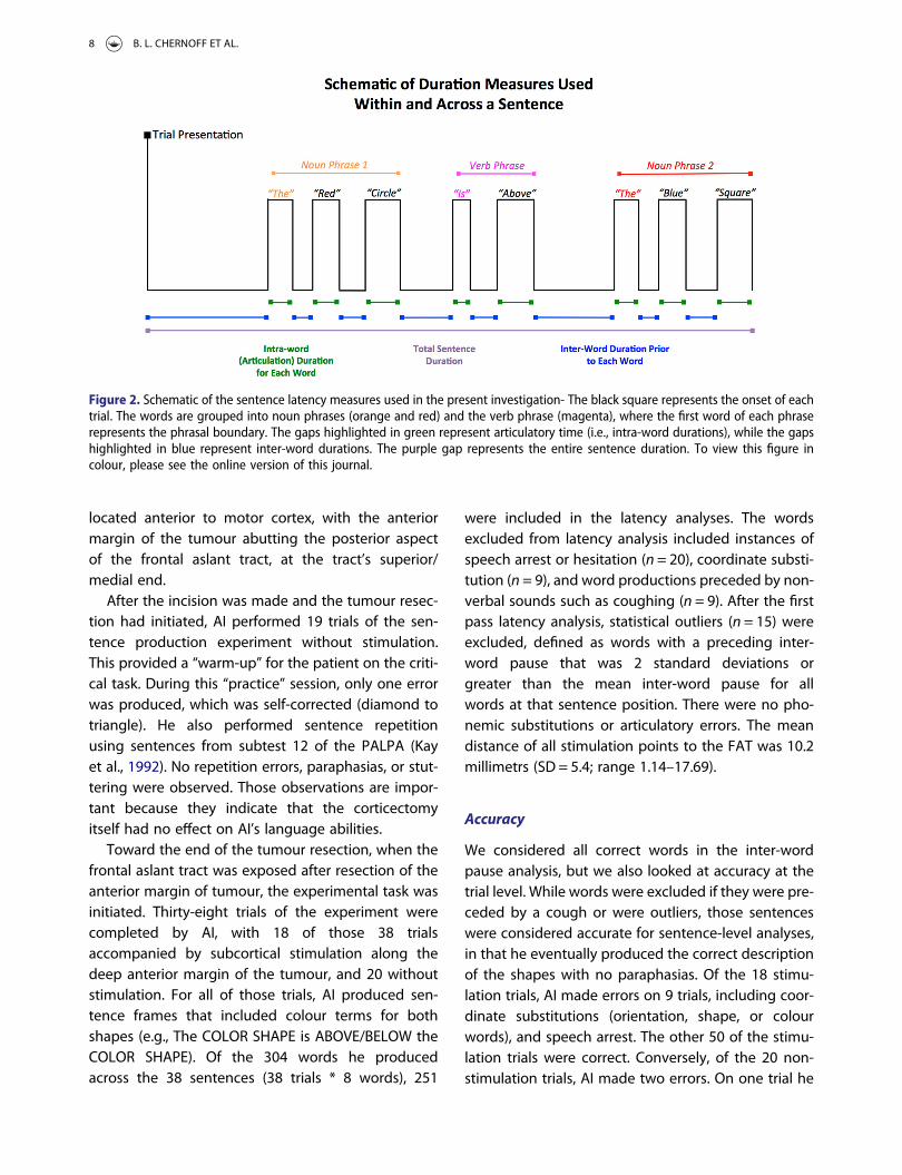
field only after performing the latency analyses). Wecoded each word with its ordinal position within thesentence (i.e., “is” is position number four; <above/below> is position number five, etc.). In addition, abinary variable was coded for each word that rep-resents whether or not the word was part of a trialwhere subcortical direct electrical stimulation wasapplied. The inter-word duration for a given wordwas defined as the difference between the offset ofthe last phoneme of the previous word, and theonset of the first phoneme of the current word. Theintra-word duration (articulatory duration) of a givenword was defined as the difference between theonset of the first phoneme of a word and the offsetof the last phoneme of that word. A schematic ofthe sentence parcellation scheme adopted herein isdepicted in Figure 2.
Determining proximity of stimulation points tothe frontal aslant tract
In order to calculate Euclidean distance from eachstimulation point to the frontal aslant tract, we usedFSL to extract the entire set of coordinates (n = 2580)that correspond to the FAT, based on pre-operativeDTI. FSL’s linear registration tool (FLIRT) (Jenkinson &Smith, 2001) was used to register the pre-operativefrontal aslant tract to the T1 anatomy used for intra-operative navigation. Separately, and as describedabove, we used BrainLab for cranial navigation in theoperating room to record the coordinate of eachlocation of intraoperative stimulation. For each stimu-lation coordinate, we calculated the Euclidean dis-tance to each of the 2580 voxels of the frontal aslanttract. The minimum value from the resulting vectorwas taken as the estimate of the Euclidean distancebetween the stimulation point and the closest pointalong the frontal aslant tract.
Results
Intra-operative mapping to identify eloquent corticalsites initiated with motor mapping and picturenaming. Direct electrical stimulation was deliveredwith a bipolar Ojemann stimulator (Nicolet). Therewere no motor- or language-positive sites in thearea of planned corticectomy. On that basis, thesurgeon began the tumour resection. From preopera-tive MRI (fMRI + DTI), we knew that the tumour was
COGNITIVE NEUROPSYCHOLOGY 7
located anterior to motor cortex, with the anteriormargin of the tumour abutting the posterior aspectof the frontal aslant tract, at the tract’s superior/medial end.
After the incision was made and the tumour resec-tion had initiated, AI performed 19 trials of the sen-tence production experiment without stimulation.This provided a “warm-up” for the patient on the criti-cal task. During this “practice” session, only one errorwas produced, which was self-corrected (diamond totriangle). He also performed sentence repetitionusing sentences from subtest 12 of the PALPA (Kayet al., 1992). No repetition errors, paraphasias, or stut-tering were observed. Those observations are impor-tant because they indicate that the corticectomyitself had no effect on AI’s language abilities.
Toward the end of the tumour resection, when thefrontal aslant tract was exposed after resection of theanterior margin of tumour, the experimental task wasinitiated. Thirty-eight trials of the experiment werecompleted by AI, with 18 of those 38 trialsaccompanied by subcortical stimulation along thedeep anterior margin of the tumour, and 20 withoutstimulation. For all of those trials, AI produced sen-tence frames that included colour terms for bothshapes (e.g., The COLOR SHAPE is ABOVE/BELOW theCOLOR SHAPE). Of the 304 words he producedacross the 38 sentences (38 trials * 8 words), 251
were included in the latency analyses. The wordsexcluded from latency analysis included instances ofspeech arrest or hesitation (n = 20), coordinate substi-tution (n = 9), and word productions preceded by non-verbal sounds such as coughing (n = 9). After the firstpass latency analysis, statistical outliers (n = 15) wereexcluded, defined as words with a preceding inter-word pause that was 2 standard deviations orgreater than the mean inter-word pause for allwords at that sentence position. There were no pho-nemic substitutions or articulatory errors. The meandistance of all stimulation points to the FAT was 10.2millimetrs (SD = 5.4; range 1.14–17.69).
Accuracy
We considered all correct words in the inter-wordpause analysis, but we also looked at accuracy at thetrial level. While words were excluded if they were pre-ceded by a cough or were outliers, those sentenceswere considered accurate for sentence-level analyses,in that he eventually produced the correct descriptionof the shapes with no paraphasias. Of the 18 stimu-lation trials, AI made errors on 9 trials, including coor-dinate substitutions (orientation, shape, or colourwords), and speech arrest. The other 50 of the stimu-lation trials were correct. Conversely, of the 20 non-stimulation trials, AI made two errors. On one trial he
Figure 2. Schematic of the sentence latency measures used in the present investigation- The black square represents the onset of eachtrial. The words are grouped into noun phrases (orange and red) and the verb phrase (magenta), where the first word of each phraserepresents the phrasal boundary. The gaps highlighted in green represent articulatory time (i.e., intra-word durations), while the gapshighlighted in blue represent inter-word durations. The purple gap represents the entire sentence duration. To view this figure incolour, please see the online version of this journal.
8 B. L. CHERNOFF ET AL.
said “below” instead of “above”, and on another trialhe failed to complete the full sentence prior to theonset of the next trial. The difference in accuracybetween stimulation and non-stimulation trials wassignificant (X2
38 = 7.37, p = 0.007).
Latency analysis
Total sentence duration was not different for correctstimulation trials compared to non-stimulation trials(t25 = 0.41, p = 0.68). Sentence duration was calculatedfrom the onset of the stimulus to the offset of the lastword.
We examined articulation time of stimulation andnon-stimulation trials, in order to test whether stimu-lation affected the time required to produce thespeech motor movements of each word. The meanarticulation time for words with stimulation was413 ms (SD = 236 ms; range = 127–885 ms) and forwords without stimulation was 449 ms (SD = 257 ms;range = 129–1162 ms); the difference was not signifi-cant (t249 = 1.15, p = 0.25, d = 0.15) (Figure 3A).
We then examined the effect of stimulation oninter-word durations. We conducted a two-way analy-sis of variance (ANOVA) to test the influence of stimu-lation (2 levels: Stimulation, no stimulation) andsentence position (8 levels: First | second | third |fourth | fifth | sixth | seventh | and eighth word inthe sentence) on inter-word durations. There weremain effects of stimulation (F(1,235) = 11.15; p = 0.001,h2p = 0.05) and position (F(7,235) = 191.52, p < 0.00001;
p < 0.02, h2p = 0.85), and an interaction between stimu-
lation and position (F(7,235) = 5.92, p < 0.00001, h2p =
0.15). Hypothesis driven tests evaluated the key ques-tion of whether inter-word durations were differen-tially prolonged at phrasal heads, including for theverb (“is”) and noun phrases (“the”). For this analysis,we binned across sentence positions according togrammatical structure. Specifically, inter-word dur-ations prior to the noun phrases (prior to “the”) andprior to the verb (“is”) were combined, while inter-word durations prior to all other words were com-bined (Figure 3B). There was a two-way interactionbetween position and stimulation (F(1,247) = 13.386, p= 0.0003, h2
p = 0.05). This interaction is reflected inthe higher mean for “the” and “is” after stimulation(mean = 1908 ms, SD = 1076 ms) compared towithout stimulation (mean = 1407 ms, SD = 699 ms),but a lower mean for all other words when produced
in the context of stimulation (mean = 221 ms, SD =91 ms) compared to without stimulation (mean =261 ms, SD = 175 ms).
Finally, we conducted t-tests for stimulation vs.non-stimulation for inter-word durations at each pos-ition in the sentence (Figure 3C). There were signifi-cant differences for words in the first ordinalposition (“The”) (t25 = 2.23, p = 0.04, d = 0.86); thethird ordinal position (shape) (t31 =−2.64, p = 0.01, d= 0.91); the fourth ordinal position (“is”) (t34 = 2.14, p= 0.04, d = 0.71); and the sixth ordinal position (“the”)(t29 = 3.08, p = 0.004, d = 1.1). There were no significantdifferences in inter-word durations between stimu-lation and non-stimulation words for the otherordinal positions.
Discussion
The frontal aslant tract is a recently described whitematter pathway that connects the inferior frontalgyrus with supplementary motor cortex, and hasbeen implicated in verbal fluency in a series ofreports. We propose a new hypothesis for under-standing the contribution of the frontal aslant tractto language production, emphasizing the role ofthis pathway in mapping grammatical specified plan-ning of sentence structure and positional level plan-ning—referred to as the Syntagmatic Constraints OnPositional Elements (SCOPE) hypothesis. One predic-tion made by this proposal is that disruption of theFAT will disrupt sentence production at the level atwhich syntagmatic relations interface with positionalspecifications of words, which by hypothesis, shouldcorrespond to the boundaries of grammaticalphrases. To test this prediction, we designed a cuedsentence production task for use during intraopera-tive awake language mapping, and analyzed bothaccuracy and the distribution of intra- and inter-word durations as a function of stimulation. Wefound that neither the overall duration of sentencesnor the articulation time (intra-word durations) ofwords was affected by stimulation. However, inter-word durations were prolonged for words precededby stimulation of the frontal aslant tract, but only ifthose words were at the boundaries of grammaticalphrases. Other words were either not significantlydifferent, or facilitated by stimulation (shorter inter-word durations for stimulation than non-stimulation).One possibility is that the facilitation observed for
COGNITIVE NEUROPSYCHOLOGY 9
inter-word durations within noun phrases is a bypro-duct of the prolonged durations at the boundary ofthe noun phrase; i.e., there was more time devotedto planning the entire phrase, and some of thattime allowed articulatory planning to proceed aswell. To evaluate this possibility, we correlated theinter-word durations for the boundary of thesecond noun phrase in each sentence with thewithin-phrase inter-word durations (also for thesecond noun phrase in each sentence). This wasdone separately for stimulation and non-stimulationtrials. If facilitation effects are a byproduct of moretime devoted to planning the phrase at its initiation,then there should be a negative correlation. In con-trast, we observed a positive correlation (r = 0.44)between inter-word durations for the head of thesecond noun phrase and inter-word durationswithin that phrase. Thus, at present, it is notobvious why stimulation would lead to shorterinter-word durations within noun phrases, and thisissue merits additional empirical scrutiny with futureresearch.
All models of speech production agree that sen-tence production involves the (i) formulation of amessage, (ii) construction of a syntactic frametogether with access to words’ grammatical proper-ties, and (iii) phonological encoding and articulation(e.g., Caramazza, 1997; Dell, Schwartz, Martin, Saffran,& Gagnon, 1997; Garrett, 1980a, 1980b; Rapp & Gold-rick, 2000). While models disagree on issues such asthe dynamics of information flow in the system,whether there are 1 or 2 lexical levels, and whetheraccess to syntactic and phonological propertiesoccurs in that order or in parallel, there is broad agree-ment that sentence production involves the trans-lation of a hierarchical representation specifyingsentence-level grammatical dependencies into aspecific surface form. Furthermore, there is generalagreement that grammatical processing is a separableprocess in language production, and there is substan-tial neural evidence as well (Friederici, Bahlmann,Heim, Schubotz, & Anwander, 2006; Hickok &Poeppel, 2007; Indefrey & Levelt, 2004; Shapiro,Shelton, & Caramazza, 2000; Shapiro & Caramazza,2003; Shapiro, Moo, & Caramazza, 2006). Thus, allmodels must posit a process whereby grammaticalspecifications are “realized” as surface forms. Our pro-posal is that the frontal aslant tract is a key componentof that interface.
Our findings are relevant as well to previousresearch on the functional consequences of damageto the cortical regions connected by the FrontalAslant Tract—the inferior frontal gyrus (IFG) and pre-supplementary motor area (pre-SMA). The IFG isassociated with agrammatism in primary progressiveaphasia, both in production (Grossman, 2012) andcomprehension (Charles et al., 2014). Lesion studieshave also implicated the IFG in syntactic processing(Grodzinsky, 2006; Kaan & Swaab, 2002). Importantly,the IFG has been implicated in a number of otheraspects of language processing, including phonologi-cal encoding, selection among alternatives andgeneral control processes (e.g., Anders, Riès, VanMaanen, & Alario, 2017; Kan, Kable, Van Scoyoc, Chat-terjee, & Thompson-Schill, 2006; Nozari & Hepner,2018; Thompson-Schill, D’Esposito, Aguirre, & Farah,1997; Thompson-Schill, Bedny, & Goldberg, 2005),and it has been argued that this brain region is com-posed of functionally distinct subregions (Amuntset al., 1999; Anwander, Tittgemeyer, von Cramon, Frie-derici, & Knösche, 2006; Heim, Eickhoff, & Amunts,2008; Sahin, Pinker, Cash, Schomer, & Halgren, 2009).If the IFG is composed of functionally distinct sub-regions, then it is important to consider if the FAT con-sists of functionally dissociable tracts with differentendpoints in different subregions of the of IFG. Parsopercularis and pars triangularis are the regions ofthe IFG most commonly described as the endpointsof the FAT (Dick et al., 2018), and an important openquestion is whether there may be different subcompo-nents of the FAT, projecting to different regions of theIFG, that support different functions.
At the other end of the FAT, the pre-SMA has beenshown to support the organization of actionsequences (Kennerley, Sakai, & Rushworth, 2004),especially when it comes to initiation (Eccles, 1982;Nachev et al., 2007 for review). There is some evidencethat the right hemisphere FAT may support theinitiation and control of motor movements generally,as damage to the right FAT can result in Foix-Chavany-Marie syndrome, an impairment of voluntarycontrol of certain facial and pharyngeal movements(e.g., laughing, coughing) with intact reflexes in thesame muscles (Brandao, Ferreria, & Leal Loureiro,2013; Martino, de Lucas, Ibanez-Plagaro, Valle-Folgu-eral, & Vazquez-Barquero, 2012). These impairmentshave not been observed after damage to the leftFAT, which may support a dissociation between the
10 B. L. CHERNOFF ET AL.
left and right hemisphere FAT (Dick et al., 2018). It isimportant to emphasize that while we have framedthe SCOPE hypothesis as applying to sentence pro-duction, there is nothing in our data that precludes arole for the FAT in relating other hierarchically orga-nized action sequences to planning processes necess-ary for their production. Whether or not the left FATsupports similar functions outside of language as itdoes, by hypothesis, in language is an important andopen empirical question.1
There are several limitations of the current investi-gation that should be considered in interpreting ourfindings and in thinking forward to future empiricalinvestigations of the FAT. First, other white matterpathways share a common endpoint with thefrontal aslant tract in the inferior frontal gyrus, suchas the Arcuate Fasciculus (AF). However, it is unlikelythose other pathways were directly affected bystimulation because all subcortical stimulationswere at the level of the superior frontal gyrus andnot the inferior frontal gyrus. It is possible, however,that the effect on behaviour of frontal aslant tractstimulation was a consequence of stimulation of U-shaped fibers that connect the superior and middlefrontal gyri (Catani et al., 2012). The functional roleof these fibers, both as considered on their own,and in support of the FAT, is an open question(Chernoff et al., 2018). To illustrate the proximity ofthese U-Fibers to the FAT in our data, Figure 1shows tractography of the FAT as well as the U-Fibers connecting the superior and middle, andmiddle and inferior frontal gyri. In addition, the con-straints of our task, which were motivated by thedesire to elicit stereotyped responses that stillrequired planning, adds some ambiguity thatinvites further consideration in future research. Inparticular, future research should involve the pro-duction of sentences that can differentiate betweencoordinate errors and errors that are specificallysemantic in nature. In the context of our task, it isnot clear if when the patient slipped for instance,between “triangle” and “square”, whether that errorconstitutes a “semantic error” or rather a coordinatesubstitution that respects task constraints. We aredeveloping a version of this task in which picturesof common objects and animals are used in placeof geometric shapes, specifically to be able to dis-tinguish whether lexical errors are coordinate substi-tutions or (proper) semantic substitutions.
It should also be emphasized that care is requiredwhen interpreting brain–behaviour relation in patientswith tumours, as there may have been reorganizationover a period of time as the tumour grew. This concernis assuaged by our ability to use functional MRI to loca-lize eloquent regions in the left frontal lobe in AI thatare similar in location to healthy participants. Inaddition, it is worth emphasizing that while AI experi-enced some weakness in his right foot after thesurgery, his language was intact both before andafter the surgery. The potential limitation associatedwith studying patients with brain tumours, whichmust be considered and managed, should not out-weigh the pragmatic reality that neurosurgerypatients represent the only opportunity to test theeffects of direct electrical stimulation on language pro-cessing in the human brain.
Prior studies using direct electrical stimulationmapping have emphasized accuracy and not latencyanalyses (e.g., Herbet, Moritz-Gasser, Lemaitre,Almairac, & Duffau, 2018; Leonard et al., 2018;Ojemann, Ojemann, Lettich, & Berger, 1989; Orena,Caldiroli, Acerbi, Barazzetta, & Papagno, 2018; Rofeset al., 2018; Sanai, Mirzadeh, & Berger, 2008), with afew notable exceptions (e.g., Hirshorn et al., 2016).There are several reasons why caution must be exer-cised when using response time in the context ofdirect electrical stimulation, including that patientsare under anesthetic agents, that the ergonomics oftesting in the operating room are highly constrained,and that typically it is not possible to obtain a largenumber of trials during an awake craniotomy. None-theless, if tasks are designed with those constraintsin mind, and patients sufficiently practiced so as tobe able to execute the task fluently, then there is noreason why response time cannot be used to infersubtle effects of direct electrical stimulation on cogni-tive function. For instance, in the current investigation,the key comparison is between stimulation and non-stimulation trials, and those trials were intermixed;thus any general effects of anesthesia would affectboth conditions equally. The current investigationconstitutes a demonstration of the potential powerof using response times to test hypotheses aboutunderlying cognitive function using direct electricalstimulation mapping during awake craniotomies.Nevertheless, reaction time analyses must be carefullymeasured and due diligence is required to ensure thatresponse times are measuring the intended aspects of
COGNITIVE NEUROPSYCHOLOGY 11
patient performance (e.g., through redundancy ofpatient recordings in the operating rooms, carefulpost-processing; for further relevant discussion, Vander Linden et al., 2014; Riès and colleagues, 2012).For instance, it should be emphasized that a key com-ponent of our approach has been to optimize thequality of the audio recordings (using directionalmicrophones) so as to be able to filter out the manyextraneous background noises in the operating room.
Conclusion
Sentence production requires the integration of syn-tactic and phonological planning with lexical retrieval.Patients with damage to the left frontal aslant tractdemonstrate an impairment in sentence production,but lexical retrieval is intact. The findings reported inthis case study, together with previous related workfrom our group (Chernoff et al., 2018), suggest ahypothesis about why that may be the case. Futurework is needed in order to integrate the SCOPEhypothesis with recent neurocognitive models oflexical access (Anders et al., 2017; Belke, 2017; Nozari& Hepner, 2018; Schnur, 2017), existing neurobiologi-cal models of speech production such as GODIVA(Bohland, Bullock, & Guenther, 2010), and networklevel hodotopic models of the language system(Duffau, 2015; Duffau, Moritz-Gasser, & Mandonnet,2014). Finally, to properly evaluate the SCOPE hypoth-esis it will be necessary to systematically test speechfluency across a wider array of grammatical structuresin the context of direct stimulation or frank injury tothe frontal aslant tract.
Note
1. We are grateful to Dr. Anthony Dick for raising thispossibility.
Acknowledgments
The authors are grateful to Dr. Michael Tanenhaus for his dis-cussion and contributions to the development of the exper-imental paradigm used during intraoperative mapping andfor his insights throughout the planning phase of this study.The authors thank Keith Parkins for software development(StrongView), Sarah Gannon, Steve Erickson, Kelly Wright, andMichael Schmidt for assistance in the operating room, andRaouf Belkhir and for assistance with the development ofresponse time measures. This research was supported by NSF
(BCS1349042) and NIH (R01NS089069, R01EY028535) grantsto BZM, a core grant to the Center for Visual Science (P30EY001319), and by support to the Department of Neurosurgeryby Norman and Arlene Leenhouts. BLC was supported by anNSF training grant (NSF DGE-1449828).
Disclosure statement
No potential conflict of interest was reported by the authors.
Funding
This work was supported by National Eye Institute [grantnumber P30EY001319, R01EY028535]; Division of GraduateEducation [grant number DGE-1449828]; National Institute ofNeurological Disorders and Stroke [grant numberR01NSO89069]; Division of Behavioral and Cognitive Sciences[grant number BCS1349042].
ORCID
Bradford Z. Mahon http://orcid.org/0000-0002-2018-4797
References
Amador, N., & Fried, I. (2004). Single-neuron activity in thehuman supplementary motor area underlying preparationfor action. Journal of Neurosurgery, 100(2), 250–259.
Amunts, K., Schleicher, A., Bürgel, U., Mohlberg, H., Uylings, H. B.,& Zilles, K. (1999). Broca’s region revisited: Cytoarchitectureand intersubject variability. The Journal of ComparativeNeurology, 412(2), 319–341.
Anders, R., Riès, S., Van Maanen, L., & Alario, F. X. (2017). Lesionsto the left lateral prefrontal cortex impair decision thresholdadjustment for lexical selection. Cognitive Neuropsychology,34(1-2), 1–20.
Andersson, J. L., Skare, S., & Ashburner, J. (2003). How to correctsusceptibility distortions in spin-echo echo-planar images:Application to diffusion tensor imaging. NeuroImage, 20(2),870–888.
Andersson, J. L., & Sotiropoulos, S. N. (2016). An integratedapproach to correction for off-resonance effects andsubject movement in diffusion MR imaging. NeuroImage,125, 1063–1078.
Anwander, A., Tittgemeyer, M., von Cramon, D. Y., Friederici, A.D., & Knösche, T. R. (2006). Connectivity-based parcellation ofBroca’s area. Cerebral Cortex, 17(4), 816–825.
Aron, A. R., Behrens, T. E., Smith, S., Frank, M. J., & Poldrack, R. A.(2007). Triangulating a cognitive control network usingdiffusion-weighted magnetic resonance imaging (MRI) andfunctional MRI. Journal of Neuroscience, 27(14), 3743–3752.
Aron, A. R., Fletcher, P. C., Bullmore, E. T., Sahakian, B. J., &Robbins, T. W. (2003). Stop-signal inhibition disrupted bydamage to right inferior frontal gyrus in humans. NatureNeuroscience, 6(2), 115–116.
12 B. L. CHERNOFF ET AL.
Barbarotto, R., Laiacona, M., Macchi, V., & Capitani, E. (2002).Picture reality decision, semantic categories and gender: Anew set of pictures, with norms and an experimentalstudy. Neuropsychologia, 40(10), 1637–1653.
Belke, E. (2017). Effects of lesions to the left lateral prefrontalcortex on task-specific top-down biases and response strat-egies in blocked-cyclic naming. Cognitive Neuropsychology,34(1-2), 26–32.
Bohland, J. W., Bullock, D., & Guenther, F. H. (2010). Neural rep-resentations and mechanisms for the performance of simplespeech sequences. Journal of Cognitive Neuroscience, 22,1504–1529.
Bozkurt, B., Yagmurlu, K., Middlebrooks, E. H., Karadag, A.,Ovalioglu, T. C., Jagadeesan, B.,…Grande, A. W. (2016).Microsurgical and tractographic anatomyof the supplementarymotor area complex inhumans.WorldNeurosurgery,95, 99–107.
Brandao, E., Ferreria, A., & Leal Loureiro, J. (2013). Anterior bio-percular syndrome caused by unilateral infarction. ActaMedica Portuguesa, 26(2), 177–179.
Brown-Schmidt, S., & Tanenhaus, M. (2006). Watching the eyeswhen talking about size: An investigation of message formu-lation and utterance planning. Journal of Memory andLanguage, 54, 592–609.
Buxbaum, L. J., & Saffran, E. M. (2002). Knowledge of objectmanipulation and object function: Dissociations in apraxicand nonapraxic subjects. Brain and Language, 82(2), 179–199.
Cai, S., Tourville, J. A., Beal, D. S., Perkell, J. S., Guenther, F. H., &Ghosh, S. S. (2014). Diffusion imaging of cerebral whitematter in persons who stutter: Evidence for network-levelanomalies. Frontiers in Human Neuroscience, 8, 1–18. doi:10.3389/fnhum.2014.00054
Caramazza, A. (1984). The logic of neuropsychological researchand the problem of patient classification in aphasia. Brainand Language, 21(1), 9–20.
Caramazza, A. (1997). How many levels of processing are therein lexical access? Cognitive Neuropsychology, 14, 177–208.
Catani, M., Dell’Acqua, F., Vergani, F., Malik, F., Hodge, H., Roy, P.,… Thiebaut de Schotten, M. (2012). Short frontal lobe con-nections of the human brain. Cortex, 48(2), 273–291.
Catani, M., Mesulam, M. M., Jakobsen, E., Malik, F., Martersteck,A., Wieneke, C.,… Rogalski, E. (2013). A novel frontalpathway underlies verbal fluency in primary progressiveaphasia. Brain, 136(8), 2619–2628.
Chang, S. E., Erickson, K. I., Ambrose, N. G., Hasegawa-Johnson,M. A., & Ludlow, C. L. (2008). Brain anatomy differences inchildhood stuttering. Neuroimage, 39(3), 1333–1344.
Charles, D., Olm, C., Powers, J., Ash, S., Irwin, D. J., McMillan, C. T.,… Grossman, M. (2014). Grammatical comprehensiondeficits in non-fluent/agrammatic primary progressiveaphasia. Journal of Neurology, Neurosurgery & Psychiatry, 85(3), 249–256.
Chen, Q., Garcea, F. E., Jacobs, R. A., & Mahon, B. Z. (2017).Abstract representations of object-directed action in theleft inferior parietal lobule. Cerebral Cortex, 28(6), 1–13.
Chen, Q., Garcea, F. E., & Mahon, B. Z. (2016). The representationof object-directed action and function knowledge in thehuman brain. Cerebral Cortex, 26, 1609–1618.
Chenausky, K., Kernbach, J., Norton, A., & Schlaug, G. (2017).White matter integrity and treatment-based change inspeech performance in minimally verbal children withautism spectrum disorder. Frontiers in Human Neuroscience,11, 1–13. doi:10.3389/fnhum.2017.00175
Chernoff, B. L., Teghipco, A., Garcea, F. E., Sims, M., Paul, D. A.,Tivarus, M.,…Mahon, B. Z. (2018). A role for the frontalaslant tract in speech planning: A neurosurgical case study.Journal of Cognitive Neuroscience, 30(5), 752–769.
Chikazoe, J., Konishi, S., Asari, T., Jimura, K., & Miyashita, Y.(2007). Activation of right inferior frontal gyrus duringresponse inhibition across response modalities. Journal ofCognitive Neuroscience, 19(1), 69–80.
Dell, G. S., Schwartz, M. F., Martin, N., Saffran, E. M., & Gagnon, D.A. (1997). Lexical access in aphasic and nonaphasic speakers.Psychological Review, 104(4), 801–838. doi:10.1037/0033-295X.104.4.801
Dick, A. S., Bernal, B., & Tremblay, P. (2014). The language con-nectome new pathways, new concepts. The Neuroscientist, 20(5), 453–467.
Dick, A. S., Garic, D., Graziano, P., & Tremblay, P. (2018). Thefrontal aslant tract (FAT) and its role in speech, languageand executive function. bioRxiv, 249912.
Duffau, H. (2015). Stimulation mapping of white matter tracts tostudy brain functional connectivity. Nature ReviewsNeurology, 11(5), 255–265.
Duffau, H., Moritz-Gasser, S., & Mandonnet, E. (2014). A re-exam-ination of neural basis of language processing: Proposal of adynamic hodotopical model from data provided by brainstimulation mapping during picture naming. Brain andLanguage, 131, 1–10.
Eccles, J. C. (1982). The initiation of voluntary movements by thesupplementary motor area. Archiv für Psychiatrie undNervenkrankheiten, 231(5), 423–441.
Fintzi, A. R., & Mahon, B. Z. (2013). A bimodal tuning curve forspatial frequency across left and right human orbitalfrontal cortex during object recognition. Cerebral Cortex, 24(5), 1311–1318.
Ford, A., McGregor, K. M., Case, K., Crosson, B., & White, K. D.(2010). Structural connectivity of Broca’s area and medialfrontal cortex. NeuroImage, 52(4), 1230–1237.
Friederici, A. D., Bahlmann, J., Heim, S., Schubotz, R. I., &Anwander, A. (2006). The brain differentiates human andnon-human grammars: Functional localization and struc-tural connectivity. Proceedings of the National Academyof Sciences of the United States of America, 103(7), 2458–2463.
Fujii, M., Maesawa, S., Motomura, K., Futamura, M., Hayashi, Y.,Koba, I., & Wakabayashi, T. (2015). Intraoperative subcorticalmapping of a language-associated deep frontal tract con-necting the superior frontal gyrus to Broca’s area in thedominant hemisphere of patients with glioma. Journal ofNeurosurgery, 122(6), 1390–1396.
Garcea, F. E., Chernoff, B. L., Diamond, B., Lewis, W., Sims, M. H.,Tomlinson, S. B.,…Mahon, B. Z. (2017). Direct electricalstimulation of the human brain disrupts music processing.Current Biology, 27(17), 2684–2691.e7.
COGNITIVE NEUROPSYCHOLOGY 13
Garcea, F. E., Dombovy, M., & Mahon, B. Z. (2013). Preserved toolknowledge in the context of impaired action knowledge:Implications for models of semantic memory. Frontiers inHuman Neuroscience, 7, 1–18.
Garcea, F. E., Kristensen, S., Almeida, J., & Mahon, B. Z. (2016).Resilience to the contralateral visual field bias as a windowinto object representations. Cortex, 81, 14–23.
Garcea, F. E., & Mahon, B. Z. (2012). What is in a tool concept?Dissociating manipulation knowledge from function knowl-edge. Memory & Cognition, 40(8), 1303–1313. doi:10.3758/s13421-012-0236-y
Garcea, F. E., & Mahon, B. Z. (2014). Parcellation of left parietaltool representations by functional connectivity.Neuropsychologia, 60, 131–143.
Garrett, M. (1980a). Levels of processing in sentence pro-duction. In B. Butterworth (Ed.), Language production (Vol.1, pp. 177–220). London: Academic Press.
Garrett, M. (1980b). The limits of accommodation: Argumentsfor independent processing levels in sentence production.In V. A. Fromkin (Ed.), Errors in linguistic performance: Slipsof the tongue, ear, pen, and hand (pp. 263–271). New York:Academic Press.
Goryainov, S. A., Kondrashov, A. V., Gol’dberg, M. F., Batalov, A. I.,Sufianov, R. A., Zakharova, N. E.,… Potapov, A. A. (2017).Long association tracts of the human white matter: An analy-sis of 18 hemisphere dissections and in vivo HARDI-CSD trac-tography. Problems of Neurosurgery Named After N.NBurdenko, 81(1), 13–25.
Grodzinsky, Y. (2006). A blueprint for a brain map of syntax. In Y.Grodzinsky & K. Amunts (Eds.), Broca’s region (pp. 83–107).New York: Oxford University Press.
Grossman, M. (2012). The non-fluent/agrammatic variant ofprimary progressive aphasia. The Lancet Neurology, 11(6),545–555.
Hampshire, A., Chamberlain, S. R., Monti, M. M., Duncan, J., &Owen, A. M. (2010). The role of the right inferior frontalgyrus: Inhibition and attentional control. Neuroimage, 50(3),1313–1319.
Heim, S., Eickhoff, S. B., & Amunts, K. (2008). Specialisation inBroca’s region for semantic, phonological, and syntacticfluency? Neuroimage, 40(3), 1362–1368.
Herbet, G., Moritz-Gasser, S., Lemaitre, A. L., Almairac, F., &Duffau, H. (2018). Functional compensation of the leftinferior longitudinal fasciculus for picture naming. CognitiveNeuropsychology, 1–18. doi:10.1080/02643294.2018.1477749
Hickok, G., & Poeppel, D. (2007). The cortical organization ofspeechprocessing.Nature ReviewsNeuroscience,8(5), 393–402.
Hirshorn, E. A., Li, Y., Ward, M. J., Richardson, R. M., Fiez, J. A., &Ghuman, A. S. (2016). Decoding and disrupting left midfusi-form gyrus activity during word reading. Proceedings of theNational Academy of Sciences, 113(29), 8162–8167.
Indefrey, P., & Levelt, W. J. (2004). The spatial and temporal sig-natures of word production components. Cognition, 92(1-2),101–144.
Jenkinson, M., & Smith, S. (2001). A global optimisation methodfor robust affine registration of brain images. Medical ImageAnalysis, 5(2), 143–156.
Kaan, E., & Swaab, T. Y. (2002). The brain circuitry of syntacticcomprehension. Trends in Cognitive Sciences, 6(8), 350–356.
Kan, I. P., Kable, J. W., Van Scoyoc, A., Chatterjee, A., &Thompson-Schill, S. L. (2006). Fractionating the left frontalresponse to tools: Dissociable effects of motor experienceand lexical competition. Journal of Cognitive Neuroscience,18(2), 267–277.
Kay, J., Coltheart, M., & Lesser, R. (1992). Palpa: Psycholinguisticasessments of language processing in aphasia. Hove, EastSusex: Psychology Press.
Kemerdere, R., de Champfleur, N. M., Deverdun, J., Cochereau, J.,Moritz-Gasser, S., Herbet, G., & Duffau, H. (2016). Role of theleft frontal aslant tract in stuttering: A brain stimulation andtractographic study. Journal of Neurology, 263(1), 157–167.
Kennerley, S. W., Sakai, K., & Rushworth, M. F. (2004).Organization of action sequences and the role of the pre-SMA. Journal of Neurophysiology, 91(2), 978–993.
Kinoshita, M., de Champfleur, N. M., Deverdun, J., Moritz-Gasser,S., Herbet, G., & Duffau, H. (2014). Role of fronto-striatal tractand frontal aslant tract in movement and speech: An axonalmapping study. Brain Structure and Function, 220(6), 3399–3412.
Koutsarnakis, C., Liakos, F., Kalyvas, A., Skandalakis, G., Komaitis,S., Christidi, F.,… Stranjalis, G. (2017). The superior frontaltranssulcal approach to the anterior ventricular system:Exploring the sulcal and subcortical anatomy using anatomicdissections and diffusion tensor imaging tractography.WorldNeurosurgery, 106, 339–354.
Kristensen, S., Garcea, F. E., Mahon, B. Z., & Almeida, J. (2016).Temporal frequency tuning reveals interactions betweenthe dorsal and ventral visual streams. Journal of CognitiveNeuroscience, 28(9), 1295–1302.
Kronfeld-Duenias, V., Amir, O., Ezrati-Vinacour, R., Civier, O., &Ben-Shachar, M. (2016a). Dorsal and ventral languagepathways in persistent developmental stuttering. Cortex,81, 79–92.
Kronfeld-Duenias, V., Amir, O., Ezrati-Vinacour, R., Civier, O., &Ben-Shachar, M. (2016b). The frontal aslant tract underliesspeech fluency in persistent developmental stuttering.Brain Structure and Function, 221(1), 365–381.
Lawes, I. N. C., Barrick, T. R., Murugam, V., Spierings, N., Evans, D.R., Song, M., & Clark, C. A. (2008). Atlas-based segmentationof white matter tracts of the human brain using diffusiontensor tractography and comparison with classical dissec-tion. NeuroImage, 39(1), 62–79.
Leonard, M. K., Desai, M., Hungate, D., Cai, R., Singhal, N. S.,Knowlton, R. C., & Chang, E. F. (2018). Direct cortical stimu-lation of inferior frontal cortex disrupts both speech andmusic production in highly trained musicians. CognitiveNeuropsychology, 1–9. doi:10.1080/02643294.2018.1472559
Li, M., Zhang, Y., Song, L., Huang, R., Ding, J., Fang, Y.,… Han, Z.(2017). Structural connectivity subserving verbal fluencyrevealed by lesion-behavior mapping in stroke patients.Neuropsychologia, 101, 85–96.
Mahon, B. Z., Kumar, N., & Almeida, J. (2013). Spatial frequencytuning reveals interactions between the dorsal and ventralvisual systems. Journal of Cognitive Neuroscience, 25, 862–871.
14 B. L. CHERNOFF ET AL.
Mandelli, M. L., Caverzasi, E., Binney, R. J., Henry, M. L., Lobach, I.,Block, N.,… Gorno-Tempini, M. L. (2014). Frontal whitematter tracts sustaining speech production in primary pro-gressive aphasia. Journal of Neuroscience, 34(29), 9754–9767.
Martino, J., de Lucas, E. M., Ibanez-Plagaro, F. J., Valle-Folgueral, J.M., & Vazquez-Barquero, A. (2012). Foix-Chavany-Marie syn-drome caused by a disconnection between the right parsopercularis of the inferior frontal gyrus and the supplemen-tary motor area. Journal of Neurosurgery, 117(5), 844–850.
Nachev, P., Wydell, H., O’Neill, K., Husain, M., & Kennard, C.(2007). The role of the pre-supplementary motor area inthe control of action. NeuroImage, 36, T155–T163. doi:10.1016/j.neuroimage.2007.03.034
Nozari, N., & Hepner, C. R. (2018). To select or to wait? Theimportance of criterion setting in debates of competitivelexical selection. Cognitive Neuropsychology, 1–15. doi:10.1080/02643294.2018.1476335
Ojemann, G., Ojemann, J., Lettich, E. R. E. E. G. T., & Berger, M.(1989). Cortical language localization in left, dominant hemi-sphere: An electrical stimulation mapping investigation in117 patients. Journal of Neurosurgery, 71(3), 316–326.
Orena, E. F., Caldiroli, D., Acerbi, F., Barazzetta, I., & Papagno, C.(2018). Investigating the functional neuroanatomy of con-crete and abstract word processing through direct electricstimulation (DES) during awake surgery. CognitiveNeuropsychology, 1–11. doi:10.1080/02643294.2018.1477748
Rapp, B., & Goldrick, M. (2000). Discreteness and interactivity inspokenwordproduction.Psychological Review,107(3), 460–499.
Riès, S., Legou, T., Burle, B., Alario, F. X., & Malfait, N. (2012). Whydoes picture naming take longer than word reading? Thecontribution of articulatory processes. Psychonomic Bulletin& Review, 19(5), 955–961.
Riley, G. (1994). Stuttering severity instrument for children andadults (3rd ed.). Austin: Pro-Ed.
Rofes, A., Mandonnet, E., de Aguiar, V., Rapp, B., Tsapkini, K., &Miceli, G. (2018). Language processing from the perspectiveof electrical stimulation mapping. Cognitive Neuropsychology,1–23. doi:10.1080/02643294.2018.1485636
Sahin, N. T., Pinker, S., Cash, S. S., Schomer, D., & Halgren, E.(2009). Sequential processing of lexical, grammatical, andphonological information within Broca’s area. Science, 326(5951), 445–449.
Sanai, N., Mirzadeh, Z., & Berger, M. S. (2008). Functionaloutcome after language mapping for glioma resection.New England Journal of Medicine, 358(1), 18–27.
Schnur, T. T. (2017). Word selection deficits and multiwordspeech. Cognitive Neuropsychology, 34(1-2), 21–25.
Shapiro, K., & Caramazza, A. (2003). Grammatical processing ofnouns and verbs in left frontal cortex? Neuropsychologia, 41(9), 1189–1198.
Shapiro, K. A., Moo, L. R., & Caramazza, A. (2006). Cortical signa-tures of noun and verb production. Proceedings of theNational Academy of Sciences, 103(5), 1644–1649.
Shapiro, K., Shelton, J., & Caramazza, A. (2000). Grammaticalclass in lexical production and morhpological processing:Evidence from a case of fluent aphasia. CognitiveNeuropsychology, 17(8), 665–682.
Sierpowska, J., Gabarrós, A., Fernandez-Coello, A., Camins, A.,Castañer, S., Juncadella, M.,… Rodríguez-Fornells, A. (2015).Morphological derivation overflow as a result of disruptionof the left frontal aslant white matter tract. Brain andLanguage, 142, 54–64.
Smith, S. M., Jenkinson, M., Woolrich, M. W., Beckmann, C. F.,Behrens, T. E., Johansen-Berg, H.,… Niazy, R. K. (2004).Advances in functional and structural MR image analysisand implementation as FSL. NeuroImage, 23, S208–S219.
Snodgrass, J., & Vanderwart, M. (1980). A standardized set of260 pictures: Norms for name agreement, image agreement,familiarity, and visual complexity. Journal of ExperimentalPsychology: Human Learning and Memory, 6(2), 174–215.
Stasenko, A., Garcea, F., Dombovy, M., & Mahon, B. (2014). Whenconcepts lose their color: A case of object-color knowledgeimpairment. Cortex, 58, 217–238.
Szmuda, T., Rogowska, M., Sloniewski, P., Abuhaimed, A.,Szmuda, M., Springer, J.,… Skorek, A. (2017). Frontal aslanttract projections to the inferior frontal gyrus. FoliaMorphologica, 76(4), 574–581.
Thiebaut de Schotten, M., Dell’Acqua, F., Valabregue, R., &Catani, M. (2012). Monkey to human comparative anatomyof the frontal lobe association tracts. Cortex, 48(1), 82–96.
Thompson-Schill, S. L., Bedny, M., & Goldberg, R. F. (2005). Thefrontal lobes and the regulation of mental activity. CurrentOpinion in Neurobiology, 15(2), 219–224.
Thompson-Schill, S. L., D’Esposito, M., Aguirre, G. K., & Farah, M.J. (1997). Role of left inferior prefrontal cortex in retrieval ofsemantic knowledge: A reevaluation. Proceedings of theNational Academy of Sciences, 94(26), 14792–14797.
Van der Linden, L., Riès, S., Legou, T., Burle, B., Malfait, N., &Alario, A. (2014). A comparison of two procedures forverbal response time fractionation. Frontiers in Psychology,section Language Sciences, 5, 1213.
Vassal, F., Boutet, C., Lemaire, J.-J., & Nuti, C. (2014, October).New insights into the functional significance of the frontalaslant tract-An anatomo-functional study using intraopera-tive electrical stimulations combined with diffusion tensorimaging-based fiber tracking. British Journal ofNeurosurgery, 28, 685–687.
Verbruggen, F., & Logan, G. D. (2008). Response inhibition in thestop-signal paradigm. Trends in Cognitive Sciences, 12(11),418–424.
Wang, R., Benner, T., Sorensen, A. G., & Wedeen, V. J. (2007).Diffusion toolkit: a software package for diffusion imagingdata processing and tractography. Proceedings of theInternational Society for Magnetic Resonance in Medicine, 15,3720–3720. https://cds.ismrm.org/ismrm-2007/files/03720.pdf
COGNITIVE NEUROPSYCHOLOGY 15