Embed Size (px)
Citation preview

Direct Atomic Force Microscopy Observation of DNA Tile CrystalGrowth at the Single-Molecule LevelConstantine G. Evans,† Rizal F. Hariadi,‡,⊥ and Erik Winfree*,§
†Physics, ‡Applied Physics, and §Computer Science, Bioengineering, and Computation and Neural Systems, California Institute ofTechnology, Pasadena, California 91125, United States
*S Supporting Information
ABSTRACT: While the theoretical implications of models of DNA tile self-assembly have been extensively researched and suchmodels have been used to design DNA tile systems for use in experiments, there has been little research testing the fundamentalassumptions of those models. In this paper, we use direct observation of individual tile attachments and detachments of twoDNA tile systems on a mica surface imaged with an atomic force microscope (AFM) to compile statistics of tile attachments anddetachments. We show that these statistics fit the widely used kinetic Tile Assembly Model and demonstrate AFM movies as aviable technique for directly investigating DNA tile systems during growth rather than after assembly.
1. INTRODUCTIONMacromolecular self-assembly is common in nature as a mecha-nism for the construction of complex structures: the con-struction of microtubules from tubulin in the cytoskeleton isone notable example. Analogous self-assembling systems havebeen designed using a variety of often biology-derived “buildingblocks” to construct large, complex structures with nanoscaleresolution suitable for a diverse range of applications.1−3
Crystals formed from DNA tiles are particularly interesting intheir ability to self-assemble based on designed, single-strandedDNA “sticky ends” that attach to complementary ends on othertiles.4 This allows crystals to be designed that have complex,programmable structure despite consisting of only a relativelysmall number of different tile types.5−8
In this regard, the tiles are similar to Wang tiles,9 which wereused as a basis for the abstract Tile Assembly Model (aTAM)of DNA tile assembly.10 In this model (see Figure 1), tiles fillempty lattice spaces if they can attach by at least a certainnumber of correct bonds, usually two. While the model ignoresthe kinetics of tile attachment and ignores detachment entirely,it does serve as a useful basis for designing the logic behindboth simple and complex tile sets. In an attempt to make amodel useful for understanding experimental results, the kineticTile Assembly Model (kTAM) was devised.10,11 In this model,free tiles in solution attach to empty binding sites at a ratedetermined only by their concentrations, and detach at a rate
determined only by the number of matching sticky-endbindings holding them to the latticein other words, an on-rate and off-rate per tile of
=
= α− Δ +◦
k k c
k k e
[ ]
andb G RT
on f
off f/se (1)
where kf is the forward rate constant, [c] is the concentrationof free tiles of the type of interest, b is the number of correctbonds holding a particular tile in place, ΔGse
° is the standardfree energy of a single bond, and α is a constant factor account-ing for other binding energies. In order to remove α, parameterskf ≡ kf e
α and dimensionless “free energy” Gmc where [c] =e−Gmc + α are used; these, along with a dimensionless energyGse ≡ ΔGse
° /RT result in
=
=
−
−
k k
k k
e
ande
G
bG
on f
off f
mc
se (2)
where both Gmc and Gse are usually positive numbers.
Received: February 13, 2012
Article
pubs.acs.org/JACS
© XXXX American Chemical Society A dx.doi.org/10.1021/ja301026z | J. Am. Chem. Soc. XXXX, XXX, XXX−XXX

Figure 1. Illustrations of the abstract (aTAM) and kinetic (kTAM) Tile Assembly Models. In the aTAM, tiles attach only in locations with at least acertain number of correct bonds (here, two) and never detach. In the kTAM, tiles attach with rates dependent only on concentration and detachwith rates dependent on the number of correct bonds. A few examples of attachment and detachment rates for given crystals are shown, omitting thecommon factor kf. The right side shows the same crystals at a later time; in the aTAM, this is the unique and final result of tile attachments, whereasin the kTAM it is one possible state that can continue to grow or shrink. In the kTAM, a crystal can also contain mismatched tiles; a mismatched tileis highlighted by a black circle in the bottom right crystal.
Figure 2. DNA tile structures and their resultant lattices for R00/S00 (left side of figure) and NAoMI-B (right side) systems. (a) Shows sequencedesigns of tiles: A, A*, B, and B* represent symbolic sticky ends, with * denoting complements and barbs indicating 3′ ends. (b) Shows examplelattice structures resulting from perfect (no-mismatch) growth, along with examples of the “short” and “long” orientations of tiles attached by twobonds. (c) Shows example AFM images of lattices.
Journal of the American Chemical Society Article
dx.doi.org/10.1021/ja301026z | J. Am. Chem. Soc. XXXX, XXX, XXX−XXXB

2. EXPERIMENTAL METHODS2.1. System Description. The systems described by the kinetic
model are typically implemented with DNA tile structures similar tothose shown in Figure 2. For example, in these experiments, we usedtwo previously published tile systems that had two different molecularstructures. The first was R00/S00, a system of two tiles that form atwo-dimensional (2D) lattice.11 The second was NAoMI-B,12 a systemwith a single tile type that assembles in solution into three-dimensional(3D) tubes, but in our experiment formed into 2D lattices owing toassociation with the mica surface.13 The R00/S00 tiles have a DAO-Emolecular structure: two crossovers between helices (Double),Antiparallel strand orientations (a strand in one helix has the oppositeorientation after crossing over to the other helix), an Odd number ofhalf-turns between crossover points in a single tile, and an Evennumber of half-turns between crossovers in adjacent tiles. NAoMI-Bhas a DAO-O structure, which is similar except with an odd number ofhalf-turns between crossovers in adjacent tiles (see Figure 2a).4,13,14 Bothof these systems have similar properties, using tiles with four binding sitescomprising five-base (R00/S00) or six-base (NAoMI-B) single-strandedsticky ends that attach to complementary sticky ends on other tiles.The kinetic model has been widely used as a quantitative model to
investigate tile systems,15−17 and with some extensions (sequence-dependent ΔG°ses, varying concentrations), has been used in numerouscomputational simulations of crystal growth.11,18−20 Algorithmicgrowth behavior suggested by the kTAM and design techniques likeproofreading15 that have been based on the model have translated wellinto experimental findings,6,7,19 but no investigation has been donedirectly into the model’s mechanistic assumptions.In particular, the kinetic model makes the following simplifying
assumptions or idealizations:10
1 Free monomer concentrations remain constant during growth.2 Crystals are perfectly rectilinear, 2D, and free of defects, with
single tiles that attach and detach one at a time.3 Attachment rates are constant and equal regardless of the
number of correct or incorrect bonds for a tile at theattachment site, as shown in eq 2.
4 Detachment rates are exponentially dependent on (a) thenumber of correct attachments the tile has to the crystal, asshown in eq 2, and (b) on nothing else.
Assumption 1, while not true for growth in general, can beapproximated by using seeded nucleation with a small enoughconcentration of seeds in comparison to tiles and a short enoughperiod of sufficiently slow growth.6,17,20 Assumption 2 is beyond thescope of this paper; it is worth mentioning that we were able to obtaindata without interference from lattice defects or multiple attachments,though violations of the assumptions have been seen.5,7,11 Assumptions 3and 4 are of primary interest to us here, as there are a number of physicaleffects that could cause experiments to deviate from the assumptions. It ispossible, for example, that electrostatic or steric effects in the molecularstructure of the lattice could impact attachment and detachment rates indifferent ways, depending upon the location of the binding sites in thelattice or other factors. Another specific example is that tiles attaching bytwo bonds in both of our tile systems can have two different orientationsof attachment to the lattice (Figure 2b); entropic or electrostatic effectscould cause these orientations to have different rates. Any of these effectscould violate 3 and 4b. Tiles with multiple bonds to the lattice could alsoattach and detach via processes that might violate the simple additiveassumption of 4a, such as detachment and attachment of a single bond ina tile with four bonds.
2.2. Atomic Force Microscopy. In our experiments, we examinedassumptions 3 and 4 by using fluid tapping-mode atomic force microscopy(AFM) to create movies of individual tiles attaching and detaching inlattices. The lattices were in a fluid environment with free monomers butwere weakly bound to mica for imaging while growing;21 whilemost systems have previously been grown with lattices in solution,there have been systems successfully grown on surfaces in a similarfashion.22−24 With a system in growth-biased conditions, we wereable to create movies of individual crystals growing rapidly on thesurface (Figure 3a and movies 3 and 4 in Supporting Information [SI]);however, attachment of individual tiles took place too quickly for us todistinguish single-tile changes, and in any case, there would have beentoo few detachments to obtain sufficient statistics. By instead imagingnear an equilibrium that we reached via adjustment of temperature, tileconcentration, and other parameters (discussed later), we were able toobserve distinct, single-tile attachments and detachments on singlecrystals, and compile statistics of these changes as a function of thenumber of bonds and tile orientation.
Throughout the experiment, we used TAE/Mg2+ buffer (40 mMTris, 20 mM acetic acid, 1 mM EDTA, 12.5 mM Mg acetate) andPAGE-purified strands (Integrated DNA Technologies). For R00/S00,
Figure 3. AFM images of R00/S00 and NAoMI-B systems: (a) shows several frames from a movie of quickly growing R00/S00 crystals. (b) withR00/S00 and (c) with NAoMI-B show adjacent frames in a near equilibrium movie (movies 1 and 2 in SI) and their interpretation: blue tiles arepresent in both frames, while green tiles attached and red tiles detached from one frame to the next.
Journal of the American Chemical Society Article
dx.doi.org/10.1021/ja301026z | J. Am. Chem. Soc. XXXX, XXX, XXX−XXXC
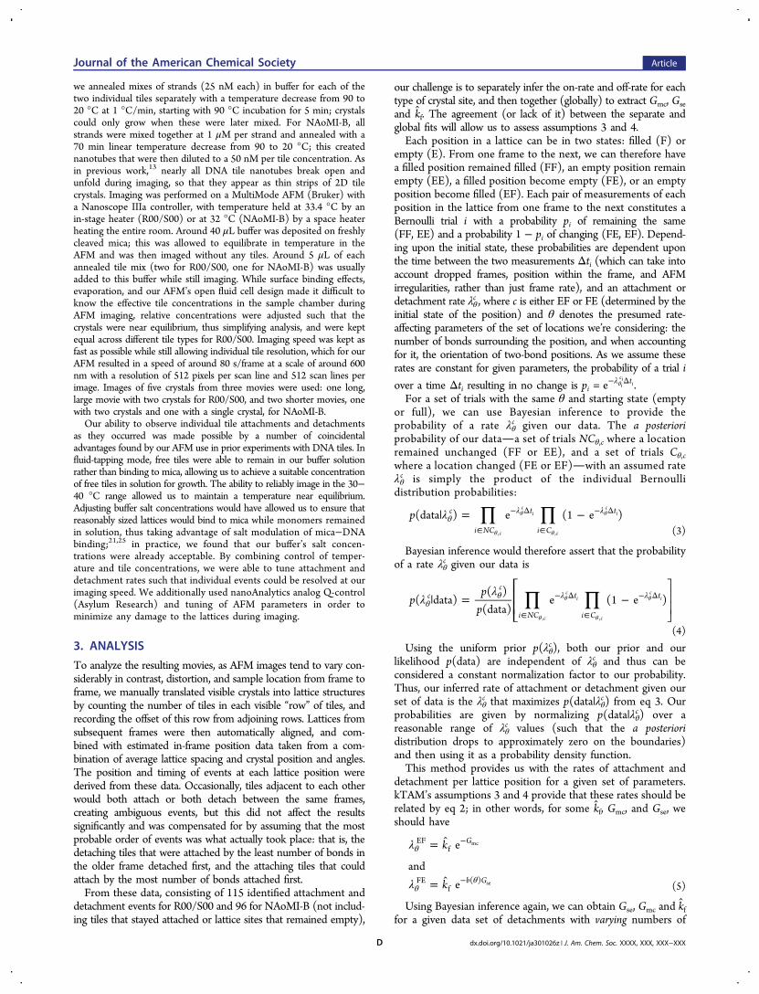
we annealed mixes of strands (25 nM each) in buffer for each of thetwo individual tiles separately with a temperature decrease from 90 to20 °C at 1 °C/min, starting with 90 °C incubation for 5 min; crystalscould only grow when these were later mixed. For NAoMI-B, allstrands were mixed together at 1 μM per strand and annealed with a70 min linear temperature decrease from 90 to 20 °C; this creatednanotubes that were then diluted to a 50 nM per tile concentration. Asin previous work,13 nearly all DNA tile nanotubes break open andunfold during imaging, so that they appear as thin strips of 2D tilecrystals. Imaging was performed on a MultiMode AFM (Bruker) witha Nanoscope IIIa controller, with temperature held at 33.4 °C by anin-stage heater (R00/S00) or at 32 °C (NAoMI-B) by a space heaterheating the entire room. Around 40 μL buffer was deposited on freshlycleaved mica; this was allowed to equilibrate in temperature in theAFM and was then imaged without any tiles. Around 5 μL of eachannealed tile mix (two for R00/S00, one for NAoMI-B) was usuallyadded to this buffer while still imaging. While surface binding effects,evaporation, and our AFM’s open fluid cell design made it difficult toknow the effective tile concentrations in the sample chamber duringAFM imaging, relative concentrations were adjusted such that thecrystals were near equilibrium, thus simplifying analysis, and were keptequal across different tile types for R00/S00. Imaging speed was kept asfast as possible while still allowing individual tile resolution, which for ourAFM resulted in a speed of around 80 s/frame at a scale of around 600nm with a resolution of 512 pixels per scan line and 512 scan lines perimage. Images of five crystals from three movies were used: one long,large movie with two crystals for R00/S00, and two shorter movies, onewith two crystals and one with a single crystal, for NAoMI-B.Our ability to observe individual tile attachments and detachments
as they occurred was made possible by a number of coincidentaladvantages found by our AFM use in prior experiments with DNA tiles. Influid-tapping mode, free tiles were able to remain in our buffer solutionrather than binding to mica, allowing us to achieve a suitable concentrationof free tiles in solution for growth. The ability to reliably image in the 30−40 °C range allowed us to maintain a temperature near equilibrium.Adjusting buffer salt concentrations would have allowed us to ensure thatreasonably sized lattices would bind to mica while monomers remainedin solution, thus taking advantage of salt modulation of mica−DNAbinding;21,25 in practice, we found that our buffer’s salt concen-trations were already acceptable. By combining control of temper-ature and tile concentrations, we were able to tune attachment anddetachment rates such that individual events could be resolved at ourimaging speed. We additionally used nanoAnalytics analog Q-control(Asylum Research) and tuning of AFM parameters in order tominimize any damage to the lattices during imaging.
3. ANALYSIS
To analyze the resulting movies, as AFM images tend to vary con-siderably in contrast, distortion, and sample location from frame toframe, we manually translated visible crystals into lattice structuresby counting the number of tiles in each visible “row” of tiles, andrecording the offset of this row from adjoining rows. Lattices fromsubsequent frames were then automatically aligned, and com-bined with estimated in-frame position data taken from a com-bination of average lattice spacing and crystal position and angles.The position and timing of events at each lattice position werederived from these data. Occasionally, tiles adjacent to each otherwould both attach or both detach between the same frames,creating ambiguous events, but this did not affect the resultssignificantly and was compensated for by assuming that the mostprobable order of events was what actually took place: that is, thedetaching tiles that were attached by the least number of bonds inthe older frame detached first, and the attaching tiles that couldattach by the most number of bonds attached first.From these data, consisting of 115 identified attachment and
detachment events for R00/S00 and 96 for NAoMI-B (not includ-ing tiles that stayed attached or lattice sites that remained empty),
our challenge is to separately infer the on-rate and off-rate for eachtype of crystal site, and then together (globally) to extract Gmc, Gseand kf. The agreement (or lack of it) between the separate andglobal fits will allow us to assess assumptions 3 and 4.Each position in a lattice can be in two states: filled (F) or
empty (E). From one frame to the next, we can therefore havea filled position remained filled (FF), an empty position remainempty (EE), a filled position become empty (FE), or an emptyposition become filled (EF). Each pair of measurements of eachposition in the lattice from one frame to the next constitutes aBernoulli trial i with a probability pi of remaining the same(FF, EE) and a probability 1 − pi of changing (FE, EF). Depend-ing upon the initial state, these probabilities are dependent uponthe time between the two measurements Δti (which can take intoaccount dropped frames, position within the frame, and AFMirregularities, rather than just frame rate), and an attachment ordetachment rate λθ
c , where c is either EF or FE (determined by theinitial state of the position) and θ denotes the presumed rate-affecting parameters of the set of locations we’re considering: thenumber of bonds surrounding the position, and when accountingfor it, the orientation of two-bond positions. As we assume theserates are constant for given parameters, the probability of a trial i
over a time Δti resulting in no change is pi =λ− Δθe t
ici
i.For a set of trials with the same θ and starting state (empty
or full), we can use Bayesian inference to provide theprobability of a rate λθ
c given our data. The a posterioriprobability of our dataa set of trials NCθ,c where a locationremained unchanged (FF or EE), and a set of trials Cθ,cwhere a location changed (FE or EF)with an assumed rateλθc is simply the product of the individual Bernoullidistribution probabilities:
∏ ∏λ| = −θλ λ
∈
− Δ
∈
− Δ
θ
θ
θ
θp(data ) e (1 e )c
i NC
t
i C
t
c
ci
c
ci
, , (3)
Bayesian inference would therefore assert that the probabilityof a rate λθ
c given our data is
∏ ∏λλ
| = −θθ λ λ
∈
− Δ
∈
− Δ
θ
θ
θ
θ
⎡
⎣⎢⎢
⎤
⎦⎥⎥p
pp
( data)( )
(data)e (1 e )c
c
i NC
t
i C
t
c
ci
c
ci
, ,
(4)
Using the uniform prior p(λθc), both our prior and our
likelihood p(data) are independent of λθc and thus can be
considered a constant normalization factor to our probability.Thus, our inferred rate of attachment or detachment given ourset of data is the λθ
c that maximizes p(data|λθc) from eq 3. Our
probabilities are given by normalizing p(data|λθc) over a
reasonable range of λθc values (such that the a posteriori
distribution drops to approximately zero on the boundaries)and then using it as a probability density function.This method provides us with the rates of attachment and
detachment per lattice position for a given set of parameters.kTAM’s assumptions 3 and 4 provide that these rates should berelated by eq 2; in other words, for some kf, Gmc, and Gse, weshould have
λ
λ
=
=
θ
θθ
−
−
k
k
e
ande
G
b G
EFf
FEf
( )
mc
se (5)
Using Bayesian inference again, we can obtain Gse, Gmc and kffor a given data set of detachments with varying numbers of
Journal of the American Chemical Society Article
dx.doi.org/10.1021/ja301026z | J. Am. Chem. Soc. XXXX, XXX, XXX−XXXD
bonds by using Gse, Gmc, and kf as parameters, using eq 5 toderive detachment rates for different θ parameters, andincluding all data in the sets NC and C:
∏
∏
λ
λ
|
=
−Δ
× − −Δ
θ
θ
∈
∈
⎡⎣⎢⎢
⎤⎦⎥⎥
p G G k
p G G kp
t G G k
t G G k
( , , data)
( , , )(data)
exp[ ( , , )]
(1 exp[ ( , , )])
i NCi
c
i Ci
c
se mc f
se mc fse mc f
se mc f
ii
ii
(6)
As we can again use a uniform prior, the probability densityfor Gse , Gmc and kf can be derived by ignoring p(ΔG°se , Gmc, kf)/p(data) and normalizing p(Gse , Gmc , kf |data) over a sufficientlylarge parameter space.In order to verify that our analysis produced accurate results
when presented with data fitting the kinetic model, the entireanalysis, with the exception of the manual data entry, wasapplied to simulated data taken from the Xgrow kTAM simu-lator.26 Parameters were chosen to match conditions in ourexperimental results, the amount of data was chosen to be similar,and the output was modified to mimic the line-scanning of anAFM. The results (Figure 4b,d) suggested our analysis methodswere sound in the face of finite scanning rates and multiple events.
4. RESULTS AND DISCUSSIONOur results, shown in Figure 4, were compiled separately forR00/S00 and NAoMI-B. Ignoring the possibility of incorrectbonds and lattice defects in the system (assumption 2), andassuming constant monomer concentrations (assumption 1),our results largely suggest that assumptions 3 (equal attach-ment rates) and 4a (detachment rates exponentially dependenton the number of bonds) of the kTAM are valid within ourexperimental error. Additionally, while observing relatively largeerror bounds owing to the smaller amount of data for eachorientation, we found that tile orientation did not alterdetachment rate beyond experimental error, and thus did notviolate assumption 4b (detachment rates dependent uponnothing but number of bonds).As for the consistency of the quantitative results, it is worth
remembering that NAoMI-B uses 6 nt sticky ends, whereasR00/S00 uses 5 nt sticky ends. Using the rough approximationthat Gse scales linearly with the number of nucleotides, thissuggests that the NAoMI-B Gse should be around 1.2 times thatof the R00/S00 Gse. Our results give a ratio of around 1.14 ±0.15, in line with this approximated expectation.There were several key differences between our exper-
imentally observed data and simulated data, many of whichlikely originate from differences in the relative numbers ofevents observed and ultimately in overall crystal “shape.” Inparticular, data from simulation, while resulting in similardetachment rates, had significantly more empty lattice locationswith three or four connecting bonds (b = 3 or b = 4). Thesesites are the result of a crystal that is, subjectively, more“spindly” and less compact than those we found experimentally,with long “arms” of tiles. Whether “spindly” crystals actuallyform is a question that would require a different experiment;however, regardless of their formation, it is quite likely thatcrystals of that shape would not cleanly bind to mica, or wouldproduce AFM images unsuitable for analysis; our experiment,therefore, inadvertently selected against crystals of this formand instead selected for relatively solid, compact crystals that
were easily imaged. In a related shape issue, the fact that imagedNAoMI-B crystals originated from nanotubes in solution meantthat all observed crystals were long, narrow ribbons, resulting inno data for short-orientation two-bond attachments, as our datanever included the ends of the crystals.Our analysis of attachment and detachment rates involved
only statistics of detachment and attachment for single tiles.The rates derived from such an analysis would ideally beindependent of crystal shape, with only the size of the errorbounds differing, owing to differences in the number of events.In some cases, however, the significantly smaller number ofevents likely resulted in anomalous observations having a largeeffect on attachment and detachment rates. For R00/S00,the attachment rate for b = 4 is calculated from only 5 events(2 empty-to-empty and 3 empty-to-full), and is significantlyhigher than expected by kTAM. For NAoMI-B, the attachmentrate for b = 3 is similarly calculated by only 10 events, and isalso significantly higher than expected. By comparison, b = 3 forR00/S00 involved 54 events, and was in line with expectations.With such events made rare by crystal shape, the chance of
the rates being affected by errors in image interpretation,anomalous imaging results, or erroneous assumptions about theorder of tile attachment is significantly increased. We expect thatthis accounts for the discrepancies seen in attachment rates, as allunexpected attachment rates have very few recorded events.In addition to the directly obtainable results, we can also give
rough estimates of other parameters. Bayesian inference sug-gested kf values of 0.024/M/s and 0.053/M/s for R00/S00and NAoMI-B, respectively. If we assume what should be areasonable order-of-magnitude estimation of a 10 nM tileconcentration, this results via eqs 1 and 2 in an α ≈ −15 andkf ≈ 106/M/s. Such a kf value is on the same order of the 6 ×10⁵ /M/s used in ref 10, but the α is very different than the+ ln(20) used there for simulations of growth in solution.10
RTα can be interpreted as a free energy adjustment thatremains constant for all attached tiles, as opposed to the ΔG°sewhich is scaled with the number of bonds. While the positive αfor growth in solution can be interpreted as due to the loss ofrotational entropy when a tile binds to the crystal, negative αcorresponds to an energetically favorable contribution to attach-ment in all cases, as could be expected for growth on mica whereall tiles involved bind to the mica surface.21
In comparing our results to other experiments, it is necessaryto convert standard free energies to our unitless Gse = ΔG°se/RT(eqs 1 and 2), and in many cases make a change in sign per ourconvention. The Gse we calculate from the statistics issignificantly lower than that found by experiments in solu-tion rather than on a mica surface; this corresponds to asignificantly “weaker” binding energy contribution for individ-ual sticky ends. In particular, Schulman et al20 measured an in-solution ΔG°2se = −9.43 ± 0.21 kcal/mol at 37 °C for twobonds, corresponding to Gse = 7.7 ± 0.17. Nangreave et al,27
using a different form of tile with 6 nt sticky ends, found a ΔG°seon the order of −3 to −6 kcal/mol, depending upon the formof attachment, resulting in a Gse of between 6 and 10.By comparison, our rates of Gse = 1.55 and Gse = 1.77 areextremely low. Indeed, a simple order of magnitude comparisonof kTAM-expected detachment rates derived from the 7.7 valueof Schulman would suggest that, were the ΔG°se in our on-micaconditions even close to so large, our entire experiment, andviewing growth with any level of detail at all, would be infeasible:the difference in rates between even tiles attached by two andthree bonds would be on the order of e−2·7.7/e−3·7.7 ≈ 2200.
Journal of the American Chemical Society Article
dx.doi.org/10.1021/ja301026z | J. Am. Chem. Soc. XXXX, XXX, XXX−XXXE

There are a number of possible factors involving DNAbinding with mica and DNA hybridization near a micasurface which could explain this discrepancy. A difference inbinding for ssDNA vs dsDNA to mica, for example, couldresult in a modified Gse for sticky ends, as two free, single-stranded sticky ends could have a stronger binding to micathan the bound, double-stranded combination of the two
ends. Also, while DNA hybridization near a mica surfacehas not been widely studied, it is reasonable to expect thatthere could be significant differences from hybridization insolution: at the very least, the “salt bridge” of divalentcations (in our case, Mg2+) between the DNA and micawould result in a very different and likely nonhomogenousionic environment.21
Figure 4. Plots of detachment and attachment rates for R00/S00 (left top) and NAoMI-B (right top) lattices. Bottom figures show simulation datawith similar parameters. Insets in detachment rate plots show activation rate/Gse parameter space, with lines representing p = 0.1, 0.5, and 0.9. Linesin attachment rate plots show mean and 90% confidence interval for Gmc.
Journal of the American Chemical Society Article
dx.doi.org/10.1021/ja301026z | J. Am. Chem. Soc. XXXX, XXX, XXX−XXXF

5. CONCLUSIONSWith this work, we have shown that we can make single-tile-resolution AFM movies of single DNA tile system crystals at asufficient frame rate to provide a consistent view of the stepsinvolved in assembly. While we have used these movies toverify some key assumptions of the kTAM, at least for growthon a surface, the technique raises the prospect of examininggrowth on a surface in general at a level that has not previouslybeen possible. For example, AFM movies could be used toexamine the growth of algorithmic tile systems, possibly allowinginsight into the mechanisms of both errors and error-reducingmethods such as proofreading15 and snaked proofreading.16
It is worth noting that if the Gse for tile detachment on a micasurface is indeed significantly lower than that in solution, as wehave found, it may significantly increase error rates for algo-rithmic tile systems grown on mica surfaces, with the kinetictrapping model10 suggesting a possible error rate of upward of30% for Gse = 1.6 and Xgrow simulations suggesting an errorrate for the binary counter system from Barish et al.6 too highto be measured. However, for the examination of themechanisms behind errors this could end up being a benefit,with the higher error rate providing significantly more data onwhat might otherwise be errors in a relatively accurate system.As algorithmic systems can often be reliant on perfect, error-free assembly, the ability to examine the systems, especiallythose that involve proofreading schemes, in an environmentthat significantly increases error rate could be vital inunderstanding how to further reduce errors.
■ ASSOCIATED CONTENT*S Supporting InformationSeveral AFM movies of nonequilibrium and equilibrium crystalgrowth:
• Movie 1: equilibrium conditions with R00/S00; 25 nMeach of R00/S00 were annealed separately, mixed in theAFM sample stage buffer during imaging, and imaged at33.4 °C; the movie has a ∼560 nm × 525 nm field ofview for 1088 s at a frame rate of ∼73 s/frame.
• Movie 2: equilibrium conditions with NAoMI-B; 50 nMof NAoMI-B was annealed and imaged at 32 °C; themovie has a ∼430 nm × 425 nm field of view for 1012 sat a frame rate of ∼126 s/frame.
• Movie 3: growth-biased conditions for R00/S00; 100 nMeach of R00 and S00-2J (S00 with added hairpins forlabeling purposes creating a striped lattice, also known asB in Figure 4a−c and Supporting Information of ref 4.)were annealed separately. Five microliters of each wasmixed in 30 μL of the AFM sample stage buffer duringimaging, and was imaged at 32 °C. The movie has a∼2 μm field of view at a frame rate of ∼70 s/frame.
• Movie 4: growth-biased conditions for NAoMI-B; ∼200 nMof NAoMI-B was annealed from 90 to 40 °C at 1 °C/min,and was then added directly to unheated buffer in the AFMand imaged at room temperature. The movie has a ∼4 μmfield of view at ∼84 s/frame. The concentration of tileswas doubled during the two frames that exhibit extensiveline artifacts.
For presentation purposes, equilibrium movie image contrastlevels were manually adjusted, with black and white being set atthe bottom of histogram peaks for surface and tile heights,respectively, and gray being set midway between the two peaks.Images were then manually shifted (but not stretched) to adjust
for AFM drift. Growth-biased movies underwent the sameprocess with the addition of manual stretching of images toadjust for AFM distortion. This material is available free ofcharge via the Internet at http://pubs.acs.org.
■ AUTHOR INFORMATION
Corresponding [email protected]
Present Address⊥Cell and Developmental Biology, University of Michigan,Ann Arbor, MI 48109-2200, United States
NotesThe authors declare no competing financial interest.
■ ACKNOWLEDGMENTSWe thank Christina Wright for prototype software for analyzingAFM movies, and Shaun Lee for discussions regarding AFMmovies of DNA tiles. We acknowledge National ScienceFoundation (NSF) Awards 0832824 (“The Molecular Pro-gramming Project”) and 0622254.
■ REFERENCES(1) Vauthey, S.; Santoso, S.; Gong, H.; Watson, N.; Zhang, S. Proc.Natl. Acad. Sci. U.S.A. 2002, 99, 5355−5360.(2) Nam, K. T.; Kim, D.-W.; Yoo, P. J.; Chiang, C.-Y.; Meethong, N.;Hammond, P. T.; Chiang, Y.-M.; Belcher, A. M. Science 2006, 312,885−888.(3) Nasalean, L.; Baudrey, S.; Leontis, N. B.; Jaeger, L. Nucleic AcidsRes. 2006, 34, 1381−1392.(4) Winfree, E.; Liu, F.; Wenzler, L. A.; Seeman, N. C. Nature 1998,394, 539−544.(5) Barish, R. D.; Rothemund, P. W. K.; Winfree, E. Nano Lett. 2005,5, 2586−2592.(6) Barish, R. D.; Schulman, R.; Rothemund, P. W. K.; Winfree, E.Proc. Natl. Acad. Sci. U.S.A. 2009, 106, 6054−6059.(7) Fujibayashi, K.; Hariadi, R.; Park, S. H.; Winfree, E.; Murata, S.Nano Lett. 2008, 8, 1791−1797.(8) Chworos, A.; Severcan, I.; Koyfman, A. Y.; Weinkam, P.;Oroudjev, E.; Hansma, H. G.; Jaeger, L. Science 2004, 306, 2068−2072.(9) Wang, H. An Unsolvable Problem on Dominoes; HarvardComputation Laboratory: Cambridge, MA, 1962.(10) Winfree, E. Simulations of Computing by Self-Assembly; CaliforniaInstitute of Technology: Pasadena, CA, 1998.(11) Rothemund, P. W. K.; Papadakis, N.; Winfree, E. PLoS Biol.2004, 2, e424.(12) Hariadi, R. F. Non-equilibrium Dynamics of DNA Nanotubes.Ph.D. Thesis; California Institute of Technology: Pasadena, CA, 2011.(13) Rothemund, P. W. K.; Ekani-Nkodo, A.; Papadakis, N.; Kumar,A.; Fygenson, D.; Winfree, E. J. Am. Chem. Soc. 2004, 126, 16344−16352.(14) Fu, T. J.; Seeman, N. C. Biochemistry 1993, 32, 3211-3220,PMID: 8461289.(15) Winfree, E.; Bekbolatov, R. In DNA Computing; Chen, J., Reif, J.,Eds.; Lecture Notes in Computer Science; Springer: Berlin/Heidelberg, 2004; Vol. 2943; pp 1980−1981.(16) Chen, H.-L.; Goel, A. In DNA Computing; Ferretti, C., Mauri,G., Zandron, C., Eds.; Lecture Notes in Computer Science; Springer:Berlin/Heidelberg, 2005; Vol. 3384; pp 702−707.(17) Schulman, R.; Winfree, E. In DNA Computing; Ferretti, C.,Mauri, G., Zandron, C., Eds.; Lecture Notes in Computer Science;Springer: Berlin/Heidelberg, 2005; Vol. 3384; pp 672.(18) Fujibayashi, K.; Murata, S. IEEE Trans. Nanotechnol. 2009, 8,361−368.(19) Chen, H.-L.; Schulman, R.; Goel, A.; Winfree, E. Nano Lett.2007, 7, 2913−2919.
Journal of the American Chemical Society Article
dx.doi.org/10.1021/ja301026z | J. Am. Chem. Soc. XXXX, XXX, XXX−XXXG
(20) Schulman, R.; Winfree, E. Proc. Natl. Acad. Sci. U.S.A. 2007, 104,15236−15241.(21) Pastre, D.; Pietrement, O.; Fusil, S.; Landousy, F.; Jeusset, J.;David, M.-O.; Hamon, L.; Le Cam, E.; Zozime, A. Biophys. J. 2003, 85,2507−2518.(22) Hamada, S.; Murata, S. Angew. Chem. 2009, 121, 6952−6955.(23) Sun, X.; Hyeon Ko, S.; Zhang, C.; Ribbe, A. E.; Mao, C. J. Am.Chem. Soc. 2009, 131, 13248-13249, PMID: 19715316.(24) Kim, B.; Amin, R.; Lee, J.; Yun, K.; Park, S. H. Chem. Commun.2011, 47, 11053−11055.(25) Bezanilla, M.; Manne, S.; Laney, D. E.; Lyubchenko, Y. L.;Hansma, H. G. Langmuir 1995, 11, 655−659.(26) The Xgrow Simulator. http://www.dna.caltech.edu/Xgrow/.(27) Nangreave, J.; Yan, H.; Liu, Y. Biophys. J. 2009, 97, 563−571.
Journal of the American Chemical Society Article
dx.doi.org/10.1021/ja301026z | J. Am. Chem. Soc. XXXX, XXX, XXX−XXXH