Embed Size (px)
Citation preview

DIPLOMARBEIT
Titel der Diplomarbeit
Interneurons in the Medial Prefrontal Cortex -
Molecular Expression Profiles, Postsynaptic Targets and
The Qest for Novel Molecular Markers
angestrebter akademischer Grad
Magister der Naturwissenschaften (Mag. rer.nat.)
Verfasser: Thomas Pollak
Matrikel-Nummer: 0103333
Studienrichtung: A490
Betreuer: Univ.-Doz.Dr. Thomas Klausberger
Wien, am 29.01.08
...dedicated to Elie Bahbah, the person I missed the most during my stay in Oxford
and to my parents, who supported me throughout my student career without
questioning what I am doing...
in memoriam of Patricia S. Goldman-Rakic, who dedicated her life to
understanding the circuitry of the prefrontal cortex

Thomas Pollak 20/01/2008
- 4 -
Content Content .............................................................................................................................4
Abstract.............................................................................................................................6
Zusammenfassung ............................................................................................................7
Introduction ......................................................................................................................8
Methods ..........................................................................................................................14
Preparing buffers ........................................................................................................14
TBS buffer ..............................................................................................................14
PB buffer ................................................................................................................14
Vibratome cutting.......................................................................................................14
Strep-Alexa488 labelling............................................................................................14
Immunoreaction..........................................................................................................15
Negative and positive controls for immunoreaction ..............................................16
Image Acquisition and Interpretation of Immunoreactions........................................20
Section preparation for Electron Microscopy.............................................................21
Freeze thaw.............................................................................................................21
DAB-reaction .........................................................................................................22
Dehydration and Uranyl acetate staining ...............................................................23
Re-embedding.............................................................................................................23
Ultra microtome cutting .............................................................................................24
Image Acquisition and Interpretation of Electron micrographs .................................25
Results ............................................................................................................................27
Immunoreaction on labelled cells...............................................................................28
PV only expressing cells ............................................................................................29
CB only expressing cells ........................................................................................30
PV/CB co-expressing cells .....................................................................................32
Other cells...............................................................................................................33
Identification of postsynaptic targets..........................................................................33
PV expressing cells.................................................................................................33
CB only expressing cells ........................................................................................35
PV/CB co-expressing cells .....................................................................................38
Other cells...............................................................................................................41

Thomas Pollak 20/01/2008
Quest for novel molecular markers............................................................................ 45
mGluR1α................................................................................................................ 46
vGLUT2................................................................................................................. 47
5-HT2c – a serotonin receptor subunit................................................................... 48
N200....................................................................................................................... 48
Kv3.1...................................................................................................................... 48
Kv4.3...................................................................................................................... 48
D1 dopamine receptor............................................................................................ 49
GluR2..................................................................................................................... 49
NO sensitive guanylyl cyclase α1 subunit............................................................. 49
CB-SM-NPY.......................................................................................................... 51
VIP ......................................................................................................................... 51
Discussion...................................................................................................................... 53
Basket cells ................................................................................................................ 53
Axo-axonic cell.......................................................................................................... 54
CB-expressing dendrite targeting cells ...................................................................... 54
Heterogeneity of mPFC interneurons ........................................................................ 55
Future directions ........................................................................................................ 56
mPFC in health and disease ....................................................................................... 57
Acknowledgement ......................................................................................................... 58
References...................................................................................................................... 60
Curiculum Vitae............................................................................................................. 68
- 5 -

Thomas Pollak 20/01/2008
Abstract During working memory/decision making tasks correlated firing between the
hippocampus and the medial prefrontal cortex (mPFC) of rats is enhanced and neurons
in the mPFC are phase-locked to hippocampal theta oscillations. In order to understand
the contribution of GABAergic interneurons of the mPFC to temporal network
organisation, the firing patterns of single interneurons in the mPFC were recorded in
anaesthetized rats and their soma, dendrites and axons specifically labelled with
neurobiotin (done by Katja Hartwich). In this diploma thesis, I have investigated the
molecular expression profile and postsynaptic targets of the recorded and labelled
interneurons. Most of the cells expressed either parvalbumin (PV), or Calbindin (CB)
or both calcium-binding proteins. Cells expressing PV only and cells expressing PV
and CB target small and apical dendrites, dendritic spines and somata of pyramidal
cells, and could therefore be identified as basket cells. In contrast, CB only expressing
cells exclusively terminate on small dendrites and spines. Additionally, an axo-axonic
cell was identified, which targets exclusively axon initial segments and does not
express PV or CB. In order to support future characterization of interneuron classes
using novel molecular markers, I have also tested several antibodies against molecules
that could further distinguish between cell classes. In conclusion, in a subset of
GABAergic interneurons in the mPFC, molecular expression profile and postsynaptic
target-specificity seems to be correlated: PV expressing, but also PV/CB co-expressing
interneurons could be identified as basket cells and CB only expressing cells as dendrite
targeting interneurons.
- 6 -

Thomas Pollak 20/01/2008
Zusammenfassung In Aufgaben, die eine Entscheidungsfindung unter Mitwirkung des
Arbeitsgedächtnisses erfordern, tritt verstärkt korreliertes Feuerungsverhalten von
Neuronen im Hippocampus und im medialen, präfrontalen Kortex (mPFC) auf und
Neurone im mPFC feuern zu einer bestimmten Phase von Theta-Oszillationen im
Hippocampus. Um zu verstehen welche Rolle Interneurone im mPFC bei diesem
Phänomen spielen, wurde das Feuerungsmuster von einzelnen Interneuronen im mPFC
aufgezeichnet und deren Soma, Dendriten und Axons mit Neurobiotin angefärbt
(durchgeführt von Katja Hartwich). In dieser Diplomarbeit habe ich die molekularen
Expressionsmuster und postsynaptischen Zielzellen dieser angefärbten Interneurone
untersucht. Der Großteil dieser angefärbten Zellen exprimiert entweder Parvalbumin
(PV) oder Calbindin (CB) oder beide dieser Calcium-bindenden Proteine. Zellen, die
nur PV exprimieren oder PV und CB co-exprimieren bilden Synapsen an kleinen und
apikalen Dendriten, dendritischen Dornen und Zellkörpern von Pyramidenzellen und
konnten daher als Korbzellen identifiziert werden. Im Gegensatz dazu bilden CB-
exprimierende Zellen Synapsen ausschließlich an kleinen Dendriten und dendritischen
Dornen von Pyramidenzellen aus. Außerdem wurde noch ein Zelltyp identifiziert, der
ausschließlich Synapsen an initialen Axonhügeln ausbildet und weder PV noch CB
exprimiert. Um zukünftig die Charakterisierung von Interneuronklassen durch neue
molekulare Marker zu unterstützen, habe ich auch diverse Antikörper getestet, die es
erlauben zwischen Zelltypen zu unterscheiden.
Als Fazit, in einer Subpopulation von GABAergen Interneuronen im mPFC scheint das
molekulare Expressionsmuster mit der Spezifität von postsynaptischen Zielzellen
korreliert zu sein: PV-exprimierende aber auch PV und CB co-exprimierende Zellen
konnten als Korbzellen und Zellen, die nur CB exprimieren, als Dendriten
innervierende Interneurone identifiziert werden.
- 7 -

Thomas Pollak 20/01/2008
Introduction Interaction of hippocampus-dependent short-term memory and neocortex-dependent
long-term memory is important for intelligent behaviour (Goldman-Rakic, 1995).
György Buzsaki (Buzsaki, 1989) put forward a model of memory consolidation in
which the animal is considered to be in a recording mode during exploratory behaviour.
This recorded information is played back in temporally compressed form during
subsequent periods of inactivity and sleep in the prefrontal cortex (Euston et al., 2007),
the hippocampus (Diba and Buzsaki, 2007) and the visual cortex (Ji and Wilson, 2007),
whereas the latter might account for vivid images during dreaming. Periodic playback
could promote a gradual integration of newly acquired knowledge into the neocortex
(Wiltgen et al., 2004). A gradual integration is important, because a rapid incorporation
of new information into a pre-existing knowledge system could cause interference and
previously acquired information would be erased (McClelland, 1995).
Once the information is consolidated in the neocortex, it becomes independent of the
hippocampus. For subsequent retrieval and integration with newly acquired information
stored in the hippocampus, the prefrontal cortex (PFC) plays a central role (Wiltgen et
al., 2004). This integration process is referred to as working memory and can be
described as retaining acquired knowledge for an impending action, which is dependent
on that knowledge (Fuster, 2001). In delayed response tasks the functioning of working
memory can be elucidated and it was shown that in rats with lesions in the
prelimbic/infralimbic medial PFC, working memory was impaired (Gisquet-Verrier and
Delatour, 2006).
The rodent PFC is divided into medial, lateral and orbital parts with the medial part
(mPFC) consisting of four sub-divisions (Ongur and Price, 2000). The most dorsal
divisions are the medial (frontal) agranular and the anterior cingulate cortices, which
receive sensorimotor input and are associated with motor behaviour. The prelimbic
(PL) and infralimbic (IL) cortices make up the medio-ventral portion of the mPFC,
receive limbic input from the hippocampus, subiculum and amygdala and are involved
in emotional, cognitive and mnemonic processes (Heidbreder and Groenewegen, 2003).
Furthermore, these brain areas are highly interconnected among themselves (Vertes,
2006). The functional analogue to the PL in primates is the dorsolateral PFC (Vertes,
- 8 -

Thomas Pollak 20/01/2008
2006) and it was reported that lesions of this brain area disrupt performance on delayed
response tasks in monkeys (Goldman and Rosvold, 1970).
The mPFC receives input from various brain regions, including the hippocampus.
Retrograde tracing has shown that PL and IL of rats are targeted by glutamatergic
afferents of the CA1 / subiculum (Hoover and Vertes, 2007) – the major output areas of
the hippocampal formation. This is consistent with anterograde tracing studies (Jay and
Witter, 1991) and has also been shown in primates with both retrograde and
anterograde tracing (Goldman-Rakic et al., 1984). Not only the mPFC but also this
Hippocampo-mPFC pathway seems to be important for working memory as it was
shown that rats with a transient inactivation of this pathway were impaired in a delayed
choice of a radial-arm maze task (Floresco et al., 1997).
The cerebral cortex contains two main types of neurons, excitatory pyramidal cells
(DeFelipe and Farinas, 1992), which are the vast majority (70-80 %) and are relatively
uniform, and interneurons, which are mostly inhibitory and have diverse
morphological, physiological, molecular and synaptic properties (Kawaguchi and
Kubota, 1997). Single pulse stimulation of the hippocampus in anaesthetized rats
induced EPSPs in mPFC pyramidal neurons of fixed latency, suggesting a
monosynaptic connection, followed by prolonged IPSPs (Degenetais et al., 2003).
Afferent projections from CA1 / subiculum not only target pyramidal cells of the mPFC
(Carr and Sesack, 1996), but also interneurons (Gabbott et al., 2002). So whilst the
IPSP could result from innervation of interneurons by these activated PFC pyramidal
cells through a feedback process, it might also result from interneurons being excited
directly by CA1 projections. Indeed, a large proportion of recorded mPFC interneurons
showed an excitatory response with a latency consistent with the conduction time of the
hippocampo-mPFC pathway, suggesting a monosynaptic connection (Tierney et al.,
2004). However, recorded and juxtacellularly labelled interneurons were not
characterised further in this study. Traditionally neurons have been classified by their
morphology (Ramon y Cajal, 1911), but because this is a highly variable feature of
neurons, morphology alone cannot reliably define a cell type (Markram et al., 2004).
It has been suggested (Somogyi and Klausberger, 2005) that cortical neurons can be
characterized and defined on application of following measures: (I) brain area- and cell
domain-specific distribution of presynaptic inputs and postsynaptic targets, (II)
expression profiles of signalling molecules and ion channels, and (III) in vivo firing
patterns.
- 9 -
Thomas Pollak 20/01/2008
Interneurons can be classified by investigating their postsynaptic targets and can be
functionally divided into axo-axonic cells targeting exclusively axon initial segments,
soma- and proximal dendrite-targeting basket cells, proximal dendrite-targeting, and
distal dendrite- and spine-targeting interneurons (Somogyi et al., 1998). Axo-axonic
cells have their boutons aligned along axon initial segments and can thus be recognized
in some cases on the light microscopic level. On the other hand, axons of basket cells,
which are soma- and proximal dendrite-targeting cells, show little directivity in their
orientation in close proximity to their somata (Somogyi and Klausberger, 2005).
Furthermore, there are subpopulations of interneurons that target preferentially other
interneurons (Acsady et al., 1996).
Molecules such as calcium binding proteins Parvalbumin (PV), Calbindin (CB), or
Calretinin (CR), or neuromodulators like neuropeptide Y (NPY), Somatostatin (SM),
Cholecystokinin (CCK), vasoactive intestinal peptide (VIP) are known to be
differentially expressed in subpopulations of interneurons in the hippocampus
(Somogyi and Klausberger, 2005). However, it has been demonstrated in the
hippocampus that the expression of a single molecule cannot define a distinct class of
interneurons as defined by synaptic connectivity, because most molecules are expressed
by several classes. For example, parvalbumin is expressed by basket, axo-axonic,
bistratified and oriens-lacunosum moleculare (O-LM) cells (Somogyi and Klausberger,
2005). In order to further characterize interneuron classes, novel molecular markers
were investigated and potential candidates are described below.
The mPFC receives dopaminergic input from the ventral tegmental area (VTA) and it
was shown that working memory performance was at its best with moderate stimulation
of D1 family dopamine receptors (Muly et al., 1998). Dopamine D1 receptors are
expressed by both pyramidal cells and GABAergic interneurons (Muly et al., 1998);
most PV-expressing and 10 % of CB-expressing interneurons express D1 as shown by
in situ hybridization (Le Moine and Gaspar, 1998). Furthermore, the effect of dopamine
on inhibition dominates over that on excitation (Bandyopadhyay and Hablitz, 2007) and
the PFC in turn acts through various pathways to excite or inhibit dopaminergic
neurons in the VTA (Gao et al., 2007). Because of the importance of dopaminergic
input for reinforcement learning mechanisms (Hazy et al., 2006), it would be interesting
to investigate, which subpopulations of PV- and/or CB-expressing interneurons also
expresses the dopamine D1 receptor.
- 10 -
Thomas Pollak 20/01/2008
The mPFC also interacts with the thalamus especially during sleep, which leads to the
generation of spindle oscillations (7-14 Hz) (Steriade and Amzica, 1998). Furthermore,
afferents from the thalamus express the vesicular glutamate transporter type 2
(vGLUT2), whereas cortical terminals either express vGLUT1 or a minor
subpopulation of cortical terminals co-express both types (Kubota et al., 2007). It
would be interesting to investigate, whether a subpopulation of PV-expressing cells
receives more thalamic input than others.
Neurons in the mPFC are also modulated by Serotonin and in fact, the 2c subunit of the
serotonin receptor (5-HT2c) is expressed particularly by neurons in deep layers of the
PL area and 50 % of these neurons were immunopositive for glutamate decarboxylase
(GAD67) (Liu et al., 2007a), the enzyme that converts glutamate into γ-aminobutyric
acid (GABA). Furthermore, this receptor subunit is predominantly expressed by PV-
expressing interneurons and to a significantly lesser extant in CB- or Calretinin-
expressing cells (Liu et al., 2007a), so it would be interesting to see which
subpopulation of PV-expressing cells also expresses 5-HT2c.
A subset of PV-expressing cells (26 %) receives about five times more boutons
immunopositive for VIP than the rest of PV-expressing cells (David et al., 2007), but it
is not known which subpopulation receives more input.
It was shown that the α1 subunit of a nitric oxide (NO)-sensitive guanylyl cyclase is
specifically expressed in interneurons, but only in a subpopulation of PV-positive cells
(75%) in the hippocampus (Szabadits et al., 2007). Nitric oxide contributes to synaptic
plasticity as a retrograde messenger and this enzyme plays an important role in this
pathway. This enzyme could therefore be used as a molecular marker for a
subpopulation of PV-expressing cells.
Voltage-gated potassium channels comprise the most divers group of heteromeric ion
channels discovered so far. There are more than 20 genes encoding these channels and
examples of interneuron specific potassium channels are Kv3.1 (Sekirnjak et al., 1997)
and Kv4.3 (Serodio and Rudy, 1998), Kv3.1 is co-expressed with PV, but not found in
other subpopulations of interneurons (Chow et al., 1999). For Kv4.3 on the other hand,
a highly uneven subcellular distribution of this potassium channel was reported (Kollo
et al., 2006). Both of these channels might be usable as a molecular marker for
characterization of cell types.
- 11 -

Thomas Pollak 20/01/2008
Another type of proteins that are differentially expressed in subsets of neurons are
neurofilaments. For example, N200 is expressed in a subpopulation of pyramidal cells
in the prefrontal cortex (Law and Harrison, 2003), but it would also be interesting to
show whether these neurofilaments are also expressed in certain types of interneurons.
The metabotropic glutamate receptor subtype 1α (mGluR1α) has been associated with
a variety of contributions to network activity, including modulation of synaptic
transmission and plasticity and is predominantly expressed by interneurons of the
hippocampal CA1 area. In particular, only a small subset of PV-expressing cells –
mostly those, which also express SM - co-express mGluR1α (Ferraguti et al., 2004).
This molecular marker could be employed to differentiate subpopulations of PV-
expressing cells.
Network oscillations of various frequency bands in the brain provide a time frame and
temporal coordination within and between distinct brain areas. Theta oscillations (4-8
Hz), for example, occur during exploratory behaviour and rapid-eye-movement sleep
(Buzsaki, 2002) and are considered to be the online state of the hippocampus.
Furthermore, during sleep the thalamus interacts with the mPFC, which leads to the
generation of spindle oscillations (7-14 Hz) during slow wave sleep (0.5-4 Hz)
(Steriade and Amzica, 1998).
It has been shown (Siapas et al., 2005) that in freely behaving rats mPFC neurons are
phase-locked to hippocampal theta oscillations with a delay of approximately 50 ms
suggesting that hippocampal activity is leading neural activity in the mPFC.
Furthermore, mPFC neurons exhibit phase precession to the hippocampal theta rhythm
(Jones and Wilson, 2005a), which indicates that the cell discharges at earlier phases of
consecutive theta cycles (O'Keefe and Recce, 1993). Moreover, correlated firing of
neurons in the hippocampus and the mPFC is selectively enhanced during behaviour
that involves working memory (Jones and Wilson, 2005b).
As it has been shown that interneurons during theta oscillations coordinate the temporal
activity of pyramidal cells in the hippocampus (Klausberger et al., 2003), interneurons
in the mPFC might therefore play an important role for spike timing and phase-locking
of mPFC neurons. Hence, Katja Hartwich in our lab has been investigating interneurons
in the PL area of the mPFC in the context of hippocampal theta oscillations in
anaesthetized rats. In order to do so, single interneurons were extracellularly recorded
simultaneous with the local field potential (LFP) in the mPFC and the LFP in the CA1
- 12 -

Thomas Pollak 20/01/2008
region of the hippocampus and subsequently juxtacellularly labelled with neurobiotin.
With the juxtacellular labelling method single-cells can be labelled specifically by
using a glass electrode filled with Neurobiotin and injecting positive current pulses. The
cell modulates its firing to these pulses and takes up the Neurobiotin, which distributes
throughout the cell including dendrites and axon (Figure 1, (Pinault, 1996).
Figure 1: modified from Pinault (1996). The left panel shows extracellular recording from a single cell with a glass electrode filled with neurobiotin from which the local field potential (frequency band up to 200 Hz; top trace) and the in vivo-firing pattern of the cell depicted (high frequency: 0.8-5 kHz; bottom trace) can be extracted. Note that the anaesthetized rat was pinched in the foot (red arrow indicating the time point), which causes a change from slow wave oscillations (0.5-4 Hz) to theta oscillations (4-8 Hz). The right panel illustrates the labelling procedure, where the glass electrode was lowered very closely to the cell and neurobiotin was ejected together with a current pulse. If the cell is modulated to the current pulse, it takes up the ejected neurobiotin.
In order to determine the synaptic connectivity, molecular expression profile and
neuronal classes of these labelled cells, I have tested their expression of various
molecular markers and identified their postsynaptic targets with electron microscopy.
- 13 -

Thomas Pollak 20/01/2008
Methods
Preparing buffers
TBS buffer
For 1 l of buffer (pH 7.4) 6.61 g TRIZMA Hydrochloride (Sigma-Aldrich Inc., St.
Louis, USA, T-3253), 0.97 g TRIZMA Base (Sigma-Aldrich Inc., St. Louis, USA, T-
1503), 9 g NaCl (BDH Laboratory Supplies, Poole, UK, 44827W) were dissolved in
ddH2O and filled up to an end volume of 1 l.
PB buffer
For 5 l of 0.2 M PB buffer 142.39 g Na2HPO4 x 2H2O (BDH Laboratory Supplies,
Poole, UK, 103834G) were dissolved in 4 l of dH20. 27.6 g NaH2PO4 x H2O were
dissolved in 1 l of dH2O, mixed with the other solution and the pH was confirmed to be
below 7.39.
Vibratome cutting
The rat was perfused through the aorta with 0.9 % saline followed by fixative (4 %
Paraformaldehyde + 15 % w/v of saturated Picric acid + 0.05 % Glutaraldehyde in 0.1
M PB, pH 7.3) for approximately 20 minutes. The brain was removed from the skull
and was washed over night in 0.1 M PB. Then the frontal part of the right hemisphere
was cut on the vibratome (Leica, Wetzlar, Germany, VT 1000S) in 70 µm thick coronal
sections. The sections were washed twice in 0.1M PB for 15 minutes on the shaker and
stored in 0.1M PB + 0.05 % Sodium Azide (VWR International Ltd., Poole, England,
103692K) at 4ºC (Maccaferri et al., 2000).
Strep-Alexa488 labelling
Sections stored in 0.1 M PB + 0.05 % Sodium Azide (VWR International Ltd., Poole,
England, 103692K) were washed twice in 0.1 M PB and once in TBS + 0.3 % Triton
X-100 (VWR International Ltd., Poole, England, 306324N) for 20 min on the shaker at
room temperature. After that they were incubated with StrepAlexa488 (1:1000) in TBS
+ 0.3 % Triton X-100 + 2 % normal horse serum for 4 hours on the shaker at room
temperature (or alternatively 24 hours at 4ºC, although this was abandoned during the
- 14 -

Thomas Pollak 20/01/2008
course of the thesis because it resulted in higher unspecific background labelling). The
sections were washed three times with TBS + 0.3 % Triton X-100 for 20 min on the
shaker at room temperature and mounted on microscopic slides (VWR International
bvba, Leuven, Belgium, 631-0117) with vectashield mounting medium for fluorescence
(Vector Laboratories Inc, Burlingame, USA, H-1000) and covered with a cover slip
(VWR International bvba, Leuven, Belgium, 631-0133) (Losonczy et al., 2002).
Immunoreaction
Sections stored in 0.1 M PB + 0.05 % Sodium Azide (VWR International Ltd., Poole,
England, 103692K) were washed twice in 0.1 M PB and once in TBS + 0.3 % Triton
X-100 (VWR International Ltd., Poole, England, 306324N) for 20 min on the shaker at
room temperature. The sections were blocked by incubating them in 20 % normal horse
serum (Vector Laboratories Inc, Burlingame, USA, S-2000) in TBS + 0.3 % Triton X-
100 for one hour on the shaker at room temperature. They were then incubated with the
primary antibodies (see
- 15 -

Thomas Pollak 20/01/2008
Table 1 for concentration) in TBS + 0.3 % Triton X-100 + 2 % normal horse serum for
48 hours on the shaker at 4ºC.
After washing the section three times with TBS + 0.3 % Triton X-100 for 20 min on the
shaker at room temperature they were incubated with the secondary antibodies (Table 2) in TBS + 0.3 % Triton X-100 + 2 % normal horse serum for 24 hours on the shaker
at 4ºC. The section were washed three times with TBS + 0.3 % Triton X-100 for 20 min
on the shaker at room temperature and mounted on microscopic slides (VWR
International bvba, Leuven, Belgium, 631-0117) with vectashield mounting medium for
fluorescence (Vector Laboratories Inc, Burlingame, USA, H-1000) and covered with a
cover slip (VWR International bvba, Leuven, Belgium, 631-0133) (Losonczy et al.,
2002).
Negative and positive controls for immunoreaction
Antibody labelling was considered to be specific, if it was comparable with labelling
previously published for this molecular marker (see
- 16 -

Thomas Pollak 20/01/2008
Table 1 for references). Some antibodies that were tested for the first time (including
vGLUT2, 5-HT2c, Kv4.3 and D1) showed unspecific labelling and therefore negative
and positive controls were applied to verify the reason for unspecific labelling. First of
all, the secondary antibodies used for these reactions were tested without primary
antibody, to verify whether the unspecific labelling stems from the secondary antibody.
Additionally, all primary antibodies were tested with two different secondary antibodies
(Alexa488 and Cy3). Furthermore, each species of secondary antibody was used in
combination with primary antibodies, often used in the lab and known to give strong
and characteristic labelling as a positive control. In all cases secondary antibodies did
not show any labelling after omitting primary antibodies, different secondary antibodies
showed the same result in combination with the same primary antibody, and all positive
controls showed specific labelling.
In order to avoid shine through – labelling can be observed in a channel with a filter of
longer wavelength – the primary and secondary antibodies should not be used too
concentrated (ideal concentration of antibodies has to be evaluated for each antibody
individually). Furthermore, also the Strep-Alexa488 labelling should not shine through
in other channels and therefore after labelling images are captured with all channels as
a negative control, before any immunoreaction are performed on the section.
- 17 -

Thomas Pollak 20/01/2008
Table 1: List of primary antibodies used in this study.
Antibody to
Host Dilution Source Reference of
characterisation Immunogens
α-actinin mouse 1:1000 Sigma-Aldrich, St. Louis, Missouri,
USA. Code No. A7811
Labelling pattern
as published with
other antibodies.
sarcomeric α-
actinin
rabbit 1:1000 Dr. A. Beggs, Howard Hughes
Medical Institute, Boston, USA. (Chan et al., 1998)
recombinant α-
actinin-2 (N-17)
5-HT2c goat 1:500
Santa Cruz Biotechnology, Inc.
Santa Cruz, CA, USA. Code No.
sc-15081
(Liu et al., 2007b) SR-2C (N-19)
CB rabbit 1:10000 Swant, Bellinzona, Switzerland.
Code No. CB-38
Labelling pattern
as published with
other antibodies.
rat CB D-28k
mouse 1:500 Swant, Bellinzona, Switzerland.
Code No. 300 (Celio et al., 1990)
chicken CB D-
28k
CCK rabbit 1:500 DiaSorin, Stillwater, USA. Code
No. 20078
Labelling pattern
as published with
other antibodies.
CCK
octapeptide
mouse 1:5000
Dr. G Ohning, CURE/Digestive
Diseases Research Center.
Antibody/RIA Core, UCLA. Code
No. 9303
(Ohning et al.,
1996) gastrin-17
COUP-TFII
mouse 1:250 Perseus Proteomics Inc., Tokyo,
Japan. Code No. PP-H7147-00 (Lee et al., 2004)
human COUP-
TF2
CR goat 1:1000 Swant, Bellinzona, Switzerland.
Code No CG1
(Schwaller et al.,
1994) human CR
D1 rat 1:5000 Sigma-Aldrich, St. Louis, Missouri,
USA. Code No. D187
(Paspalas and
Goldman-Rakic,
2005)
C-terminal 97
aa of human
D1 receptor
GAD mouse 1:100
Dr. D. Gottlieb, Dept. of Anatomy &
Neurobiology, Washington
University School of Medicine, St.
Louis, USA
(Chang and
Gottlieb, 1988)
rat brain GAD
(65kDa)
GCα1 rabbit 1:10000 Sigma-Aldrich, St. Louis, Missouri,
USA. Code No. G4280
(Szabadits et al.,
2007)
synthetic
peptide (673-
690) of rat GC α1
GluR2 mouse 1:100 Antibodies Inc., Davis, CA, USA.
Code No. 75-002
Labelling pattern
as published with
other antibodies.
(Fujiyama et al.,
2004)
monoclonal,
clone L21/32
- 18 -

Thomas Pollak 20/01/2008
Kv4.3 rabbit 1:1000 Alomone Labs Ltd. Jerusalem,
Israel. Code No. APC-017A (Yang et al., 2001)
Human Kv4.3
451-467
goat 1:1000
Santa Cruz Biotechnology, Inc.
Santa Cruz, CA, USA. Code No.
sc-11686
(Kollo et al., 2006)
peptide of
internal region
of human Kv4.3
mGluR1α Guinea
pig 1:1000
Dr. M. Watanabe, Department of
Anatomy, Hokkaido University
School of
Medicine, Sapporo, Japan
(Tanaka et al.,
2000)
rat mGluR1α
(945-1127)
MOR guinea
pig 1:1000
Chemicon, Hampshire, United
Kingdom. Code No. AB1774
(Rodriguez et al.,
2001)
synthetic
peptide (384-
398) from rat
MOR-1
N200 mouse 1:1000 Sigma-Aldrich, St. Louis, Missouri,
USA. Code No. 0142 N52
(Law and Harrison,
2003)
Phosphorylated
and non-
phosphorylated
N200
NOS rabbit 1:1000 Chemicon, Hampshire, United
Kingdom. Code No. AB5380
Labelling pattern
as published with
other antibodies.
(Moro et al., 1998)
recombinant
human nNOS
sheep 1:500 Chemicon, Hampshire, United
Kingdom. Code No. AB1529
Labelling pattern
as published with
other antibodies.
rat NOS (1409-
1429)
NPY sheep 1:500 Chemicon, Hampshire, United
Kingdom. Code No. 1017
Labelling pattern
as published with
other antibodies.
(Blessing et al.,
1986)
PV mouse 1:10000 Swant, Bellinzona, Switzerland.
Code No. 235 (Celio et al., 1988) carp muscle PV
Reelin mouse 1:1000 Chemicon, Hampshire, United
Kingdom. Code No. MAB5364
(Demyanenko et
al., 2004)
Recombinant
reelin amino
acids 164-496
SOM rat 1:500 Chemicon, Hampshire, United
Kingdom. Code No. MAB354
Monoclonal,
labelling pattern as
published with
other antibodies,
(Vincent et al.,
1985)
synthetic 1-14
cyclic SOM
vGLUT2 guinea
pig 1:2500
Chemicon, Hampshire, United
Kingdom. Code No. AB5907
Labelling pattern
as published with
other antibodies,
(Fremeau et al.,
Synthetic
peptide from rat
vGLUT2
- 19 -

Thomas Pollak 20/01/2008
2001)
VIP rabbit 1:50,000 Dr. TJ Görcs, Dept. Anatomy, Sote,
Hungary
(Gulyas et al.,
1990)
synthetic
porcine VIP
Table 2: List of secondary antibodies used in this study
Antibody/Streptavidin Fluorophore Amax Supplier
Donkey Anti-Goat Alexa 350 346nm Molecular Probes, Inc., Eugene, OR, USA
Donkey Anti-Sheep Alexa 350 346nm Molecular Probes, Inc., Eugene, OR, USA
Donkey Anti-Mouse IgG AMCA 350nm Jackson Immuno Research Laboratories, Inc.,
West Grove, PA, USA
Streptavidin AMCA 350nm Jackson Immuno Research Laboratories, Inc.,
West Grove, PA, USA
Streptavidin Alexa488 495nm Molecular Probes, Inc., Eugene, OR, USA
Donkey Anti-Goat Alexa488 495nm Molecular Probes, Inc., Eugene, OR, USA
Donkey Anti-Guinea Pig Alexa488 495nm Molecular Probes, Inc., Eugene, OR, USA
Donkey Anti-Mouse Alexa488 495nm Molecular Probes, Inc., Eugene, OR, USA
Donkey Anti-Rabbit Alexa488 495nm Molecular Probes, Inc., Eugene, OR, USA
Donkey Anti-Mouse Cy3 550nm Jackson Immuno Research Laboratories, Inc.,
West Grove, PA, USA
Donkey Anti-Guinea Pig Cy3 550nm Jackson Immuno Research Laboratories, Inc.,
West Grove, PA, USA
Donkey Anti-Rabbit Cy3 550nm Jackson Immuno Research Laboratories, Inc.,
West Grove, PA, USA
Donkey Anti-Rat Cy3 550nm Jackson Immuno Research Laboratories, Inc.,
West Grove, PA, USA
Donkey Anti-sheep Cy3 550nm Jackson Immuno Research Laboratories, Inc.,
West Grove, PA, USA
Donkey Anti-Goat Cy3 550nm Jackson Immuno Research Laboratories, Inc.,
West Grove, PA, USA
Donkey Anti-Guinea Pig Cy5 650nm Jackson Immuno Research Laboratories, Inc.,
West Grove, PA, USA
Donkey Anti-Goat Cy5 650nm Jackson Immuno Research Laboratories, Inc.,
West Grove, PA, USA
Donkey Anti-Mouse Cy5 650nm Jackson Immuno Research Laboratories, Inc.,
West Grove, PA, USA
Image Acquisition and Interpretation of Immunoreactions
The immunoreaction was examined with a Leitz fluorescence microscope (Leica,
Wetzlar, Germany, DMRB) with the following filter blocks: A4 (excitation filter band
pass (BP), 360/40nm; reflection short-pass (SP) filter, 400nm: suppression filter BP,
- 20 -

Thomas Pollak 20/01/2008
470/40nm), L5 (excitation filter BP, 480/40nm; reflection SP, 505nm; suppression filter
BP, 527/30nm), Y3 (excitation filter BP, 545/30 nm; reflection SP, 565 nm;
suppression filter BP, 610/75 nm), and Y5 (excitation filter BP, 620/60nm; reflection
SP, 660 nm; suppression filter BP, 700/75 nm). Images were acquired with a CCD
camera (Hamamatsu Photonics, Welwyn Garden City, UK, C4742-80) using the
software package openlab 5.0.2 (Improvision, Coventry, UK) (Ferraguti et al., 2004).
For each image the exposure time had to be adjusted depending on the reaction quality,
the objective used and the channel (A4: 500-2000ms, L5: 200-500 ms, Y3: 20-150 ms,
Y5: 1000-3500 ms). If images had to be taken from more than two channels, a macro
programmed by Wai-Yee Suen was used.
In order to show whether a neurobiotin-labelled cell expresses a certain marker or not, a
co-localisation study with sections containing either soma, dendrites, or axons of the
labelled cell was performed. For soma sections, images with at least three different
focuses were acquired for each channel. For co-localisation in dendrites, images were
taken of at least three individual dendrites of the same cell with as many focuses
required for an unequivocal judgment. For co-localisation in axons, images were
acquired from at least ten boutons of at least 3 different axons. If there was a co-
localisation, the cell was called “immuno-positive” for this marker. If no co-localisation
was observed with the tested marker, the cell was called “immuno-negative”, although
weak expression cannot be excluded with this method. In case of ambiguous results, the
cell was called “not tested” for this marker.
Section preparation for Electron Microscopy
Freeze thaw
Sections containing axons of neurobiotin-labelled cells, which have not been used for
immunofluorescence and were stored in 0.1 M PB + 0.05 % Sodium Azide (VWR
International Ltd., Poole, England, 103692K) were washed four times for 15 minutes in
0.1 M PB on the shaker at room temperature. Then they were incubated for 15 minutes
in 0.1 M PB + 10 % Sucrose (Sigma-Aldrich Inc., St. Louis, USA, S7903-1KG) and for
at least 2 hour in 0.1 M PB + 20 % Sucrose or until they sank to the bottom of the well
without pushing them down. Afterwards, the sections, one by one, were transferred into
a well of a TC-plate six well (Greiner bio-one Ltd., Stonehouse, UK, 657 160) and any
remaining buffer discarded. The plate was dipped into liquid nitrogen until the section
- 21 -

Thomas Pollak 20/01/2008
was frozen as indicated by turning into a white colour. The section was then thawed as
quickly as possible by pouring 0.1 M PB (room temperature) into the well and rubbing
with a finger underneath the section. Then the sections were washed twice with 0.1 M
PB and once with TBS for 15 minutes at room temperature on the shaker. Meanwhile,
component A and component B of the Vectastain ABC kit elite (Vector Laboratories
Inc, Burlingame, USA, PK-6100) were mixed both 1:100 in TBS and pre-incubated for
30 minutes at room temperature. The sections were incubated in the pre-incubated
mixture for 48 hours at 4ºC on the shaker.
DAB-reaction
In order to prepare the DAB solution, 10 mg of DAB (3,3’-Diaminobenzidine tetra-
hydrochloride, approx. 97 %, Sigma-Aldrich Inc., St. Louis, USA, D5637-1G) were
dissolved in 9.6 ml distilled water and mixed with 10 ml 0.2 M PB, 200 µl of 20 %
Glucose (BDH Laboratory Supplies, Poole, UK, 10117) in distilled water + 200 µl of
0.4 % Ammonium Chloride (Sigma-Aldrich Inc., St. Louis, USA, A-4514-100G) in
distilled water. The solution was divided in two portions and one was used to pre-
incubate the sections in 0.5 ml per well of a TC-plate 24 well (Greiner bio-one Ltd.,
Stonehouse, UK, 662 160) for 15 minutes at room temperature in the fume hood (all
following steps were carried out in the fume hood as well). To the other portion, 15 µl
of Glucose-oxidase (0.00542 g / ml, Sigma-Aldrich Inc., St. Louis, USA, G-2133) were
added. After pre-incubation, the sections were incubated with the DAB solution
containing Glucose-oxidase at room temperature for approximately 45 minutes,
although the progress of the reaction had to be monitored carefully and checked
regularly under the microscope. This glucose oxidase reaction was used to produce
H2O2 in situ, which is required for the DAB reaction. To stop the reaction the sections
were washed three times in 0.1 M PB at room temperature. The sections were then
incubated for 45 minutes in Osmium tetroxide (4 % w/v solution, TAAB Laboratories
Equipment Ltd., Reading, UK, O012) with a concentration between 0.5 to 2 % in 0.1 M
PB according to the colour and the level of unspecific background labelling of the
section after the DAB reaction. The sections were washed twice in 0.1 M PB for ten
minutes and three times rinsed with distilled water in order to remove phosphate, which
would form a precipitate with uranium in subsequent steps.
- 22 -
Thomas Pollak 20/01/2008
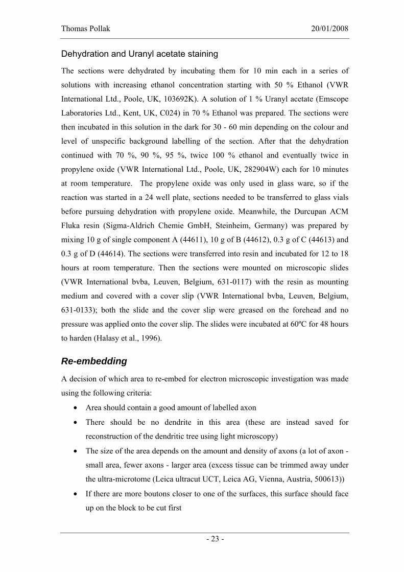
Dehydration and Uranyl acetate staining
The sections were dehydrated by incubating them for 10 min each in a series of
solutions with increasing ethanol concentration starting with 50 % Ethanol (VWR
International Ltd., Poole, UK, 103692K). A solution of 1 % Uranyl acetate (Emscope
Laboratories Ltd., Kent, UK, C024) in 70 % Ethanol was prepared. The sections were
then incubated in this solution in the dark for 30 - 60 min depending on the colour and
level of unspecific background labelling of the section. After that the dehydration
continued with 70 %, 90 %, 95 %, twice 100 % ethanol and eventually twice in
propylene oxide (VWR International Ltd., Poole, UK, 282904W) each for 10 minutes
at room temperature. The propylene oxide was only used in glass ware, so if the
reaction was started in a 24 well plate, sections needed to be transferred to glass vials
before pursuing dehydration with propylene oxide. Meanwhile, the Durcupan ACM
Fluka resin (Sigma-Aldrich Chemie GmbH, Steinheim, Germany) was prepared by
mixing 10 g of single component A (44611), 10 g of B (44612), 0.3 g of C (44613) and
0.3 g of D (44614). The sections were transferred into resin and incubated for 12 to 18
hours at room temperature. Then the sections were mounted on microscopic slides
(VWR International bvba, Leuven, Belgium, 631-0117) with the resin as mounting
medium and covered with a cover slip (VWR International bvba, Leuven, Belgium,
631-0133); both the slide and the cover slip were greased on the forehead and no
pressure was applied onto the cover slip. The slides were incubated at 60ºC for 48 hours
to harden (Halasy et al., 1996).
Re-embedding
A decision of which area to re-embed for electron microscopic investigation was made
using the following criteria:
• Area should contain a good amount of labelled axon
• There should be no dendrite in this area (these are instead saved for
reconstruction of the dendritic tree using light microscopy)
• The size of the area depends on the amount and density of axons (a lot of axon -
small area, fewer axons - larger area (excess tissue can be trimmed away under
the ultra-microtome (Leica ultracut UCT, Leica AG, Vienna, Austria, 500613))
• If there are more boutons closer to one of the surfaces, this surface should face
up on the block to be cut first
- 23 -

Thomas Pollak 20/01/2008
Pictures were then taken of the selected area of interest and the cover slip was removed
by using a double-edged razor blade (Wilkinson Sword).
A droplet of Durcupan ACM Fluka resin (Sigma-Aldrich Chemie GmbH, Steinheim,
Germany) (usually leftover resin from embedding, which was stored in the frigde and
can be reused for a few weeks for this purpose only) was put in the lid of the
embedding capsule (TAAB Laboratories equipment limited, Aldermaston, UK,
MS9051). Using a surgical blade type 26 (Swann-Morton, Sheffield, UK, 0213) the
first cut into the tissue was made in an area, away from the selected area of interest in
order to see how the tissue was cutting. In some cases the tissue was very brittle in
which case the slide was placed on a heating plate (Agar scientific Ltd., Stansted, UK)
at 60°C for a couple of minutes to soften the resin. The area was then cut out and
transferred to the droplet of resin with the preferred surface face down. An antistatic
gun Zerostat (Discwasher, England, 3997817) was used with all metal equipment, to
prevent the loss of the cut piece (usually around 1 mm2) due to electrostatic forces.
Another security measure was to put a finger close to the area of cutting. If the piece
would jump, it would often be trapped on the fingertip. The embedding capsule was
then labelled with a number, filled up with resin, covered with a cover slip and
incubated at 60°C for 48 hours to harden. After removing the cover slip and the
embedding capsule, the block could be used for ultra microtome cutting (Halasy et al.,
1996).
Ultra microtome cutting
The excess resin around the area of interest of the block was trimmed away using a
double bladed razor blade (Wilkinson Sword) to make a trapezoid shaped area on top of
a pyramid. A glass knife was then made with the knife maker (LKB-Produkter AB,
Stockholm, Sweden, 7801B) and aligned to the surface of the block by following
adjustments:
• The base of the trapezoid and the edge of the knife should be parallel
• The left and right side of the surface should be equidistant from the edge of the
knife (can be adjusted by aligning the edge of the knife with the reflection of the
edge of the knife on the surface of the block)
• The top and bottom of the surface should be equidistant from the edge of the
knife (if the distance between edge of the knife and the reflection of the edge of
- 24 -

Thomas Pollak 20/01/2008
the knife on the surface of the block doesn’t change when moving the block up
and down, then top and bottom are equidistant)
When the block was aligned the resin above the tissue was trimmed away. Then final
cuts around the area of interest were made to get a block as small as possible without
losing any labelled axon (empty tissue can be trimmed away) by using a double-edged
razor blade. After aligning the block again with a glass knife and trimming a few µm of
the surface away, the glass knife was replaced by a diamond knife Diatome (TAAB
Laboratory equipment limited, Aldermaston, UK, MS9051) and realigned. Then the
water bath was filled with ddH2O, such that the water level was slightly lower then the
edge of the knife (can be adjusted by watching the reflection of light on the water
surface). After setting the cutting window, the thickness (desired thickness was 70nm,
which can be judged by the reflection colour of the sections on the water surface after
cutting; so after cutting a few sections, settings were adjusted accordingly) and the
speed (around 1mm/s, but optimal speed varied between blocks) cutting was started and
serial sections were collected on Piloform coated copper grids (3.05mm, slot 2mm x
1mm, Agar scientific Ltd., Stansted, UK, G2500C) (Halasy et al., 1996).
Image Acquisition and Interpretation of Electron micrographs
Images were acquired with either CM10 or CM100 Transmission Electron Microscope
(Philips, Eindhoven, The Netherlands) using DigitalMicrograph™ 3.9.3 (Gatan
Software Team, Pleasanton, CA, USA). In order to achieve optimal image quality,
magnification, focus and beam size were set by eye, the camera was then inserted and
the image acquired with auto exposure. To get an optimal image of a synapse where the
synaptic cleft is clearly visible, the specimen holder was tilted accordingly. In some
cases, the copper grid had to be rotated, since tilting of the specimen holder was only
possible in one dimension.
In order to find labelled boutons – the most likely part of an axon to find a synapse –
the first section was searched in regions where labelled axon could be seen at the light
microscopic level. When an axon was found, it was followed through subsequent
sections until it forms a bouton. In order to find a synapse on either side of the bouton,
it was necessary to tilt the specimen holder until both plasma membranes – the one of
the bouton and the one of the neighbouring structure – became clearly visible. Then the
following criteria were used to identify Type II synapses according to Gray (Gray,
1959):
- 25 -

Thomas Pollak 20/01/2008
• Widening between the two parallel running membranes (synaptic cleft)
containing electron-dense protein (appearing grey in the electron microscope)
• Vesicles on the presynaptic side (sometimes difficult to jugde, since the bouton
appears completely black in some cases (because of the DAB labelling) but
there should at least not be a mitochondrion in direct apposition to the putative
synapse on the presynaptic side)
• A small postsynaptic density (compared to a nearby Gray Type I synapse)
Having identified a synapse, the next step was to identify, whether the target was a
dendritic shaft (proximal or distal), spine, soma or axon initial segment of a pyramidal
cell or an interneuron.
The criteria for the identification of spines were:
• Gets smaller in one direction (spine neck) and ends in the other when followed
through serial sections
• Contains spine apparatus (looks like smooth endoplasmatic reticulum)
• Does not contain mitochondria or microtubuli
• Also receives a single Gray Type I synapse (not observable when synapse
parallel to cutting plane)
A dendritic shaft, on the other hand, does contain mitochondria and microtubules. If the
diameter exceeds 500 nm, it was considered as an apical dendrite. A soma can usually
be identified by the presence of the nucleus, but rough endoplasmatic reticulum and
ribosomes are also only found in the soma. Axon initial segments have a characteristic
undercoating beneath the plasma membrane, receive a high number of Type II but no
Type I synapses, do not emit spines and contain bundles of microtubules (Kosaka,
1980).
In order to distinguish between pyramidal cells and interneurons, the following criteria
were applied:
• Dendrites of pyramidal cells have spines, whereas most interneurons have a
smooth dendritic shaft
• Pyramidal cells predominantly receive Gray Type I synapses on spines (in the
hippocampus exclusively), very rarely on their dendritic shaft and not on soma
• Pyramidal cells contain non-membrane bound protein aggregations (dendrites
and soma) (Halasy et al., 1996).
- 26 -

Thomas Pollak 20/01/2008
- 27 -
Results In order to identify prefrontal interneurons that are involved in the temporal
coordination of the prefrontal cortex and hippocampus, single cells were extracellularly
recorded with glass electrodes in the medial prefrontal cortex together with the local
field potential (LFP) in the same region and in the hippocampal CA1 area of
anaesthetized rats. The LFP is an electroencephalogram (EEG) recorded extracellularly
within certain brain areas and information about brain oscillations of different
frequency bands (theta (4-8 Hz; during exploratory behaviour and rapid-eye-movement
sleep (Buzsaki, 2002)), gamma (at 30-80 Hz and associated with working memory and
attention (Engel et al., 2001)), slow wave or delta (0.5-4 Hz; a certain stage of sleep is
referred to as slow wave sleep (SWS) (Steriade and Amzica, 1998)), hippocampal sharp
wave-associated ripple oscillations (120-200 Hz; associated with memory consolidation
(Ylinen et al., 1995)), spindles (12-16 Hz and occur during SWS (Steriade and Amzica,
1998))) can be extracted from the LFP. After electrophysiological data was acquired,
the cell was juxtacellularly labelled with Neurobiotin, by ejecting a small volume of
Neurobiotin from the recording electrode and by stimulating the cell with a current
pulse. The Neurobiotin is then specifically taken up by the stimulated cell only. The
Neurobiotin was allowed to be distributed throughout the cell (including axons and
dendrites), and the rat was perfused with fixative via the aorta only after 2-4h; the brain
was removed and cut into 70 µm thick sections with a vibratome1.
For organisation purposes each cell has a code number, for example K125a. K for Katja
(the recorder), the rat brains are consecutively numbered and each recording attempt in
each brain gets a small letter (not every recorded cell was successfully labelled).
Next, every third section was incubated with Streptavidin-Alexa488 and investigated
under the fluorescence microscope. On average, the dendrites of a cell can be observed
in 5-10 sections around the soma, the axon of an interneuron usually spans in 10-15
sections.
Depending on the electrophysiological data and the dendritic and axonal arborisation a
decision was made on which molecular markers to test first. In most cases the calcium
1 All in vivo experiments – from recording to perfusion - were performed by Katja
Hartwich.

Thomas Pollak 20/01/2008
- 28 -
binding proteins Parvalbumin (PV) and Calbindin (CB) were tested initially, because
they are both expressed by many cell types and so the result of this experiment narrows
down the possibilities for further investigations2.
Immunoreaction on labelled cells3
Most of the cells that were recorded in the prelimbic medial prefrontal cortex in fact
express either PV or CB or both (Table 3). In contrast to the hippocampus, where PV
and CB are not co-expressed (unpublished observation), most PV cells in layer II/III of
the frontal cortex are either weakly (80%) or strongly (11%) immunopositive for CB
(Kawaguchi and Kubota, 1997).
Table 3: Molecular expression profile of in vivo recorded and labelled GABAergic interneurons
PV CB SM CCK GABAARα1 GAD CR mGLUR1α VIP NPY NOS
K19a + - - + - - - K48a + - - - - K64a + - K79a + - + K94e + - - K105a + - K109a + - K126c + - K131g + - K133a + - K135b + - K137c + - K146c + -
2 Cutting the brain, Strep-Alexa488 labelling, Immunoreactions on labelled cells and
image acquisition were performed by both Katja Hartwich and me. In this thesis only
figures are shown were I did the reaction, the image acquisition and editing, if not
explicitly stated otherwise. 3 My contribution to this part of the project was about 30%. A lot of the data was
already acquired before I joined the project and Katja Hartwich and I did both the
immunoreactions and image acquisition in parallel.

Thomas Pollak 20/01/2008
K148c + - K155a + - + K35a + + + K60a - + - - K70b - + - - - K125a - + - - + - K65b + + - K72a + + + K74d + + K75a + + + K101a + + - K107b + + K122c + + - K123a + + K142a + + K145a + + K157a + + K158c + + K47b - - - + - K58a - - - - K93d - - - K104a - - K108d - - - K110b - - - - K119a - + K149d - - K153b - - - K154b - - -
PV only expressing cells
Out of 27 labelled PV expressing cells, 15 were immunonegative for CB. Importantly,
immunonegativity cannot distinguish between the total absence of expression of this
- 29 -

Thomas Pollak 20/01/2008
- 30 -
protein and a very weak expression that falls below the detection threshold of this
method. Therfore we cannot exclude that all PV immuno-positive cells might express
low levels of CB. An example of a PV immunopositive cell is shown in Figure 2
(K146a); this cell was tested on a proximal dendrite. Of the PV only expressing cells
K19a was additionally tested for somatostatin (SM), calretinin (CR), neuropeptide
tyrosin (NPY), nitric oxide-synthase (NOS), and the α1 subunit of the GABAA repector
(GABAARα1) of which only the latter one was immunopositive. Cell K48a was
immunonegative for CCK, GABAARα1 and CR, whereas K79a was immunopositive
for GABAARα1. K94e was tested negative for mGluR1α and K155a positive for GAD.
Figure 2: Fluorescence micrograph shows a dendrite of a GABAergic interneuron (K146a) labelled with Neurobiotin. The dendrite is immunopositive for Parvalbumin (PV), but immunonegative for Calbindin (CB).
Almost all PV expressing cells fire rhythmically in relation to local and hippocampal
network oscillations and are correlated to the spindle trough and the hippocampal theta
peak, as tested by Katja Hartwich. One exception was K19a, which is correlated to the
theta trough, but that might be due to problems with the theta detection during the
analysis of firing patterns.
CB only expressing cells
Three (K60a, K70b, K125a) CB only positive cells were observed in our sample
(Figure 3). Two of the cells (K60a and K125a) had remarkably spiny dendrites (shown
for K60a in Figure 4), whereas K70b had rather smooth dendrites.

Thomas Pollak 20/01/2008
Figure 3: Fluorescence micrograph showing the dendrite of a Neurobiotin-labelled cell (K125a) immunopositive for Calbindin (CB), but immunonegative for Parvalbumin (PV).
Figure 4: Light micrograph showing a spiny dendrite captured from K60a – a CB only expressing cell.
All of them were tested for PV, SM, and CCK and K70b and K125a additionally for
VIP; all reactions tested immunonegative for these cells. The cell K125a was also
immunopositive for GAD (Figure 5) and all three cells form Gray Type II synapses, as
defined by Gray (Gray, 1959) and usually attributed to GABAergic synapses. The most
striking feature of these three cells is that they are not correlated to theta oscillations
recorded in the hippocampus and spindle oscillations detected locally (recorded and
tested by Katja Hartwich), whereas all PV and PV/CB expressing cells are correlated to
spindle oscillations and hippocampal theta. One CB expressing cell (K35a), which was
immunopositive for SM and NPY, has not been characterized further yet.
- 31 -

Thomas Pollak 20/01/2008
Figure 5: Fluorescence micrograph showing the axon of cell K125a (CB only expressing cell) labelled with Neurobiotin, which expresses L-Glutamic Acid Decarboxylase (GAD).
PV/CB co-expressing cells
Out of twelve labelled PV/CB co-expressing cells K65b was the only cell that was
labelled weakly for PV and strongly for CB, whereas most cells exhibit
immunoreactivity for PV and CB at comparable levels (as jugded in comparision to
nearby immunopositive cells), with three exceptions that were only weakly labelled for
CB (K72a, K75a, K123a). An example of a PV/CB co-expressing cell is shown in
Figure 6 (K145a).
Figure 6: Fluorescence micrograph showing the dendrite of neuron (K145a) labelled with Neurobiotin. The cell is immunopositive for both, Parvalbumin (PV) and Calbindin (CB).
Cell K65b was also tested for NPY and is immunonegative for this marker, whereas
two cells (K72a and K75a) were immunopositive for GABAARα1. Cell K122c was
negative for CR and K101a for mGluR1α.
These co-expressing cells are mostly correlated to network oscillations similar to PV
only expressing cells, but there is some heterogeneity within this group. One way to
- 32 -

Thomas Pollak 20/01/2008
- 33 -
further characterize these cells would be to find molecular markers that are
differentially expressed by these PV/CB co-expressing cells.
Other cells
Additionally, eight interneurons recorded and labelled in vivo tested negative for both
PV and CB. One of them expressed SM (K119a), one CCK (K47b) and six other cells
do not express any of the markers tested so far. As for the PV/CB co-expressing cells,
testing of additional molecular markers will be required in the future.
Identification of postsynaptic targets4
The majority of all labelled cells expressed PV, CB or both and in order to test whether
these cells have distinct synaptic connectivity, their postsynaptic targets were identified
by randomly sampling synapses of the labelled cells using electron microscopy.
PV expressing cells
Labelled axons were traced through serial ultrathin sections using an electron
microscope until a synapse was identified and then the postsynaptic target was
identified using criteria described in the method section.
The results are described here for each cell in detail and summarized in Figure 16. Of
23 identified synapses originating from cell K79a, which expressed PV and
GABAARα1, ten synapses targeted small dendritic shafts (including distal and oblique
dendrites), seven targeted spines, four made synapses onto apical dendrites, and two
synapses innervated the soma of pyramidal cells. Most of the dendrites could be
identified to belong to a pyramidal cell, whereas two of them were identified as
interneuron dendrites and one dendrite could not be clearly identified (shown in Figure 7).
4 My contribution to this part of the project was about 95%. Katja Hartwich prepared
some of the sections for electron microscopy, but I did the the re-embedding, the
cutting and image acquisition with the electron microscope.

Thomas Pollak 20/01/2008
Figure 7: Electron micrographs showing a labelled bouton (b) forming a Typ II synapse (arrow; B) onto the dendritic shaft (d) of a target neuron (A) which receives two Type I synapses (arrowhead) on its shaft (arrow and C, rarely for pyramidal cells), but also has a sessile spine with a synapse (D) unclear if Typ I or II. B is from the neighbouring section of A, whereas C is about 300nm in one direction and D about 550nm in the other direction on a part of the target neuron not depicted in A (figure derived from K79a – a PV-expressing cell).
Furthermore, the postsynaptic targets of PV only expressing cells - K19a, K48a, and
K64a - were tested; all cells target apical dendrites and/or somata of predominantly
pyramidal cells, additionally to distal and oblique dendrites. In more detail, K19a
targeted seven distal dendritic shafts, six apical dendrites (Figure 8), three spines, and
one soma of 17 identified synapses in total.
Figure 8: Electron micrographs (A-F) derived from K19a (a PV-expressing cell) showing serial sections (one section about 70nm) of a Type II synapse of a labelled bouton onto an apical dendrite of a pyramidal cell. Synapse (between arrow heads) was observed in 4 sections (arrows) and therefore spanning 280nm perpendicularly.
- 34 -

Thomas Pollak 20/01/2008
Boutons of cell K48a formed synapses onto four distal or oblique dendritic shafts, three
apical dendrites, nine spines and one soma out of 17 identified synapses. For K64a
eleven synapses were identified of which four targeted somata of pyramidal cells and
seven targeted small dendritic shafts.
Interneurons of the cerebral cortex which target somata (20-30%), apical and basal
dendrites (35-50%) and dendritic spines (20-30%) have previously been classified as
basket cells in the neocortex of cats (Kisvarday et al., 1985; Somogyi et al., 1983a).
Accordingly, all PV expressing cells analysed so far in this study are identified as
basket cells, because they target apical dendrites (15-35%) and somata (5-35%) of
pyramidal cells (Figure 16), which is also consistent with findings in the rat neocortex
(Kubota et al., 2007).
CB only expressing cells
The postsynaptic targets of the CB expressing cell K70b were investigated and of the
14 identified synapses, ten targeted small dendrites on the shaft (Figure 9) and four
targeted spines of pyramidal cells. Eleven of the targets could be identified as
pyramidal cells and three of the dendritic shaft targets could not be verified as
originating from pyramidal cells or interneurons.
- 35 -

Thomas Pollak 20/01/2008
Figure 9: Electron micrograph showing a labelled bouton targeting the dendritic shaft of a pyramidal cell (A). Two spines (circles) emerge from the target dendrite (B-E) (captured from K70b – a CB-expressing cell).
The other two CB expressing cell also targeted exclusively small dendrites (K60a
eleven of 15 in total, K125a eight of twelve) and spines (K60a three of 15, K125a four
of twelve). One bouton was observed in direct apposition to a soma and another axon in
direct proximity to an apical dendrite, but both axons did not form a synapse. This
suggests that these cells specifically target distal and oblique dendrites and dendritic
spines of predominantly pyramidal cells and avoid somata and apical dendrites. All CB
expressing cells form Gray Type II synapses (Figure 10). In contrast to PV only
expressing basket cells, CB only expressing cells are not correlated to theta and spindle
oscillations, differ in molecular expression profile, and exclusively target small dndrites
and spines.
- 36 -

Thomas Pollak 20/01/2008
Figure 10: Electron micrographs (A-D) captured from cell K60a (a CB only expressing cell) showing serial sections of a Gray Type II synapse (between arrow heads). As a comparison E shows a Type I synapse from the same area. The target was classified as a pyramidal cell dendrite, because it emits two spines within a few sections (not shown).
- 37 -

Thomas Pollak 20/01/2008
PV/CB co-expressing cells
The postsynaptic targets of four PV and CB co-expressing cells were tested. Sixteen
synapses of cell K65b, which expresses PV weakly and CB strongly (in contrast to all
other co-expressing cells, which show either opposite expression levels or equal
expression levels), were identified of which five targeted distal dendritic shafts, one an
apical dendrite, six spines and four somata (three boutons targeted the same soma) of
mainly pyramidal cells (two distal dendritic shafts remained unidentified). Therefore,
this cell was classified as a basket cell.
Boutons sampled of cell K72a formed synapses onto four small dendritic shafts, three
apical dendrites, nine spines and one soma (in total 17 identified synapses) of
pyramidal cells (only one target remained unidentified). From cell K75a one synapses
targeted a distal dendritic shaft, two apical dendrites, three spines and three somata
(nine synapses in total) (Figure 11).
- 38 -

Thomas Pollak 20/01/2008
- 39 -

Thomas Pollak 20/01/2008
Figure 11: Electron micrograph showing a labelled bouton targeting an apical dendrite of a pyramidal cell (A). B-F serial sections of a Typ II synapse (B) and an axon between the labelled bouton and its target. Approximately 600 nm away, the same bouton forms another synapse onto the same apical dendrite (X) (electron micrographs derived from K75a – a PV and CB-co-expressing cell).
For cell K101a not many synapses could be identified because of poor ultrastructural
preservation of the tissue, but an interesting finding with this cell was that one bouton
targeted two somata simultaneously (Figure 12). Additionally, one synapse onto a
spine and two synapses onto distal dendritic shafts of a pyramidal cell or interneuron
were identified for this neuron.
In conclusion, the PV/CB co-expressing cells investigated so far could also be
identified as basket cells, because they target apical dendrites and somata of pyramidal
cells.
- 40 -

Thomas Pollak 20/01/2008
Figure 12: Electron micrograph shows a bouton of a labelled neuron targeting two somata (A). B, the synapse onto the left soma shown in A; C, synapse onto the right soma in panel A (electron micrographs captured from K101a – a PV and CB-co-expressing cell).
Other cells
Cell K104a belongs to a very distinct type of cells, because their axon runs along axon
initial segment, which appears in the light microscopic level as chandeliers (Figure 13).
- 41 -
Thomas Pollak 20/01/2008
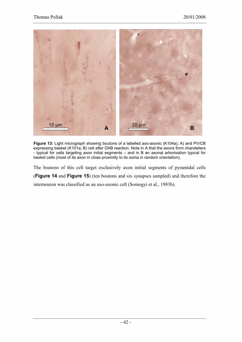
Figure 13: Light micrograph showing boutons of a labelled axo-axonic (K104a); A) and PV/CB expressing basket (K101a; B) cell after DAB reaction. Note in A that the axons form chandeliers - typical for cells targeting axon initial segments – and in B an axonal arborisation typical for basket cells (most of its axon in close proximity to its soma in random orientation).
The boutons of this cell target exclusively axon initial segments of pyramidal cells
(Figure 14 and Figure 15) (ten boutons and six synapses sampled) and therefore the
interneuron was classified as an axo-axonic cell (Somogyi et al., 1983b).
- 42 -

Thomas Pollak 20/01/2008
Figure 14: Electron micrographs showing a labelled bouton targeting an axon initial segment. These can be recognized in the electron microscope by their undercoating beneath the plasma membrane (A, opposite of labelled bouton), bundles of microtubules (B in box) and their innervation with several Gray Type II synapses (four synapses in figure A, whereas the top two release specialisations are from the same bouton and might belong to the same synapse).
- 43 -

Thomas Pollak 20/01/2008
Figure 15: Light and electron micrographs of axo-axonic cell K104a showing labelled axons forming chandeliers along axon initial segments. For each bouton that forms a synapse onto the same axon initial segment, the synapse is shown with high magnification. For one of the boutons no synapse could be observed and one of the boutons is not shown in the low magnification electron micrograph. All synapses are within 20 ultrathin sections of 70 nm thickness.
- 44 -

Thomas Pollak 20/01/2008
- 45 -
In conclusion, the cells that were investigated for their postsynaptic targets can be
divided into three distinct groups: Axo-axonic cells targeting exclusively axon initial
segments, CB-expressing cells targeting small dendrites and spines, and PV expressing
and PV/CB co-expressing basket cells, which target also apical dendrites and somata of
predominantly pyramidal cells (Figure 16). Although there is some variation within
the latter group, the postsynaptic target analysis does not distinguish between PV only
expressing and PV/CB co-expressing cells.
Figure 16: Diagram showing the percentage of the different types of targets for each cell. Cells expressing only CB exclusively target small dendrites and spines, PV-expressing cells and PV/CB co-expressing cells, additionally target apical dendrites and somata and were therefore identified as PV- and PV/CB-expressing basket cells. On the right the number of identified synapses is depicted for each cell.
Quest for novel molecular markers5
In order to identify additional moelcular markers that could further distinguish
interneurons, antibodies against various molecules, for example, known to be expressed
in subsets of PV expressing cells were tested for their suitability in our conditions (for
instance the perfusion/fixation cannot be changed retrospectively). Some of these
5 My contribution to this part of the project was about 95%. Katja Hartwich did two of
the immunoreactions (mGluR1α, PV, CB and SM-co-localisation and CB, SM and
NPY-co-localisation) before I arrived.

Thomas Pollak 20/01/2008
antibodies can be used in the future to further investigate PV/CB co-expressing cells
and cells that were not positiv for markers tested so far.
mGluR1α
A co-localisation study was performed using antibodies of the metabotropic glutamate
receptor 1α subunit (mGluR1α), PV, SM, and CB raised in different species,
respectively. Of 33 cells that were immunopositive for at least one of the markers, 18
were immunopositive for PV and seven of these cells also expressed mGluR1α, five
cells expressed PV, mGluR1α and CB and one cell expressed PV and CB, whereas five
cells only expressed PV. Additionally, two cells were observed that express both CB
and mGluR1α, four cells expressing CB, SM and mGluR1α and three cells which only
express CB.
In summary, PV, CB and mGluR1α can be expressed in all sorts of combinations,
except PV and SM are never co-expressed. In contrast to this observation in the medial
prefrontal cortex (mPFC), bistratified and O-LM (oriens-lacunosum moleculare) cells
in the hippocampal CA1 co-express PV and SM (Klausberger et al., 2003; Klausberger
et al., 2004). An additional difference between mPFC and hippocampus is indicated by
the observation that PV and mGluR1α are much more frequently co-expressed in the
mPFC (Figure 17) than in the hippocampus (Ferraguti et al., 2004).
- 46 -

Thomas Pollak 20/01/2008
Figure 17: Fluorescence micrographs showing a co-localisation between PV and mGluR1α in the same neurons. In the first row two PV-positive cells are depicted which also express mGluR1α, whereas one cell expresses only mGluR1α, but not PV. In the second row a PV-expressing cell is shown, which is mGluR1α-negative.
vGLUT2
An antibody against the vesicular glutamate transporter type 2 (vGLUT2) raised in
guinea pig was tested in dilutions of 1:2500 and 1:5000, which both showed a dotted
labelling pattern, supposedly boutons, throughout the CA1 area of the hippocampus,
but remarkably dense in the stratum lacunosum-moleculare. The labelling in the
prefrontal cortex was much weaker in the 1:5000 dilution, so for the co-localisation
with PV and CB a concentration of 1:2500 was used. The high number of vGLUT2-
positive dots, presumably representing axonal boutons from the thalamus (Kubota et
al., 2007) did not allow to judge at the light microscopic level, whether these boutons
form synapses preferentially onto the soma or dendrites of certain PV-expressing cells
and not on others. To investigate if terminals of certain PV- and/or CB-expressing cells
target spines, which receive a Gray Type I synapse of a terminal immunopositive for
vGLUT2 and therefore most likely thalamic input, was impossible at the light
microscopic level, but this question could be addressed in the future by immunogold
labelling using electron microscopy.
- 47 -

Thomas Pollak 20/01/2008
5-HT2c – a serotonin receptor subunit
Another antibody against 5-HT2c – the 2c subunit of a serotonin receptor – was tested
with various dilutions ranging from 1:20 to 1:500. However, only a very weak nuclear
labelling in all cells was observed, although a receptor-like labelling of the cell
membrane was expected. Interestingly, when this antibody was also tested in a much
higher dilution (1:10000 and 1:100000), a specific labelling of the plasma membranes
of neurons was observed in layer VI of the prefrontal cortex, but no labelling in
superficial layers or the hippocampus.
N200
The co-localisation of PV and N200 – a neurofilament - was tested as well, but of 28
counted cells in layer II and III of the mPFC only one PV-expressing cell was negative
for N200. Further investigations may verify whether this molecular marker could be
used to distinguish between subpopulations of PV-expressing cells.
Kv3.1
An antibody against the potassium channel Kv3.1 was tested for co-localisation with
PV and of 164 observed cells only 3 PV positive cells did not express this potassium
channel.
Kv4.3
A Kv4.3 antibody raised in rabbit was tested as well, but instead of an expression
pattern on the cell surface a dotted nuclear labelling was observed in all concentration
ranging from 1:500 to 1:100000. A co-localisation study with PV indicated that the
labelling was not on the cell surface, but in the nucleus (Figure 18). Additionally, an
antibody raised in goat was also tested, but showed similar non-specific results as the
rabbit antibody.
- 48 -

Thomas Pollak 20/01/2008
Figure 18: Fluorescence micrographs showing a PV (A,C,E,G) positive cell in different focuses (from top to bottom) acquired with a conventional fluorescence microscope and the corresponding images showing Kv4.3 labelling (B,D,F,H). The Kv4.3 labelling cannot be encountered on the cell surface, but in the nucleus.
D1 dopamine receptor
A subtype specific antibody against the D1 dopamine receptor was tested in
concentrations ranging from 1:20 to 1:100000 (optimal concentration between 1:1000
and 1:10000) and showed strong labelling in the substantia nigra and the striatum, but
only a few dots (presumably boutons) in the prefrontal cortex.
GluR2
A GluR2 antibody was also tested and showed dotted labelling throughout the section
(like boutons) and also some dotted nuclear labelling of interneurons (somata in stratum
radiatum and stratum oriens of the hippocampal CA1 region were labelled). Also
somata in the prefrontal cortex had this unspecific nuclear labelling and therefore, this
antibody cannot be used in our material.
NO sensitive guanylyl cyclase α1 subunit
Another promising candidate was a nitric oxide (NO) sensitive guanylyl cyclase α1
subunit (GCα1) specific antibody, which was used at an optimal dilution of 1:10000
and showed receptor-like labelling in both the hippocampus and the prefrontal cortex.
A co-localisation study with PV and Somatostatin (SM) was performed, but
- 49 -

Thomas Pollak 20/01/2008
unfortunately, the immunoreaction of the GCα1 only seemed to have worked in the
hippocampus (Figure 19), but not in the prefrontal cortex, although both other
antibodies worked in the prefrontal cortex as well. Therefore, the prefrontal cortex of
the same brain as the hippocampal sections was cut and the immunoreaction repeated
on those sections. This time the reaction worked in both the hippocampal and the
prefrontal section, although it was somewhat weaker in the mPFC. Many cells
expressed this protein at relatively low level especially in layer II/III, whereas in layer
V fewer cells were strongly immunopositve for this molecular marker.
In conclusion, this molecular marker might allow to distinguish subpopulations of PV-
expressing cells in the hippocampus (possibly axo-axonic and basket cells), but whether
it can be used in the mPFC is a matter of further investigations.
- 50 -

Thomas Pollak 20/01/2008
Figure 19: Fluorescence micrographs showing co-immunolabelling for PV in blue, SM in red and GCα1 in green. The first row depicts a cells in stratum pyramidale of the hippocampal CA1 area immunopositive for PV, but immunonegative for SM and GCα1. In the second row a cell in stratum oriens is shown that is positive for all three molecules tested in this co-localisation study. In the third row a cell is shown, which is positive for PV and SM, but negative for GCα1 and another cell positive for GCα1 and PV (weak), but negative for SM captured from stratum oriens.
CB-SM-NPY
Furthermore, to further investigate CB expressing cells, a co-localisation of CB, SM
and Neuropeptide tyrosin (NPY) was carried out. Of 41 CB expressing cells 30 % also
expressed SM, 10 % also NPY, 10 % both markers, whereas 50 % expressed CB only.
Out of the four in vivo labelled CB expressing cells, one expresses SM and NPY,
whereas the other three only expressed CB; this reflects the ratio of the co-localisation
study.
VIP
In order to verify whether distinct subsets of PV expressing cells receive differential
amounts of input from vasoactive intestinal peptide (VIP) expressing interneurons as
suggested previously (David et al., 2007), a co-localisation of PV and VIP was
performed. In Figure 20 an example of a bouton expressing VIP forming a putative
synapse onto a dendrite expressing PV is shown, but only very few similar examples
were observed.
In conclusion, mGluR1α, N200 and GC α1 could be used to further characterize
labelled cells in the mPFC, and vGLUT2- and VIP-input could be investigated using
confocal or electron microscopy.
- 51 -

Thomas Pollak 20/01/2008
Figure 20: Fluorescence micrograph showing VIP positive boutons (orange, in circle) in close proximity to and putatively forming synapses onto a PV positive dendrite (blue).
- 52 -

Thomas Pollak 20/01/2008
Discussion In the present thesis I have analysed the molecular expression profile and postsynaptic
targets of GABAergic interneurons in the medial prefrontal cortex. Of twelve labelled
cells the postsynaptic targets were identified and these cells can be divided into three
categories: axo-axonic cells, which target exclusively axon initial segments, CB-
expressing dendrite targeting cells and basket cells, which either express PV or co-
express PV and CB and target apical dendrites and somata additionally to small
dendrites and spines of pyramidal cells. Approximately 25% of synapses of
interneurons target dendritic spines in the cat visual cortex (Beaulieu et al., 1992) and
these spines usually also receive excitatory input (Jones and Powell, 1969). Innervation
of spines has been found for basket and dendrite targeting cells, but not the axo-axonic
cell. Because spines are electrically compact, GABAergic synapses are in a unique
position to shape the excitatory input onto the spine (Dehay et al., 1991) and it has been
shown that interneurons target predominantly spines that receive thalamo-cortical input
(Kubota et al., 2007). These spines are larger and express AMPA receptors in contrast
to spines that receive cortico-cortical input (Kubota et al., 2007) and co-innervation of
interneurons of spines receiving thalamo-cortical input might be a mechanism to
compensate for reduced synaptic plasticity of these terminals. Interestingly, it seems
that from the interneurons investigated in this study all of them innervate dendritic
spines apart from axo-axonic cells.
Basket cells
About 50% of all inhibitory interneurons are considered to be basket cells (Markram et
al., 2004) and the percentage of the different target cell domains in this study for basket
cells is consistent with previous findings in the neocortex (Kisvarday et al., 1985;
Kubota et al., 2007; Somogyi et al., 1983a). In the CA1 area of the hippocampus basket
cells express either PV, or CCK together with either VIP or vGLUT3 (Klausberger et
al., 2005), whereas in the neocortex basket cells can express both PV and CB and many
neuropeptides including CCK, NPY and occassionally SM or VIP (Markram et al.,
2004). At least some cells co-express PV and CB in the mPFC and could be identified
as basket cells in this study. There is one recorded cell that expresses CCK, which
might also be a basket cell, but its postsynaptic targets have not been investigated yet.
- 53 -

Thomas Pollak 20/01/2008
Basket cells in the hippocampus are considered to provide temporal coordination for
pyramidal cells (Klausberger et al., 2003) and considering that basket cells in the mPFC
are also correlated to network oscillations, they might have the same function.
Axo-axonic cell
In this study one axo-axonic cell has been investigated. In the hippocampus axo-axonic
cells are considered to express PV (Katsumaru et al., 1988) and in the neocortex to
express either PV or CB (Markram et al., 2004), but this particular cell was tested for
both molecular markers and turned out to be negative for both of them. Another
calcium-binding protein expressed in certain GABAergic interneurons is Calretinin
(Acsady et al., 1996), but this molecular marker was not tested yet.
Axo-axonic cells target exclusively axon initial segments (Somogyi et al., 1983b) and
are therefore in the unique position to control the output of postsynaptic pyramidal cells
(Buhl et al., 1994). The electrophysiological data of this recorded axo-axonic cell is not
evaluated yet, but in the hippocampus it has been shown that axo-axonic cells fire just
after the peak during theta oscillation and at the beginning of sharp wave bursts, but are
silent subsequently, whereas basket cells fire at the descending phase of theta and are
modulated to sharp wave oscillations (Klausberger et al., 2003). This suggests a
differential contribution of basket cells and axo-axonic cells to the temporal
coordination of pyramidal cells with the latter considered as controlling the output of
pyramidal cells through inhibition. In contrast, it was reported that axo-axonic cells can
depolarize pyramidal cells dependent on their membrane potential and chloride
concentration in the axon-initial segment (Szabadics et al., 2006). Therefore, the
function of axo-axonic cells and their contribution to network oscillations is not only
dependent on their input, but also on the electrophysiological state of the postsynaptic
target.
CB-expressing dendrite targeting cells
On the other hand, CB only expressing cells exclusively target small dendrites and
dendritic spines and they are interestingly not correlated to local spindle and CA1 theta
oscillations. Two of these cells have remarkably spiny dendrites and considering that
most interneurons have fewer spines than pyramidal cells (Fairen et al., 1984), this
might suggest that these cells mature slower than other cells (Wang et al., 2004).
Martinotti cells in the neocortex also have spiny dendrites (Wang et al., 2004), but they
- 54 -

Thomas Pollak 20/01/2008
always express SM and for oriens-lacunosum moleculare (O-LM) cells – the pendant to
Martinotti cells in the hippocampus – it has been shown that they are correlated to the
trough of hippocampal theta oscillations (Klausberger et al., 2003). Since these cells
have not been reconstructed yet, it is difficult to compare them with classes of cells
already described in the literature or to suggest possible functions of these cells. But
considering that most pyramidal cells are correlated to local spindle oscillations
(Steriade and Amzica, 1998), there might be a subpopulation that is not correlated to
these network oscillation. These CB-expressing cells might provide feedback inhibition
for such pyramidal cells.
In conclusion, interneurons innervating distinct domains of pyramidal cells – axon
initial segments; somata and apical dendrites; distal dendrites – shape the firing of
pyramidal cells in different ways, with axo-axonic cells having the strongest influence
on the output of their target cells (Buhl et al., 1994).
Heterogeneity of mPFC interneurons
The mPFC integrates input from several different brain regions including the limbic
system (hippocampus and amygdala), the midline thalamus, various cortical areas and
monoaminergic nuclei of the brain stem (Hoover and Vertes, 2007), whereas the CA1
area of the hippocampus has fewer extrinsic inputs and therefore less variation amongst
its cell population (Somogyi and Klausberger, 2005).
An example to illustrate the heterogeneity of interneurons in the mPFC is a co-
localisation study of PV, SM, mGluR1α and CB, which showed that PV and mGluR1α
are much more frequently co-localized in the mPFC than in the CA1 of the
hippocampus (Ferraguti et al., 2004). Furthermore, in the mPFC PV, CB and mGluR1α
can be expressed either by themselves or in all sorts of combinations, except for PV and
SM, which are not co-expressed. In contrast, bistratified and O-LM cells in the CA1
region of the hippocampus co-express PV and SM (Klausberger et al., 2003;
Klausberger et al., 2004).
Examples for different expression in different layers of the mPFC are the serotonin
receptor subunit 2c, which is predominantly expressed by cells in deeper layers (Liu et
al., 2007a), vGLUT2, which is localized in boutons mainly in layer I/III/V (unpublished
observation) and is mainly expressed in thalamic terminals, and GCα1 (NO sensitive
guanylyl cyclase subunit α1), which shows high expression levels in relatively few
- 55 -

Thomas Pollak 20/01/2008
layer V neurons (similar to hippocampal CA1 (Szabadits et al., 2007)), whereas in layer
II/III many cells seem to express this molecular marker at relatively low levels.
The above mentioned examples illustrate which complexity of interneuron types can be
expected in the mPFC and that it is not always straightforward to classify these cells.
Therefore, not only molecular markers, but also postsynaptic targets, in vivo-firing
patterns, dendritic arborisation and presynaptic input (expression profile of boutons
innervating dendrites and/or soma of a labelled cell, but also postsynaptic receptor
expression) are considered to reliably define cell types (Markram et al., 2004; Somogyi
and Klausberger, 2005).
Future directions
Because the recording and labelling of single cells in freely-moving animals has not yet
been possible for technical reasons, all recordings were performed in anaesthetized rats.
How does that relate to decision making? During this drug induced brain state of rats,
network oscillations occur that were also observed during sleep, like spindle
oscillations (7-14 Hz) during slow wave sleep (0.5-4 Hz) in the neocortex (Steriade and
Amzica, 1998), or theta oscillations (4-12 Hz) in the hippocampus (Buzsaki, 2002),
which also occur during exploratory behaviour. Therefore, single cell recordings
simultaneous with the LFP of the mPFC and hippocampus - to obtain information about
oscillatory brain activity - allow investigating the in vivo-firing patterns of single cells,
but also the anatomy including postsynaptic targets and molecular expression profiles
of these recorded cells. In order to relate these results to behaving rats there are two
possible ways. First of all, these results can be used to identify certain types of
interneurons according to their in vivo-firing patterns alone obtained by other recording
methods, which in fact is currently being done by Timothy Senior, MRC Anatomical
Neuropharmacology Unit, Oxford, UK. Secondly, after technical difficulties are
circumvented single cell recording methods could be applied to behaving animals to
verify the results from anaesthetized animals.
Furthermore, molecular expression profiles can be used to classify neurons in different
brain areas. In addition, genetic manipulation of subpopulations of neurons expressing a
certain marker could be used to measure the contribution of these subpopulations to
network oscillation. After a promoter that controls the expression of a certain protein in
a certain subpopulations of interneurons is identified, this promoter could be used in a
construct to control the expression of genes that allow the manipulation of the cell’s
- 56 -

Thomas Pollak 20/01/2008
activity with, for example, light (channelrhodopsin-2 (Deisseroth et al., 2006)) or
reversible manipulation with a pharmacological drug such as zolpidem (Wulff et al.,
2007). This would be a way to investigate causality – which subpopulations contribute
to which kind of oscillations and to which degree – which is not possible with the
experimental procedure used in this study.
mPFC in health and disease
Understanding the circuitry of the mPFC and the role of interneurons in these circuits is
not only important to comprehend working memory and decision making in the healthy
brain, but also has implications for brain dysfunction. It was suggested that alteration in
GABAergic inhibition originating from distinct interneuron subclasses, in particular
axo-axonic cells, is associated with schizophrenia (Lewis et al., 1999) and that
abnormal synchronization of hippocampus and mPFC might contribute to attention
deficit/hyperactivity disorder (Willis and Weiler, 2005) as well as to schizophrenia
(Friston and Frith, 1995).
In conclusion, PV- and PV/CB-basket cells in the mPFC contribute to the temporal
coordination of local pyramidal cells, whereas CB-expressing dendrite targeting cells
might provide feedback inhibition of certain pyramidal cells that are not correlated to
rhythmic brain activity. In order to further characterize these cells, in particular PV/CB
co-expressing cells and cells that were not positive for any markers tested so far, they
could be tested for some of the novel molecular markers. In order to understand the
contribution of certain subpopulations of interneurons to local and distributed network
oscillations during behaviour, these results can be used to support and help the
understanding of results from experiments in behaving animals. A combination of
behavioural experiments and anatomical analysis will eventually lead to an
understanding which cells or cell types contribute to which kind of brain activity and
resulting behaviour.
- 57 -

Thomas Pollak 20/01/2008
Acknowledgement This thesis would not have been possible without the support of a number of people.
First of all, I would like to say a general thank you to all members of the MRC
Anatomical Neuropharmacology Unit at Oxford University. All of you were very
supportive and had an open ear for questions of all kinds and gave good advice. In
particular, I would like to acknowledge my supervisor, Thomas Klausberger, who gave
me the chance to do my diploma thesis in the Unit and organized financial support for
the duration of my work. He was always very supportive and prepared me quite well
for my future as a scientist. I would also like to say thank you to Katja Hartwich, who
took the risk to let a relatively inexperienced student work with her precious labelled
cells, in which she already put a lot of hard work. I think we were quite a good team
and collaborated very well. Furthermore, I would like to thank Peter Somogyi, the
Director of the Unit, who, although he was not my supervisor, always gave me good
advice.
In order to learn ultra microtome cutting and electron microscopy (EM) I have quite a
long list of supervisors. Richard Hewer showed me for the first time how to use an ultra
microtome and he did a really great job, although he also just learned this technique a
couple of months ago. But I also learned a lot from David Roberts, who for example
showed me how to use some features of the EM and explained to me in the EM, how to
recognize axon initial segments. From Jojanneke Huck I learned re-embedding and
from Yannis Dalezios, a visiting Professor from the University of Crete, I got a lot of
very helpful technical advice.
A lab without technicians would not work at all, but it works well, if you have great
technicians as in Peter Somogyi’s lab. From Wai-Yee Suen I learned a lot about
immunohistochemistry, Freeze/thaw and DAB reaction. Kristina Detzner as a German
tries to bring some organisation into the chaos and is doing a great job. She was always
very supportive and when you ask her for a favour, for example, to wash your sections,
she would even mount them. It was a pleasure to work with both of them.
I also made lots of great friends in the lab and just to mention a few of them Katja, who
invited me to her place for a Christmas dinner after knowing me a couple of weeks,
John, with whom I went to the gym and had lots of great discussions about literally
random stuff, Pablo, with whom I went for a run during lunch breaks at least once a
- 58 -

Thomas Pollak 20/01/2008
week, Jerome, who is just simply a nice guy, Wai-Yee, who was a great friend and with
whom I cooked together and I improved my Chinese cooking, Kristina, with whom I
could chat in German in the lab about anything, Romana, a fellow Austrian, who taught
me how to bake bread, the “Mexican Cristina”, with whom I could talk about anything
and we had lots of fun together, Tim, with whom I made some music (unfortunately, at
the end we were both too stressed with science that we could not give a concert
together), Laurence, the only native speaker in the lab, who had the honour to proof-
read parts of this thesis, John Huxter, Amy, Adrian, Jeff, and all the other members of
our coffee table, where we had intense discussions about god and the world. I could go
on, but I can’t mention all 50 or so unit members by name, so I would like to say thank
you all again for bearing me for a year. It was a pleasure for me.
I would also like to say thank you to some people at the University of Vienna, who
supported me to do my thesis abroad. First of all, Prof. Michael Kiebler, who had the
idea that doing my diploma thesis with Thomas Klausberger would be the ideal
preparation for my future as a scientist and allows to brigde the gap from molecular
biology to cognitive neuroscience, what I am going to do for my PhD. And Prof.
Barbara Hamilton, the study-programme coordinator of molecular biology, who made
an exception to the usual policy and allowed me to do my thesis partially abroad.
- 59 -

Thomas Pollak 20/01/2008
References Acsady, I., Gorcs, T.J., and Freund, T.F. (1996). Different populations of vasoactive intestinal polypeptide-immunoreactive interneurons are specialized to control pyramidal cells or interneurons in the hippocampus. Neuroscience 73, 317-334.
Bandyopadhyay, S., and Hablitz, J.J. (2007). Dopaminergic Modulation of Local Network Activity in Rat Prefrontal Cortex. J Neurophysiol 97, 4120-4128.
Beaulieu, C., Kisvarday, Z., Somogyi, P., Cynader, M., and Cowey, A. (1992). Quantitative distribution of GABA-immunopositive and-immunonegative neurons and synapses in the monkey striate cortex (area 17). Cerebral Cortex 2, 295-309.
Blessing, W.W., Howe, P.R., Joh, T.H., Oliver, J.R., and Willoughby, J.O. (1986). Distribution of tyrosine hydroxylase and neuropeptide Y-like immunoreactive neurons in rabbit medulla oblongata, with attention to colocalization studies, presumptive adrenaline-synthesizing perikarya, and vagal preganglionic cells. Journal of Comparative Neurology 248, 285-300.
Buhl, E.H., Halasy, K., and Somogyi, P. (1994). Diverse sources of hippocampal unitary inhibitory postsynaptic potentials and the number of synaptic release sites. Nature 368, 823-828.
Buzsaki, G. (1989). Two-stage model of memory trace formation: A role for "noisy" brain states. Neuroscience 31, 551-570.
Buzsaki, G. (2002). Theta oscillations in the hippocampus. Neuron 33, 325-340.
Carr, D.B., and Sesack, S.R. (1996). Hippocampal afferents to the rat prefrontal cortex: Synaptic targets and relation to dopamine terminals. The Journal of Comparative Neurology 369, 1-15.
Celio, M.R., Baier, W., Scharer, L., de Viragh, P.A., and Gerday, C. (1988). Monoclonal antibodies directed against the calcium binding protein parvalbumin. Cell Calcium 9, 81-86.
Celio, M.R., Baier, W., Scharer, L., Gregersen, H.J., de Viragh, P.A., and Norman, A.W. (1990). Monoclonal antibodies directed against the calcium binding protein Calbindin D-28k. Cell Calcium 11, 599-602.
Chan, Y.-m., Tong, H.-Q., Beggs, A.H., and Kunkel, L.M. (1998). Human Skeletal Muscle-Specific [alpha]-Actinin-2 and -3 Isoforms Form Homodimers and Heterodimersin Vitroandin Vivo. Biochemical and Biophysical Research Communications 248, 134-139.
Chang, Y.C., and Gottlieb, D.I. (1988). Characterization of the proteins purified with monoclonal antibodies to glutamic acid decarboxylase. J. Neurosci. 8, 2123-2130.
- 60 -

Thomas Pollak 20/01/2008
Chow, A., Erisir, A., Farb, C., Nadal, M.S., Ozaita, A., Lau, D., Welker, E., and Rudy, B. (1999). K(+) channel expression distinguishes subpopulations of parvalbumin- and somatostatin-containing neocortical interneurons. J Neurosci 19, 9332-9345.
David, C., Schleicher, A., Zuschratter, W., and Staiger, J.F. (2007). The innervation of parvalbumin-containing interneurons by VIP-immunopositive interneurons in the primary somatosensory cortex of the adult rat. European Journal of Neuroscience 25, 2329-2340.
DeFelipe, J., and Farinas, I. (1992). The pyramidal neuron of the cerebral cortex: Morphological and chemical characteristics of the synaptic inputs. Progress in Neurobiology 39, 563-607.
Degenetais, E., Thierry, A.-M., Glowinski, J., and Gioanni, Y. (2003). Synaptic Influence of Hippocampus on Pyramidal Cells of the Rat Prefrontal Cortex: An In Vivo Intracellular Recording Study. Cereb. Cortex 13, 782-792.
Dehay, C., Douglas, R.J., Martin, K.A., and Nelson, C. (1991). Excitation by geniculo-cortical synapses is not "vetoed" at the level of dendritic spines in cat visual cortex. Journal of Physiology 440, 723-734.
Deisseroth, K., Feng, G., Majewska, A.K., Miesenbock, G., Ting, A., and Schnitzer, M.J. (2006). Next-generation optical technologies for illuminating geneticall targeted brain circuits. Journal of Neuroscience 26, 10380-10386.
Demyanenko, G.P., Schachner, M., Anton, E., Schmid, R., Feng, G., Sanes, J., and Maness, P.F. (2004). Close Homolog of L1 Modulates Area-Specific Neuronal Positioning and Dendrite Orientation in the Cerebral Cortex. Neuron 44, 423-437.
Diba, K., and Buzsaki, G. (2007). Forward and reverse hippocampal place-cell sequences during ripples. Nat Neurosci 10, 1241-1242.
Engel, A.K., Fries, P., and Singer, W. (2001). Dynamic prodictions: oscillations and synchrony in top-down processing. Nature Reviews Neuroscience 2, 704-716.
Euston, D.R., Tatsuno, M., and McNaughton, B.L. (2007). Fast-Forward Playback of Recent Memory Sequences in Prefrontal Cortex During Sleep. Science 318, 1147-1150.
Fairen, A., DeFelipe, J., and Regidor, J. (1984). Cerebral Cortex: Cellular Components of the Cerebral Cortex (New York: Plenum Press).
Ferraguti, F., Cobden, P., Pollard, M., Cope, D., Shigemoto, R., Watanabe, M., and Somogyi, P. (2004). Immunolocalization of metabotropic glutamate receptor 1alpha (mGluR1alpha) in distinct classes of interneuron in the CA1 region of the rat hippocampus. Hippocampus 14, 193-215.
- 61 -

Thomas Pollak 20/01/2008
Floresco, S.B., Seamans, J.K., and Phillips, A.G. (1997). Selective Roles for Hippocampal, Prefrontal Cortical, and Ventral Striatal Circuits in Radial-Arm Maze Tasks With or Without a Delay. J. Neurosci. 17, 1880-1890.
Fremeau, J.R.T., Troyer, M.D., Pahner, I., Nygaard, G.O., Tran, C.H., Reimer, R.J., Bellocchio, E.E., Fortin, D., Storm-Mathisen, J., and Edwards, R.H. (2001). The Expression of Vesicular Glutamate Transporters Defines Two Classes of Excitatory Synapse. Neuron 31, 247-260.
Friston, K.J., and Frith, C.D. (1995). Schizophrenia: a disconnection syndrome? Clin Neurosci 3, 89-97.
Fujiyama, F., Kuramoto, E., Okamoto, K., Hioki, H., Furuta, T., Zhou, L., Nomura, S., and Kaneko, T. (2004). Presynaptic localization of an AMPA-type glutamate receptor in corticostriatal and thalamostriatal axon terminals. European Journal of Neuroscience 20, 3322-3330.
Fuster, J.M. (2001). The Prefrontal Cortex--An Update: Time Is of the Essence. Neuron 30, 319-333.
Gabbott, P., Headlam, A., and Busby, S. (2002). Morphological evidence that CA1 hippocampal afferents monosynaptically innervate PV-containing neurons and NADPH-diaphorase reactive cells in the medial prefrontal cortex (Areas 25/32) of the rat. Brain Research 946, 314-322.
Gao, M., Liu, C.-L., Yang, S., Jin, G.-Z., Bunney, B.S., and Shi, W.-X. (2007). Functional Coupling between the Prefrontal Cortex and Dopamine Neurons in the Ventral Tegmental Area. J. Neurosci. 27, 5414-5421.
Gisquet-Verrier, P., and Delatour, B. (2006). The role of the rat prelimbic/infralimbic cortex in working memory: Not involved in the short-term maintenance but in monitoring and processing functions. Neuroscience 141, 585-596.
Goldman, P.S., and Rosvold, H.E. (1970). Localization of function within the dorsolateral prefrontal cortex of the rhesus monkey. Experimental Neurology 27, 291-304.
Goldman-Rakic, P.S. (1995). Cellular basis of working memory. Neuron 14, 477-485.
Goldman-Rakic, P.S., Selemon, L.D., and Schwartz, M.L. (1984). Dual pathways connecting the dorsolateral prefrontal cortex with the hippocampal formation and parahippocampal cortex in the rhesus monkey. Neuroscience 12, 719-743.
Gray, E.G. (1959). Axo-somatic and axo-dendritic synapses of the cerebral cortex: an electron microscope study. Journal of Anatomy 93, 420-433.
Gulyas, A.I., Gorcs, T.J., and Freund, T.F. (1990). Innervation of different peptide-containing neurons in the hippocampus by gabaergic septal afferents. Neuroscience 37, 31-44.
- 62 -

Thomas Pollak 20/01/2008
Halasy, K., Buhl, E.B., Lörinczi, Z., Tamás, G., and Somogyi, P. (1996). Synaptic target selectivity and input of GABAergic basket and bistratified interneurons in the CA1 area of the rat hippocampus. Hippocampus 6, 306-329.
Hazy, T.E., Frank, M.J., and O'Reilly, R.C. (2006). Banishing the homunculus: Making working memory work. Neuroscience 139, 105-118.
Heidbreder, C.A., and Groenewegen, H.J. (2003). The medial prefrontal cortex in the rat: evidence for a dorso-ventral distinction based upon functional and anatomical characteristics. Neuroscience & Biobehavioral Reviews 27, 555-579.
Hoover, W.B., and Vertes, R.P. (2007). Anatomical analysis of afferent projections to the medial prefrontal cortex in the rat. Brain Structure and Function 212, 149-179.
Jay, T.M., and Witter, M.P. (1991). Distribution of hippocampal CA1 and subicular efferents in the prefrontal cortex of the rat studied by means of anterograde transport of Phaseolus vulgaris-leucoagglutinin. J Comp Neurol 313, 574-586.
Ji, D., and Wilson, M.A. (2007). Coordinated memory replay in the visual cortex and hippocampus during sleep. Nat Neurosci 10, 100-107.
Jones, E.G., and Powell, T.P. (1969). Morphological variations in the dendritic spines of the neocortex. Journal of Cell Science 5, 509-529.
Jones, M.W., and Wilson, M.A. (2005a). Phase precession of medial prefrontal cortical activity relative to the hippocampal theta rhythm. Hippocampus 15, 867-873.
Jones, M.W., and Wilson, M.A. (2005b). Theta Rhythms Coordinate Hippocampal-Prefrontal Interactions in a Spatial Memory Task. PLoS Biology 3, e402.
Katsumaru, H., Kosaka, T., Heizmann, C.W., and Hama, K. (1988). Gap junctions on GABAergic neurons containing the calcium-binding protein parvalbumin in the rat hippocampus (CA1 region). Experimental Brain Research 72, 363-370.
Kawaguchi, Y., and Kubota, Y. (1997). GABAergic Cell Subtypes and their Synaptic Connections in Rat Frontal Cortex. Cerebral Cortex 7, 476-486.
Kisvarday, Z.F., Martin, K.A., Whitteridge, D., and Somogyi, P. (1985). Synaptic connections of intracellularly filled clutch cells: a type of small basket cell in the visual cortex of the cat. Journal of Comparative Neurology 241, 111-137.
Klausberger, T., Magill, P.J., Marton, L.F., Roberts, J.D., Cobden, P.M., Buzsaki, G., and Somogyi, P. (2003). Brain-state- and cell-type-specific firing of hippocampal interneurons in vivo. Nature 421, 844-848.
- 63 -

Thomas Pollak 20/01/2008
Klausberger, T., Marton, L.F., Baude, A., Roberts, J.D., Magill, P.J., and Somogyi, P. (2004). Spike timing of dendrite-targeting bistratified cells during hippocampal network oscillations in vivo. Nat Neurosci 7, 41-47.
Kollo, M., Holderith, N.B., and Nusser, Z. (2006). Novel Subcellular Distribution Pattern of A-Type K+ Channels on Neuronal Surface. J. Neurosci. 26, 2684-2691.
Kosaka, T. (1980). The axon initial segment as a synaptic site: ultrastructure and synaptology of the initial segment of the pyramidal cell in the rat hippocampus (CA3 region). Journal of Neurocytology 9, 861-882.
Kubota, Y., Hatada, S., Kondo, S., Karube, F., and Kawaguchi, Y. (2007). Neocortical inhibitory terminals innervate dendritic spines targeted by thalamocortical afferents. J Neurosci 27, 1139-1150.
Law, A.J., and Harrison, P.J. (2003). The distribution and morphology of prefrontal cortex pyramidal neurons identified using anti-neurofilament antibodies SMI32, N200 and FNP7. Normative data and a comparison in subjects of schizophrenia, bipolar disorder or major depression. Journal of Psychiatric Research 37, 487-499.
Le Moine, C., and Gaspar, P. (1998). Subpopulations of cortical GABAergic interneurons differ by their expression of D1 and D2 dopamine receptor subtypes. Molecular Brain Research 58, 231-236.
Lee, C.T., Li, L., Takamoto, N., Martin, J.F., DeMayo, F.J., Tsai, M.-J., and Tsai, S.Y. (2004). The Nuclear Orphan Receptor COUP-TFII Is Required for Limb and Skeletal Muscle Development. Mol. Cell. Biol. 24, 10835-10843.
Lewis, D.A., Pierri, J.N., Volk, D.W., Melchitzky, D.S., and Woo, T.-U.W. (1999). Altered GABA neurotransmission and prefrontal cortical dysfunction in schizophrenia. Biological Psychiatry 46, 616-626.
Liu, S., Bubar, M.J., Lanfranco, M.F., Hillman, G.R., and Cunningham, K.A. (2007a). Serotonin2C receptor localization in GABA neurons of the rat medial prefrontal cortex: implications for understanding the neurobiology of addiction. Neuroscience In Press, Corrected Proof.
Liu, S., Bubar, M.J., Lanfranco, M.F., Hillman, G.R., and Cunningham, K.A. (2007b). Serotonin2C receptor localization in GABA neurons of the rat medial prefrontal cortex: Implications for understanding the neurobiology of addiction. Neuroscience 146, 1677-1688.
Losonczy, A., Zhang, L., Shigemoto, R., Somogyi, P., and Nusser, Z. (2002). Cell-type dependence and variability in the short-term plasticity of EPSCs in identified mouse hippocampal interneurones. Journal of Physiology 542, 193-210.
Maccaferri, G., Roberts, J.D.B., Szucs, P., Cottingham, C.A., and Somogyi, P. (2000). Cell surface domain specific postsynaptic currents exoked by identified
- 64 -

Thomas Pollak 20/01/2008
GABAergic neurones in rat hippocampus in vitro. Journal of Physiology 524, 91-116.
Markram, H., Toledo-Rodriguez, M., Wang, Y., Gupta, A., Silberberg, G., and Wu, C. (2004). Interneurons of the neocortical inhibitory system. Nat Rev Neurosci 5, 793-807.
McClelland, J.L., Mc Naughton, B.L., and O'Reilly, R.C. (1995). Why there are complementary learning systems in the hippocampus and neocortex: insights from the successes and failures of connectionist models of learning and memory. Psychol. Rev. 102, 419-457.
Moro, M.A., De Alba, J., Leza, J.C., Lorenzo, P., Fernandez, A.P., Bentura, M.L., Bosca, L., Rodrigo, J., and Lizasoain, I. (1998). Neuronal expression of inducible nitric oxide synthase after oxygen and glucose deprivation in rat forebrain slices. European Journal of Neuroscience 10, 445-456.
Muly, E.C., 3rd, Szigeti, K., and Goldman-Rakic, P.S. (1998). D1 receptor in interneurons of macaque prefrontal cortex: distribution and subcellular localization. J Neurosci 18, 10553-10565.
O'Keefe, J., and Recce, M.L. (1993). Phase relationship between hippocampal place units and the EEG theta rhythm. Hippocampus 3, 317-330.
Ohning, G.V., Wong, H.C., Lloyd, K.C., and Walsh, J.H. (1996). Gastrin mediates the gastric mucosal proliferative response to feeding. Am J Physiol Gastrointest Liver Physiol 271, G470-476.
Ongur, D., and Price, J.L. (2000). The Organization of Networks within the Orbital and Medial Prefrontal Cortex of Rats, Monkeys and Humans. Cereb. Cortex 10, 206-219.
Paspalas, C.D., and Goldman-Rakic, P.S. (2005). Presynaptic D1 Dopamine Receptors in Primate Prefrontal Cortex: Target-Specific Expression in the Glutamatergic Synapse. J. Neurosci. 25, 1260-1267.
Pinault, D. (1996). A novel single-cell staining procedure performed in vivo under electrophysiological control: morpho-functional features of juxtacellularly labeled thalamic cells and other central neurons with biocytin or Neurobiotin. Journal of Neuroscience Methods 65, 113-136.
Ramon y Cajal, S. (1911). Histology of the Nervous System (New York: Oxford University Press).
Rodriguez, J.J., Mackie, K., and Pickel, V.M. (2001). Ultrastructural Localization of the CB1 Cannabinoid Receptor in µ-Opioid Receptor Patches of the Rat Caudate Putamen Nucleus. J. Neurosci. 21, 823-833.
Schwaller, B., Buchwald, P., Blümke, I., and Celio, M.R. (1994). Characterization of a polyclonal antiserum against the purified human recombinant calcium-binding protein calretinin. Cell Calcium 14, 639-648.
- 65 -

Thomas Pollak 20/01/2008
Sekirnjak, C., Martone, M.E., Weiser, M., Deerinck, T., Bueno, E., Rudy, B., and Ellisman, M. (1997). Subcellular localization of the K+ channel subunit Kv3.1b in selected rat CNS neurons. Brain Res 766, 173-187.
Serodio, P., and Rudy, B. (1998). Differential expression of Kv4 K+ channel subunits mediating subthreshold transient K+ (A-type) currents in rat brain. J Neurophysiol 79, 1081-1091.
Siapas, A.G., Lubenov, E.V., and Wilson, M.A. (2005). Prefrontal phase locking to hippocampal theta oscillations. Neuron 46, 141-151.
Somogyi, P., Kisvarday, Z.F., Martin, K.A., and Whitteridge, D. (1983a). Synaptic connections of morphologically identified and physiologically characterized large basket cells in the striate cortex of cat. Neuroscience 10, 261-294.
Somogyi, P., and Klausberger, T. (2005). Defined types of cortical interneurone structure space and spike timing in the hippocampus. J Physiol 562, 9-26.
Somogyi, P., Nunzi, M.G., Gorio, A., and Smith, A.D. (1983b). A new type of specific interneuron in the monkey hippocampus forming synapses exclusively with the axon initial segments of pyramidal cells. Brain Research 259, 137-142.
Somogyi, P., Tamas, G., Lujan, R., and Buhl, E.H. (1998). Salient features of synaptic organisation in the cerebral cortex. Brain Research Reviews 26, 113-135.
Steriade, M., and Amzica, F. (1998). Coalescence of sleep rhythms and their chronology in corticothalamic networks. Sleep Research Online 1, 1-10.
Szabadics, J., Varga, C., Molnar, G., Olah, S., Barzo, P., and Tamas, G. (2006). Excitatory effect of GABAergic axo-axonic cells in cortical microcircuits. Science 311, 233-235.
Szabadits, E., Cserep, C., Ludanyi, A., Katona, I., Gracia-Llanes, J., Freund, T.F., and Nyiri, G. (2007). Hippocampal GABAergic Synapses Possess the Molecular Machinery for Retrograde Nitric Oxide Signaling. J. Neurosci. 27, 8101-8111.
Tanaka, J., Nakagawa, S., Kushiya, E., Yamasaki, M., Fukaya, M., Iwanaga, T., Simon, M.I., Sakimura, K., Kano, M., and Watanabe, M. (2000). Gq protein alpha subunits Galphaq and Galpha11 are localized at postsynaptic extra-junctional membrane of cerebellar Purkinje cells and hippocampal pyramidal cells. European Journal of Neuroscience 12, 781-792.
Tierney, P.L., Degenetais, E., Thierry, A.M., Glowinski, J., and Gioanni, Y. (2004). Influence of the hippocampus on interneurons of the rat prefrontal cortex. Eur J Neurosci 20, 514-524.
Vertes, R.P. (2006). Interactions among the medial prefrontal cortex, hippocampus and midline thalamus in emotional and cognitive processing in the rat. Neuroscience 142, 1-20.
- 66 -

Thomas Pollak 20/01/2008
Vincent, S.R., McIntosh, C.H., Buchan, A.M., and Brown, J.C. (1985). Central somatostatin systems revealed with monoclonal antibodies. Journal of comparative Neurology 238, 169-186.
Wang, Y., Toledo-Rodriguez, M., Gupta, A., Wu, C., Silberberg, G., Luo, J., and Markram, H. (2004). Anatomical, physiological and molecular properties of Martinotti cells in the somatosensory cortex of the juvenile rat. Journal of Physiology 561, 65-90.
Willis, W.G., and Weiler, M.D. (2005). Neural Substrates of Childhood Attention-Deficit/Hyperactivity Disorder: Electroencephalographic and Magnetic Resonance Imaging Evidence. Developmental Neuropsychology 27, 135-182.
Wiltgen, B.J., Brown, R.A.M., Talton, L.E., and Silva, A.J. (2004). New Circuits for Old Memories: The Role of the Neocortex in Consolidation. Neuron 44, 101-108.
Wulff, P., Goetz, T., Leppä, E., Linden, A.M., Renzi, M., Swinny, J.D., Vekovischeva, O.Y., Sieghart, W., Somogyi, P., Korpi, E.R., et al. (2007). From synapse to behavior: rapid modulation of defined neuronal types with engineered GABAA receptors. Nature Neuroscience 10, 923-929.
Yang, E.-K., Alvira, M.R., Levitan, E.S., and Takimoto, K. (2001). Kvbeta Subunits Increase Expression of Kv4.3 Channels by Interacting with Their C Termini. J. Biol. Chem. 276, 4839-4844.
Ylinen, A., Bragin, A., Nádasdy, Z., Jandó, G., Szabó, I., Sik, A., and G., B. (1995). Sharp wave-associated highfriquency oscillation (200 Hz) in the intact hippocampus: Network and intracellular mechanisms. Journal of Neuroscience 15, 30-46.
- 67 -

[email protected] - 68 -
Curiculum Vitae
Personal Information
Nationality: Austria Date and place of Birth: 15th January 1981, Zwettl
Education
Since October 2002 University of ViennaMolecular Biology 1. diploma passed (equivalent to BSc) diploma project at Oxford University, 2. diploma exam will be at the end of January 2008 (equivalent to MSc)
From October 2001 to January 2005 University of ViennaNutritional Science 1. diploma passed
From 1995 to 2000 ViennaSecondary College for Chemical Technology passed with distinction
From 1991 to 1995 Stift ZwettlSecondary Modern School with Emphasis on Music passed with distinction
From 1987 to 1991 JahringsElementary School passed with distinction
Abilities
Experience in working in laboratories:
Chemistry: photometry, HPLC, titrimetry,…
Molecular Biology: genotyping (mouse & plant
tissue), northern & western
blotting, antibody purification,
tissue culture,…

[email protected] - 69 -
Neuroscience: dissecting of anatomical structures,
Immunohistochemistry,
Electron Microscopy
of serial, ultrathin sections,…
Computer know-how (Word, Excel, Power Point,
Adobe Photoshop and Illustrator, Endnotes,…)
Language skills: English: 7.5 points on the IELTS-Test,
lived in Australia and England for the last 20 months
Hobbies
Playing clarinet in: Oxford University Wind Orchestra,
Oxford Millennium Orchestra,
Also playing piano, saxophone, accordeon, keyboards,
bassoon,
Sports: jogging, gym, scuba diving, horse riding, skiing,...
reading, attending the opera and the theatre
Experiences
Visiting Student at the MRC Anatomical Neuropharmacology Unit, Oxford University, in Dr. Thomas Klausberger’s lab: from December 2006 until
November 2007 working on a project with the aim to
characterize GABAergic Interneurons in the prefrontal cortex
Exchange Student at the University of Queensland: in
semester 1 in 2006 and additionally in July and August 2006
Work experience student in Prof. Linda Richards’ lab
involved in a project with the aim to characterize a point
mutation in the human leader sequence of the DCC gene
Work experience student in Dr. Peter Schlögelhofer’s lab: in September and October 2005 working on project to
characterize a mutation in the FANCD2 gene in Arabidopsis
thaliana
Work experience student in Prof. Gerhard Wiche’s lab: in
July 2005 doing antibody purification, cell transfection and
[email protected] - 70 -
immunolabelling, and protein expression in Escherichia coli
Study trip to Costa Rica: organised by the University of
Vienna (12.07.04 – 16.08.04): 2 weeks guided tours through
the different kinds of rainforests and a 3 week project
concerning Bromeliaceae at the tropical research station in
the ”Rainforest of the Austrians”
ofi-Austrian Research Institute for Chemistry and Technology: occupied from August 2002 until February 2006
in the department for analytical chemistry (one day per week),
where I did different standard analysis on all sorts of plastic
materials, and in November 2006 in the QLAB (the
department for food quality control)
Military Service: from July 2000 until August 2001 at the
Gardemusik Wien where I played bass clarinet and clarinet in
B flat
Wilhelmsburger Lederfabrik: occupied in August 1999
Ankerbrot AG: occupied in August 1998 doing food quality
control
Herberts (now Dupont): occupied in August 1997 in the
laboratory
Scholarships
FWF-scholarship: for diploma thesis (£3,849.00 for one
year)
MRC living allowance: for diploma thesis (£3,900.00 for one
year)
Joint Study Scholarship: for study abroad at the University
of Queensland (£1,312.00 for one term)
TOP-Scholarship: for study abroad at the University of
Queensland (£583.00 for one term)