Embed Size (px)
Citation preview
CHAPTER IX
DIOXOURANIUM(VI) COMPLEXES OF A SCHIFF
BASE AND AN AZO DYE DERIVED FROM 4-
AMINOANTIPYRINE
There has been considerable interest on theoretical and experimental
chemistry of metal oxocations. Dioxouranium(VI) is one of the stable
oxocations (Greenwood and Earnshaw,1986) and the complexes of
dioxouranium(VI) have been studied extensively (Tabushi et al.,1980;Shinkai
et al.,1986), because of the theoretical interest in the linear O=U=O group,
different structures, detection of uranium compounds in sea water and its
importance in relation to energy problems. The uranyl ion is quite peculiar both
in its own structure and in its coordination chemistry. It has a linear shape
forming complexes with 4 to 8 donor atoms in the equatorial plane (Arora
et al., 1980; Cattalini et al., 1971; Dash et al., 2006). Dioxouranium (VI) is one
of the most studied oxocations for which a large number of complexes with
varying geometries are possible (Maurya M.R and Maurya R.C, 1995).
The present study involves the preparation and characterization of
dioxouranium(VI) complexes with a Schiff base (APES), derived from 4-
aminoantipyrine and 3-ethoxysalicylaldehyde and an azo dye
isoeuginolazoantipyrine, IEAP on the basis of elemental analysis, molar
conductance measurements, infrared, 1H NMR,mass spectral studies,powder X-
ray diffraction and thermogravimetric techniques.
EXPERIMENTAL
Details of the reagents used and method of preparation of the ligands
APES and IEAP are given in chapter III.

Dioxouranium(vi) complexes of a Schiff base and an azo dye derived from 4-aminoantipyrine
195
Synthesis of the complexes
a. Synthesis of nitrate and acetate complexes of dioxouranium(VI)
The nitrate and acetate complexes were prepared by the following
general method. Uranyl nitrate/Uranyl acetate in hot methanol (2 mmol, 20 mL)
was added in small quantities with stirring to a hot methanolic solution (50 mL) of
the corresponding ligands, APES(4mmol) and IEAP (2mmol) containing 1mL of
NaOAc/HOAc buffer (pH = ~6). A distinct colour change was noticed on the
addition of the ligand. The resulting mixture was refluxed on a water bath for
~5 h. It was then concentrated to reduce the volume. The solid complexes
formed were filtered and washed with aqueous methanol and finally with ~15
mL of dry ether. The complexes were dried over P4O10 in vacuo.
b. Synthesis of thiocyante complexes of dioxouranium(VI)
The following general method was adopted for the preparation of
thiocyanate complexes. Uranyl nitrate in hot methanol (2mmol, 20 mL)
containing 2 mmol of NH4CNS was added to a hot methanolic solution
(50mL) of the corresponding ligands, APES(4 mmol) and IEAP(2mmol)
containing 1mL of NaOAc/HOAc buffer (pH = ~6) .The complexes were
precipitated on heating the mixture at 40 °C for ~30 min. The precipitated
complexes were suction filtered, washed with aqueous methanol followed by
dry ether and dried over P4O10 in vacuo.
C. Synthesis of chloride and bromide complexes of dioxouranium(VI)
Uranyl carbonate was precipitated by adding AR Na2CO3 to a saturated
solution of uranyl nitrate. Dioxouranium carbonate was dissolved in minimum
quantity of appropriate dilute acids to get solutions of chloride and bromide.
Dioxouranium(vi) complexes of a Schiff base and an azo dye derived from 4-aminoantipyrine
196
These solutions were evaporated on a water bath to get crystals of the salts. A
hot methanolic solution of the metal salt (2mmol, 20 mL) was added to a hot
methanolic solution of the ligands, APES (4mmol) and IEAP (2mmol)
containing 1mL of NaOAc/HOAc buffer (pH = ~6) with good stirring. Intensely
coloured solution obtained in each case was refluxed for 5-6 h. The solid
complex separated on volume reduction was filtered off, washed successively
with aqueous methanol followed by dry ether and dried over P4O10 in vacuo.
General properties
All complexes are coloured, stable, non hygroscopic solids at room
temperature and possess good keeping qualities. They are soluble in common
organic solvents like methanol, nitrobenzene, DMF and DMSO and sparingly
soluble in acetonitrile, acetone and chloroform and insoluble in water.
Analysis and physico-chemical studies
Physico-chemical studies were carried out as described in chapter III.
RESULTS AND DISCUSSION
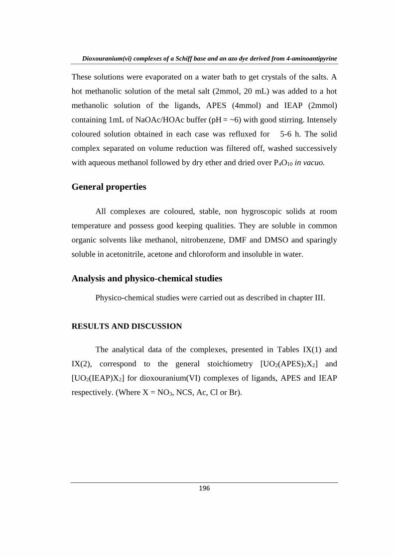
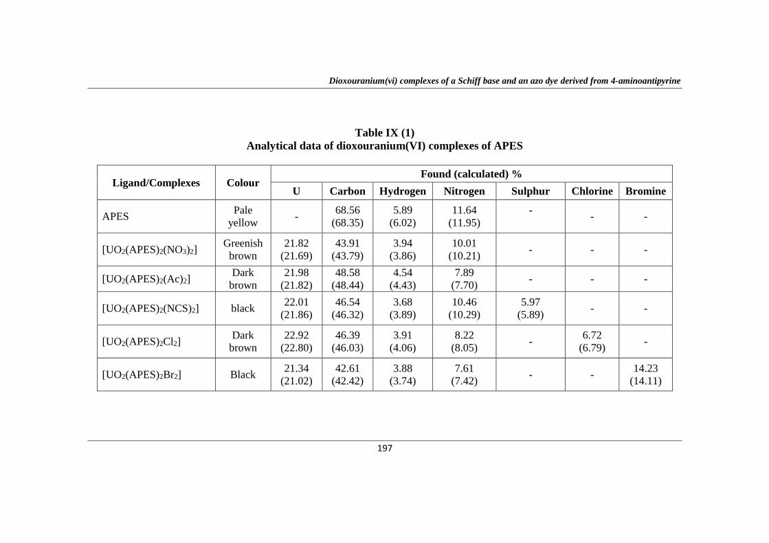
The analytical data of the complexes, presented in Tables IX(1) and
IX(2), correspond to the general stoichiometry [UO2(APES)2X2] and
[UO2(IEAP)X2] for dioxouranium(VI) complexes of ligands, APES and IEAP
respectively. (Where X = NO3, NCS, Ac, Cl or Br).
Dioxouranium(vi) complexes of a Schiff base and an azo dye derived from 4-aminoantipyrine
197
Table IX (1)
Analytical data of dioxouranium(VI) complexes of APES
Ligand/Complexes Colour Found (calculated) %
U Carbon Hydrogen Nitrogen Sulphur Chlorine Bromine
APES Pale
yellow -
68.56
(68.35)
5.89
(6.02)
11.64
(11.95)
-
- -
[UO2(APES)2(NO3)2] Greenish
brown
21.82
(21.69)
43.91
(43.79)
3.94
(3.86)
10.01
(10.21) - - -
[UO2(APES)2(Ac)2] Dark
brown
21.98
(21.82)
48.58
(48.44)
4.54
(4.43)
7.89
(7.70) - - -
[UO2(APES)2(NCS)2] black 22.01
(21.86)
46.54
(46.32)
3.68
(3.89)
10.46
(10.29)
5.97
(5.89) - -
[UO2(APES)2Cl2] Dark
brown
22.92
(22.80)
46.39
(46.03)
3.91
(4.06)
8.22
(8.05) -
6.72
(6.79) -
[UO2(APES)2Br2] Black 21.34
(21.02)
42.61
(42.42)
3.88
(3.74)
7.61
(7.42) - -
14.23
(14.11)
Dioxouranium(vi) complexes of a Schiff base and an azo dye derived from 4-aminoantipyrine
198
Table IX (2)
Analytical data of dioxouranium(VI) complexes of IEAP
Ligand/Complexes Colour Found (calculated) %
U Carbon Hydrogen Nitrogen Sulphur Chlorine Bromine
IEAP Light
brown -
66.48
(66.45)
5.79
(5.86)
14.76
(14.81) - - -
[UO2(IEAP)(NO3)2] Reddish
brown
31.69
(31.42)
33.52
(33.30)
3.12
(3.06)
7.28
(7.39) - - -
[UO2(IEAP)(Ac)2] Dark
brown
31.79
(31.67)
39.78
(39.96)
3.56
(3.89)
7.63
(7.46) - - -
[UO2(IEAP)(NCS)2] Black 31.58
(31.75)
36.96
(36.85)
3.24
(3.09)
11.45
(11.21)
8.64
(8.56) -
[UO2(IEAP)Cl2] Reddish
brown
33.95
(33.79)
35.92
(35.81)
3.48
(3.29)
7.79
(7.96) -
10.24
(10.07) -
[UO2(IEAP)Br2] Brown 30.16
(30.01)
32.02
(31.79)
2.86
(2.92)
7.18
(7.06) - -
20.26
(20.15)
Dioxouranium(vi) complexes of a Schiff base and an azo dye derived from 4-aminoantipyrine
199
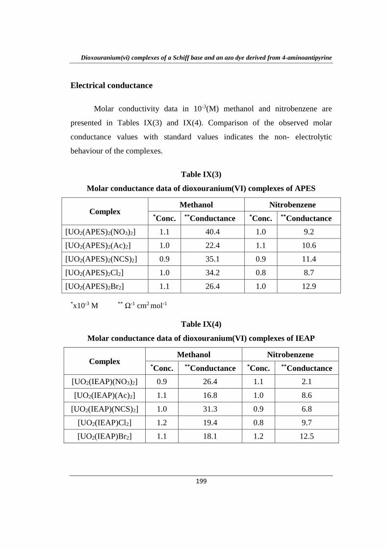
Electrical conductance
Molar conductivity data in 10-3(M) methanol and nitrobenzene are
presented in Tables IX(3) and IX(4). Comparison of the observed molar
conductance values with standard values indicates the non- electrolytic
behaviour of the complexes.
Table IX(3)
Molar conductance data of dioxouranium(VI) complexes of APES
Complex Methanol Nitrobenzene
*Conc. **Conductance *Conc. **Conductance
[UO2(APES)2(NO3)2] 1.1 40.4 1.0 9.2
[UO2(APES)2(Ac)2] 1.0 22.4 1.1 10.6
[UO2(APES)2(NCS)2] 0.9 35.1 0.9 11.4
[UO2(APES)2Cl2] 1.0 34.2 0.8 8.7
[UO2(APES)2Br2] 1.1 26.4 1.0 12.9
*x10-3 M ** Ω-1 cm2 mol-1
Table IX(4)
Molar conductance data of dioxouranium(VI) complexes of IEAP
Complex Methanol Nitrobenzene
*Conc. **Conductance *Conc. **Conductance
[UO2(IEAP)(NO3)2] 0.9 26.4 1.1 2.1
[UO2(IEAP)(Ac)2] 1.1 16.8 1.0 8.6
[UO2(IEAP)(NCS)2] 1.0 31.3 0.9 6.8
[UO2(IEAP)Cl2] 1.2 19.4 0.8 9.7
[UO2(IEAP)Br2] 1.1 18.1 1.2 12.5
Dioxouranium(vi) complexes of a Schiff base and an azo dye derived from 4-aminoantipyrine
200
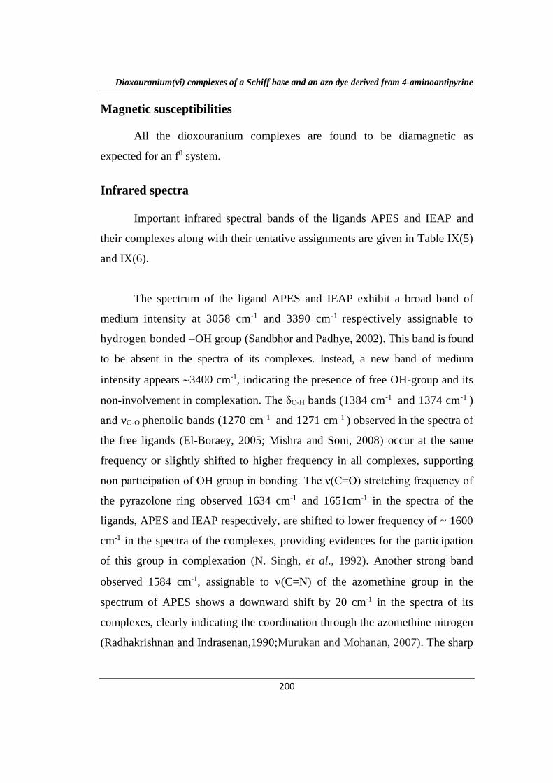
Magnetic susceptibilities
All the dioxouranium complexes are found to be diamagnetic as
expected for an f0 system.
Infrared spectra
Important infrared spectral bands of the ligands APES and IEAP and
their complexes along with their tentative assignments are given in Table IX(5)
and IX(6).
The spectrum of the ligand APES and IEAP exhibit a broad band of
medium intensity at 3058 cm-1 and 3390 cm-1 respectively assignable to
hydrogen bonded –OH group (Sandbhor and Padhye, 2002). This band is found
to be absent in the spectra of its complexes. Instead, a new band of medium
intensity appears 3400 cm-1, indicating the presence of free OH-group and its
non-involvement in complexation. The δO-H bands (1384 cm-1 and 1374 cm-1 )
and νC-O phenolic bands (1270 cm-1 and 1271 cm-1 ) observed in the spectra of
the free ligands (El-Boraey, 2005; Mishra and Soni, 2008) occur at the same
frequency or slightly shifted to higher frequency in all complexes, supporting
non participation of OH group in bonding. The ν(C=O) stretching frequency of
the pyrazolone ring observed 1634 cm-1 and 1651cm-1 in the spectra of the
ligands, APES and IEAP respectively, are shifted to lower frequency of ~ 1600
cm-1 in the spectra of the complexes, providing evidences for the participation
of this group in complexation (N. Singh, et al., 1992). Another strong band
observed 1584 cm-1, assignable to (C=N) of the azomethine group in the
spectrum of APES shows a downward shift by 20 cm-1 in the spectra of its
complexes, clearly indicating the coordination through the azomethine nitrogen
(Radhakrishnan and Indrasenan,1990;Murukan and Mohanan, 2007). The sharp
Dioxouranium(vi) complexes of a Schiff base and an azo dye derived from 4-aminoantipyrine
201
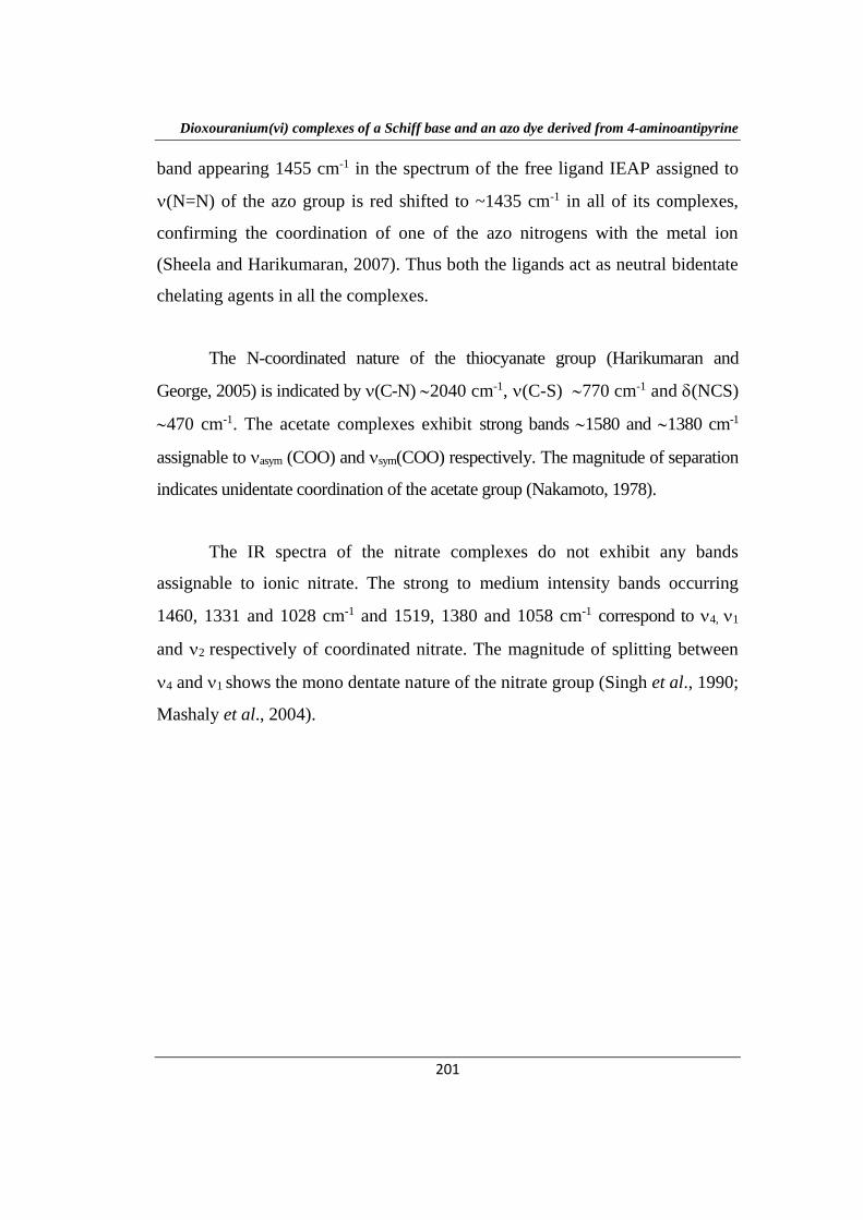
band appearing 1455 cm-1 in the spectrum of the free ligand IEAP assigned to
(N=N) of the azo group is red shifted to ~1435 cm-1 in all of its complexes,
confirming the coordination of one of the azo nitrogens with the metal ion
(Sheela and Harikumaran, 2007). Thus both the ligands act as neutral bidentate
chelating agents in all the complexes.
The N-coordinated nature of the thiocyanate group (Harikumaran and
George, 2005) is indicated by (C-N) 2040 cm-1, (C-S) 770 cm-1 and (NCS)
470 cm-1. The acetate complexes exhibit strong bands 1580 and 1380 cm-1
assignable to asym (COO) and sym(COO) respectively. The magnitude of separation
indicates unidentate coordination of the acetate group (Nakamoto, 1978).
The IR spectra of the nitrate complexes do not exhibit any bands
assignable to ionic nitrate. The strong to medium intensity bands occurring
1460, 1331 and 1028 cm-1 and 1519, 1380 and 1058 cm-1 correspond to 4, 1
and 2 respectively of coordinated nitrate. The magnitude of splitting between
4 and 1 shows the mono dentate nature of the nitrate group (Singh et al., 1990;
Mashaly et al., 2004).
Dioxouranium(vi) complexes of a Schiff base and an azo dye derived from 4-aminoantipyrine
202
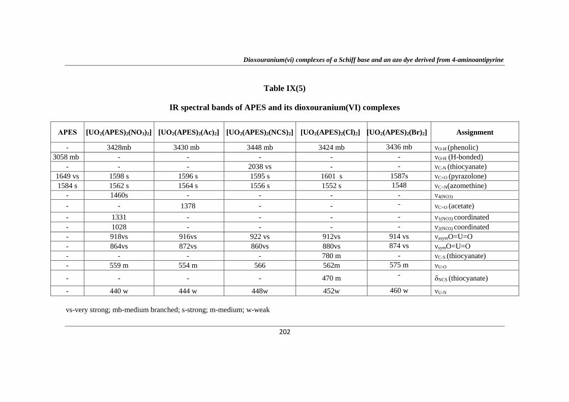
Table IX(5)
IR spectral bands of APES and its dioxouranium(VI) complexes
APES [UO2(APES)2(NO3)2] [UO2(APES)2(Ac)2] [UO2(APES)2(NCS)2] [UO2(APES)2(Cl)2] [UO2(APES)2(Br)2] Assignment
- 3428mb 3430 mb 3448 mb 3424 mb 3436 mb νO-H (phenolic)
3058 mb - - - - - νO-H (H-bonded)
- - - 2038 vs - - νC-N (thiocyanate)
1649 vs 1598 s 1596 s 1595 s 1601 s 1587s νC=O (pyrazolone)
1584 s 1562 s 1564 s 1556 s 1552 s 1548 νC=N(azomethine)
- 1460s - - - - ν4(NO3)
- - 1378 - - - νC=O (acetate)
- 1331 - - - - ν1(NO3) coordinated
- 1028 - - - - ν2(NO3) coordinated
- 918vs 916vs 922 vs 912vs 914 vs νasymO=U=O
- 864vs 872vs 860vs 880vs 874 vs νsymO=U=O
- - - - 780 m - νC-S (thiocyanate)
- 559 m 554 m 566 562m 575 m νU-O
- - - - 470 m -
δNCS (thiocyanate)
- 440 w 444 w 448w 452w 460 w νU-N
vs-very strong; mb-medium branched; s-strong; m-medium; w-weak
Dioxouranium(vi) complexes of a Schiff base and an azo dye derived from 4-aminoantipyrine
203
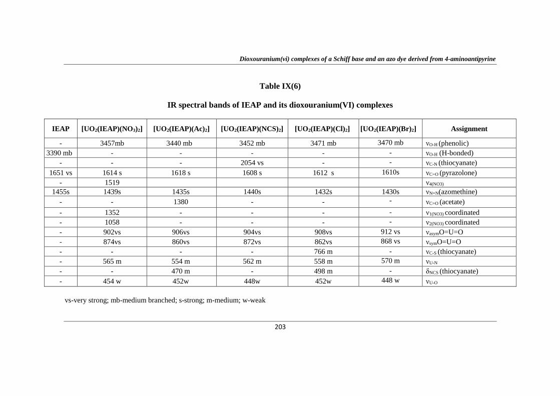
Table IX(6)
IR spectral bands of IEAP and its dioxouranium(VI) complexes
IEAP [UO2(IEAP)(NO3)2] [UO2(IEAP)(Ac)2] [UO2(IEAP)(NCS)2] [UO2(IEAP)(Cl)2] [UO2(IEAP)(Br)2] Assignment
- 3457mb 3440 mb 3452 mb 3471 mb 3470 mb νO-H (phenolic)
3390 mb - - - - - νO-H (H-bonded)
- - - 2054 vs - - νC-N (thiocyanate)
1651 vs 1614 s 1618 s 1608 s 1612 s 1610s νC=O (pyrazolone)
- 1519 ν4(NO3)
1455s 1439s 1435s 1440s 1432s 1430s νN=N(azomethine)
- - 1380 - - - νC=O (acetate)
- 1352 - - - - ν1(NO3) coordinated
- 1058 - - - - ν2(NO3) coordinated
- 902vs 906vs 904vs 908vs 912 vs νasymO=U=O
- 874vs 860vs 872vs 862vs 868 vs νsymO=U=O
- - - - 766 m - νC-S (thiocyanate)
- 565 m 554 m 562 m 558 m 570 m νU-N
- - 470 m - 498 m - δNCS (thiocyanate)
- 454 w 452w 448w 452w 448 w νU-O
vs-very strong; mb-medium branched; s-strong; m-medium; w-weak
Dioxouranium(vi) complexes of a Schiff base and an azo dye derived from 4-aminoantipyrine
204
The IR spectra of all complexes exhibit strong bands 900-925 cm-1 and
860-880 cm-1 assignable to asym(O=U=O) and sym(O=U=O) respectively
indicates trans UO22+ moiety in the complexes (Maurya, M.R and Maurya, R.C,
1995).The non-ligand bands in the range 550-575 cm-1 and 440-460 cm-1 can be
assigned to (U-O) and (U-N) respectively.
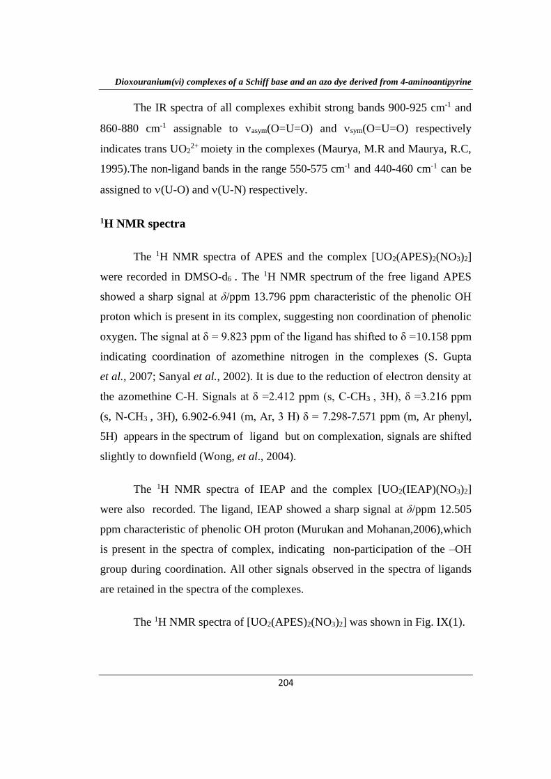
1H NMR spectra
The 1H NMR spectra of APES and the complex [UO2(APES)2(NO3)2]
were recorded in DMSO-d6 . The 1H NMR spectrum of the free ligand APES
showed a sharp signal at δ/ppm 13.796 ppm characteristic of the phenolic OH
proton which is present in its complex, suggesting non coordination of phenolic
oxygen. The signal at δ = 9.823 ppm of the ligand has shifted to δ =10.158 ppm
indicating coordination of azomethine nitrogen in the complexes (S. Gupta
et al., 2007; Sanyal et al., 2002). It is due to the reduction of electron density at
the azomethine C-H. Signals at δ =2.412 ppm (s, C-CH3 , 3H), δ =3.216 ppm
(s, N-CH3 , 3H), 6.902-6.941 (m, Ar, 3 H) δ = 7.298-7.571 ppm (m, Ar phenyl,
5H) appears in the spectrum of ligand but on complexation, signals are shifted
slightly to downfield (Wong, et al., 2004).
The 1H NMR spectra of IEAP and the complex [UO2(IEAP)(NO3)2]
were also recorded. The ligand, IEAP showed a sharp signal at δ/ppm 12.505
ppm characteristic of phenolic OH proton (Murukan and Mohanan,2006),which
is present in the spectra of complex, indicating non-participation of the –OH
group during coordination. All other signals observed in the spectra of ligands
are retained in the spectra of the complexes.
The 1H NMR spectra of [UO2(APES)2(NO3)2] was shown in Fig. IX(1).
Dioxouranium(vi) complexes of a Schiff base and an azo dye derived from 4-aminoantipyrine
205
Applied magnetic field (G)
Fig. IX (1)
1H NMR spectrum of [UO2(APES)2(NO3)2]
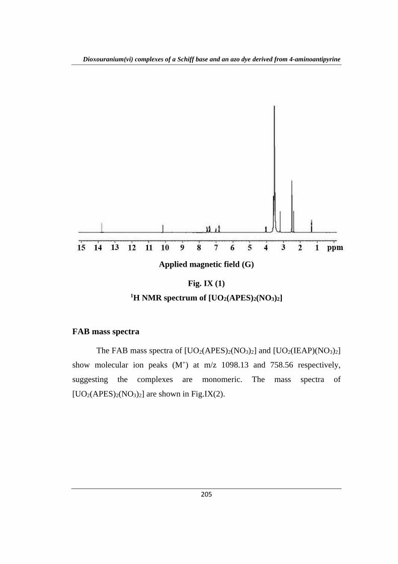
FAB mass spectra
The FAB mass spectra of [UO2(APES)2(NO3)2] and [UO2(IEAP)(NO3)2]
show molecular ion peaks (M+) at m/z 1098.13 and 758.56 respectively,
suggesting the complexes are monomeric. The mass spectra of
[UO2(APES)2(NO3)2] are shown in Fig.IX(2).
Dioxouranium(vi) complexes of a Schiff base and an azo dye derived from 4-aminoantipyrine
206
Fig. IX (2)
Mass spectrum of [UO2(APES)2(NO3)2]
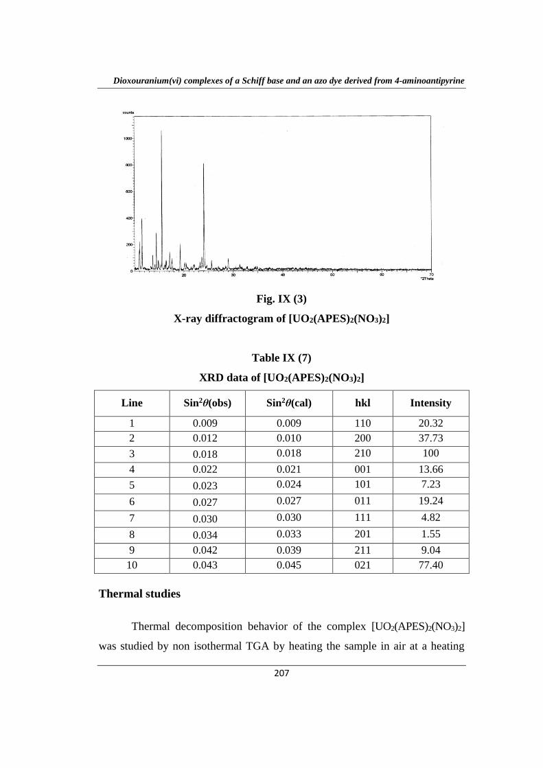
X-ray diffraction studies
The X-ray powder diffraction patterns of the complexes
[UO2(APES)2(NO3)2] and [UO2(IEAP)(NO3)2] were recorded using CuKα
radiation (λ=1.54056Ǻ) and the X-ray diffractogram of [UO2(APES)2(NO3)2]
was shown in Fig. IX(3). The diffraction patterns were indexed by the method
developed by Hesse (1948) and Lipson’s (1949) procedure and the complex
was found to be orthorhombic.
The calculated and observed sin2 values for the corresponding h k l
values with the relative intensities of the peaks of the complexes are listed in
Table IX (7). A careful comparison of the sin2 values of the complexes reveals
that there is a good agreement between the calculated and observed values. The
lattice constants for [UO2(APES)2(NO3)2] are A=0.003,B=0.006 and C=0.021
and the unit cell dimensions were a=14.06 Å,b=9.94 Å and c=5.32 Å. The X-
ray powder photograph obtained for [UO2(IEAP)(NO3)2] recorded only very
few reflections and hence could not be indexed. This may be an indication of
the amorphous nature of the complex.
Dioxouranium(vi) complexes of a Schiff base and an azo dye derived from 4-aminoantipyrine
207
Fig. IX (3)
X-ray diffractogram of [UO2(APES)2(NO3)2]
Table IX (7)
XRD data of [UO2(APES)2(NO3)2]
Line Sin2θ(obs) Sin2θ(cal) hkl Intensity
1 0.009 0.009 110 20.32
2 0.012 0.010 200 37.73
3 0.018 0.018 210 100
4 0.022 0.021 001 13.66
5 0.023 0.024 101 7.23
6 0.027 0.027 011 19.24
7 0.030 0.030 111 4.82
8 0.034 0.033 201 1.55
9 0.042 0.039 211 9.04
10 0.043 0.045 021 77.40
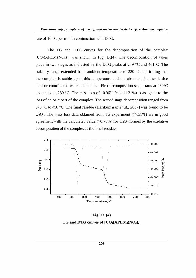
Thermal studies
Thermal decomposition behavior of the complex [UO2(APES)2(NO3)2]
was studied by non isothermal TGA by heating the sample in air at a heating
Dioxouranium(vi) complexes of a Schiff base and an azo dye derived from 4-aminoantipyrine
208
rate of 10 °C per min in conjunction with DTG.
The TG and DTG curves for the decomposition of the complex
[UO2(APES)2(NO3)2] was shown in Fig. IX(4). The decomposition of takes
place in two stages as indicated by the DTG peaks at 249 °C and 461°C .The
stability range extended from ambient temperature to 220 °C confirming that
the complex is stable up to this temperature and the absence of either lattice
held or coordinated water molecules . First decomposition stage starts at 230°C
and ended at 280 °C. The mass loss of 10.96% (calc.11.31%) is assigned to the
loss of anionic part of the complex. The second stage decomposition ranged from
370 °C to 490 °C. The final residue (Harikumaran et al., 2007) was found to be
U3O8. The mass loss data obtained from TG experiment (77.31%) are in good
agreement with the calculated value (76.76%) for U3O8 formed by the oxidative
decomposition of the complex as the final residue.
100 200 300 400 500 600 700 800
2.4
2.6
2.8
3.0
3.2
3.4
Mas
s,m
g
Temperature,0C
100 200 300 400 500 600 700 800
-0.012
-0.010
-0.008
-0.006
-0.004
-0.002
0.000
Mas
s lo
ss,m
g/0 C
0 2 4 6 8 10
Fig. IX (4)
TG and DTG curves of [UO2(APES)2(NO3)2]
Dioxouranium(vi) complexes of a Schiff base and an azo dye derived from 4-aminoantipyrine
209
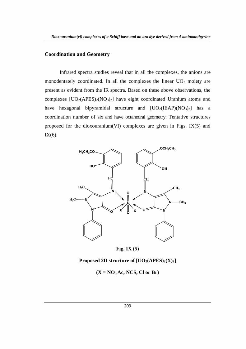
Coordination and Geometry
Infrared spectra studies reveal that in all the complexes, the anions are
monodentately coordinated. In all the complexes the linear UO2 moiety are
present as evident from the IR spectra. Based on these above observations, the
complexes [UO2(APES)2(NO3)2] have eight coordinated Uranium atoms and
have hexagonal bipyramidal structure and [UO2(IEAP)(NO3)2] has a
coordination number of six and have octahedral geometry. Tentative structures
proposed for the dioxouranium(VI) complexes are given in Figs. IX(5) and
IX(6).
Fig. IX (5)
Proposed 2D structure of [UO2(APES)2(X)2]
(X = NO3,Ac, NCS, Cl or Br)
Dioxouranium(vi) complexes of a Schiff base and an azo dye derived from 4-aminoantipyrine
210
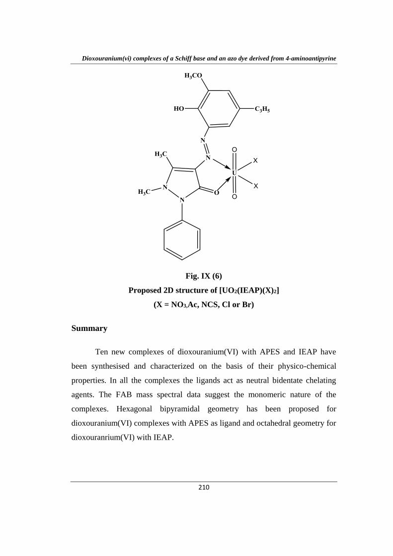
Fig. IX (6)
Proposed 2D structure of [UO2(IEAP)(X)2]
(X = NO3,Ac, NCS, Cl or Br)
Summary
Ten new complexes of dioxouranium(VI) with APES and IEAP have
been synthesised and characterized on the basis of their physico-chemical
properties. In all the complexes the ligands act as neutral bidentate chelating
agents. The FAB mass spectral data suggest the monomeric nature of the
complexes. Hexagonal bipyramidal geometry has been proposed for
dioxouranium(VI) complexes with APES as ligand and octahedral geometry for
dioxouranrium(VI) with IEAP.


![Hetero-binuclear Schiff Base Complexes of Copper(II ...nopr.niscair.res.in/bitstream/123456789/49062/1... · Hetero-binuclear schiff base complexes of the types [Cu Ll \1X1] and ["liL'\1X1J](https://img.pdfslide.us/doc/110x75/5fab3d32647cdd491452f6fc/hetero-binuclear-schiff-base-complexes-of-copperii-nopr-hetero-binuclear-schiff.jpg)