Embed Size (px)
Citation preview
RESEARCH ARTICLE
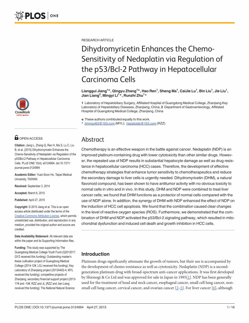
Dihydromyricetin Enhances the Chemo-Sensitivity of Nedaplatin via Regulation ofthe p53/Bcl-2 Pathway in HepatocellularCarcinoma CellsLianggui Jiang1☯, Qingyu Zhang1☯, Hao Ren1, Sheng Ma1, CaiJie Lu1, Bin Liu1, Jie Liu1,Jian Liang2, Mingyi Li1*, Runzhi Zhu1*
1 Laboratory of Hepatobiliary Surgery, Affiliated Hospital of Guangdong Medical College, Zhanjiang KeyLaboratory of Hepatobiliary Diseases, Zhanjiang, China, 2 Department of Gastroenterology, AffiliatedHospital of Guangdong Medical College, Zhanjiang, China
☯ These authors contributed equally to this work.* [email protected] (MYL); [email protected] (RZZ)
AbstractChemotherapy is an effective weapon in the battle against cancer. Nedaplatin (NDP) is an
improved platinum-containing drug with lower cytotoxicity than other similar drugs. Howev-
er, the repeated use of NDP results in substantial hepatocyte damage as well as drug resis-
tance in hepatocellular carcinoma (HCC) cases. Therefore, the development of effective
chemotherapy strategies that enhance tumor sensitivity to chemotherapeutics and reduce
the secondary damage to liver cells is urgently needed. Dihydromyricetin (DHM), a natural
flavonoid compound, has been shown to have antitumor activity with no obvious toxicity to
normal cells in vitro and in vivo. In this study, DHM and NDP were combined to treat liver
cancer cells; we found that DHM functions as a protector of normal cells compared with the
use of NDP alone. In addition, the synergy of DHM with NDP enhanced the effect of NDP on
the induction of HCC cell apoptosis. We found that the combination caused clear changes
in the level of reactive oxygen species (ROS). Furthermore, we demonstrated that the com-
bination of DHM and NDP activated the p53/Bcl-2 signaling pathway, which resulted in mito-
chondrial dysfunction and induced cell death and growth inhibition in HCC cells.
IntroductionPlatinum drugs significantly attenuate the growth of tumors, but their use is accompanied bythe development of chemo-resistance as well as cytotoxicity. Nedaplatin (NDP) is a second-generation platinum drug with broad-spectrum anti-cancer applications. It was first developedby Shionogi & Co Ltd and was approved for sale in Japan in 1995[1]. NDP has been generallyused for the treatment of head and neck cancer, esophageal cancer, small cell lung cancer, non-small cell lung cancer, cervical cancer, and ovarian cancer [2–5]. For liver cancer [6], although
PLOSONE | DOI:10.1371/journal.pone.0124994 April 27, 2015 1 / 16
OPEN ACCESS
Citation: Jiang L, Zhang Q, Ren H, Ma S, Lu C, LiuB, et al. (2015) Dihydromyricetin Enhances theChemo-Sensitivity of Nedaplatin via Regulation of thep53/Bcl-2 Pathway in Hepatocellular CarcinomaCells. PLoS ONE 10(4): e0124994. doi:10.1371/journal.pone.0124994
Academic Editor: Yuan-Soon Ho, Taipei MedicalUniversity, TAIWAN
Received: September 3, 2014
Accepted: March 6, 2015
Published: April 27, 2015
Copyright: © 2015 Jiang et al. This is an openaccess article distributed under the terms of theCreative Commons Attribution License, which permitsunrestricted use, distribution, and reproduction in anymedium, provided the original author and source arecredited.
Data Availability Statement: All relevant data arewithin the paper and its Supporting Information files.
Funding: This study was supported by TheGuangdong Medical College Youth Fund (Q2012017,QYZ received this funding); Outstanding master'sthesis cultivation project of Guangdong MedicalCollege (2014-12#, LGJ received this funding); KeyLaboratory of Zhanjiang project (2013A402-4, MYLreceived this funding); competitive projects ofZhanjiang, secondary financial support project (2013-17# and -10#, RZZ and JL (RZZ and Jian Liangreceived this funding); The National Natural Science
NDP had fewer side effects than first-generation platinum drugs, it still causes a variety of ad-verse reactions such as nausea, vomiting and nephrotoxicity [7–9]. We often administer NDPin combination with other chemotherapy drugs such as 5-fluorouracil, paclitaxel, and strepto-mycin. Synergistic use can produce more pronounced anti-cancer effects [10–13], but multi-drug resistance and cell cytotoxicity also develop.
The enhancement of drug sensitivity by combination chemotherapy has been extensivelystudied. However, few studies have sought to develop a novel compound that protects normalcell functions when combined with platinum drugs.[14–17] Dihydromyricetin is the main ac-tive ingredient of flavonoids and has many functions, such as scavenging free radicals as well asanti-oxidation, antithrombotic, antitumor and anti-inflammatory effects. Our lab has demon-strated that DHM is an inducer of apoptosis in HCC cells but a protector of normal liver cells[18]; our previous research has shown that DHM inhibits HCC cell HepG2 by activation of thep53/Bax pathway in vitro. In addition, DHM has an impact on cancer cell migration, prolifera-tion and cell autophagy [19–21]. However, DHM actually has no significant cytotoxicity innormal cells in vitro and in vivo[22]. Based on these findings, we sought to determine whetherthe combination of NDP and DHM increases the sensitivity of cancer cells to NDP whileavoiding obvious injury to normal cells. Here, we perform a prospective study to demonstratethat DHM enhances the curative effects and reduces the damage to normal cells and to eluci-date the underlying molecular mechanisms.
Materials and Methods
2.1 Drugs and reagentsDHMwas purchased from Sigma-Aldrich and solubilized in dimethylsulfoxide (DMSO) to afinal stock concentration of 50 mM and stored at -20°C in the freezer. NDP was purchasedfrom Nanjing East Express Pharmaceutical Corporation and was solubilized in sterilized H2Oto a final stock concentration of 5 mg/ml and stored at -4°C. Antibodies specific for p53, Bcl-2,Bad, Bax, Bak and GAPDH were purchased from Cell Signaling Technology. Secondary anti-bodies were obtained from Earthox (Cat: E030120-01).
2.2 Cell lines and cultureThe SMMC7721 and QGY7701 human hepatocellular carcinoma cell lines and the HL7702 he-patic immortal cell line were kind gifts from Professor Yi Cao (Molecular Pathology Laborato-ry, Kunming Institution of Zoology, Chinese Academy of Science, Kunming, China). Cellswere cultured in RPMI 1640 medium (Gibco, Grand Island, NY) supplemented with 10% heat-inactivated fetal bovine serum (GIBICO, NY), penicillin 100 U/ml and streptomycin 100 U/ml.Cells were maintained in a humidified atmosphere of 95% air with 5% CO2 in an incubator at37°C. These three cell types were grown in standard media, and when the proliferation of thecells was 40%–60%, the cells were treated with different concentrations of DHM and NDP.
2.3. Cell morphology assessmentCells were seeded in 6-well plates with 1.5x10⁵ cells per well. After treatment with DHM andNDP, cells were observed (100×objective) with an inverted microscope (Leica, Germany)and photographed.
2.4. Cell inhibition and cytotoxicity assaysMTT preliminary experiments: Cell densities were adjusted to 1×10⁴ cells per 100 μl. Cellswere seeded on a 96-well plate with 1×10⁴ cells per well and cultured in an incubator overnight.
Dihydromyricetin Enhances the Chemo-Sensitivity of Nedaplatin
PLOS ONE | DOI:10.1371/journal.pone.0124994 April 27, 2015 2 / 16
Funds 81041099 and Guangdong Province NaturalScience Funds S2011010003750 (MYL receivedthese fundings). The funders had no role in studydesign, data collection and analysis, decision topublish, or preparation of the manuscript.
Competing Interests: All the authors have reviewedthis manuscript and declare that no conflicts ofinterest exist.
To select the most effective concentration and time point, QGY7701 and HL7702 cells weretreated with 0, 20, 40, 60, 80, 100, 120, 140 and 160 μMDHM for 6, 12 and 24 h. SMMC7721cells were treated with 0, 25, 50, 75, 100, 125, 150, 175 and 200 μMDHM for 12, 24 and 48 h.The three cell lines (QGY7701, SMMC7721, and HL7702) were pretreated with 0, 2, 4, 6, 8, 10,12, 14, 16, 18 and 20 μg/ml NDP for 12, 24 and 48 h.
According to the data above, we selected 100 μMDHM combined with 10 μg/ml NDP forthe QGY7701 and HL7702 cells; 200 μMDHM was combined with 15 μg/ml NDP for theSMMC7721 cells. After drug exposure, 10 μL MTT solutions (5 mg/ml) were added, and thecells were incubated for 2 hrs. The cell supernatants were carefully removed, and 150 μlDMSO was transferred to each well for resolving of the MTT crystals. The optical density (OD570 nm) values were recorded by a spectrophotometer.
2.5. Colony formation assayA 0.8% base agar layer and an upper soft agarose (0.7%) layer were pre-coated in 6-well platesin a 37°C incubator for 12 hrs. Cells were pretreated with drugs for 24 hrs. Then, the cells wereharvested and plated in each well at 1000 cells per well. After 10 days of culture, the cells werestained with crystal violet, the clones were counted, and cell morphology was photographed.
2.6. Apoptosis assayBriefly, cells were plated in the 6-well plates (1×10⁵ cells per well); 12 h later, the cells weretreated with the drugs. After various exposure times, the cells were collected and washed twicewith cold D-Hanks buffer solution. Apoptotic cells were quantified using an Annexin V-FITC/PI cell apoptosis detection kit (BD Pharmingen, USA) and monitored using flow cytometry(FACSCalibur, Becton Dickinson). Our experiment strictly complied with the manufacturer’sprotocol.
2.7. Reactive oxygen species detectionWe seeded cells into 6-well plates, which were placed in an incubator overnight to allow for at-tachment and recovery. Cells were treated with the drugs at different exposure times. Then, weadded drugs, and two repeated dosing groups were set up in each group. We set up three con-trols: a blank control, a positive control (ROSUP), and a DCFH control. We replaced themedium with serum-free medium, and 10 μMDCFH was added per well. After incubation for20 min in a 37°C incubator, the cells were harvested and analyzed using flow cytometry.
2.8. Western blot analysisBriefly, cells were collected and washed with ice-cold PBS once. The cells were lysed with lysisbuffer (10 μl PMSF (100 mM), was added to 1 ml RIPA). The soluble cell lysates were collectedafter centrifugation at 13000 g for 5 min. Equivalent amounts of protein from each lysate wereresolved in 10% SDS–polyacrylamide gels. Bands were transferred to PVDF membranes, whichwere blocked with 5% skim milk in PBS containing 0.1% Tween 20 (TBST) and then incubatedovernight with the specific antibodies at a dilution of 1:1,000 at 4°C. After washing with TBSTthree times (10 min each), the membranes were incubated at room temperature for 2 hrs withthe secondary antibody at a dilution of 1:2,000. Chemiluminescent substrate (ECL, GE Health-care) was added to the membranes, which were photographed using a protein analysis system(ProteinSimple, USA).
Dihydromyricetin Enhances the Chemo-Sensitivity of Nedaplatin
PLOS ONE | DOI:10.1371/journal.pone.0124994 April 27, 2015 3 / 16
2.9. Mitochondrial stainingMito-tracker green was purchased from Beyotime Corporation (Shanghai, China), and thecells were incubated with mito-tracker green probes for 30 min. Once the mitochondria werelabeled, the cells were fixed with 4% paraformaldehyde, and the cell nuclei were stained withDAPI for 5 min. Detailed procedures were performed according to the manufacturer’sprotocol.
2.10. Statistical analysisThe statistical analyses were performed using GraphPad Prism 5 software. All results are repre-sented as the mean ± SD. Significant differences were evaluated using Student’s t-test and wereconsidered significant at the � P<0.05, �� P<0.01 or ��� P<0.001 level. All data in the figuresshown in this article were obtained from at least three independent experiments.
Results
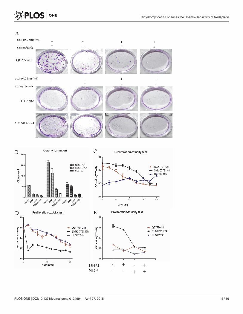
3.1 The combination of DHM and NDP showed a synergistic effect onthe inhibition of tumor cell proliferation but no significant impact on non-tumor cellsFor the hepatoma cells, the effect of the combination of NDP and DHM showed a greatereffect in the inhibition of colony formation ability than the use of DHM or NDP individually.In normal liver cells, DHM possesses functions in preserving cell colony formation capabilityand reducing the inhibitory effect induced by NDP (Fig 1-A and 1-B). The three cells linesshowed a time-concentration dependence on NDP, and we selected the 10 μg/ml concentrationfor the subsequent experiments in the QGY7701 and HL7702 cells. For the SMMC7721 cells, a15 μg/ml concentration of NDP was adopted for the subsequent experiments (Fig 1-C). TheQGY7701 and SMMC7721 cell proliferation showed a time/concentration-dependence onDHM, but this phenomenon was not observed in the HL7702 cells. We selected a sensitive con-centration for the QGY7701 and HL7702 cells of 100 μMDHM and a concentration for theSMMC7721 cells of 200 μMDHM (Fig 1-D). We found that the DHM combination with NDPshowed a synergy effect to inhibit cancer cell proliferation. Interestingly, DHM reduced the in-hibition of proliferation induced by NDP treatment in the HL7702 cells. (Fig 1-E)
3.2 DHM enhanced NDP–induced tumor cell apoptosis but protectednon-tumor cells from cell apoptosisThe combination of DHM with NDP produced a clear synergistic effect on the promotion ofapoptosis in the QGY7701 and SMMC7721 cells. The cell morphology showed obviouschanges, which were photographed with the microscope. Remarkably, DHM exhibited an obvi-ous protective effect on the HL7702 cells and significantly reduced cell apoptosis induced byNDP treatment (Fig 2-A). Cell apoptosis was detected by FITC Annexin V-PI Apoptosis detec-tion kit (BD Pharmingen, USA) and measured using flow cytometry (Fig 2-B). Cells apoptosisratios of drug treatment were calculated and presented as a statistical figure (Fig 2-C). The datashowed that DHM combined with NDP increasingly induced cell apoptosis in hepatic cancercells but reduce the side-effect to the normal cells.
Dihydromyricetin Enhances the Chemo-Sensitivity of Nedaplatin
PLOS ONE | DOI:10.1371/journal.pone.0124994 April 27, 2015 4 / 16
Dihydromyricetin Enhances the Chemo-Sensitivity of Nedaplatin
PLOS ONE | DOI:10.1371/journal.pone.0124994 April 27, 2015 5 / 16
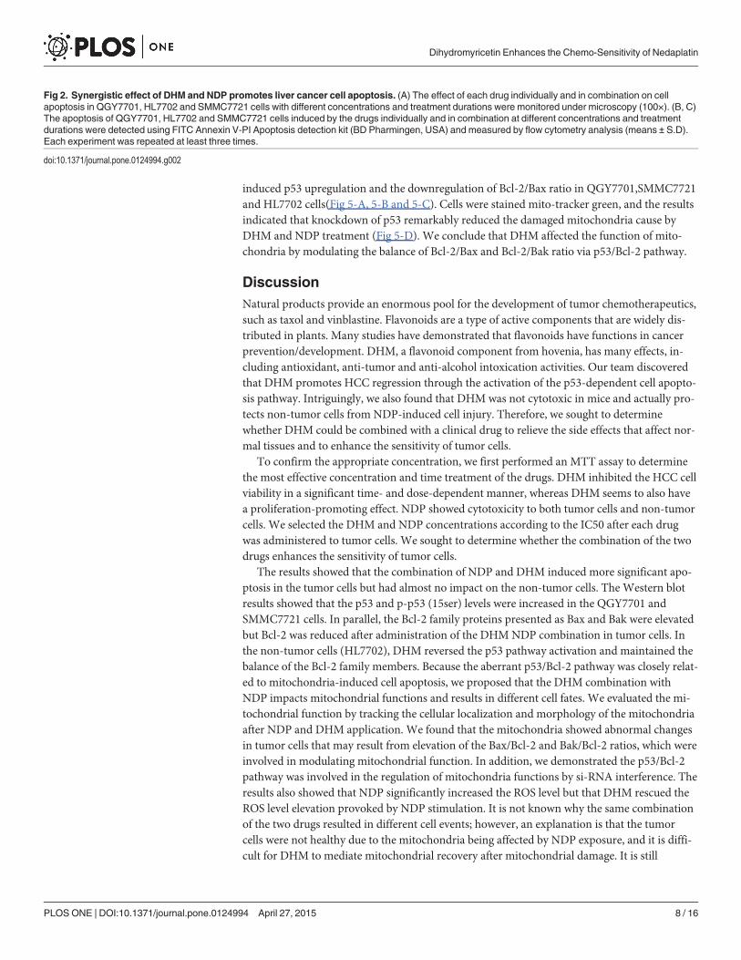
3.3 DHM enhanced the activation of the p53/Bcl-2 signaling pathwayinduced by NDP treatmentThe protein levels of Bak, Bax and Bad were clearly enhanced in response to the combinationof NDP and DHM in the QGY7701 and SMMC7721 cells. In parallel, the combination pro-duced an inhibitory effect on the Bcl-2 protein levels. In the HL7702 cells, the opposite trendwas observed. In addition, DHM combined with NDP promoted the activation of p53 and thephosphorylation of the 15-serine of p53 in the QGY7701 and SMMC7721 cells. However, theseeffects induced by NDP in tumor cells were rescued by DHM in the non-tumor HL7702 cells.(Fig 3-A). Furthermore, the interaction of Bcl-2 with Bax and Bak modulates mitochondrialfunctions and determines the cell fate of tumors. A higher ratio of Bcl-2/Bax or Bak always re-sults in cell apoptosis. Our study found that the Bcl-2/Bax or Bcl-2/Bak ratios were downregu-lated in QGY7701 and SMMC7721 cells but were upregulated in HL7702 cells by the DHMand NDP combination (Fig 3-B and 3-C).These results indicated that DHM could promoteNDP-induced hepatoma cells apoptosis via activation of the p53/Bcl-2 pathways and relievethe NDP induced damage by maintaining the Bcl-2/Bax or Bcl-2/Bak balance in normalliver cells.
3.4 DHM inhibited ROS production in the HL7702 and SMMC7721 cellsbut had no effect on the ROS level in the QGY7701 cells after NDPtreatmentROS play a crucial role in chemotherapy for cancer prevention. Higher levels of ROS damageDNA and induce cell apoptosis. The combination of the two drugs showed no elevation inROS compared to the use of NDP alone. Notably, DHM reduced the ROS level induced byNDP treatment in the Smmc7721 and HL7702 cells (Fig 4-A, 4-B and 4-C). DHM reduced theROS level but the ROS levels in the tumor cells were still higher than that in the non-tumorcells. Therefore, ROS production and the balance of ROS in cells play an important role in cellfate determination. DHM regulates the ROS production with NDP treatment in different cellsand determinates different cell fate.
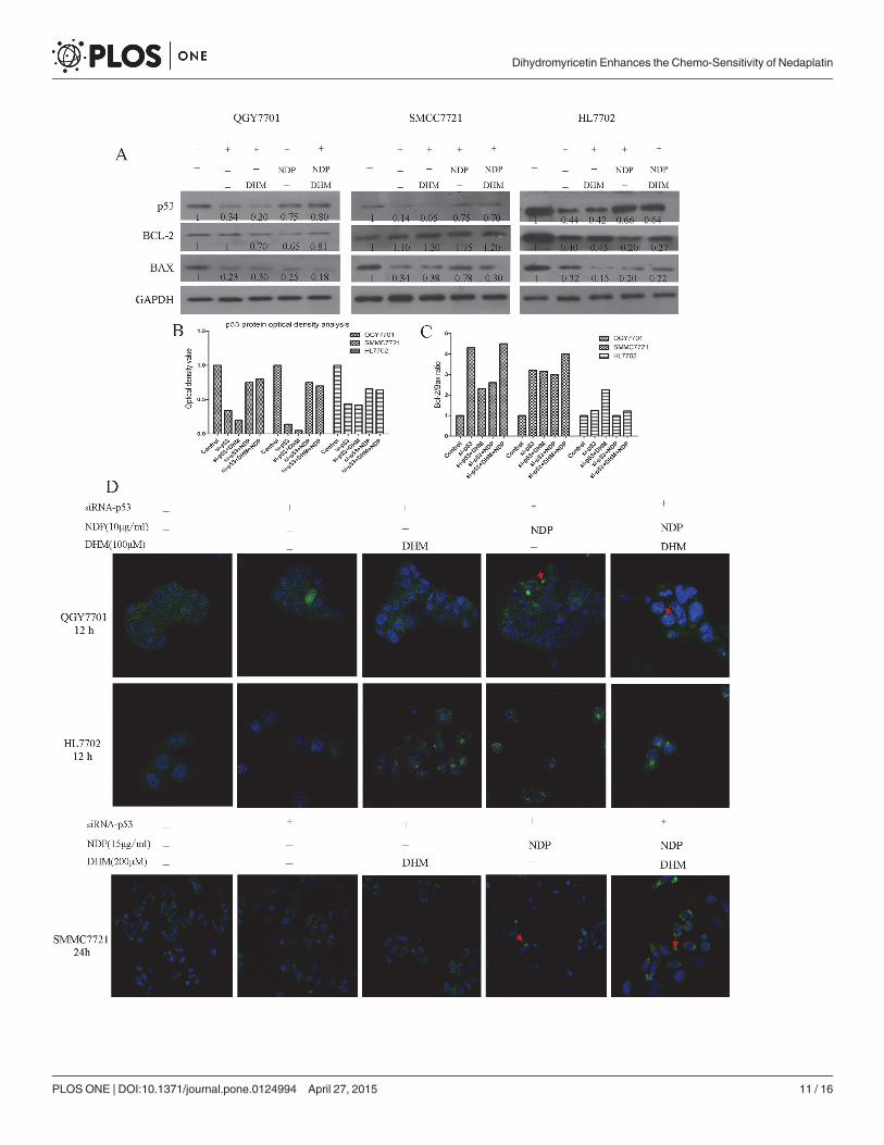
3.5 DHM attenuated mitochondria damage resulting from NDP treatmentin the HL7702 cells but did not have a positive effect on the QGY7701and SMMC7721 HCC cellsMany chemotherapeutic agents induce tumor cell apoptosis by affecting mitochondrial func-tion. Furthermore, disruption of Bcl-2/Bax and Bcl-2/Bak always results in dysfunction of mi-tochondria. We sought to determine whether the synergistic effect of DHM and NDP resultedfrom mitochondrial dysfunction. We stained the mitochondria and found that the morpholog-ical changes induced by NDP and DHM treatment, and the combination of the two drugssynergetically increased the numbers of abnormal cells (Fig 4-D). To demonstrate the role ofp53/Bcl-2 pathway in the synergetic effect of DHM with NDP in deregulation of mitochondria,we design si-RNA(JiMa, Shanghai, China) specific to p53 mRNA for down-regulation of p53expression. p53 si-RNA transfection rescued DHM and NDP combined administration
Fig 1. DHM combination with NDP inhibits the ability of colony formation of liver cancer cells. (A) Colony formation capability assay with differenttreatments of DHM and NDP in HCC cells. The images were captured with a Nikon camera (Japan). (B) The clones were quantified and presented as astatistical figure. (C, D) The proliferation-toxicity of NDP or DHM in QGY7701, HL7702 and SMMC7721 cells with various drug concentrations and treatmentdurations were assayed using the MTT method (means ± S.D). (D) Proliferation and toxicity of different drug combinations were measured using the MTTassay (means ± S.D). Each experiment was repeated at least three times.
doi:10.1371/journal.pone.0124994.g001
Dihydromyricetin Enhances the Chemo-Sensitivity of Nedaplatin
PLOS ONE | DOI:10.1371/journal.pone.0124994 April 27, 2015 6 / 16
Dihydromyricetin Enhances the Chemo-Sensitivity of Nedaplatin
PLOS ONE | DOI:10.1371/journal.pone.0124994 April 27, 2015 7 / 16
induced p53 upregulation and the downregulation of Bcl-2/Bax ratio in QGY7701,SMMC7721and HL7702 cells(Fig 5-A, 5-B and 5-C). Cells were stained mito-tracker green, and the resultsindicated that knockdown of p53 remarkably reduced the damaged mitochondria cause byDHM and NDP treatment (Fig 5-D). We conclude that DHM affected the function of mito-chondria by modulating the balance of Bcl-2/Bax and Bcl-2/Bak ratio via p53/Bcl-2 pathway.
DiscussionNatural products provide an enormous pool for the development of tumor chemotherapeutics,such as taxol and vinblastine. Flavonoids are a type of active components that are widely dis-tributed in plants. Many studies have demonstrated that flavonoids have functions in cancerprevention/development. DHM, a flavonoid component from hovenia, has many effects, in-cluding antioxidant, anti-tumor and anti-alcohol intoxication activities. Our team discoveredthat DHM promotes HCC regression through the activation of the p53-dependent cell apopto-sis pathway. Intriguingly, we also found that DHM was not cytotoxic in mice and actually pro-tects non-tumor cells from NDP-induced cell injury. Therefore, we sought to determinewhether DHM could be combined with a clinical drug to relieve the side effects that affect nor-mal tissues and to enhance the sensitivity of tumor cells.
To confirm the appropriate concentration, we first performed an MTT assay to determinethe most effective concentration and time treatment of the drugs. DHM inhibited the HCC cellviability in a significant time- and dose-dependent manner, whereas DHM seems to also havea proliferation-promoting effect. NDP showed cytotoxicity to both tumor cells and non-tumorcells. We selected the DHM and NDP concentrations according to the IC50 after each drugwas administered to tumor cells. We sought to determine whether the combination of the twodrugs enhances the sensitivity of tumor cells.
The results showed that the combination of NDP and DHM induced more significant apo-ptosis in the tumor cells but had almost no impact on the non-tumor cells. The Western blotresults showed that the p53 and p-p53 (15ser) levels were increased in the QGY7701 andSMMC7721 cells. In parallel, the Bcl-2 family proteins presented as Bax and Bak were elevatedbut Bcl-2 was reduced after administration of the DHMNDP combination in tumor cells. Inthe non-tumor cells (HL7702), DHM reversed the p53 pathway activation and maintained thebalance of the Bcl-2 family members. Because the aberrant p53/Bcl-2 pathway was closely relat-ed to mitochondria-induced cell apoptosis, we proposed that the DHM combination withNDP impacts mitochondrial functions and results in different cell fates. We evaluated the mi-tochondrial function by tracking the cellular localization and morphology of the mitochondriaafter NDP and DHM application. We found that the mitochondria showed abnormal changesin tumor cells that may result from elevation of the Bax/Bcl-2 and Bak/Bcl-2 ratios, which wereinvolved in modulating mitochondrial function. In addition, we demonstrated the p53/Bcl-2pathway was involved in the regulation of mitochondria functions by si-RNA interference. Theresults also showed that NDP significantly increased the ROS level but that DHM rescued theROS level elevation provoked by NDP stimulation. It is not known why the same combinationof the two drugs resulted in different cell events; however, an explanation is that the tumorcells were not healthy due to the mitochondria being affected by NDP exposure, and it is diffi-cult for DHM to mediate mitochondrial recovery after mitochondrial damage. It is still
Fig 2. Synergistic effect of DHM and NDP promotes liver cancer cell apoptosis. (A) The effect of each drug individually and in combination on cellapoptosis in QGY7701, HL7702 and SMMC7721 cells with different concentrations and treatment durations were monitored under microscopy (100×). (B, C)The apoptosis of QGY7701, HL7702 and SMMC7721 cells induced by the drugs individually and in combination at different concentrations and treatmentdurations were detected using FITC Annexin V-PI Apoptosis detection kit (BD Pharmingen, USA) and measured by flow cytometry analysis (means ± S.D).Each experiment was repeated at least three times.
doi:10.1371/journal.pone.0124994.g002
Dihydromyricetin Enhances the Chemo-Sensitivity of Nedaplatin
PLOS ONE | DOI:10.1371/journal.pone.0124994 April 27, 2015 8 / 16
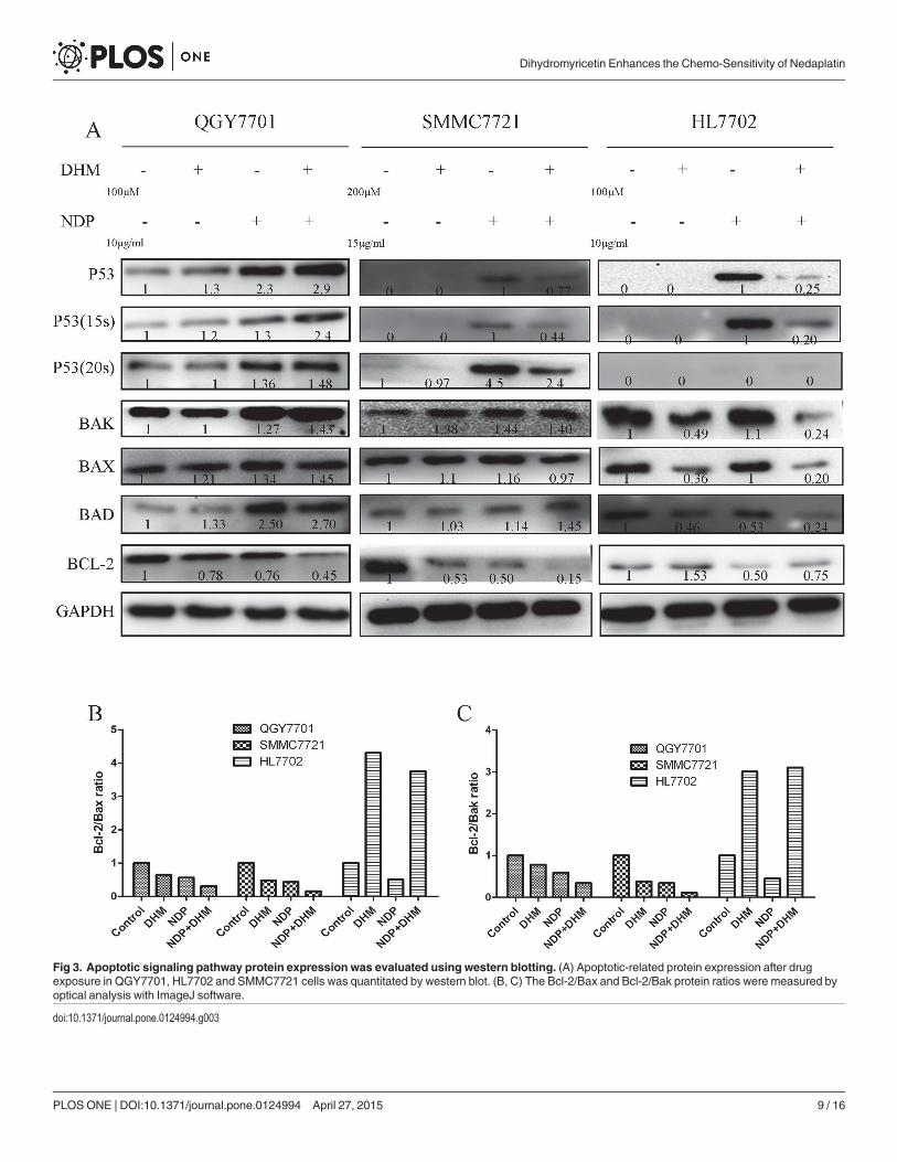
Fig 3. Apoptotic signaling pathway protein expression was evaluated using western blotting. (A) Apoptotic-related protein expression after drugexposure in QGY7701, HL7702 and SMMC7721 cells was quantitated by western blot. (B, C) The Bcl-2/Bax and Bcl-2/Bak protein ratios were measured byoptical analysis with ImageJ software.
doi:10.1371/journal.pone.0124994.g003
Dihydromyricetin Enhances the Chemo-Sensitivity of Nedaplatin
PLOS ONE | DOI:10.1371/journal.pone.0124994 April 27, 2015 9 / 16
unknown why DHM alone also induces cell death in tumor cells. We think that DHM is a re-ducibility compound that inhibits the ROS production that is necessary for tumor cell growthand returns the ROS levels to a normal level.
Fig 4. The alteration of ROS in normal liver cells and hepatoma cells. (A,B,C) The level of ROS changes in QGY7701, HL7702 and SMMC7721 cellsafter treatment with DHM and NDP were detected using the DCFH assay. (B) The morphological changes in the mitochondria in QGY7701, HL7702 andSMMC7721 cells after treatment with the drug individually and in combination were detected using mito-tracker green. Each experiment was repeated atleast three times.
doi:10.1371/journal.pone.0124994.g004
Dihydromyricetin Enhances the Chemo-Sensitivity of Nedaplatin
PLOS ONE | DOI:10.1371/journal.pone.0124994 April 27, 2015 10 / 16
Dihydromyricetin Enhances the Chemo-Sensitivity of Nedaplatin
PLOS ONE | DOI:10.1371/journal.pone.0124994 April 27, 2015 11 / 16
In conclusion, we found that the DHM combination promoted the NDP chemotherapeuticsensitivity of liver cancer cells and also reduced the cytotoxicity to normal liver cells in vitro.We discovered that DHM application played a role in the regulation of the p53/Bcl-2 pathway,impacted the function of mitochondria and modulated ROS production. Although a prospec-tive target is that DHM could regulate the ROS production induced by NDP treatment andpromote the ROS-induced tumor cells apoptosis with less side-effect to the normal cells of pa-tients, in vivo studies should be performed to test the advantages of the DHM and NDP combi-nation. We believe that DHM is a potential compound that will be clinically effective fortumor prevention.
Supporting InformationS1 Fig. Combination of DHM and NDP inhibits the colony formation ability of QGY7701cells. Colony formation ability was measured by plate colony formation experiment.(TIF)
S2 Fig. Combination of DHM and NDP inhibits the colony formation ability ofSMMC7721 cells. Colony formation ability was measured by plate colonyformation experiment.(TIF)
S3 Fig. Combination of DHM attenuates impairing of colony formation ability resultedfrom NDP treatment in HL7702 cells. Colony formation ability was measured by plate colonyformation experiment.(TIF)
S4 Fig. Combination of DHM and NDP affect the colony formation ability of three celllines (QGY7701, SMMC7721, and HL7702). Colony numbers were counted and presented asa statistical figure.(TIF)
S5 Fig. NDP decreased the cell viability of three cell lines (QGY7701, SMMC7721, andHL7702). Cells were treated with various concentrations NDP and the cell viability was mea-sured by MTT.(TIF)
S6 Fig. DHM decreased the cell viability of hepatoma cell lines (QGY7701, SMMC7721)and affects weakly to non-hepatoma cell HL7702. Cells were treated with various concentra-tions NDP and the cell viability was measured by MTT.(TIF)
S7 Fig. Combination of DHM with NDP inhibited the cell viability of hepatoma cell lines(QGY7701, SMMC7721) and reduced the NDP treatment mediated cell viability inhibitionin HL7702 cells. Cells were treated with various concentrations NDP and the cell viability wasmeasured by MTT.(TIF)
Fig 5. p53/Bcl-2 pathway was implicated in DHM and NDP induced dysfunction of mitochondria. (A, B) p53-siRNA was used to knockdown of p53 andrescued the upregulation of p53 in hepatoma cells after DHM and NDP treatment. (C) Knockdown of p53 inhibited the downregulation of Bcl-2/Bax ratiocaused by DHM and NDP treatment. (D) After si-RNA interference was performed, the morphological alteration of mitochondria in QGY7701, HL7702 andSMMC7721 cells after treatment with the drug individually and in combination were detected by using mito-tracker green.
doi:10.1371/journal.pone.0124994.g005
Dihydromyricetin Enhances the Chemo-Sensitivity of Nedaplatin
PLOS ONE | DOI:10.1371/journal.pone.0124994 April 27, 2015 12 / 16
S8 Fig. QGY7701cells were treated with NDP and DHM separately and combination. Thecell morphology was monitored by a Leica inverted microscope.(TIF)
S9 Fig. HL7702 cells were treated with NDP and DHM separately and combination. Thecell morphology was monitored by a Leica inverted microscope.(TIF)
S10 Fig. SMMC7721 Cells were treated with NDP and DHM separately and combination.The cell morphology was monitored by a Leica inverted microscope.(TIF)
S11 Fig. The apoptosis of QGY7701 cells induced by the DHM and NDP individually andcombination at different concentrations and treatment durations. The apoptosis of cellswere measured by flow cytometry analysis.(TIF)
S12 Fig. The apoptosis of HL7702 cells induced by the DHM and NDP individually andcombination at different concentrations and treatment durations. The apoptosis of cellswere measured by flow cytometry analysis.(TIF)
S13 Fig. The apoptosis of SMMC7721 cells induced by the DHM and NDP individually andcombination at different concentrations and treatment durations. The apoptosis of weremeasured by flow cytometry analysis.(TIF)
S14 Fig. A statistical figure for apoptosis rate induced by DHM and NDP synergic or indi-vidual treatment in three cell lines.(TIF)
S15 Fig. Combination of DHM with NDP activated the p53/Bcl-2 pathway in QGY7701cells. The apoptotic proteins were detected by western blot in QGY7701 cells.(TIF)
S16 Fig. Combination of DHM with NDP attenuated the activation of p53/Bcl-2 pathwayin HL7702 cells. The apoptotic proteins were detected by western blot in HL7702 cells.(TIF)
S17 Fig. Combination of DHM with NDP activated the p53/Bcl-2 pathway in SMMC7721cells. The apoptotic proteins were detected by western blot in SMMC7721 cells.(TIF)
S18 Fig. DHM reduced the ROS level increased by NDP treatment in three cell lines. Reac-tive oxygen species were detected by using the DCFH assay in three cell lines (QGY7701,SMMC7721, and HL7702).(TIF)
S19 Fig. Combination of DHM with NDP affected the mitochondria morphology inQGY7701 cells.Mitochondria morphology was evaluated by mito-tracker green staining afterdrugs treatment in QGY7701 cells.(TIF)
S20 Fig. DHM reduced the mitochondria morphology damage caused by NDP treatmentin HL7702 cells.Mitochondria morphology was evaluated by mito-tracker green staining after
Dihydromyricetin Enhances the Chemo-Sensitivity of Nedaplatin
PLOS ONE | DOI:10.1371/journal.pone.0124994 April 27, 2015 13 / 16
drugs treatment in HL7702 cells.(TIF)
S21 Fig. Combination of DHM with NDP affected the mitochondria morphology inSMMC7721 cells.Mitochondria morphology was evaluated by mito-tracker green stainingafter drugs treatment in SMMC7721 cells.(TIF)
S22 Fig. Knockdown p53 relieved the p53/Bcl-2 pathway activation in QGY7701 cells. Theapoptotic proteins were detected by western blot after p53 was knockdown in QGY7701 cells.(TIF)
S23 Fig. Knockdown p53 relieved the p53/Bcl-2 pathway activation in SMMC7721 cells.The apoptotic proteins were detected by western blot after p53 was knockdown inSMMC7721 cells.(TIF)
S24 Fig. Knockdown p53 relieved the p53/Bcl-2 pathway activation in HL7702 cells. Theapoptotic proteins were detected by western blot after p53 was knockdown in HL7702 cells.(TIF)
S25 Fig. Protein content was evaluated by optical density analysis using ImageJ software.(TIF)
S26 Fig. Knockdown p53 relieved the p53/Bcl-2 pathway activation. Bcl-2/Bax ratio werecalculated using optical density value(TIF)
S27 Fig. Knockdown p53 attenuated the NDP/DHM treatment caused mitochondria mor-phology injury in QGY7701 cells.Mitochondria morphology was evaluated by mito-trackergreen staining after drugs treatment in QGY7701while p53 was knockdown bysiRNA transfection.(TIF)
S28 Fig. Knockdown p53 attenuated the NDP/DHM treatment caused mitochondria mor-phology injury in HL7702 cells.Mitochondria morphology was evaluated by mito-trackergreen staining after drugs treatment in HL7702 while p53 was knockdown bysiRNA transfection.(TIF)
S29 Fig. Knockdown p53 attenuated the NDP/DHM treatment caused mitochondria mor-phology injury in SMMC7721 cells.Mitochondria morphology was evaluated by mito-trackergreen staining after drugs treatment in SMMC7721 while p53 was knockdown bysiRNA transfection.(TIF)
Author ContributionsConceived and designed the experiments: MYL RZZ. Performed the experiments: LGJ HR CJLSM BL J. Liu. Analyzed the data: QYZ LGJ. Contributed reagents/materials/analysis tools: J.Liang. Wrote the paper: RZZ QYZ LGJ.
References1. Ota K. Nedaplatin. Cancer Chemoth. 1996; 23:379–387.
Dihydromyricetin Enhances the Chemo-Sensitivity of Nedaplatin
PLOS ONE | DOI:10.1371/journal.pone.0124994 April 27, 2015 14 / 16
2. Sato D, Kogashiwa Y, Tsukahara K, Yamauchi K, Kohno N. Phase I study of nedaplatin prior to S-1 inpatients with locally advanced head and neck squamous cell carcinoma. Chemotherapy. 2013;59:314–318. doi: 10.1159/000357469 PMID: 24480865
3. Hirata K, Kodaira T, Tomita N, Ohshima Y, Ito J, Tachibana H, et al. Clinical Efficacy of Alternating Che-moradiotherapy by Conformal Radiotherapy Combined with Intracavitary Brachytherapy for High-riskCervical Cancer. JPN J Clin Oncol. 2014; 44:556–563. doi: 10.1093/jjco/hyu048 PMID: 24755546
4. Nishioka K, Shimizu S, Shinohara N, Ito YM, Abe T, Maruyama S, et al. Prospective phase II study ofimage-guided local boost using a real-time tumor-tracking radiotherapy (RTRT) system for locally ad-vanced bladder cancer. JPN J Clin Oncol. 2014; 44:28–35. doi: 10.1093/jjco/hyt182 PMID: 24302759
5. Li CH, Liu MY, Liu W, Li DD, Cai L. Randomized control study of nedaplatin or cisplatin concomitantwith other chemotherapy in the treatment of advanced non-small cell lung cancer. Asian Pac J CancerP. 2014; 15:731–736. PMID: 24568487
6. Fukui T, Itoh Y, Yoshioka T, Takeda H, Kawada S. A case of advanced esophageal cancer with multipleliver metastases accompanying poorly general conditions and serious liver dysfunctions, successfullytreated using concurrent radiotherapy and chemotherapy with low-dose nedaplatin and 5-fluorouracil.JPN J Gastro-enterol. 2011; 108:813–818.
7. Kubala M, Geleticova J, Huliciak M, Zatloukalova M, Vacek J, Sebela M. Na/K-ATPase inhibition by cis-platin and consequences for cisplatin nephrotoxicity. Biomed Pap. 2014; 158:194–200. doi: 10.5507/bp.2014.018 PMID: 24781046
8. Shaili E. Platinum anticancer drugs and photochemotherapeutic agents: recent advances and futuredevelopments. Sci Prog. 2014; 97:20–40. PMID: 24800467
9. Zaidi Y, Arjmand F, Zaidi N, Usmani JA, Zubair H, Akhtar K, et al. A comprehensive biological insight oftrinuclear copper(ii)-tin(iv) chemotherapeutic anticancer drug entity: in vitro cytotoxicity and in vivo sys-temic toxicity studies. Metallomics. 2014; 6:1469–1479. doi: 10.1039/c4mt00035h PMID: 24817323
10. Brunner TB, Sauer R, Fietkau R. Gemcitabine/cisplatin versus 5-fluorouracil/mitomycin C chemora-diotherapy in locally advanced pancreatic cancer: a retrospective analysis of 93 patients. Radiat Oncol.2011; 6:e88.
11. Deng L, Guindon J, Vemuri VK, Thakur GA, White FA, Makriyannis A, et al. The maintenance of cisplat-in-and paclitaxel-induced mechanical and cold allodynia is suppressed by cannabinoid CB2 receptoractivation and independent of CXCR4 signaling in models of chemotherapy-induced peripheral neurop-athy. Mol Pain. 2012; 8:e71.
12. Gamarra-Luques CD, Goyeneche AA, Hapon MB, Telleria CM. Mifepristone prevents repopulation ofovarian cancer cells escaping cisplatin-paclitaxel therapy. BMC Cancer. 2012; 12:e200.
13. Sims JT, Ganguly S, Fiore LS, Holler CJ, Park ES, Plattner R. STI571 sensitizes breast cancer cells to5-fluorouracil, cisplatin and camptothecin in a cell type-specific manner. Biochem Pharmacol. 2009;78:249–60. doi: 10.1016/j.bcp.2009.04.007 PMID: 19427998
14. Chu DJ, Yao DE, Zhuang YF, Hong Y, Zhu XC, Fang ZR, et al. Azithromycin enhances the favorable re-sults of paclitaxel and cisplatin in patients with advanced non-small cell lung cancer. Genet Mol Res.2014; 13:2796–805. doi: 10.4238/2014.April.14.8 PMID: 24782093
15. Srinivasan KN, Rauthan A, Gopal R. Combination therapy of albumin-bound Paclitaxel and Carboplatinas first line therapy in a patient with ovarian cancer. Case Rep Oncological Med. 2014; 2014:940591.doi: 10.1155/2014/940591 PMID: 24804130
16. Takamoto D, Kasuga J, Yumura Y, Koizumi M, Hanai T, Ishida H, et al. Neoadjuvant and adjuvant che-motherapy with paclitaxel, cisplatin and 5-fluorouracil for advanced penile cancer: a case report. Hinyo-kika Kiyo Acta Urologica Japonica. 2014; 60:95–98. PMID: 24755822
17. Gamarra-Luques CD, HaponMB, Goyeneche AA, Telleria CM. Resistance to cisplatin and paclitaxeldoes not affect the sensitivity of human ovarian cancer cells to antiprogestin-induced cytotoxicity. JOvarian Res. 2014; 7:230–240.
18. Shen Y, Lindemeyer AK, Gonzalez C, Shao XM, Spigelman I, Olsen RW, et al. Dihydromyricetin as anovel anti-alcohol intoxication medication. J Neurosci. 2012; 32:390–401. doi: 10.1523/JNEUROSCI.4639-11.2012 PMID: 22219299
19. Wu S, Liu B, Zhang Q, Liu J, ZhouW, Wang C, et al. Dihydromyricetin reduced Bcl-2 expression viap53 in human hepatoma HepG2 cells. PloS One. 2013; 8:e76886. doi: 10.1371/journal.pone.0076886PMID: 24223706
20. Huang H, Hu M, Zhao R, Li P, Li M. Dihydromyricetin suppresses the proliferation of hepatocellular car-cinoma cells by inducing G2/M arrest through the Chk1/Chk2/Cdc25C pathway. Oncol Rep. 2013;30:2467–2475. doi: 10.3892/or.2013.2705 PMID: 24002546
Dihydromyricetin Enhances the Chemo-Sensitivity of Nedaplatin
PLOS ONE | DOI:10.1371/journal.pone.0124994 April 27, 2015 15 / 16

21. Xia J, Guo S, Fang T, Feng D, Zhang X, Zhang Q, et al. Dihydromyricetin induces autophagy in HepG2cells involved in inhibition of mTOR and regulating its upstream pathways. Food Chem Toxicol. 2014;66:7–13. doi: 10.1016/j.fct.2014.01.014 PMID: 24444546
22. Zhang Q, Liu J, Liu B, Xia J, Chen N, Chen X, et al. Dihydromyricetin promotes hepatocellular carcino-ma regression via a p53 activation-dependent mechanism. Sci Rep. 2014; 4:e4628.
Dihydromyricetin Enhances the Chemo-Sensitivity of Nedaplatin
PLOS ONE | DOI:10.1371/journal.pone.0124994 April 27, 2015 16 / 16