Embed Size (px)
DESCRIPTION
Citation preview
Alfarabi college .. Radiology .. Level 7 Group one
Dr . Islam kassem
DigitalX ray
Level 7 Group one
أسامة أحمد المصري 20091109حازم الخطيب
يوسف السنيديعبدالعزيز المزروع
رياض العنزي 200911027أحمد محمد أمين
يحيى المفرجياسر الداهش
200911354 فراس محمد بلبل عبدالله سمارة 200911634
خالد أحمد عبدالله الحربي
Introduction
For decades now, unlike any major medical imaging methods such as ultrasound, nuclear medicine etc, all of which are digital, conventional x-ray imaging remains a largely analog technology.
Why digital?
Making the transition from analog to digital could bring several advantages to x-ray imaging. These would include improvement in contrast and other aspects of image quality by means of image processing: radiological images could be compared more with those obtained from other imaging modalities, electronic distribution of images within hospitals could make remote access and archiving possible, highly qualified personnel could service remote or poorly populated regions from a central facility by means of "teleradiology"; and radiologists could use computers more effectively to help with diagnosis.
The second phosphor based approach is a stimulable phosphor system, in which the phosphor contains traps for electrons excited by incident x-rays. The latent image formed by the trapped electrons is then brought out, in the form of a blue-light image, by illuminating the phosphor, point to point, with a red laser. Unlike the intensifier system, the stimulable phosphor system cannot produce instant images, for the cassette must be carried to a laser scanner for readout by a photo-multiplier, which performs the digitization. The third commercial digital system is based on using an amorphous selenium photoconductive layer to convert x-ray photons directly to charge carriers. It uses an amorphous selenium photoconductor sensitized by depositing charges on its surface by a corona discharge, as in xerography. After exposure to x-rays, the image resides as a charge distribution on the a-Se surface, which is read out electronically and then digitized. With the use of flat panel detectors in the a-Se method the x-ray image is captured and is converted directly to a digital signal for display, processing, and storage. Even a higher resolution can be achieved due to the use of these flat panel detectors in the a-Se method.
Digital radiography is currently practiced through the use of three commercial approaches, two of which also depend on phosphor screens. The first phosphor based approach is to digitize the signal from a video camera that is optically coupled to an x-ray image intensifier to provide an instant readout
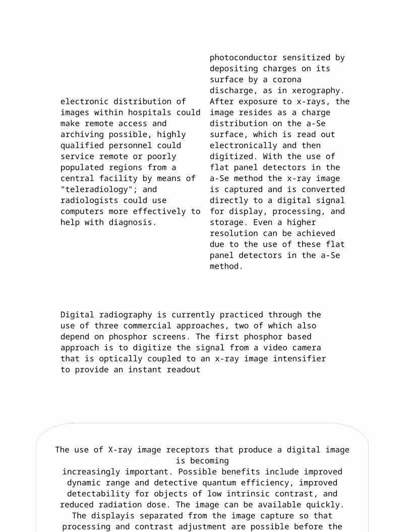
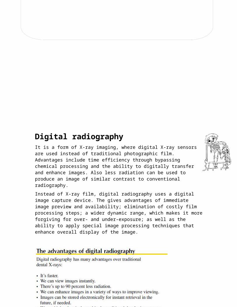
Digital radiographyIt is a form of X-ray imaging, where digital X-ray sensors are used instead of traditional photographic film. Advantages include time efficiency through bypassing chemical processing and the ability to digitally transfer and enhance images. Also less radiation can be used to produce an image of similar contrast to conventional radiography.
Instead of X-ray film, digital radiography uses a digital image capture device. The gives advantages of immediate image preview and availability; elimination of costly film processing steps; a wider dynamic range, which makes it more forgiving for over- and under-exposure; as well as the ability to apply special image processing techniques that enhance overall display of the image.
The use of X-ray image receptors that produce a digital image is becomingincreasingly important. Possible benefits include improved dynamic range and detective quantum efficiency, improved detectability for objects of low intrinsic
contrast, and reduced radiation dose. The image can be available quickly. The displayis separated from the image capture so that processing and contrast adjustment are
possible before the image is viewed. The availability of a digital image means ready input into PACS and opens up the possibility of computer-aided detection and
classification of abnormality. Possible drawbacks of digital systems include high cost, limited high contrast resolution and the fact that their clinical value is sometimes not
proven in comparison with conventional, analogue techniques. The high contrast resolution attainable with such systems is discussed and the problem of sampling
limitations and aliasing considered. The properties and limitations of digital systems using computed radiography, caesium iodide plus CCDs and active matrix arrays with either caesium iodide or selenium detectors are demonstrated. Examples are given of digital systems for mammography and general radiography and their performance is demonstrated in terms of clinical assessment and measurements of the modulation
transfer function and detective quantum efficiency.
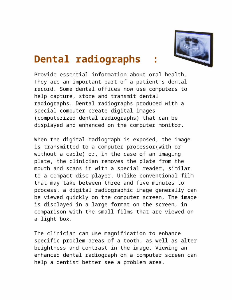
Dental radiographs :
Provide essential information about oral health. They are an important part of a patient’s dental record. Some dental offices now use computers to help capture, store and transmit dental radiographs. Dental radiographs produced with a special computer create digital images (computerized dental radiographs) that can be displayed and enhanced on the computer monitor.
When the digital radiograph is exposed, the image is transmitted to a computer processor(with or without a cable) or, in the case of an imaging plate, the clinician removes the plate from the mouth and scans it with a special reader, similar to a compact disc player. Unlike conventional film that may take between three and five minutes to process, a digital radiographic image generally can be viewed quickly on the computer screen. The image is displayed in a large format on the screen, in comparison with the small films that are viewed on a light box.
The clinician can use magnification to enhance specific problem areas of a tooth, as well as alter brightness and contrast in the image. Viewing an enhanced dental radiograph on a computer screen can help a dentist better see a problem area.
Digital X-Ray Machine and Camera System
Parts of a Digital X-Ray Machine:
The digital X-Ray machine consists of an X-Ray tube and driver to source X-Ray. The X-Ray passes through the patient's body and the digital camera (located on the other side of the patient) captures the resulting image. The main base station controls the X-Ray tube, analyzes the image and displays the image on the CRT.
Digital Camera:The digital camera converts the received image into digital signal and transfers the digitized image to the base station through a fiber optic link.
Main Base StationThe functions of the main base station are as follows:
Drive and control the X-Ray tube Communicate with the digital camera system through Fiber Store the pictures on the hard disk for image retrieval and processing Interface to an operator console for overall system control and image
manipulation Analyze and enhance the image stored on the hard disk and display the
enhanced X-Ray image on a CRT monitor
Principles :
Conventional imaging
Conventional intra-oral radiographic film consists of silver halide grains in a gelatine matrix. When this film is exposed to X-ray photons the silver halide crystals are sensitized and are reduced to black during the developing process. The film acts as both the radiation detector and the image display.
With extra-oral films indirect action receptors are used to help record the image. This type of film is sensitive to light photons which are emitted by adjacent intensifying screens. Although the film is constructed of silver halide crystals these are primarily sensitive to light rather than X-rays. The use of intensifying screens reduces the dose and can be used where fine detail is not required.
Digital imaging
In digital radiography, instead of the silver halide grain the image is constructed using pixels or small light sensitive elements. These pixels can be a range of shades of grey depending on the exposure, and are arranged in grids and rows on the sensor, unlike the random distribution of the crystals in standard film. However,
unlike film the sensors are only the radiation detector and the image is displayed on a monitor.
The signal that is produced by the sensor is an analogue signal, i.e. a voltage that varies as a function of time. The sensor is connected to the computer and the signal is sampled at regular intervals. The output of each pixel is quantified and converted to numbers by a frame grabber within the computer. The range of numbers is normally from 0 to 256 with 0 representing black, 256 representing white and all others are shades of grey.
The number of grey levels relates to contrast resolution and the size of the pixels is related to spatial resolution. Together these determine the overall resolution (i.e. the ability to distinguish between small objects close together) of the image. Resolution can also be expressed in line pairs per millimetre. Most conventional E speed films have a resolution of 20 LP/mm whereas with digital images the resolution ranges from 7–10 LP/mm. The reduced resolution should not interfere with clinical diagnosis.
Image acquisition :There are two ways to acquire a digital image.
Indirect acquisition
A digital image can be produced by scanning conventional radiographs using a flatbed scanner and a transparency adaptor, or by using a charged coupled device camera instead of the flatbed scanner. This image can then be manipulated using software packages or be passed on to a second party via a modem.
Direct digital imaging
There are two systems available, one produces the image immediately on the monitor post-exposure and is therefore called Direct Imaging. The second has an intermediate phase, whereby the image is produced on the monitor following scanning by laser. This is known as semi-direct imaging.
Semi-direct image plate systems. The image plate method involves the use of a phosphor storage plate (PSP). This plate stores energy after exposure to radiation and emits light when scanned by a laser. The scanner stimulates the phosphor plate and stores a record of the number of light photons detected.
Loading of the scanners generally only requires subdued lighting as the plates are slightly sensitive to visible light. However, some products are more light sensitive than others. The lasers used are centred around the 600-nm band and are usually of the helium-neon variety. Scanners, the size of a breadmaker, can accommodate multiple image plates at any one time. The exact numbers varies between manufacturers. There is a delay while the image is ‘developed’ before it appears on the monitor. Up to eight bitewing radiographs take about 90 seconds and a panoramic image can take approximately 3 minutes to be scanned. Again, the scan times do vary between manufacturers. Although the plate can store energy for a number of days, information starts to be lost within minutes after exposure and it is advised to scan the plates quite quickly to optimize the image recovered. To fully remove the latent image the plate should be exposed to high intensity light (as found on viewing boxes).
Image plates are available in exactly the same sizes as conventional film and come with disposable plastic barriers. They have no wires attached and are reusable for thousands of

exposures, but do need careful handling to avoid surface damage. Current systems have a spatial resolution of 6–8 LP/mm.
Direct sensor systems. The sensor for the radiation image is usually a Charge Coupled Device (CCD). It consists of silicon crystals arranged in a lattice and converts light energy into an electronic signal. This technology is widely used in video cameras. The sensor cannot store information and must be connected via fibre optic wires to the monitor, which can make the sensor bulky and awkward to use.
The greatest advantage of the direct sensor system is the gain in time. The image is directly projected onto the computer screen. Originally, the active areas of the sensors were smaller than conventional film, which increased the incidence of ‘coning off’ and required repeat exposures to capture all the desired information. Recent innovations have produced sensors approaching or equal to standard film sizes.
Extra-oral digital imaging :Extra-oral digital imaging is available using both systems. However, the larger CCD sensors are extremely expensive and usually requires the purchase of new X-ray generators, although a ‘retro-fit’ system has been developed in the USA. These constrictions effectively mean that the PSP method is the one most commonly used.
Panoramic radiography
The PSP method of panoramic digital imaging is very similar to conventional film. The film and intensifying screen are replaced by a storage phosphor plate. The plate is scanned after exposure,
which can take up to 3 minutes or longer depending on the product used. The resolution of these systems is greater than 4 LP/mm.
Cephalometric radiography
Naslund et al. investigated the effect of dose reduction obtained with PSP on the identification of cephalometric landmarks and concluded that dose reductions of up to 75 per cent did not effect the localization of cephalometric landmarks.1 It is also worth noting that with CCD sensors the image is acquired over 15 seconds as the sensor and narrow X-ray beam move up the facial bones and could lead to an increase in the incidence of movement artefact.
Advantages of digital imaging :
Dose reduction
Dose reductions of up to 90 per cent compared to E-speed film have been reported by some authors in the diagnosis of caries.2 Although some researchers do claim dose reductions compared with conventional extra-oral film, in practice the background noise rises to unacceptable levels. It is now accepted that there is no appreciable reduction compared with films used in conjunction with rare earth intensifying screens.
Image manipulation
This is perhaps the greatest advantage of digital imaging over conventional film. It involves selecting the information of greatest diagnostic value and suppressing the rest. Manufacturers provide
software programmes with many different processing tools, however some are more useful than others and these include:
Contrast enhancement. This can effectively compensate for over or under exposure of the digital image. It has been shown that contrast enhancement of CCD devices were more accurate than E-speed film for detecting simulated caries under orthodontic bands.3
Measurements. Digital callipers, rulers and protractors are some of the many tools available for image analysis. Many authors have reported on their application in cephalometric analysis.4,5 The images can also be superimposed onto each other and onto digital photographs.
3-D reconstruction. This application can be theoretically used to reconstruct intra- and extra-oral images. The uses range from profiling root canals to visualizing facial fractures in all three dimensions.
Filtration. The addition of filters to the airspace around the face can clarify the soft tissue profile if the original soft tissue image was poor.
Time
Much time is gained especially with the CCD system where the image is displayed at the chairside immediately post exposure. Although a lag time between scanning and the appearance of an image exists with the PSP method it is still substantially faster than conventional developing processes in general use.
Storage
Storage was initially a problem before the development of DVDs and CD ROMs as three peri-apical images would fill a floppy disc.
However, now a CD ROM can hold over 30,000 images. This means that images can be stored cheaply and indefinitely.
Teleradiology
The digital image file can be further reduced in size by compression techniques, and sent via a modem and telephone line to colleagues for review. This had the advantages of not losing radiographs in the post and saving time if an urgent appointment is required. The operator at the other end can also manipulate the image if desired.
Environmentally friendly
No processing chemicals are used or disposed of. Both CCD sensors and the PSP plates are capable of being reused for many thousands of exposures. They can, however, become scratched and damaged if not handled carefully.
Disadvantages of digital imagingThe majority of the disadvantages are associated with the CCD system.
Cost
Currently, the cost of converting from intra-oral film to digital imaging is approximately 6600 Euros. This initial outlay should be offset against the time saved and the efficiency of storage of the images.
Sensor dimensions
These are still quite bulky for the CCD system and awkward to position due to trailing fibre optic wires. The original problem of small sensor active areas has been rectified and the same amount of information can be captured as conventional film.
Cross-infection control
Each intra-oral sensor and plate must be covered by a plastic bag, and this bag is changed between patients. However, if they become directly contaminated there is no way of sterilizing them and they should be discarded regardless of expense.
Medicolegal
Concerns have been raised in the past about the ability to manipulate the images for fraudulent purposes. Manufacturers of software programmes have installed ‘audit trails’, which can track down and recover the original image. Many insurance companies in the USA are accepting digital images as valid attachments when the claims are electronically claimed.
ConclusionsThe technology is now available to run a practice almost paper free. It is theoretically possible to store clinical notes, photographs, radiographs, and study models on disc, and refer or consult online. The future of digital imaging could include the testing and upgrade of X-ray equipment and software on-line. Research is also continuing into the development of a credit card sized ‘smart card’, which could carry a patient's medical and dental notes along with their radiographic images. It is important that advances in technology are accepted and the benefits that they produce utilized in order that clinical practice and patient care continue to improve.
ReferencesHMS. Healthcare Management Systems, Inc. (2003). Retrieved July 16, 2009,
Web site: http://www.hmstn.com
Lattice Semiconductor Corporation. (n.d.)Digital X-Ray Machine and Camera System. (2009). Retrieved June 20, 2009,
Web site: http://www.vantis.com/solutions/marketsolutions/medical/digitalxra ymachinecameras.cfm
JPI America Inc. (n.d.) Enhancing Radiology. Retrieved July 5, 2009, Web site: http://www.jpiamerica.com/imaging-solutions
McGonigle, D., & Mastrian, K., (2009). Nursing Informatics and the Foundation of Knowledge (1st ed.). Jones & Bartlett Publishers, Sudbury, Massachusetts.
Mosby's Medical Dictionary Online, 8th edition. © 2009, Elsevier. Retrieved June 10, 2009, Web site: http://medicaldictionary.thefreedictionary.com
Sprawls, P. (n.d.) Physical Principles of Medical Imaging Online. Retrieved June 5, 2009, Web site: www.sprawls.org/resources