Embed Size (px)
Citation preview
See discussions, stats, and author profiles for this publication at: https://www.researchgate.net/publication/309426680
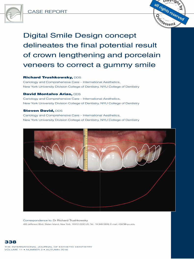
Digital Smile Design concept delineates the final potential result of crown
lengthening and porcelain veneers to correct a gummy smile
Article in The International Journal of Esthetic Dentistry · July 2016
CITATIONS
25READS
6,623
1 author:
David Montalvo Arias
Apa Aesthetic and Cosmetic Dental Centre, Dubai, UAE
3 PUBLICATIONS 55 CITATIONS
SEE PROFILE
All content following this page was uploaded by David Montalvo Arias on 26 October 2016.
The user has requested enhancement of the downloaded file.
338THE INTERNATIONAL JOURNAL OF ESTHETIC DENTISTRY
CASE REPORT
Digital Smile Design concept
delineates the final potential result
of crown lengthening and porcelain
veneers to correct a gummy smile
Richard Trushkowsky, DDS
Cariology and Comprehensive Care – International Aesthetics,
New York University Division College of Dentistry, NYU College of Dentistry
David Montalvo Arias, DDS
Cariology and Comprehensive Care – International Aesthetics,
New York University Division College of Dentistry, NYU College of Dentistry
Steven David, DDS
Cariology and Comprehensive Care – International Aesthetics,
New York University Division College of Dentistry, NYU College of Dentistry
Correspondence to: Dr Richard Trushkowsky
483 Jefferson Blvd, Staten Island, New York, 10312-2332 US; Tel.: 18-948-5808; E-mail: [email protected]
339THE INTERNATIONAL JOURNAL OF ESTHETIC DENTISTRY
TRUSHKOWSKY ET AL
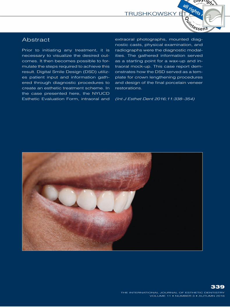
Abstract
Prior to initiating any treatment, it is
necessary to visualize the desired out-
comes. It then becomes possible to for-
mulate the steps required to achieve this
result. Digital Smile Design (DSD) utiliz-
es patient input and information gath-
ered through diagnostic procedures to
create an esthetic treatment scheme. In
the case presented here, the NYUCD
Esthetic Evaluation Form, intraoral and
extraoral photographs, mounted diag-
nostic casts, physical examination, and
radiographs were the diagnostic modal-
ities. The gathered information served
as a starting point for a wax-up and in-
traoral mock-up. This case report dem-
onstrates how the DSD served as a tem-
plate for crown lengthening procedures
and design of the final porcelain veneer
restorations.
(Int J Esthet Dent 2016;11:338–354)

340THE INTERNATIONAL JOURNAL OF ESTHETIC DENTISTRY
CASE REPORT
The upper third may fluctuate due to the
variability of the hairline. The middle and
lower thirds are more involved in esthetic
perception. The midface is measured
from the glabella to the subnasale (the
most protruding area on the forehead
between the eyebrows and the point di-
rectly under the nose). The lower face is
measured from the subnasale to the soft
tissue menton. Often, VME can be treat-
ed by orthognathic surgery. A short up-
per lip (determined by measuring from
the subnasale to the inferior border of
the upper lip) can also cause a gummy
smile. HUL is the result of hyperfunction
of the lip elevator muscles and is usu-
ally the cause of a gummy smile if the
lip length is within normal limits and the
lower third of the face is in proportion
to the remaining two thirds. Yet another
cause of a gummy smile is altered pas-
sive eruption (APE), which is due to a
deviation in normal development result-
ing in a large amount of the anatomic
crown being covered by the gingiva,
and minimal scalloping. APE has been
classified into two types: Type 1 is a
result of a disproportionate amount of
gingiva measured from the free gingival
margin to the mucogingival junction. In
Type 2, there is a normal dimension of
gingiva when measured from the free
gingival margin to the mucogingival
junction, but the gingiva extends over
the coronal portion of the tooth. Based
on an anatomic histological foundation,
Type 1 can be categorized into 1A – an
excessive amount of keratinized gingiva
with normal alveolar crest-to-cementoe-
namel junction (CEJ) relationship; and
1B – an excessive amount of keratinized
gingiva with the osseous crest at the CEJ
level. The association of the osseous
Introduction
Esthetic dental concerns have become
more widespread among people with rel-
atively affluent lifestyles in at least some
segments of the population in almost all
countries. Patients’ esthetic awareness
and expectations have increased, so
that close to what are perceived to be
ideal outcomes are required. Long-term
stability necessitates dental restorations
that are congruent with the periodon-
tium and occlusion.1 An esthetic smile
consists of three main constituents: the
teeth, the lip framework, and the gingi-
val scaffold.2 An ideal smile has the fol-
lowing properties: minimal gingival dis-
play, symmetry and harmony between
the maxillary gingiva and the upper lip,
healthy gingival tissue filling the entire
interproximal spaces, harmony between
the anterior and posterior segments,
teeth with correct form and position,
proper tooth color and shade, and the
lower lip parallel to the incisal edges of
the maxillary anterior teeth and to an im-
aginary line going through the contact
points of these teeth.3,4 When a smile
displays a disproportionate amount of
gingiva, this phenomenon is referred to
as a gummy smile.5
At rest, young women usually display
3 to 4 mm of the maxillary central inci-
sors, and young men display an aver-
age of 2 mm or less. Extraoral causes
of a gummy smile are vertical maxillary
excess (VME), hypermobile upper lip
(HUL) or a short upper lip. Face height
is usually measured by dividing the face
into thirds. A visual diagnosis of VME
can be made when, on cephalometric
analysis, the lower third of the face is
longer than the middle and upper thirds.
341THE INTERNATIONAL JOURNAL OF ESTHETIC DENTISTRY
TRUSHKOWSKY ET AL
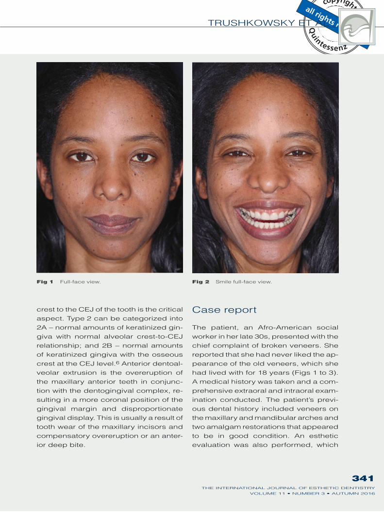
Fig 1 Full-face view. Fig 2 Smile full-face view.
crest to the CEJ of the tooth is the critical
aspect. Type 2 can be categorized into
2A – normal amounts of keratinized gin-
giva with normal alveolar crest-to-CEJ
relationship; and 2B – normal amounts
of keratinized gingiva with the osseous
crest at the CEJ level.6 Anterior dentoal-
veolar extrusion is the overeruption of
the maxillary anterior teeth in conjunc-
tion with the dentogingival complex, re-
sulting in a more coronal position of the
gingival margin and disproportionate
gingival display. This is usually a result of
tooth wear of the maxillary incisors and
compensatory overeruption or an anter-
ior deep bite.
Case report
The patient, an Afro-American social
worker in her late 30s, presented with the
chief complaint of broken veneers. She
reported that she had never liked the ap-
pearance of the old veneers, which she
had lived with for 18 years (Figs 1 to 3).
A medical history was taken and a com-
prehensive extraoral and intraoral exam-
ination conducted. The patient’s previ-
ous dental history included veneers on
the maxillary and mandibular arches and
two amalgam restorations that appeared
to be in good condition. An esthetic
evaluation was also performed, which
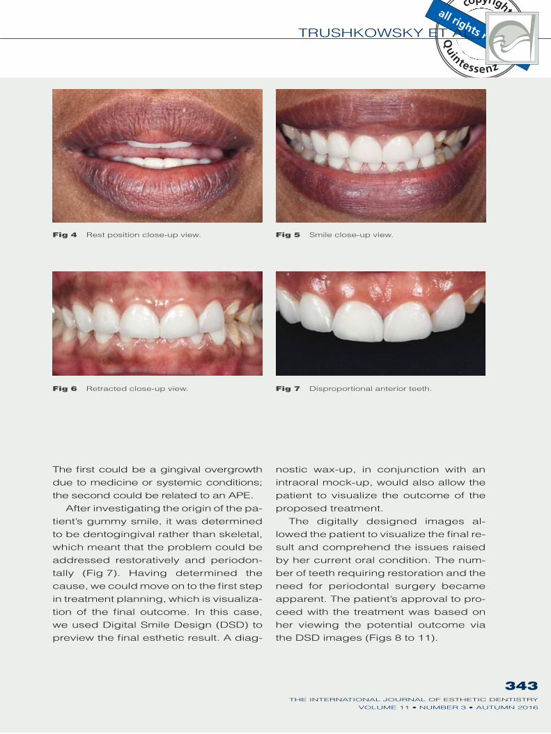
342THE INTERNATIONAL JOURNAL OF ESTHETIC DENTISTRY
CASE REPORT
included mounted models, radiographs,
photographs, and an esthetic evaluation
form incorporating the changes desired
by the patient (Figs 4 to 6).
The following problem list was cre-
ated from the gathered data:
Excessive maxillary gingival
display;
Broken veneers on teeth 11, 12, and
21;
Margins of veneers broken or stained
on teeth 5 to 10, and 22 to 28;
Unattractive proportions of the
anterior teeth.
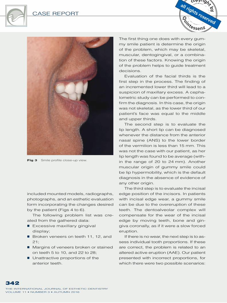
Fig 3 Smile profile close-up view.
The first thing one does with every gum-
my smile patient is determine the origin
of the problem, which may be skeletal,
muscular, dentogingival, or a combina-
tion of these factors. Knowing the origin
of the problem helps to guide treatment
decisions.
Evaluation of the facial thirds is the
first step in the process. The finding of
an incremented lower third will lead to a
suspicion of maxillary excess. A cepha-
lometric study can be performed to con-
firm the diagnosis. In this case, the origin
was not skeletal, as the lower third of our
patient’s face was equal to the middle
and upper thirds.
The second step is to evaluate the
lip length. A short lip can be diagnosed
whenever the distance from the anterior
nasal spine (ANS) to the lower border
of the vermilion is less than 15 mm. This
was not the case with our patient, as her
lip length was found to be average (with-
in the range of 20 to 24 mm). Another
muscular origin of gummy smile could
be lip hypermobility, which is the default
diagnosis in the absence of evidence of
any other origin.
The third step is to evaluate the incisal
edge position of the incisors. In patients
with incisal edge wear, a gummy smile
can be due to the overeruption of these
teeth. The dentoalveolar complex will
compensate for the wear of the incisal
edge by moving teeth, bone and gin-
giva coronally, as if it were a slow forced
eruption.
If there is no wear, the next step is to as-
sess individual tooth proportions. If these
are correct, the problem is related to an
altered active eruption (AAE). Our patient
presented with incorrect proportions, for
which there were two possible scenarios:
343THE INTERNATIONAL JOURNAL OF ESTHETIC DENTISTRY
TRUSHKOWSKY ET AL
Fig 4 Rest position close-up view. Fig 5 Smile close-up view.
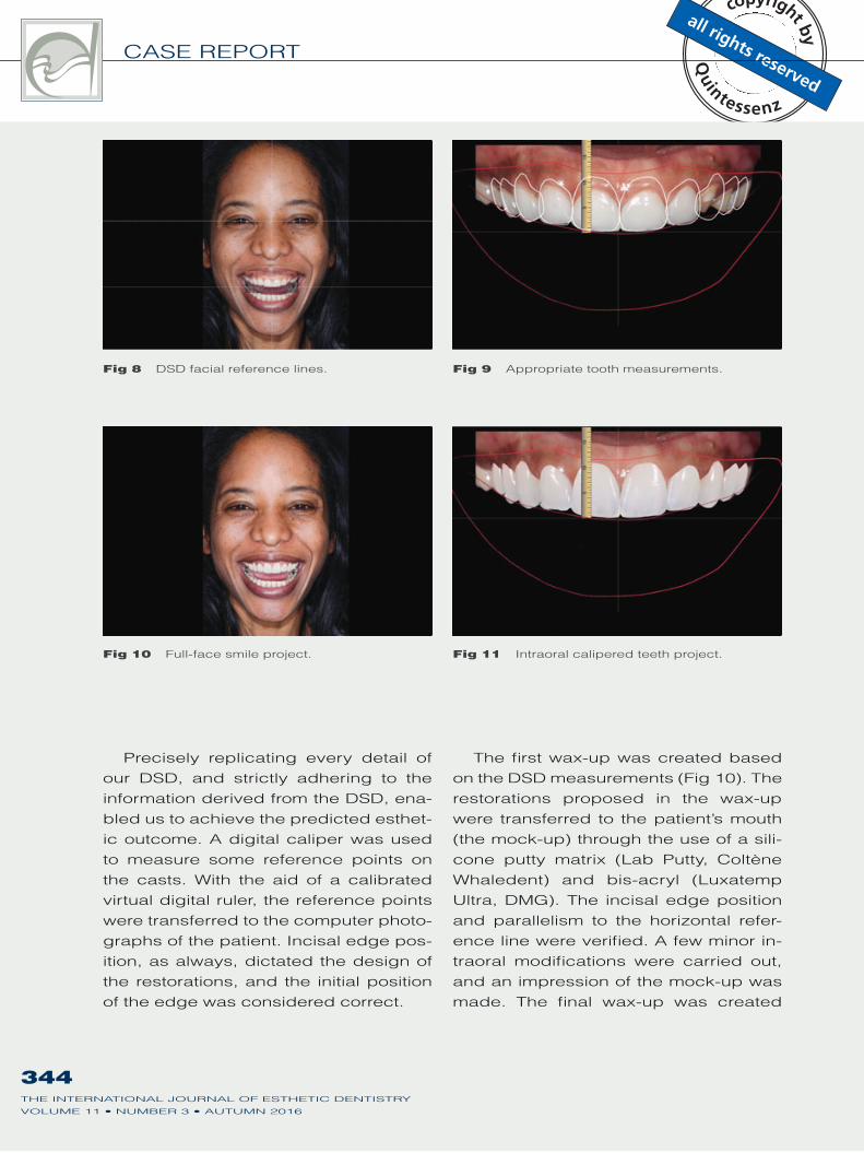
Fig 6 Retracted close-up view. Fig 7 Disproportional anterior teeth.
The first could be a gingival overgrowth
due to medicine or systemic conditions;
the second could be related to an APE.
After investigating the origin of the pa-
tient’s gummy smile, it was determined
to be dentogingival rather than skeletal,
which meant that the problem could be
addressed restoratively and periodon-
tally (Fig 7). Having determined the
cause, we could move on to the first step
in treatment planning, which is visualiza-
tion of the final outcome. In this case,
we used Digital Smile Design (DSD) to
preview the final esthetic result. A diag-
nostic wax-up, in conjunction with an
intraoral mock-up, would also allow the
patient to visualize the outcome of the
proposed treatment.
The digitally designed images al-
lowed the patient to visualize the final re-
sult and comprehend the issues raised
by her current oral condition. The num-
ber of teeth requiring restoration and the
need for periodontal surgery became
apparent. The patient’s approval to pro-
ceed with the treatment was based on
her viewing the potential outcome via
the DSD images (Figs 8 to 11).
344THE INTERNATIONAL JOURNAL OF ESTHETIC DENTISTRY
CASE REPORT
Precisely replicating every detail of
our DSD, and strictly adhering to the
information derived from the DSD, ena-
bled us to achieve the predicted esthet-
ic outcome. A digital caliper was used
to measure some reference points on
the casts. With the aid of a calibrated
virtual digital ruler, the reference points
were transferred to the computer photo-
graphs of the patient. Incisal edge pos-
ition, as always, dictated the design of
the restorations, and the initial position
of the edge was considered correct.
The first wax-up was created based
on the DSD measurements (Fig 10). The
restorations proposed in the wax-up
were transferred to the patient’s mouth
(the mock-up) through the use of a sili-
cone putty matrix (Lab Putty, Coltène
Whaledent) and bis-acryl (Luxatemp
Ultra, DMG). The incisal edge position
and parallelism to the horizontal refer-
ence line were verified. A few minor in-
traoral modifications were carried out,
and an impression of the mock-up was
made. The final wax-up was created
Fig 8 DSD facial reference lines. Fig 9 Appropriate tooth measurements.
Fig 10 Full-face smile project. Fig 11 Intraoral calipered teeth project.
345THE INTERNATIONAL JOURNAL OF ESTHETIC DENTISTRY
TRUSHKOWSKY ET AL
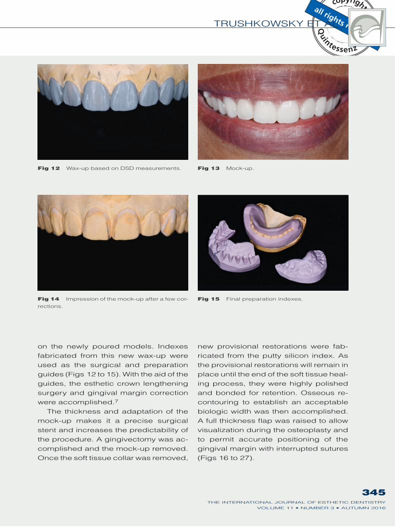
Fig 12 Wax-up based on DSD measurements. Fig 13 Mock-up.
Fig 14 Impression of the mock-up after a few cor-
rections.
Fig 15 Final preparation indexes.
on the newly poured models. Indexes
fabricated from this new wax-up were
used as the surgical and preparation
guides (Figs 12 to 15). With the aid of the
guides, the esthetic crown lengthening
surgery and gingival margin correction
were accomplished.7
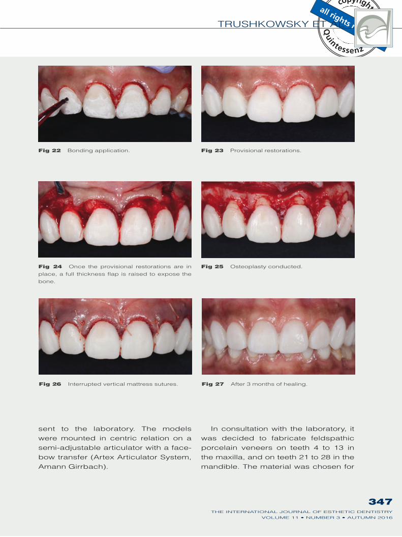
The thickness and adaptation of the
mock-up makes it a precise surgical
stent and increases the predictability of
the procedure. A gingivectomy was ac-
complished and the mock-up removed.
Once the soft tissue collar was removed,
new provisional restorations were fab-
ricated from the putty silicon index. As
the provisional restorations will remain in
place until the end of the soft tissue heal-
ing process, they were highly polished
and bonded for retention. Osseous re-
contouring to establish an acceptable
biologic width was then accomplished.
A full thickness flap was raised to allow
visualization during the osteoplasty and
to permit accurate positioning of the
gingival margin with interrupted sutures
(Figs 16 to 27).
346THE INTERNATIONAL JOURNAL OF ESTHETIC DENTISTRY
CASE REPORT
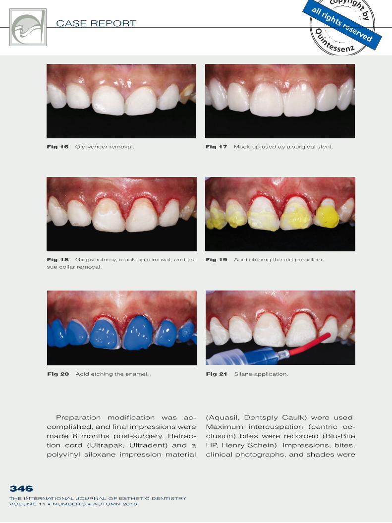
Fig 16 Old veneer removal. Fig 17 Mock-up used as a surgical stent.
Fig 18 Gingivectomy, mock-up removal, and tis-
sue collar removal.
Fig 19 Acid etching the old porcelain.
Fig 20 Acid etching the enamel. Fig 21 Silane application.
Preparation modification was ac-
complished, and final impressions were
made 6 months post-surgery. Retrac-
tion cord (Ultrapak, Ultradent) and a
polyvinyl siloxane impression material
(Aquasil, Dentsply Caulk) were used.
Maximum intercuspation (centric oc-
clusion) bites were recorded (Blu-Bite
HP, Henry Schein). Impressions, bites,
clinical photographs, and shades were
347THE INTERNATIONAL JOURNAL OF ESTHETIC DENTISTRY
TRUSHKOWSKY ET AL
Fig 22 Bonding application. Fig 23 Provisional restorations.
Fig 24 Once the provisional restorations are in
place, a full thickness flap is raised to expose the
bone.
Fig 25 Osteoplasty conducted.
Fig 26 Interrupted vertical mattress sutures. Fig 27 After 3 months of healing.
sent to the laboratory. The models
were mounted in centric relation on a
semi-adjustable articulator with a face-
bow transfer (Artex Articulator System,
Amann Girrbach).
In consultation with the laboratory, it
was decided to fabricate feldspathic
porcelain veneers on teeth 4 to 13 in
the maxilla, and on teeth 21 to 28 in the
mandible. The material was chosen for

348THE INTERNATIONAL JOURNAL OF ESTHETIC DENTISTRY
CASE REPORT
Fig 29 Feldspathic veneers.
Fig 28 Final preparations.
its esthetic qualities and due to the ab-
sence of contraindicating occlusal is-
sues. Although not as strong as pressed
veneers, feldspathic veneers provide
better color control. Moreover, less re-
duction is required. When the veneers
were returned from the laboratory, they
were inspected for conformity to the final
wax-up. They were then tried in using a
transparent shade try-in gel (Variolink II,
Ivoclar Vivadent). The patient was given
the opportunity to see the restorations
in her mouth and consented to their ce-
mentation. A water rinse was used to
remove all traces of the try-in gel from
the restorations. The internal surfaces of
the restorations were scrubbed for 15 s
with a 35% phosphoric acid solution
(Ultra-Etch, Ultradent) and ultrasonically
cleaned in alcohol for 1 min. Silane prim-
er (Ultradent) was placed on the internal
surface of the veneers and allowed to air
dry. Bonding agent (Prime & Bond NT,
Dentsply) was applied, allowing 30 s for
the solvent to evaporate. The veneered
teeth were isolated with rubber dam,
etched with Ultra-Etch for 15 s, then
rinsed with water for 30 s. Prime & Bond
NT bonding agent was applied to the
internal surface of the veneers. The res-
torations were then loaded with the base
shade (Variolink II cement transparent)
and seated on the teeth. A small brush

349THE INTERNATIONAL JOURNAL OF ESTHETIC DENTISTRY
TRUSHKOWSKY ET AL
Fig 30 Complete isolation cementation.
as well as floss was used to remove the
excess cement before light curing for
40 s. A final check of the occlusion was
made with articulating paper (AccuFilm,
Parkell) and minor adjustments were
performed (Figs 28 to 31).
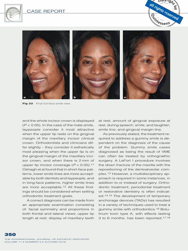
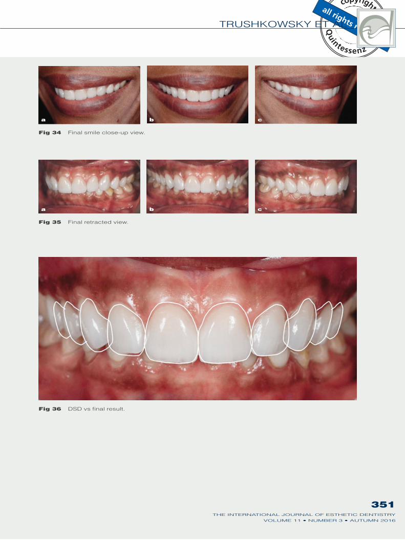
Due to its dentogingival origin, we
were able to completely correct the
patient’s gummy smile. The final result
achieved in this case demonstrates
what may be accomplished by using a
systematic interdisciplinary approach,
assisted by DSD (Figs 32 to 36).
Discussion
Excessive gingival display or gummy
smile represents an emotionally charged
esthetic concern for many patients and
a technique-sensitive challenge for clin-
icians. The clinician must understand
the various causes, determine the cor-
rect diagnosis, and formulate a clinically
predictable esthetic treatment plan. The
diagnosis of gummy smile is not rare; the
incidence of excessive gingival display
is 10% of the population between 20 and
30 years of age, and is more commonly
diagnosed in women.8,9 In their study,
Peck et al found a significant gender
dimorphism in the vertical lip–tooth–jaw
relationship: the upper lip of the females
in the study was positioned on average
1.5 mm more superiorly at maximum
smile than that of the males (P < 0.01).10
The gingival smile line is the smile at its
fullest and exposes the gingiva super-
ior to the maxillary anterior teeth.10 Ac-
cording to orthodontists, clinicians and
laypeople, the most attractive female
smile is when the upper lip rests on the
gingival margin of the maxillary incisor
Fig 31 Incisal characterization of the veneers.
Fig 32 Final profile close-up view.
350THE INTERNATIONAL JOURNAL OF ESTHETIC DENTISTRY
CASE REPORT
a b c
Fig 33 Final full-face smile view.
and the whole incisor crown is displayed
(P < 0.05). In the case of the male smile,
laypeople consider it most attractive
when the upper lip rests on the gingival
margin of the maxillary incisor clinical
crown. Orthodontists and clinicians dif-
fer slightly – they consider it esthetically
most pleasing when the upper lip is on
the gingival margin of the maxillary inci-
sor crown, and when there is 2 mm of
upper lip incisor coverage (P < 0.05).11
Oshagh et al found that in short-face pat-
terns, lower smile lines are more accept-
able by both dentists and laypeople, and
in long-face patterns, higher smile lines
are more acceptable.12 All these find-
ings should be considered when setting
orthodontic treatment goals.
A correct diagnosis can be made from
an appropriate examination consisting
of: facial symmetry and proportions in
both frontal and lateral views; upper lip
length at rest; display of maxillary teeth
at rest; amount of gingival exposure at
rest, during speech, smile, and laughter;
smile line; and gingival margin line.
As previously stated, the treatment re-
quired to address a gummy smile is de-
pendent on the diagnosis of the cause
of the problem. Gummy smile cases
diagnosed as being the result of VME
can often be treated by orthognathic
surgery. A LeFort I procedure involves
the down fracture of the maxilla with the
repositioning of the dentoalveolar com-
plex.13 However, a multidisciplinary ap-
proach is required in some instances, in
addition to or instead of surgery. Ortho-
dontic treatment, periodontal treatment
or restorative dentistry is often indicat-
ed.14,15 The development of temporary
anchorage devices (TADs) has resulted
in a variety of techniques used to treat a
gummy smile with orthodontics.16 Botu-
linum toxin type A, with effects lasting
3 to 6 months, has been reported.17,18
351THE INTERNATIONAL JOURNAL OF ESTHETIC DENTISTRY
TRUSHKOWSKY ET AL
a b c
Fig 34 Final smile close-up view.
a b c
Fig 35 Final retracted view.
Fig 36 DSD vs final result.
352THE INTERNATIONAL JOURNAL OF ESTHETIC DENTISTRY
CASE REPORT
Reestablishing the depth of the vesti-
bule to treat a short upper lip has also
been reported. Similarly, a surgical pro-
cedure to limit the movement of the el-
evator muscles has also been recom-
mended.19
In this case, the origin of the gummy
smile was determined to be dentogin-
gival. Once that assessment had been
made, DSD was used to visualize the fi-
nal esthetic result. The key to successful
treatment was then to select the appro-
priate techniques to correct the anatom-
ic problems, maintain the biologic width,
and achieve the visualized final esthetic
result. Based on the work of Gargiulo et
al, the biological width is defined as the
dimension of the soft tissue that is at-
tached to the portion of the tooth coro-
nal to the crest of the alveolar bone.20
After evaluating 171 cadaver tooth sur-
faces, Vacek et al reported the follow-
ing mean dimensions: a sulcus depth
of 0.69 mm; an epithelial attachment of
0.97 mm and a connective tissue attach-
ment of 1.07 mm; observed mean meas-
urements of 1.34 mm for sulcus depth;
1.14 mm for epithelial attachment; and
0.77 mm for connective tissue attach-
ment.21 Esthetic crown lengthening is
categorized as being either Type I, II,
III, or IV. In Type I cases, there is suf-
ficient attached gingiva coronal to the
osseous crest, and the need for osse-
ous recontouring is therefore obviated.
A simple gingivectomy that simultane-
ously sculpts, contours and maintains
appropriate zeniths may be all that is
required.22 Type II crown lengthening
is categorized by gingival proportions
that permit an apical positioning of the
gingival margin, do not reveal the osse-
ous crest, but violate the biologic width.
In these cases, the body will spontane-
ously attempt to reestablish the correct
biologic width. Recession will occur in
thin biotypes due to crestal resorption,
and protracted inflammation will occur
in thick biotypes. Either scenario would
be detrimental to any restoration placed
in this environment. Osseous recontour-
ing is required to rectify the violation of
the biologic width. As in this case, this
can be accomplished by placing pro-
visionals at the preferred crown length
and then waiting for soft tissue healing.
A flap is later raised while the papilla is
maintained, and osseous recontouring is
accomplished, with the provisional res-
toration providing a surgical template.
The flap is then replaced in its previous
position. Type III crown lengthening is
needed when repositioning of the gingi-
val margin would result in disclosure of
the osseous crest. To encourage rees-
tablishment of a healthy biologic width,
a surgical template is required to assist
in appropriate bone reshaping under the
elevated flap. The gingival margin is re-
positioned coronally to conserve soft tis-
sue. Type IV esthetic crown lengthening
is required when inadequate attached
gingiva is present. An apically pos-
itioned flap is required with a definitive
margin, and provisional construction ac-
complished at a later date.
For this patient, a Type III one-stage
surgical crown lengthening procedure
was selected as being the most benefi-
cial. According to Sonick et al,23 a sin-
gle-stage crown lengthening procedure
often results in a 1 to 3 mm rebound of
the free gingival margin 6 months to 1
year post-surgery. This would especially
apply to patients with a thick biotype.
The procedure allowed for the gingivec-
353THE INTERNATIONAL JOURNAL OF ESTHETIC DENTISTRY
TRUSHKOWSKY ET AL
tomy and placement of the provisional
veneers at the same visit. Sonick et al
recommend a two-phase crown length-
ening procedure, in which an ostectomy
is initially accomplished and, several
weeks later, a gingivectomy is performed
subsequent to initial attachment and
bone healing. In Lee’s opinion,22 since
the reaction of the soft tissue to violation
of the biologic width is not immediate,
restorations can be placed immediately
after a gingivectomy, and osseous re-
contouring surgery can be done later.
This allows precise placement of the os-
seous crest relative to the margins of the
provisional restoration so as to reestab-
lish the biologic width.
Conclusion
Precise treatment planning is essential to
achieve a long-lasting esthetic outcome
for patients presenting with a desire to
correct a gummy smile. DSD is a pow-
erful tool that can be used to expedite
the analysis of the patient’s facial and
dental features and assist in determin-
ing how the finished case will look. The
proper diagnosis of a gummy smile is a
prerequisite to any restorative treatment
that may be required.
Acknowledgment
The authors wish to thank Jason J. Kim, CDT (Jason
J. Kim Oral Design Center) for the excellent labora-
tory work.
References
1. Jorgensen MG, Nowzari
H. Aesthetic crown length-
ening. Periodontol 2000
2001;27:45–58.
2. Garber DA, Salama MA. The
aesthetic smile: diagnosis
and treatment. Periodontol
2000 1996;11:18–28.
3. de Castro MV, Santos NC,
Ricardo LH. Assessment of
the “golden proportion” in
agreeable smiles. Quintes-
sence Int 2006;37:597–604.
4. Landsberg CJ, Sarne O.
Management of excessive
gingival display following
adult orthodontic treatment:
a case report. Pract Proced
Aesthet Dent 2006;18:89–94.
5. Silberberg N, Goldstein M,
Smidt A. Excessive gin-
gival display – etiology,
diagnosis, and treatment
modalities. Quintessence Int
2009;40:809–818.
6. Coslet JG, Vanarsdall R,
Weisgold A. Diagnosis and
classification of delayed pas-
sive eruption of the dentog-
ingival junction in the adult.
Alpha Omegan 1977;70:24–
28.
7. Gurrea J, Bruguera A. Wax-
up and mock-up. A guide
for anterior periodontal and
restorative treatments. Int J
Esthet Dent 2014;9:146–162.
8. Tjan AH, Miller GD, The
JG. Some esthetic factors
in a smile. J Prosthet Dent
1984;51:24–28.
9. Peck S, Peck L, Kataja M.
The gingival smile line. Angle
Orthod 1992;62:91–100.
10. Peck S, Peck L, Kataja
M. Some vertical linea-
ments of lip position. Am J
Orthod Dentofacial Orthop
1992;101:519–524.
11. Dutra, MB, Ritter DE, Borgat-
to A, Derech CDA, Rocha R.
Influence of gingival expo-
sure on the smile esthet-
ics. Dental Press J Orthod
2011;16:111–118.
12. Oshagh M, Moghadam T,
Dashlibrun YN. Perceptions
of laypersons and dentists
regarding the effect of tooth
and gingival display on smile
attractiveness in long- and
short-face individuals. Eur J
Esthet Dent 2013;8:570–581.
13. Kim SG, Park SS. Incidence
of complications and prob-
lems related to orthognathic
surgery. J Oral Maxillofac
Surg 2007;65:2438–2444.
14. Robbins JW. Differential
diagnosis and treatment
of excess gingival display.
Pract Periodontics Aesthet
Dent 1999;11;265–272.

354THE INTERNATIONAL JOURNAL OF ESTHETIC DENTISTRY
CASE REPORT
15. Humayun N, Kolhatkar S,
Souiyas J, Bhola M. Mucosal
coronally positioned flap for
the management of exces-
sive gingival display in the
presence of hypermobility
of the upper lip and verti-
cal maxillary excess: a
case report. J Periodontol
2010;81:1858–1863.
16. Shu R, Huang L, Bai D.
Adult Class II Division 1
patient with severe gummy
smile treated with temporary
anchorage devices. Am J
Orthod Dentofacial Orthop
2011;140:97–105.
17. Polo M. Botulinum toxin type
A in the treatment of exces-
sive gingival display. Am J
Orthod Dentofacial Orthop
2005;127:214–218.
18. Polo M. Botulinum toxin type
A (Botox) for the neuromus-
cular correction of excessive
gingival display on smil-
ing (gummy smile). Am J
Orthod Dentofacial Orthop
2008;133:195–203.
19. Rosenblatt A, Simon Z. Lip
repositioning for reduction of
excessive gingival display:
A clinical report. Int J Peri-
odontics Restorative Dent
2006;26:433–437.
20. Gargiulo AW, Wentz FM,
Orban B. Mitotic activity
of human oral epithelium
exposed to 30 per cent
hydrogen peroxide. Oral
Surg Oral Med Oral Path
1961;14:474–492.
21. Vacek JS, Gher ME, Assad
DA, Richardson AC, Giam-
barresi LI. The dimensions
of the human dentogin-
gival junction. Int J Peri-
odontics Restorative Dent
1994;14:154–165.
22. Lee EA. Aesthetic crown
lengthening: classification,
biologic rationale, and treat-
ment planning considera-
tions. Pract Proced Aesthet
Dent 2004;16:769–778.
23. Sonick M. Esthetic crown
lengthening for maxil-
lary anterior teeth. Com-
pend Contin Educ Dent
1997;18:807–812.

Copyright of International Journal of Esthetic Dentistry is the property of QuintessencePublishing Company Inc. and its content may not be copied or emailed to multiple sites orposted to a listserv without the copyright holder's express written permission. However, usersmay print, download, or email articles for individual use.
View publication statsView publication stats