Embed Size (px)
Citation preview

Digestive SystemDigestive System

SALIVARY GLANDSSALIVARY GLANDS
Produce salivaProduce saliva– Names of some salivary glands:Names of some salivary glands:
Parotid (largest). Mumps is a virus that attacks here.Parotid (largest). Mumps is a virus that attacks here.
SubmandibularSubmandibular
SublingualSublingual
– Functions of salivary glandsFunctions of salivary glandsTo moisten food so you can swallow, especially crackers. To moisten food so you can swallow, especially crackers. The mucus in the saliva is what moistens the food.The mucus in the saliva is what moistens the food.
To inhibit growth of bacteria (which like dark, warm, moist To inhibit growth of bacteria (which like dark, warm, moist areas). What does this are the antibodies, enzymes, and areas). What does this are the antibodies, enzymes, and
macrophages in the saliva.macrophages in the saliva.

STRUCTURE OF TOOTHSTRUCTURE OF TOOTHGINGIVAGINGIVA are the gums are the gumsCROWNCROWN is the area above the gingiva is the area above the gingivaROOTROOT is embedded in a socket in the bone. In the is embedded in a socket in the bone. In the maxilla, the root can extend into the maxillary sinus. maxilla, the root can extend into the maxillary sinus. Damage to the sinus can be a lot of problems.Damage to the sinus can be a lot of problems.ENAMELENAMEL is the external layer of the tooth. It is stronger is the external layer of the tooth. It is stronger than bone, but does wear out. It is suppose to be ivory than bone, but does wear out. It is suppose to be ivory color, not white. Whitening procedures scrape away color, not white. Whitening procedures scrape away outer oxidized layer, to expose the layer underneath, outer oxidized layer, to expose the layer underneath, which is white, but it will oxidize, too.which is white, but it will oxidize, too.DENTINDENTIN is deep to the enamel. It is like bone, with living is deep to the enamel. It is like bone, with living tissues and cells.tissues and cells.PULP CAVITYPULP CAVITY with with PULPPULP is deep to the dentin. It has is deep to the dentin. It has blood vessels and nerves. blood vessels and nerves. PERIODONTAL LIGAMENTPERIODONTAL LIGAMENT attaches the tooth to the attaches the tooth to the bone. It’s like periosteum. Disease of this structure is bone. It’s like periosteum. Disease of this structure is the most common cause of tooth loss in adults.the most common cause of tooth loss in adults.

Tooth Tooth StructureStructure
Figure 22.11

Tooth ProblemsTooth Problems
When bacteria eat away at the enamel, what’s it called? When bacteria eat away at the enamel, what’s it called? CAVITYCAVITYThe dentist removes a larger area where the bacteria The dentist removes a larger area where the bacteria are, and fills it in.are, and fills it in.If the cavity extends into the pulp cavity, there is no way If the cavity extends into the pulp cavity, there is no way to clean it up. The treatment is to make a bigto clean it up. The treatment is to make a big hole, hole, scrape out the pulp, and fill up the whole thing = scrape out the pulp, and fill up the whole thing = ROOT ROOT CANAL.CANAL. This is a dead tooth, but still there. This is a dead tooth, but still there.Bacteria between the gingiva and tooth causes Bacteria between the gingiva and tooth causes inflammation of the gingiva = inflammation of the gingiva = GINGIVITISGINGIVITIS..When it gets worse, the gingiva pulls away from the When it gets worse, the gingiva pulls away from the tooth and the bacteria extends down to the periodontal tooth and the bacteria extends down to the periodontal ligament =ligament = PERIODONTITIS PERIODONTITIS. This is the major cause of . This is the major cause of tooth loss. The tooth loosens and falls out. That’s why tooth loss. The tooth loosens and falls out. That’s why you need to floss.you need to floss.

Layers of GI TubeLayers of GI Tube
There are four layers:There are four layers:1. MUCOSA1. MUCOSA (inner layer). The lining varies from region to (inner layer). The lining varies from region to region.region.– EpitheliumEpithelium– Lamina Propria: Loose connective tissueLamina Propria: Loose connective tissue– Muscularis mucosae: very thin smooth muscle, causes little Muscularis mucosae: very thin smooth muscle, causes little
twitches within the mucosa.twitches within the mucosa.
2. SUBMUCOSA2. SUBMUCOSA (moderate dense connective tissue). (moderate dense connective tissue). Lots of elastic fibers, blood vessels, and lymphatic vessels.Lots of elastic fibers, blood vessels, and lymphatic vessels.3. MUSCULARIS EXTERNA 3. MUSCULARIS EXTERNA (smooth muscle layer with (smooth muscle layer with two parts:two parts:– Circular Layer (inner)Circular Layer (inner)– Longitudinal layer (outer)Longitudinal layer (outer)
4. Serosa4. Serosa

Mucosa
Submucosa
Muscularis Externa
Serosa

3. Muscularis Externa3. Muscularis Externa
Muscularis Externa is extremely important for Muscularis Externa is extremely important for digestion. digestion. It allows for 2 types of actions:It allows for 2 types of actions:
a. a. PERISTALSIS PERISTALSIS: a rhythmic contraction to push : a rhythmic contraction to push something along. This pushes food down by smooth something along. This pushes food down by smooth muscle contraction. muscle contraction.
b. b. SEGMENTATIONSEGMENTATION: A back-and-forth : A back-and-forth squeezing of the muscle to grind up food. Food moves squeezing of the muscle to grind up food. Food moves forward then backward a little, then forward again. forward then backward a little, then forward again. Function is to churn up the food inside. Function is to churn up the food inside. Some areas have thicker smooth muscle = Some areas have thicker smooth muscle = SPHINCTERSPHINCTER. . Circular muscles open and closes an opening. Circular muscles open and closes an opening. – Controls the flow of food from one region to another.Controls the flow of food from one region to another.

Layers of GI TubeLayers of GI Tube
4. SEROSA4. SEROSA is not in all regions (none in is not in all regions (none in esophagus).esophagus).
– Simple squamous epithelium Simple squamous epithelium – Loose connective tissueLoose connective tissue
– From internal to external, the layers of From internal to external, the layers of this tube are the mucosa, submucosa, this tube are the mucosa, submucosa, muscularis, serosa.muscularis, serosa.

EsophagusEsophagus
Extends from the oropharynx to the stomach, Extends from the oropharynx to the stomach, about 25 cm long. The things that are about 25 cm long. The things that are specialized in the esophagus are:specialized in the esophagus are:1. 1. MUCOSAL EPITHELIUMMUCOSAL EPITHELIUM (non-keratinized (non-keratinized stratified squamous epithelium). stratified squamous epithelium).
Why? It protects against things you swallow; Why? It protects against things you swallow; pointy potato chips, etc. Cuboidal would slough.pointy potato chips, etc. Cuboidal would slough.2. 2. MUSCULARIS EXTERNUMMUSCULARIS EXTERNUM in upper half = in upper half = skeletal muscle. Lower half = smooth muscle. skeletal muscle. Lower half = smooth muscle. Why? The upper half, skeletal muscle, is under Why? The upper half, skeletal muscle, is under voluntary control. Smooth muscle is not voluntary control. Smooth muscle is not voluntary. Food gets caught in the lower half voluntary. Food gets caught in the lower half because it hasn’t started peristalsis.because it hasn’t started peristalsis.

Cardiac SphincterCardiac Sphincter
The esophagus goes through the thoracic The esophagus goes through the thoracic cavity.cavity.
It needs to go through the diaphragm’s It needs to go through the diaphragm’s opening (esophageal hiatus).opening (esophageal hiatus).
It empties to the stomach through a It empties to the stomach through a CARDIAC SPHINCTERCARDIAC SPHINCTER = a thickening of = a thickening of the muscularis externa. This is NOT A the muscularis externa. This is NOT A TRUE SPHINCTER.TRUE SPHINCTER.

Stomach AnatomyStomach Anatomy

Stomach: FunctionsStomach: FunctionsStore FoodStore Food
Mechanically churns food into a paste called Mechanically churns food into a paste called CHYMECHYME
Kill bacteriaKill bacteria
Some digestion: of proteinsSome digestion: of proteins
Some absorption: of water, alcoholSome absorption: of water, alcohol
Gastric emptying is the release of food from the stomach into the duodenum; the process is tightly controlled with liquids being emptied much more quickly than solids..

STOMACH FUNCTIONSSTOMACH FUNCTIONS
1. 1. Store Food,Store Food, so it can be slowly released into a small so it can be slowly released into a small intestine. Your whole Thanksgiving dinner can take your intestine. Your whole Thanksgiving dinner can take your stomach diameter from 2” to 8” diameter.stomach diameter from 2” to 8” diameter.22. Mechanically Churns food.. Mechanically Churns food. Secretions from the Secretions from the stomach is added, turns everything into a gooey paste. stomach is added, turns everything into a gooey paste. When you throw up, you can see the enzyme secretions When you throw up, you can see the enzyme secretions = = CHYMECHYME..3. 3. Kill bacteria.Kill bacteria. The stomach is very acidic (pH 1) like The stomach is very acidic (pH 1) like battery acid. Chyme will even eat through clothing.battery acid. Chyme will even eat through clothing.4. 4. Some digestion: of proteins.Some digestion: of proteins.5. 5. Some absorptionSome absorption: of water, alcohol (alcohol is : of water, alcohol (alcohol is absorbed in the mouth, too!)absorbed in the mouth, too!)Food takes four hours to completely leave the stomach.Food takes four hours to completely leave the stomach.

The StomachThe Stomach
Figure 22.15a-c

Stomach CellsStomach Cells
PARIETAL CELLSPARIETAL CELLS in the stomach secrete in the stomach secrete hydrochloric acid and digestive enzymes which hydrochloric acid and digestive enzymes which kill bacteria in the stomach. kill bacteria in the stomach.
They also secrete intrinsic factor, which is They also secrete intrinsic factor, which is needed to absorb vitamin B12.needed to absorb vitamin B12.
CHIEF CELLSCHIEF CELLS secrete an enzyme called secrete an enzyme called pepsinogen. When pepsinogen is exposed to pepsinogen. When pepsinogen is exposed to hydrochloric acid (HCl), it is cleaved into pepsin, hydrochloric acid (HCl), it is cleaved into pepsin, its active form. Pepsin digests proteins.its active form. Pepsin digests proteins.

Intrinsic FactorIntrinsic Factor
The parietal cells in the stomach secrete a The parietal cells in the stomach secrete a substance called substance called INTRINSIC FACTOR.INTRINSIC FACTOR. Vitamin B12 requires intrinsic factor in order to Vitamin B12 requires intrinsic factor in order to be absorbed. be absorbed. A person who lacks intrinsic factor (such as A person who lacks intrinsic factor (such as those who have a stomach stapling procedure or those who have a stomach stapling procedure or gastric bypass) will not be able to absorb vitamin gastric bypass) will not be able to absorb vitamin B12 and they will get a type of anemia called B12 and they will get a type of anemia called pernicious anemia. pernicious anemia. Treatment is injectable B12 shots monthly for Treatment is injectable B12 shots monthly for the rest of their lives.the rest of their lives.

Gastric Gastric glandgland
Figure 22.15a-c

Two major causes Two major causes of Peptic Ulcers:of Peptic Ulcers:
1) 60% of gastric and up to 90% of duodenal ulcers are 1) 60% of gastric and up to 90% of duodenal ulcers are due to a bacterium called due to a bacterium called Helicobacter pyloriHelicobacter pylori..– The body responds by increasing gastrin secretion, The body responds by increasing gastrin secretion,
which erodes the stomach lining.which erodes the stomach lining.
2) NSAIDs (non-steroidal anti-inflammatory drugs, such 2) NSAIDs (non-steroidal anti-inflammatory drugs, such as aspirin) block prostaglandin synthesis. as aspirin) block prostaglandin synthesis. – Prostaglandins promote the inflammatory reaction. Prostaglandins promote the inflammatory reaction.
They also are found in the stomach, protecting it from They also are found in the stomach, protecting it from erosion.erosion.
1919
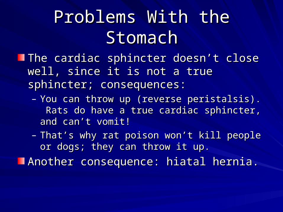
Problems With the StomachProblems With the Stomach
The cardiac sphincter doesn’t close well, since it The cardiac sphincter doesn’t close well, since it is not a true sphincter; consequences:is not a true sphincter; consequences:– You can throw up (reverse peristalsis). Rats do have You can throw up (reverse peristalsis). Rats do have
a true cardiac sphincter, and can’t vomit! a true cardiac sphincter, and can’t vomit! – That’s why rat poison won’t kill people or dogs; they That’s why rat poison won’t kill people or dogs; they
can throw it up.can throw it up.
Another consequence: hiatal hernia.Another consequence: hiatal hernia.

HIATAL HERNIAHIATAL HERNIA
Part of the stomach, protrudes through Part of the stomach, protrudes through esophageal hiatus, causing pain and esophageal hiatus, causing pain and difficulty swallowing.difficulty swallowing.It is the most common of all hernias.It is the most common of all hernias. There is a great amount of acid reflux; There is a great amount of acid reflux; erodes walls of esophagus, causing erodes walls of esophagus, causing ulcerations of esophagus. ulcerations of esophagus. Treatment is surgical; pull down the Treatment is surgical; pull down the stomach, and tighten the hernia in a stomach, and tighten the hernia in a laparoscopic procedure.laparoscopic procedure.


The Small IntestineThe Small Intestine
Figure 22.17a-c
Crypt of Lieberkuhn

Small Intestine RegionsSmall Intestine Regions
Duodenum “12 finger widths long”Duodenum “12 finger widths long”
Jejunum “hungry when empty”Jejunum “hungry when empty”
Ileum “twisted”Ileum “twisted”

DUODENUMDUODENUM
This is the shortest region, only one foot This is the shortest region, only one foot long. long. It receives chyme from the stomach. This It receives chyme from the stomach. This is where digestion begins. There are two is where digestion begins. There are two ducts at the beginning of the duodenum ducts at the beginning of the duodenum from the pancreas and gallbladder. It is from the pancreas and gallbladder. It is the site of action of liver secretions (bile) the site of action of liver secretions (bile) and pancreas secretions (digestive and pancreas secretions (digestive enzymes and bicarbonate).enzymes and bicarbonate).

PancreasPancreas
PANCREASPANCREAS is an endocrine gland, and also is an endocrine gland, and also participates in digestion. Most of the digestive participates in digestion. Most of the digestive enzymes are made here. They go out the enzymes are made here. They go out the PANCREATIC DUCTPANCREATIC DUCT to enter the small to enter the small intestine. intestine.
It also produces It also produces BICARBONATEBICARBONATE (from a (from a hormone called hormone called SECRETINSECRETIN) to increase the pH ) to increase the pH (decrease the acidity) of the chyme coming from (decrease the acidity) of the chyme coming from the stomach. If there is too much acid there, get the stomach. If there is too much acid there, get a a DUODENAL ULCERDUODENAL ULCER..

PANCREASPANCREAS
ACINAR CELLS: ACINAR CELLS: secretes digestive secretes digestive enzymesenzymes
ISLETS OF LANGERHANS: ISLETS OF LANGERHANS: secretes secretes insulin and glucagoninsulin and glucagon

PancreasPancreas
Acinar cells (secrete enzymes)
Islet of Langerhans (secretes insulin and glucagon)

GALL BLADDERGALL BLADDERThis is located inferior to the liver, and its function This is located inferior to the liver, and its function is to store and concentrate bile.is to store and concentrate bile.Bile is a detergent/soap (not an enzyme) which Bile is a detergent/soap (not an enzyme) which emulsifies fat: It breaks down the fat into emulsifies fat: It breaks down the fat into microscopic droplets which can be broken down microscopic droplets which can be broken down by pancreatic enzymes. by pancreatic enzymes. It does NOT make or secrete bile; that is done by It does NOT make or secrete bile; that is done by the liver.the liver.Bile is made in the liver from Hemoglobin (Hgb), Bile is made in the liver from Hemoglobin (Hgb), and also contains cholesterol and other things.and also contains cholesterol and other things.The function of bile is to break down lipids (fats) so The function of bile is to break down lipids (fats) so they can be digested.they can be digested.

JejunumJejunum
JEJUNUMJEJUNUM (“empty”) (“empty”)
This is the part of the small intestine where This is the part of the small intestine where most digestion and absorption occurs. most digestion and absorption occurs.
It is 3 feet long. It is 3 feet long.

IleumIleum
ILEUMILEUM (“twisted”) is 5-10 feet long. It is (“twisted”) is 5-10 feet long. It is the terminal portion of the small intestine.the terminal portion of the small intestine.
Much of the absorption takes place here. Much of the absorption takes place here.
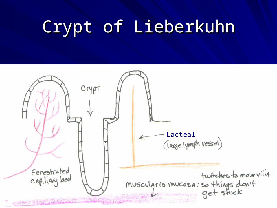
Crypt of LieberkuhnCrypt of Lieberkuhn
Lacteal
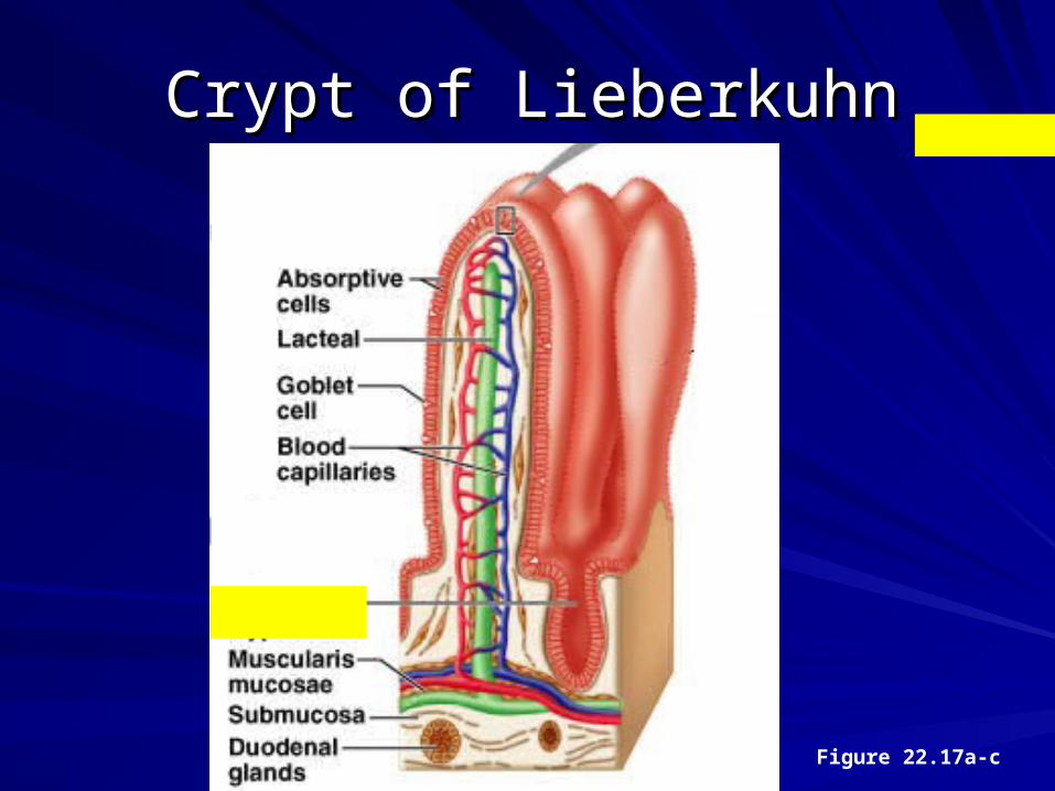
Crypt of LieberkuhnCrypt of Lieberkuhn
Figure 22.17a-c

Absorption in Small IntestineAbsorption in Small Intestine
In the villis is a fenestrated capillary bed, which In the villis is a fenestrated capillary bed, which needs to absorb a lot of material.needs to absorb a lot of material.
The small intestine absorbs carbohydrates, fats, The small intestine absorbs carbohydrates, fats, and proteins (although protein enzymes have and proteins (although protein enzymes have already begun working earlier in the digestive already begun working earlier in the digestive tract in the stomach). tract in the stomach).
The walls of the small intestine secrete most of The walls of the small intestine secrete most of the digestive enzymes that are active in its the digestive enzymes that are active in its lumen.lumen.

Lymphatics of Small IntestineLymphatics of Small Intestine
There are also large lymphatic capillaries There are also large lymphatic capillaries in each villis called in each villis called LACTEALSLACTEALS, whose , whose function is to absorb breakdown products function is to absorb breakdown products of fat.of fat. The vessel is large so it won’t get The vessel is large so it won’t get clogged up. clogged up.
Under all this are the Under all this are the MUSCULARIS MUSCULARIS MUCOSAMUCOSA muscles which can twitch to muscles which can twitch to move the villa so food does not get stuck. move the villa so food does not get stuck.

Problem with Small IntestineProblem with Small Intestine
Crohn’s DiseaseCrohn’s Disease– Autoimmune disease of the GI tractAutoimmune disease of the GI tract– Most common area affected is small intestineMost common area affected is small intestine
Celiac disease Celiac disease (Sprue; gluten intolerance)(Sprue; gluten intolerance)

Large Intestine Large Intestine (Colon, or large bowel) (Colon, or large bowel)
This is about 5 feet long, diameter of 4”.This is about 5 feet long, diameter of 4”.Absorbs a LOT of water and saltsAbsorbs a LOT of water and salts
Absorbs electrolytes (Na, K, etc)Absorbs electrolytes (Na, K, etc)
Stores feces for defecation (terminal portion)Stores feces for defecation (terminal portion)
Contains abundant bacteria (E. coli):Contains abundant bacteria (E. coli):– Make vitamins (B5, K, biotin)Make vitamins (B5, K, biotin)– Allow material to move through large intestine easierAllow material to move through large intestine easier– Keep out harmful bacteriaKeep out harmful bacteria– They eat things you can’t digestThey eat things you can’t digest
FiberFiber
Some sugars that we don’t have enzymes forSome sugars that we don’t have enzymes for

Regions of the Large IntestineRegions of the Large Intestine
CecumCecum
Ascending colonAscending colon
Transverse colonTransverse colon
Descending colonDescending colon
Sigmoid colonSigmoid colon
RectumRectum
AnusAnus

Figure 22.18a
Large IntestineLarge Intestine

Problems with Large IntestineProblems with Large Intestine
DIVERTICULITITSDIVERTICULITITS
INFLAMMATORY BOWEL DISEASEINFLAMMATORY BOWEL DISEASE– Crohn’s Disease Crohn’s Disease – Ulcerative colitisUlcerative colitis
IRRITABLE BOWEL SYNDROMEIRRITABLE BOWEL SYNDROME
COLON CANCERCOLON CANCER– SIGMOIDOSCOPYSIGMOIDOSCOPY or a or a COLONOSCOPYCOLONOSCOPY
POLYPSPOLYPS
HEMORRHOIDSHEMORRHOIDS

DIVERTICULITITSDIVERTICULITITS
DIVERTICULUM DIVERTICULUM (Diverticula is plural) can (Diverticula is plural) can form, a small pouch in the large intestine.form, a small pouch in the large intestine.
They can become inflamed, usually from a They can become inflamed, usually from a small, hard piece of feces, causes the small, hard piece of feces, causes the condition known as condition known as DIVERTICULITITS.DIVERTICULITITS.
These are painful and often need to be These are painful and often need to be surgically removed. surgically removed.
May be caused by lack of fiber, causing May be caused by lack of fiber, causing increased pressure in the colon.increased pressure in the colon.

Inflammatory Bowel Disease (IBD)Inflammatory Bowel Disease (IBD)
IBD is a group of inflammatory conditions IBD is a group of inflammatory conditions of the colon and small intestine. of the colon and small intestine.
The major types of IBD are Crohn's The major types of IBD are Crohn's disease and ulcerative colitisdisease and ulcerative colitis

IRRITABLE BOWEL SYNDROME (IBS)IRRITABLE BOWEL SYNDROME (IBS)
IBS is a diagnosis of exclusion.IBS is a diagnosis of exclusion.
Symptoms are chronic abdominal pain, bloating, Symptoms are chronic abdominal pain, bloating, and alteration of bowel habits in the absence of and alteration of bowel habits in the absence of any detectable organic cause.any detectable organic cause.
May manifest as diarrhea or constipation or may May manifest as diarrhea or constipation or may alternate between the two.alternate between the two.
May be caused by infection, stress, or onset of May be caused by infection, stress, or onset of maturitymaturity
No cure; treatments attempt to relieve symptoms, No cure; treatments attempt to relieve symptoms, including dietary adjustments, medication and including dietary adjustments, medication and psychological interventions. psychological interventions.

COLON CANCERCOLON CANCERThis is the #1 most deadly cancer (kills more This is the #1 most deadly cancer (kills more people) because it metastasizes and there are people) because it metastasizes and there are no symptoms. It can be diagnosed by seeing no symptoms. It can be diagnosed by seeing blood in the stool; this is an easy test, but not blood in the stool; this is an easy test, but not very accurate. very accurate. A more accurate test is a A more accurate test is a SIGMOIDOSCOPYSIGMOIDOSCOPY. A . A tube is inserted into the sigmoid colon, done in tube is inserted into the sigmoid colon, done in the doctor’s office. The tube has a light, and the doctor’s office. The tube has a light, and they look for growths on the walls of the intestine they look for growths on the walls of the intestine == POLYPS POLYPS, which are pre-cancerous growths. , which are pre-cancerous growths. A colonoscopy is done under general anesthesia A colonoscopy is done under general anesthesia since the tube has to go through the entire since the tube has to go through the entire colon, but it’s more effective. colon, but it’s more effective.
Hepatic Portal SystemHepatic Portal System
Almost all of the blood coming from the digestive Almost all of the blood coming from the digestive system drains into a special venous circulation system drains into a special venous circulation called the portal circulation. called the portal circulation. Before these absorbed substances can go into Before these absorbed substances can go into the systemic circulation (the main blood the systemic circulation (the main blood circulation in the body), it must be filtered first to circulation in the body), it must be filtered first to remove or detoxify toxic substances first. remove or detoxify toxic substances first. This filtering and detoxification is one of the 500+ This filtering and detoxification is one of the 500+ functions of the liver.functions of the liver.

LiverLiver
Makes bloodMakes bloodMakes blood proteins (clotting factors)Makes blood proteins (clotting factors)Makes bileMakes bileRegulates glucose levelsRegulates glucose levelsProcesses fats Processes fats Makes cholesterolMakes cholesterolProcesses amino acidsProcesses amino acidsDetoxifies chemicals Detoxifies chemicals

LiverLiver
Figure 22.23a, c, d
Hepatic Triad: Vein, Artery, Bile Duct

Liver: sinusoids and hepatocytes

Blood Flow in the LiverBlood Flow in the LiverBlood flow to the liver is unique in that it receives both Blood flow to the liver is unique in that it receives both oxygenated and deoxygenated blood. oxygenated and deoxygenated blood.
Nutrient-rich, oxygen-poor blood from the intestine enters Nutrient-rich, oxygen-poor blood from the intestine enters the liver by the hepatic portal vein. It flows through the the liver by the hepatic portal vein. It flows through the sinusoids for detoxification.sinusoids for detoxification.
Oxygen-rich blood enters the liver by the hepatic artery. It Oxygen-rich blood enters the liver by the hepatic artery. It flows through the sinusoids to supply them with oxygen.flows through the sinusoids to supply them with oxygen.
All of the blood mixes together, and when the oxygen All of the blood mixes together, and when the oxygen demand of the hepatocytes is satisfied, and the toxins have demand of the hepatocytes is satisfied, and the toxins have been removed, the oxygen-depleted blood collects in a been removed, the oxygen-depleted blood collects in a central vein within each lobule, which drains into the central vein within each lobule, which drains into the hepatic vein. The hepatic vein subsequently drains into the hepatic vein. The hepatic vein subsequently drains into the inferior vena cava and back to the heart.inferior vena cava and back to the heart.

Function of HepatocytesFunction of Hepatocytes
Detoxification of poisonsDetoxification of poisons
Picking up and processing of nutrients Picking up and processing of nutrients from the portal bloodfrom the portal blood– This includes picking up glucose from the nutrient-This includes picking up glucose from the nutrient-
rich blood coming from the small intestine and rich blood coming from the small intestine and stores it as glycogen (the storage form of glucose) stores it as glycogen (the storage form of glucose) for when the body needs it later.for when the body needs it later.
Storage of some vitaminsStorage of some vitamins
Kupffer CellsKupffer Cells
Within the sinusoids are Within the sinusoids are KUPFFER CELLSKUPFFER CELLS, , which are macrophages. As blood flows through which are macrophages. As blood flows through the sinusoids, they phagocytize old erythrocytes. the sinusoids, they phagocytize old erythrocytes. The released Hgb is given to the hepatocytes, The released Hgb is given to the hepatocytes, which convert it to bilirubin, one of the main which convert it to bilirubin, one of the main components of BILE.components of BILE.
Bile flows through a series of channels called Bile flows through a series of channels called the the BILE CANNICULI BILE CANNICULI to the bile duct.to the bile duct.
Problems with the LiverProblems with the Liver
HEPATITISHEPATITIS
CIRRHOSISCIRRHOSIS
JAUNDICEJAUNDICE

Liver ProblemsLiver Problems
Infection of the liver = Infection of the liver = HEPATITISHEPATITIS (can be (can be deadly)deadly)
CIRRHOSISCIRRHOSIS is when the hepatocytes die is when the hepatocytes die and are replaced by connective tissue. and are replaced by connective tissue. This is often from alcoholism, which kills This is often from alcoholism, which kills the hepatocytes.the hepatocytes.

JaundiceJaundice
One of the symptoms from any liver One of the symptoms from any liver disorder is a connection of the bile disorder is a connection of the bile canaliculi and the sinusoid so some canaliculi and the sinusoid so some bilirubin can enter the blood. bilirubin can enter the blood.
Bilirubin is yellow-green (later in its Bilirubin is yellow-green (later in its degradation it will turn brown and that is degradation it will turn brown and that is what gives the feces its color). what gives the feces its color).
The yellow color of bilirubin in the skin is The yellow color of bilirubin in the skin is known as known as JAUNDICEJAUNDICE. .

GREATER OMENTUMGREATER OMENTUM
GREATER OMENTUM is flat, and is in GREATER OMENTUM is flat, and is in front of the intestines like an apron. Its front of the intestines like an apron. Its function is to store fat, especially in function is to store fat, especially in men. men.

GI PhysiologyGI Physiology
Part 1: Metabolic PathwaysPart 1: Metabolic Pathways
Part 2: GI PhysiologyPart 2: GI Physiology
5656

Simple and Complex Simple and Complex CarbohydratesCarbohydrates
There are three main simple sugars (AKA There are three main simple sugars (AKA monosaccharides or simple carbohydrates)monosaccharides or simple carbohydrates)– GlucoseGlucose– FructoseFructose– GalactoseGalactose
If you join a glucose to any of these, you get a If you join a glucose to any of these, you get a disaccharidedisaccharide– Glucose + Glucose = MaltoseGlucose + Glucose = Maltose– Glucose + Galactose = LactoseGlucose + Galactose = Lactose– Glucose + Fructose = SucroseGlucose + Fructose = Sucrose
5757

Simple and Complex Simple and Complex CarbohydratesCarbohydrates
If you join many monosaccharides and/or If you join many monosaccharides and/or disaccharides together, it is called a disaccharides together, it is called a polysaccharide (AKA complex carbohydrate).polysaccharide (AKA complex carbohydrate).
These are stored in the liver as glycogen. These are stored in the liver as glycogen. They can be broken down later into glucose They can be broken down later into glucose as needed.as needed.
The storage form in plants is called starch.The storage form in plants is called starch.
When we eat starch, we covert it to glycogen When we eat starch, we covert it to glycogen and then store it.and then store it.
5858

Glucagon and InsulinGlucagon and InsulinGlucagon, a hormone secreted by the pancreas, raises blood glucose Glucagon, a hormone secreted by the pancreas, raises blood glucose levels. levels.
Its effect is opposite that of insulin, which lowers blood glucose levels.Its effect is opposite that of insulin, which lowers blood glucose levels.
The pancreas releases glucagon when blood sugar (glucose) levels The pancreas releases glucagon when blood sugar (glucose) levels fall too low. fall too low.
Glucagon causes the liver to beak down the stored glycogen into Glucagon causes the liver to beak down the stored glycogen into glucose, which is released into the bloodstream. Since glycogen is glucose, which is released into the bloodstream. Since glycogen is being broken down, this process is called glycogenolysis. Don’t being broken down, this process is called glycogenolysis. Don’t confuse this with glycolysis (break down of glucose to ATP)!confuse this with glycolysis (break down of glucose to ATP)!
High blood glucose levels stimulate the release of insulin.High blood glucose levels stimulate the release of insulin.
Insulin allows glucose to be taken up and used by insulin-dependent Insulin allows glucose to be taken up and used by insulin-dependent tissues. tissues.
Thus, glucagon and insulin are part of a feedback system that keeps Thus, glucagon and insulin are part of a feedback system that keeps blood glucose levels at a stable level. blood glucose levels at a stable level.
5959

GlycolysisGlycolysis
Glycolysis is the process where cells take in glucose and Glycolysis is the process where cells take in glucose and break it down into pyruvate, and ATP is released. break it down into pyruvate, and ATP is released.
This is how we get ATP from glucose.This is how we get ATP from glucose.
Fructose and galactose can also be broken down into Fructose and galactose can also be broken down into pyruvate and ATP.pyruvate and ATP.
During glycolysis, NAD (an energy molecule) is reduced During glycolysis, NAD (an energy molecule) is reduced to NADH. If you run out of NAD, glycolysis will stop. to NADH. If you run out of NAD, glycolysis will stop. Therefore, we need to oxidize NADH to convert it back Therefore, we need to oxidize NADH to convert it back into NAD.into NAD.
This can be done by aerobic or anaerobic respiration, or This can be done by aerobic or anaerobic respiration, or fermentation.fermentation.
6060

Aerobic vs. Anaerobic RespirationAerobic vs. Anaerobic Respiration
Aerobic respiration Aerobic respiration
(in the (in the mitochondria)will mitochondria)will result in 6 ATP’s.result in 6 ATP’s.
Anaerobic Anaerobic respiration (in our respiration (in our cytoplasm) will cytoplasm) will result in only 2 result in only 2 ATP’s.ATP’s.
6161More importantly, we get our NAD back, so glycolysis can continue.

Making ATP by Aerobic Making ATP by Aerobic RespirationRespiration
Takes place in the Takes place in the mitochondriamitochondria
Requires Requires oxygenoxygen
Breaks down glucose to produce ATPBreaks down glucose to produce ATP
Waste products are Waste products are CO2 CO2 andand H2O H2O (we exhale them)(we exhale them)
The good thing about making ATP from our mitochondria The good thing about making ATP from our mitochondria is that we can make a is that we can make a LOTLOT of it. of it.
The bad things are that it takes The bad things are that it takes longer longer to make it, and it to make it, and it requires oxygen, and a muscle cell may have used up all requires oxygen, and a muscle cell may have used up all the oxygen during a sprinting run.the oxygen during a sprinting run.

Making ATP by Anaerobic Making ATP by Anaerobic RespirationRespiration
Takes place in the Takes place in the cytoplasmcytoplasm
Does not require oxygenDoes not require oxygen
Breaks down glucose to produce ATPBreaks down glucose to produce ATP
Waste product is Waste product is lactic acidlactic acid
The good thing about making ATP this way is The good thing about making ATP this way is that we can make it that we can make it FASTFAST. .
The bad thing is that it The bad thing is that it does not make much does not make much ATPATP, and we deplete the reserves quickly., and we deplete the reserves quickly.

Lactic Acid Build-upLactic Acid Build-upDuring strenuous workouts where oxygen becomes deficient, the During strenuous workouts where oxygen becomes deficient, the pyruvate product of glycolysis does not have enough oxygen to use pyruvate product of glycolysis does not have enough oxygen to use for aerobic respiration, so it has to undergo anaerobic respiration.for aerobic respiration, so it has to undergo anaerobic respiration.
The enzyme lactate dehydrogenase (LDH) is used to transfer The enzyme lactate dehydrogenase (LDH) is used to transfer hydrogen from the NADH molecule to the pyruvate molecule.hydrogen from the NADH molecule to the pyruvate molecule.
Pyruvate with the extra hydrogen is called lactate. Pyruvate with the extra hydrogen is called lactate.
Lactic acid is formed from lactate. This causes muscle aches and Lactic acid is formed from lactate. This causes muscle aches and fatigue. fatigue.
Lactic acid is deactivated by the addition of oxygen to it. Therefore, Lactic acid is deactivated by the addition of oxygen to it. Therefore, breathing heavily adds the oxygen to our system to deactivate lactic breathing heavily adds the oxygen to our system to deactivate lactic acid, and the muscle pains go away. Warm water or ultrasound will acid, and the muscle pains go away. Warm water or ultrasound will also increase oxygenated blood to the muscles, easing muscle also increase oxygenated blood to the muscles, easing muscle cramps from lactic acid.cramps from lactic acid.
6464

ATP and Creatinine PhosphateATP and Creatinine Phosphate
What do we do when we run out of ATP?What do we do when we run out of ATP?
Muscle fibers cannot stockpile ATP in preparation for Muscle fibers cannot stockpile ATP in preparation for future periods of activity.future periods of activity.
However, they can store another high energy molecule However, they can store another high energy molecule called called creatinine phosphatecreatinine phosphate..
Creatine phosphate is made from the excess ATP that we Creatine phosphate is made from the excess ATP that we accumulate when we are resting.accumulate when we are resting.
During short periods of intense exercise, the small During short periods of intense exercise, the small reserves of ATP existing in a cell are used first. reserves of ATP existing in a cell are used first.
Then creatinine phosphate is broken down to produce Then creatinine phosphate is broken down to produce ATP.ATP.

Aerobic vs. Anaerobic Aerobic vs. Anaerobic RespirationRespiration
When do we use aerobic respiration?When do we use aerobic respiration?– Resting (can breathe easily)Resting (can breathe easily)– Running marathons (can breathe easily on long runs)Running marathons (can breathe easily on long runs)
Marathon runners Marathon runners want to make sure there will be enough want to make sure there will be enough readily available energy for the muscles, so they readily available energy for the muscles, so they eat a lot of eat a lot of carbohydrates carbohydrates over a two-day period before the marathon. over a two-day period before the marathon. That’s why they load up on pasta before a marathon.That’s why they load up on pasta before a marathon.
When do we use anaerobic respiration?When do we use anaerobic respiration?– Sprint running (can’t talk while sprinting!)Sprint running (can’t talk while sprinting!)

GluconeogenesisGluconeogenesis
Gluconeogenesis is a metabolic pathway that results in Gluconeogenesis is a metabolic pathway that results in the generation of new glucose from non-carbohydrate the generation of new glucose from non-carbohydrate carbon substrates such as lactate, glycerol, and amino carbon substrates such as lactate, glycerol, and amino acids. Therefore, if we do not have enough glucose in acids. Therefore, if we do not have enough glucose in our body, we will break down proteins (muscles) to make our body, we will break down proteins (muscles) to make glucose.glucose.
It is one of the two main mechanisms to keep blood It is one of the two main mechanisms to keep blood glucose levels from dropping too low (hypoglycemia). glucose levels from dropping too low (hypoglycemia).
The other means of maintaining blood glucose levels is The other means of maintaining blood glucose levels is through the degradation of glycogen (glycogenolysis).through the degradation of glycogen (glycogenolysis).
6767
6868
Part 2Part 2GI PhysiologyGI Physiology
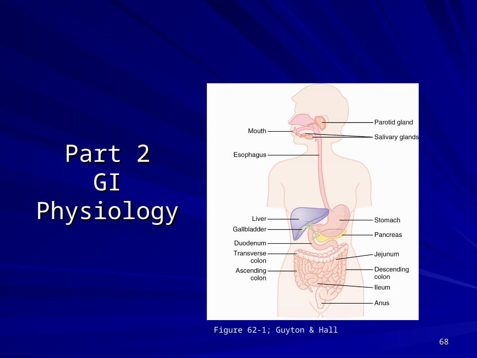
Figure 62-1; Guyton & Hall

SalivaSaliva
The saliva serves to clean the oral cavity and The saliva serves to clean the oral cavity and moisten the food.moisten the food.
It also contains digestive enzymes such as It also contains digestive enzymes such as salivary amylase, which aids in the chemical salivary amylase, which aids in the chemical breakdown of polysaccharides such as starch breakdown of polysaccharides such as starch into disaccharides such as maltose. into disaccharides such as maltose.
It also contains mucus, a glycoprotein which It also contains mucus, a glycoprotein which helps soften the food and form it into a bolus. helps soften the food and form it into a bolus.
6969

StomachStomach
The stomach is responsible for the digestion of protein The stomach is responsible for the digestion of protein and ionization of minerals. and ionization of minerals.
Mucous cells (in the stomach) secrete mucous. The Mucous cells (in the stomach) secrete mucous. The pancreas secretes bicarbonate. Mucous, bicarbonate, pancreas secretes bicarbonate. Mucous, bicarbonate, and prostaglandins protect the stomach lining from being and prostaglandins protect the stomach lining from being digested. digested.
The parietal cells of the stomach secrete hydrochloric The parietal cells of the stomach secrete hydrochloric acid (gastric acid) and intrinsic factor.acid (gastric acid) and intrinsic factor.
Hydrochloric acid (HCl), along with pepsin (from the chief Hydrochloric acid (HCl), along with pepsin (from the chief cells), breaks down proteins to their individual amino cells), breaks down proteins to their individual amino acids.acids.
7070

Downloaded from: StudentConsult (on 23 April 2010 06:51 PM)
© 2005 Elsevier
Stomach Protection and Stomach Protection and DamageDamage

Downloaded from: StudentConsult (on 23 April 2010 06:51 PM)
© 2005 Elsevier

© 2005 Elsevier

Stomach AcidStomach Acid
The acid itself does not break down food The acid itself does not break down food molecules.molecules.
It provides an optimum pH for the activation of It provides an optimum pH for the activation of pepsin, and kills many microorganisms that are pepsin, and kills many microorganisms that are ingested with the food. ingested with the food.
It can also denature proteins. It can also denature proteins.
The parietal cells of the stomach also secrete a The parietal cells of the stomach also secrete a glycoprotein called intrinsic factor, which enables glycoprotein called intrinsic factor, which enables the absorption of vitamin B-12. the absorption of vitamin B-12.
7474

Stomach Acid DiseasesStomach Acid Diseases
HypochlorhydriaHypochlorhydria
HyperchlorhydriaHyperchlorhydria
7575

Small IntestineSmall IntestineDuodenumDuodenum– Absorption of mineralsAbsorption of minerals– Receives pancreatic digestive enzymesReceives pancreatic digestive enzymes– Secretes hormones when acidic chyme enters duodenumSecretes hormones when acidic chyme enters duodenum
SecretinSecretin– Tells pancreas to secrete bicarbonateTells pancreas to secrete bicarbonate– Tells liver to make bileTells liver to make bile
Cholecystokinin (CCK)Cholecystokinin (CCK)– Tells pancreas to release protein-digesting enzymes Tells pancreas to release protein-digesting enzymes – Tells the gallbladder to release stored bile.Tells the gallbladder to release stored bile.– Therefore, it stimulates digestion of fat and protein. Therefore, it stimulates digestion of fat and protein.
GIPGIP– stimulates insulin secretionstimulates insulin secretion
MotilinMotilin– Initiates peristalsis (increases GI motility)Initiates peristalsis (increases GI motility)– Tells the Chief cells to secrete pepsinogen Tells the Chief cells to secrete pepsinogen
– Secretes enzymes to break down polysaccharidesSecretes enzymes to break down polysaccharides
Maltase: breaks maltose down into glucoseMaltase: breaks maltose down into glucose
Lactase: breaks lactose down to galactose plus glucoseLactase: breaks lactose down to galactose plus glucose
Sucrase: breaks sucrose down into fructose plus glucoseSucrase: breaks sucrose down into fructose plus glucose7676

Small IntestineSmall IntestineDuodenumDuodenum– When there is no more chyme entering the When there is no more chyme entering the
duodenum, it secretes glucose-dependent duodenum, it secretes glucose-dependent insulinotropic peptide (GIP).insulinotropic peptide (GIP).
– GIP is synthesized by K cells, which are found in the GIP is synthesized by K cells, which are found in the duodenum and jejunum.duodenum and jejunum.
– GIP stimulates insulin secretion.GIP stimulates insulin secretion.– Insulin is in the blood stream. It takes the absorbed Insulin is in the blood stream. It takes the absorbed
sugars and pulls them into cells that need it. sugars and pulls them into cells that need it. – GIP also stimulates lipoprotein lipase activity in GIP also stimulates lipoprotein lipase activity in
adipocytes. This causes fat to be broken down into adipocytes. This causes fat to be broken down into fatty acids.fatty acids.
7777

Lipid digestion and absorption• Lipid digestion utilizes lingual
and pancreatic lipases, to release fatty acids and monoglycerides.– Bile salts improve chemical
digestion by emulsifying lipid drops
– Lipid-bile salt complexes called micelles are formed
78
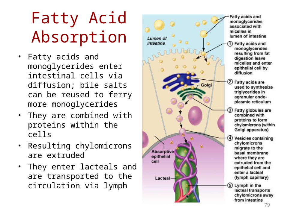
Fatty Acid Absorption
• Fatty acids and monoglycerides enter intestinal cells via diffusion; bile salts can be reused to ferry more monoglycerides
• They are combined with proteins within the cells
• Resulting chylomicrons are extruded
• They enter lacteals and are transported to the circulation via lymph
79

Small IntestineSmall Intestine
JejunumJejunum– Absorbs water-soluble vitamins, Absorbs water-soluble vitamins,
protein and carbohydrates. protein and carbohydrates. – The proteins began to be broken The proteins began to be broken
down into amino acids in the down into amino acids in the stomach by pepsin and acid. stomach by pepsin and acid.
– Proteins are further broken down Proteins are further broken down into amino acids in the duodenum into amino acids in the duodenum by trypsin and chymotrypsin (made by trypsin and chymotrypsin (made by the pancreas and secreted into by the pancreas and secreted into the duodenum).the duodenum).
– The carbohydrates are broken The carbohydrates are broken down in the duodenum by down in the duodenum by enzymes from the pancreas and enzymes from the pancreas and liver into sugars. liver into sugars.
8080
Small IntestineSmall Intestine
IleumIleum
– Absorbs fat-soluble vitamins, fat, cholesterol, Absorbs fat-soluble vitamins, fat, cholesterol, and bile salts. and bile salts.
– Fats are broken down into fatty acids in the Fats are broken down into fatty acids in the duodenum.duodenum.
First, bile emulsifies the fat (breaks it down into First, bile emulsifies the fat (breaks it down into droplets). droplets).
Then, lipase (made in the pancreas) breaks the fat Then, lipase (made in the pancreas) breaks the fat into fatty acids, which are small enough to be into fatty acids, which are small enough to be absorbed.absorbed.
8181

Pancreas EnzymesPancreas Enzymes
The pancreas secretes about one and a half liters of The pancreas secretes about one and a half liters of pancreatic juice a day!pancreatic juice a day!
Pancreatic juice secretion is regulated by the hormones Pancreatic juice secretion is regulated by the hormones secretin and cholecystokinin, which is produced by the secretin and cholecystokinin, which is produced by the walls of the duodenum upon detection of acid food, walls of the duodenum upon detection of acid food, proteins and fats.proteins and fats.
The enzymes produced by the pancreas includeThe enzymes produced by the pancreas include
– Lipases Lipases – Amylases Amylases – ProteasesProteases
8282

Pancreas EnzymesPancreas Enzymes
Lipases Lipases – Digestion of fats, oils, and fat-soluble vitaminsDigestion of fats, oils, and fat-soluble vitamins
AmylasesAmylases– Break down starch molecules into smaller sugars. Break down starch molecules into smaller sugars. – Break down carbohydrates into maltoseBreak down carbohydrates into maltose
ProteasesProteases– Break down protein into smaller amino acidsBreak down protein into smaller amino acids– Proteases include trypsin, chromotrypsin and carboxypeptidase. Proteases include trypsin, chromotrypsin and carboxypeptidase. – Proteases are also responsible for keeping the small intestine free Proteases are also responsible for keeping the small intestine free
from parasites (intestinal worms, yeast overgrowth and bacteria).from parasites (intestinal worms, yeast overgrowth and bacteria).– A lack of proteases can cause incomplete digestion that can lead A lack of proteases can cause incomplete digestion that can lead
to allergies and the formation of toxins. to allergies and the formation of toxins.
8383

Regulation of Pancreatic Secretion• Secretin and CCK are released
when fatty or acidic chyme enters the duodenum
• CCK and secretin enter the bloodstream
• Upon reaching the pancreas:– CCK induces the secretion of
enzyme-rich pancreatic juice– Secretin causes secretion of
bicarbonate-rich pancreatic juice • Vagal stimulation also causes
release of pancreatic juice
84

8585
The PancreasThe Pancreas
Exocrine function (98%)Exocrine function (98%)– Acinar cellsAcinar cells make, store, make, store,
and secrete pancreatic and secrete pancreatic enzymesenzymes
Endocrine function – Endocrine function – cells (delta cells) release cells (delta cells) release
somatostatin (inhibitory to somatostatin (inhibitory to gastrin and insulin and gastrin and insulin and glucagon)glucagon)
– ββ-cells –release insulin-cells –release insulin– αα-cells-Release glucagon-cells-Release glucagon

8686
The Pancreas as an Endocrine The Pancreas as an Endocrine GlandGland
InsulinInsulin– Beta cellsBeta cells
– Promotes glucose uptakePromotes glucose uptake
– Prevents fat and glycogen Prevents fat and glycogen breakdown and inhibits breakdown and inhibits gluconeogenesisgluconeogenesis
– Increases protein synthesisIncreases protein synthesis
– Promotes fat storagePromotes fat storage
Picture from:http://www.dkimages.com/discover/Home/Health-and-Beauty/Human-Body/Endocrine-System/Pancreas/Pancreas-1.html
Epi/Norepi inhibit insulin!Help maintain glucose levels during times of stress and increase lipase activity in order to conserve glucose levels

8787
The Pancreas as an Endocrine The Pancreas as an Endocrine GlandGland
GlucagonGlucagon– Increases blood glucose levelsIncreases blood glucose levels– Maintains blood glucose between Maintains blood glucose between
meals and during periods of fasting meals and during periods of fasting by breaking down glycogen (stored by breaking down glycogen (stored in liver) into glucose.in liver) into glucose.
– Initiates glycogenolysis in liver Initiates glycogenolysis in liver (within minutes). (within minutes).
– Stimulates gluconeogenesis. This Stimulates gluconeogenesis. This process involves breaking down process involves breaking down amino acids (proteins) into glucose.amino acids (proteins) into glucose.
– Stimulates amino acid transport to Stimulates amino acid transport to liver to stimulate gluconeogenesisliver to stimulate gluconeogenesis
– Nervous tissue (brain) does not Nervous tissue (brain) does not need insulin; but is heavily need insulin; but is heavily dependent on glucose levels!dependent on glucose levels! Image from: http://www.dkimages.com/discover/previews/768/74261.JPG
Liver and Gallbladder Liver and Gallbladder
The liver produces bile that is either stored by the The liver produces bile that is either stored by the gallbladder or secreted into the small intestine. gallbladder or secreted into the small intestine. – Bile emulsifies fats and fat-soluble vitamins. Bile emulsifies fats and fat-soluble vitamins. – It also helps keep the small intestine free from It also helps keep the small intestine free from
parasites. parasites.
The liver does not make the digestive enzymes for The liver does not make the digestive enzymes for carbohydrates, amino acids and proteins (the pancreas carbohydrates, amino acids and proteins (the pancreas and small intestine do that), but the liver does metabolize and small intestine do that), but the liver does metabolize proteins, carbohydrates and cholesterol.proteins, carbohydrates and cholesterol.
It also is responsible for the detoxification of toxins, It also is responsible for the detoxification of toxins, drugs and hormones. drugs and hormones.
8888

Large Intestine Large Intestine
The large intestine absorbs water, The large intestine absorbs water, electrolytes and some of the final products electrolytes and some of the final products of digestion.of digestion.
Food products that cannot go through the Food products that cannot go through the villi, such as cellulose (dietary fiber), are villi, such as cellulose (dietary fiber), are mixed with other waste products from the mixed with other waste products from the body and become hard and concentrated body and become hard and concentrated feces.feces.
8989

Physiology of the large intestine
– Reabsorption of water and electrolytes
– Coliform bacteria make: Vitamins – K, biotin, and B5
– Organic wastes are left in the lumen
90

Phases of gastric secretionPhases of gastric secretion
Cephalic phase Cephalic phase
Gastric phase Gastric phase
Intestinal phase Intestinal phase
9191

Cephalic phase Cephalic phase
This phase occurs before food enters the stomach and involves This phase occurs before food enters the stomach and involves preparation of the body for eating and digestion. preparation of the body for eating and digestion.
Sight and thought stimulate the cerebral cortex. Taste and smell Sight and thought stimulate the cerebral cortex. Taste and smell stimulus is sent to the hypothalamus and medulla oblongata. stimulus is sent to the hypothalamus and medulla oblongata.
After this it is routed through the vagus nerve and release of After this it is routed through the vagus nerve and release of acetylcholine. acetylcholine.
9292
Gastric secretion at this phase rises to 40% of maximum rate. Acidity in the stomach is not buffered by food at this point and thus acts to inhibit parietal (secretes acid) and G cell (secretes gastrin) activity via D cell secretion of somatostatin.

G cell secretion of gastrinG cell secretion of gastrinD cell secretion of somatostatinD cell secretion of somatostatin
9393

G cells and GastrinG cells and Gastrin
G cells are found deep within the gastric glands of the stomach.G cells are found deep within the gastric glands of the stomach.
When food arrives in the stomach, the parasympathetic nervous When food arrives in the stomach, the parasympathetic nervous system is activated. This causes the vagus nerve to release a system is activated. This causes the vagus nerve to release a neurotransmitter called Gastrin-releasing peptide onto the G cells in neurotransmitter called Gastrin-releasing peptide onto the G cells in the stomach.the stomach.
Gastrin-releasing peptide, as well as the presence of amino acids in Gastrin-releasing peptide, as well as the presence of amino acids in the stomach, stimulates the release of gastrin from the G cells. the stomach, stimulates the release of gastrin from the G cells.
Gastrin tells parietal cells to increase HCl secretion, and it also Gastrin tells parietal cells to increase HCl secretion, and it also stimulates other special cells to release histamine.stimulates other special cells to release histamine.
Gastrin also tells the chief cells to produce pepsinogen.Gastrin also tells the chief cells to produce pepsinogen.
Gastrin is inhibited by low pH (acid) in the stomach. When enough Gastrin is inhibited by low pH (acid) in the stomach. When enough acid is present, it turns off.acid is present, it turns off.
9494

GastrinGastrin
Gastrin is released in response toGastrin is released in response to
– Stomach distensionStomach distension– Vagus nerve stimulationVagus nerve stimulation– The presence of proteins or amino acidsThe presence of proteins or amino acids
Gastrin release is inhibited byGastrin release is inhibited by
– The presence of enough HCl in the stomach The presence of enough HCl in the stomach (negative feedback)(negative feedback)
– Somatostatin also inhibits the release of Somatostatin also inhibits the release of gastringastrin
9595

D cellsD cells
D cells can be found in the stomach, D cells can be found in the stomach, intestine and the Islets of Langerhans in intestine and the Islets of Langerhans in the pancreas.the pancreas.
When gastrin is present, D cells increase When gastrin is present, D cells increase somatostatin output.somatostatin output.
When D cells are stimulated by Ach, they When D cells are stimulated by Ach, they decrease somatostatin output.decrease somatostatin output.
9696

SomatostatinSomatostatin
Somatostatin is also known as growth hormone-inhibiting hormone.Somatostatin is also known as growth hormone-inhibiting hormone.
It suppresses the release of gastrointestinal hormonesIt suppresses the release of gastrointestinal hormones– GastrinGastrin– Cholecystokinin (CCK)Cholecystokinin (CCK)– SecretinSecretin– GIPGIP
It suppresses the release of pancreatic hormones.It suppresses the release of pancreatic hormones.
It slows down the digestive process.It slows down the digestive process.
It inhibits insulin release. It inhibits insulin release.
It inhibits the release of glucagon.It inhibits the release of glucagon.
9797

Gastric phaseGastric phase
This phase takes 3 to 4 hours. It is stimulated by distension This phase takes 3 to 4 hours. It is stimulated by distension of the stomach, presence of food in stomach and decrease of the stomach, presence of food in stomach and decrease in pH.in pH.
This activates the release of acetylcholine which stimulates This activates the release of acetylcholine which stimulates the release of more gastric juices. the release of more gastric juices.
As protein enters the stomach, it binds to hydrogen ions, As protein enters the stomach, it binds to hydrogen ions, which raises the pH of the stomach. which raises the pH of the stomach.
Inhibition of gastrin and gastric acid secretion is lifted. Inhibition of gastrin and gastric acid secretion is lifted.
This triggers G cells to release gastrin, which in turn This triggers G cells to release gastrin, which in turn stimulates parietal cells to secrete gastric acid. stimulates parietal cells to secrete gastric acid.
Acid release is also triggered by acetylcholine and Acid release is also triggered by acetylcholine and histamine.histamine.
9898

Intestinal phaseIntestinal phase
This phase has 2 opposing actions: the This phase has 2 opposing actions: the excitatory and the inhibitory. excitatory and the inhibitory.
Partially digested food fills the duodenum.Partially digested food fills the duodenum.
This triggers gastrin to be released. This triggers gastrin to be released.
It also triggers the enterogastric reflex, which It also triggers the enterogastric reflex, which inhibits the Vagus nerve.inhibits the Vagus nerve.
This activates the sympathetic nervouse system, This activates the sympathetic nervouse system, which causes the pyloric sphincter to tighten to which causes the pyloric sphincter to tighten to prevent more food from entering the duodenum.prevent more food from entering the duodenum.
9999

100
Digestive Enzymes
Salivary glands-amylase
lingual lipase
Stomachpepsin
Intestinal Mucosasucrase maltase lactase
Pancreas amylase trypsin chymotrypsincarboxypeptidaselipasecholesterolesterase

The Activities of Major Digestive Tract Hormones
101Figure 24.22

102
Organ Region of the Organ Substances FunctionPancreas Acinar cells Amylase (enzyme) Breaks down starch and carbohydrates into glucose
Acinar cells Lipase (enzyme) Breaks down fat into fatty acids
Acinar cellsProtease enzymes (trypsin,
chymotrypsin, carboxypeptidase)Breaks down proteins into amino acids and also kills intestinal parasites and bacteria
Acinar cells Bicarbonate (not an enzyme) Raises pH in duodenum
Islet of Langerhans; Alpha
cells glucagon (hormone)
Causes glycogenolysis, the process which breaks down glycogen into glucose to raise blood glucose. Also causes gluconeogenesis to make new glucose molecules
Islet of Langerhans; Beta
cells insulin (hormone)Removes glucose in bloodstream and brings it into cells. Lowers blood glucose levels.
Islet of Langerhans; Delta
cells Somatostatin (hormone) Inhibits gastrin, insulin, and glucagon (inhibits digestive system)
Liver Bile (a detergent)
Salivary glands Amylase (enzyme) Breaks down starch and carbohydrates into glucose
Stomach Mucous (not an enzyme) Protect the stomach lining
Prostaglandins (not an enzyme) Protect the stomach lining
Parietal cells HCl (not an enzyme) Allows Pepsinogen to be converted to pepsin, and it also kills bacteria
Parietal cells Intrinsic factor (not an enzyme)Allows Vit B12 to be absorbed, which is needed to make RBCs. Without it, you get megaloblastic (pernicous) anemia.
Chief cells Pepsinogen --> pepsin (enzyme) Breaks proteins into amino acids G cells Gastrin (hormone) Tells parietal cells to secrete HCl
Duodenum Secretin (hormone) Tells pancreas to secrete bicarbonate
CCK (hormone)Tells pancrease to secrete proteases, and tells gallbladder to release stored bile (stimulates fat and protein digestion)
K cells GIP (hormone)Tells pancreas to release insulin and also causes fat to be broken down into fatty acids
Motilin (hormone) Initiates perstalsis and tells Chief cells to secrete pepsinogen
Maltase, Lactase, Sucrase (enzymes) Break down complex carbohydrates into glucose

The OvariesThe OvariesSmall, almond-shaped organs, each 1 ½” x 1”Small, almond-shaped organs, each 1 ½” x 1”Within the peritoneal cavity on the posterior body wallWithin the peritoneal cavity on the posterior body wallCovered by a superficial epithelium called the Covered by a superficial epithelium called the VISCERAL VISCERAL PERITONEUMPERITONEUM. . Held in place by mesentery called Held in place by mesentery called MESOVARIUMMESOVARIUMAlso held in place by ligaments Also held in place by ligaments – BROAD LIGAMENTBROAD LIGAMENT: where the mesentery attaches to the : where the mesentery attaches to the
uterine (fallopian) tube; this is an extension of the mesovarium.uterine (fallopian) tube; this is an extension of the mesovarium.– SUSPENSORY LIGAMENT: SUSPENSORY LIGAMENT: holds the ovary superiorlyholds the ovary superiorly– OVARIAN LIGAMENT: OVARIAN LIGAMENT: connects ovary to the uterusconnects ovary to the uterus
Ovarian arteries – arterial supply through the mesentery to the Ovarian arteries – arterial supply through the mesentery to the ovaryovary

The Female Reproductive The Female Reproductive SystemSystem
Figure 24.11a

Female Internal Reproductive Female Internal Reproductive OrgansOrgans
Figure 24.10

OvaryOvary
Medulla
Cortex
Tunica albuginia
Primary follicle
Secondary follicle
Graafian follicle

Primary Follicle
• The oocyte is surrounded by a group of cells called FOLLICULAR CELLS.
• The whole structure is called the PRIMARY FOLLICLE.
• At puberty there is a change in hormones which causes development of some of these oocytes.
Oocyte
Follicular cells
Primary Follicle

The Ovarian CycleThe Ovarian Cycle
Ovulation – occurs about halfway through Ovulation – occurs about halfway through each ovarian cycleeach ovarian cycle– Oocyte exits from one ovary (it is now called an Oocyte exits from one ovary (it is now called an
ovum)ovum)Enters the peritoneal cavityEnters the peritoneal cavity
– Is swept into the uterine tubeIs swept into the uterine tube
Luteal Phase – occurs after ovulationLuteal Phase – occurs after ovulation– Remaining follicle becomes a corpus luteum Remaining follicle becomes a corpus luteum
Secretes progesteroneSecretes progesterone
Acts to prepare for implantation of an embryoActs to prepare for implantation of an embryo

Figure 24.13
corpus luteum
Primary follicles with oocyte
Secondary follicles with oocyte
Graafian follicle with oocyte
ovum
The Ovarian Cycle

The average ovarian cycle is 28 days.The average ovarian cycle is 28 days.
Day 1Day 1
This is the first day of menstruation. The This is the first day of menstruation. The primary follicle begins to develop.primary follicle begins to develop.
The female sex cycle begins on the first The female sex cycle begins on the first day of menstruation.day of menstruation.

OVARIAN CYCLEOVARIAN CYCLE
Day 1-7Day 1-7
The oocyte develops and the follicle cells The oocyte develops and the follicle cells grow and divide.grow and divide.
The adenohypophysis secretes FSH The adenohypophysis secretes FSH (follicle stimulating hormone). This causes (follicle stimulating hormone). This causes 20-30 eggs to be stimulated in both 20-30 eggs to be stimulated in both ovaries.ovaries.

Day 1 Day 7Day 1 Day 7

SECONDARY FOLLICLESECONDARY FOLLICLE
It has now become a It has now become a SECONDARY SECONDARY FOLLICLEFOLLICLE, which starts to produce the , which starts to produce the hormone hormone ESTROGEN.ESTROGEN.
Estrogen causes a build up the lining of Estrogen causes a build up the lining of the uterus and also inhibits the the uterus and also inhibits the development of the follicles. development of the follicles.

The Ovarian Cycle
Figure 24.13
corpus luteum
Primary follicles with oocyte
Secondary follicles with oocyte
Graafian follicle with oocyte
ovum
Day 14: GRAAFIAN FOLLICLEDay 14: GRAAFIAN FOLLICLE
The follicle is fully mature = The follicle is fully mature = GRAAFIAN GRAAFIAN FOLLICLE.FOLLICLE.The oocyte is now fully mature = ovumThe oocyte is now fully mature = ovumThe ovum is surrounded by a ring called The ovum is surrounded by a ring called the the CORONA RADIATA.CORONA RADIATA.It is then surrounded by a space = It is then surrounded by a space = ANTRUM, ANTRUM, which contains a clear fluid.which contains a clear fluid.The antrum is surrounded by the follicular The antrum is surrounded by the follicular cells.cells.

OVULATIONOVULATION
The mature follicle is still producing estrogen. It has The mature follicle is still producing estrogen. It has become so big that it forms a blister on the outside of become so big that it forms a blister on the outside of the ovary.the ovary.
The adenohypophysis secretes another hormone The adenohypophysis secretes another hormone called called LHLH (leuteinizing hormone). (leuteinizing hormone).
LH causes fluid to rapidly flow into the antrum, which LH causes fluid to rapidly flow into the antrum, which then expands and pops, which also breaks through then expands and pops, which also breaks through the tunica albuginia. The egg and corona radiata are the tunica albuginia. The egg and corona radiata are released into the peritoneum. This process is called released into the peritoneum. This process is called OVULATIONOVULATION. Can be some pain.. Can be some pain.

OvulationOvulation

OVULATIONOVULATION
The follicle cells that are leftover remain in The follicle cells that are leftover remain in the ovary and are called the the ovary and are called the CORPUS CORPUS LUTEUM LUTEUM (“yellow body”).(“yellow body”).
After a pregnancy the corpus luteum After a pregnancy the corpus luteum disintegrates into dead tissue; a white scar disintegrates into dead tissue; a white scar calledcalled the CORPUS ALBICANS the CORPUS ALBICANS (“white (“white body”).body”). In autopsy, you can see how many In autopsy, you can see how many of these scars are present to determine the of these scars are present to determine the number of pregnancies she had.number of pregnancies she had.

The Ovarian Cycle
Figure 24.13
corpus luteum
CORPUS ALBICANS
Day 14-21Day 14-21
The egg takes a week to make its way The egg takes a week to make its way down to the entrance of the uterus. down to the entrance of the uterus.
The follicular cells continue to grow and The follicular cells continue to grow and now they make progesterone, which builds now they make progesterone, which builds the uterus lining so it’s ready for the egg the uterus lining so it’s ready for the egg by the time it gets there.by the time it gets there.

Ovarian CycleOvarian Cycle
Day 23Day 23
If no fertilization, the egg starts to break If no fertilization, the egg starts to break down.down.
Day 27Day 27
There is no more estrogen.There is no more estrogen.
Day 28Day 28
Menstruation starts as the uterine lining Menstruation starts as the uterine lining breaks down breaks down Day 1 Day 1

FERTILITY PILLSFERTILITY PILLS
Women who have trouble conceiving take Women who have trouble conceiving take fertility pills = fertility pills = FSHFSH, which causes 100 , which causes 100 follicles to develop, 4-5 of which may follicles to develop, 4-5 of which may mature mature multiple births. multiple births.

BIRTH CONTROL PILLSBIRTH CONTROL PILLS
Birth Control Pills are made of estrogen, Birth Control Pills are made of estrogen, so they inhibit the development of the so they inhibit the development of the follicles, but the uterine lining still grows. follicles, but the uterine lining still grows.
They are taken for 3 weeks, then one They are taken for 3 weeks, then one week is taken off to allow for menstruation. week is taken off to allow for menstruation.
Some of the new estrogen pills can cause Some of the new estrogen pills can cause a period only every 3 months instead, but a period only every 3 months instead, but there are side effects.there are side effects.

EstrogenEstrogen
The estrogen allows for deposition of The estrogen allows for deposition of subcutaneous fat, which is what gives subcutaneous fat, which is what gives women their curves. women their curves.
In pregnancy, the breasts get larger, the In pregnancy, the breasts get larger, the mammary glands get bigger. mammary glands get bigger.

FertilizationFertilization
If the egg is fertilized, the corpus luteum If the egg is fertilized, the corpus luteum grows until the pregnancy is over and then grows until the pregnancy is over and then disintegrates into the disintegrates into the CORPUS CORPUS ALBICANSALBICANS, which is a scar that can be , which is a scar that can be seen on autopsy; reveals the number of seen on autopsy; reveals the number of pregnancies she had.pregnancies she had.
UTERINE (FALLOPIAN) TUBESUTERINE (FALLOPIAN) TUBES
The ovary is in the peritoneal cavity, The ovary is in the peritoneal cavity, surrounded by the peritoneum, with an surrounded by the peritoneum, with an egg releasing. egg releasing.
The uterine tube has The uterine tube has FIMBRIAEFIMBRIAE (“fingers”) (“fingers”) that surround the ovary. When the egg is that surround the ovary. When the egg is released, it goes into the peritoneal cavity, released, it goes into the peritoneal cavity, but the but the CILIACILIA that line the uterine tube that line the uterine tube create a current that drags the egg in.create a current that drags the egg in.

Uterine TubeUterine Tube

UTERINE(FALLOPIAN ) UTERINE(FALLOPIAN ) TUBESTUBES
The uterine (fallopian) tubes are held up The uterine (fallopian) tubes are held up by the broad ligament and the suspensory by the broad ligament and the suspensory ligament.ligament.
The uterine tubes are about 10cm long The uterine tubes are about 10cm long (3”), but only 7/10cm in diameter, and the (3”), but only 7/10cm in diameter, and the actual lumen where the tube enters the actual lumen where the tube enters the uterus is tiny. uterus is tiny.

The Uterine TubesThe Uterine Tubes
Figure 24.11a

The Uterine TubesThe Uterine Tubes
The uterine tube is made of the The uterine tube is made of the INFUNDIBULUMINFUNDIBULUM (funnel), the (funnel), the AMPULLAAMPULLA (most of the tube), and the (most of the tube), and the ISTHMUSISTHMUS (the part of the tube closest to the uterus). (the part of the tube closest to the uterus). The ampulla is where fertilization usually occurs. If the The ampulla is where fertilization usually occurs. If the egg implants outside of the uterus or on the external egg implants outside of the uterus or on the external surface of the wall of the uterus, it is called an surface of the wall of the uterus, it is called an ECTOPIC ECTOPIC PREGNANCYPREGNANCY. The most common location for an ectopic . The most common location for an ectopic pregnancy is the uterine tubes. pregnancy is the uterine tubes.

ECTOPIC PREGNANCYECTOPIC PREGNANCY
The egg is normally fertilized in the uterine tube, The egg is normally fertilized in the uterine tube, goes down into the uterus and implants there. If it goes down into the uterus and implants there. If it implants anywhere else, it is called an implants anywhere else, it is called an ECTOPIC ECTOPIC PREGNANCY.PREGNANCY.If it implants in the uterine tube = If it implants in the uterine tube = TUBAL TUBAL PREGNANCY, PREGNANCY, a type of ectopic pregnancy. a type of ectopic pregnancy. The uterine tube is the most common location for an The uterine tube is the most common location for an ectopic pregnancy. ectopic pregnancy. Ectopic pregnancies are fatal to the mother and Ectopic pregnancies are fatal to the mother and embryo, but nowadays there are few deaths of the embryo, but nowadays there are few deaths of the mother because it is very painful, so she will go to mother because it is very painful, so she will go to the ER and they will do surgery. the ER and they will do surgery.

PELVIC INFLAMMATORY DISEASEPELVIC INFLAMMATORY DISEASE
Sperm swim out of the opening of the uterine Sperm swim out of the opening of the uterine tube and into the peritoneal cavity.tube and into the peritoneal cavity.That means any STD can also enter there, That means any STD can also enter there, causing causing PELVIC INFLAMMATORY DISEASEPELVIC INFLAMMATORY DISEASE (PID), which is when it spreads to the ovaries. (PID), which is when it spreads to the ovaries. It could then continue to all organs in the pelvis It could then continue to all organs in the pelvis EXCEPT those organs which are EXCEPT those organs which are retroperitoneal (Kidney, ureter, and urethra). It retroperitoneal (Kidney, ureter, and urethra). It includes includes SALPINGITIS SALPINGITIS (inflammation of the (inflammation of the uterine tube). uterine tube).

PIDPID
The most common cause of PID and The most common cause of PID and infertility in women is STD, usually infertility in women is STD, usually Chlamydia or gonorrhea. Chlamydia or gonorrhea.
The inflammation and scarring closes off The inflammation and scarring closes off the uterine tube; although PID does not the uterine tube; although PID does not inhibit ovulation, it can lead to sterility. inhibit ovulation, it can lead to sterility.

UTERUS
Endometrium

UTERUSUTERUS
It is held in place by by It is held in place by by the the ROUND ROUND LIGAMENTLIGAMENT and and mesentery = the mesentery = the BROAD LIGAMENTBROAD LIGAMENT..
When a woman stands When a woman stands upright, the uterus sits upright, the uterus sits on top of the urinary on top of the urinary bladder bladder

The Female Reproductive The Female Reproductive SystemSystem
Figure 24.11a

Layers of the UterusLayers of the Uterus
ENDOMETRIUMENDOMETRIUM (two layers) (two layers)– STRATUM FUNCTIONALESTRATUM FUNCTIONALE– STRATUM BASALE: STRATUM BASALE: the deeper layer, can the deeper layer, can
divide and grow to replace itself.divide and grow to replace itself.

UTERUSUTERUS
The stratum functionale develops with the The stratum functionale develops with the hormone cycle, which causes it to grow, hormone cycle, which causes it to grow, along with its glands and blood vessels.along with its glands and blood vessels.
When the hormones stop, the stratum When the hormones stop, the stratum functionale breaks down, leaving only the functionale breaks down, leaving only the stratum basale.stratum basale.

UTERUSUTERUS
Deep to the endometrium is the Deep to the endometrium is the MYOMETRIUMMYOMETRIUM, made of smooth muscles , made of smooth muscles which contract during birth.which contract during birth.
The The PERIMETRIUMPERIMETRIUM (Or Epimetrium) is (Or Epimetrium) is the name of the visceral perineum.the name of the visceral perineum.

ENDOMETRIOSISENDOMETRIOSIS
Pieces of the endometrium are supposed Pieces of the endometrium are supposed to fall down the vagina, but sometimes its to fall down the vagina, but sometimes its cells go up the uterine tube and enter the cells go up the uterine tube and enter the peritoneal cavity. peritoneal cavity.
They can lodge anywhere; on top of the They can lodge anywhere; on top of the fundus, even on the lung pleura. fundus, even on the lung pleura.
One lady got a collapsed lung every One lady got a collapsed lung every month!month!

FIBROIDSFIBROIDS
These are benign tumors like scar tissue These are benign tumors like scar tissue in the myometrium. They can get large in the myometrium. They can get large and be painful, especially during and be painful, especially during contraction of menses and pregnancy. contraction of menses and pregnancy.
Fibroids are the most common reason for Fibroids are the most common reason for hysterectomy (surgical removal of the hysterectomy (surgical removal of the uterus).uterus).

EXTERNAL GENITALIA = VULVAEXTERNAL GENITALIA = VULVA
Parts of the vulvaParts of the vulvaMONS PUBISMONS PUBIS is a pad of adipose tissue above the pubic is a pad of adipose tissue above the pubic symphysis, covered with pubic hair. symphysis, covered with pubic hair. LABIA MAJORALABIA MAJORA is an extension on either side of the is an extension on either side of the vestibule, also with pubic hair. vestibule, also with pubic hair. It is the female It is the female equivalent of the scrotumequivalent of the scrotum. . LABIA MINORALABIA MINORA is medial to the labia majora. They are is medial to the labia majora. They are thin folds of tissue and erectile tissue. thin folds of tissue and erectile tissue. CLITORISCLITORIS (equivalent of the penis), which also has (equivalent of the penis), which also has erectile tissue. The clitoris is covered by a erectile tissue. The clitoris is covered by a PREPUCEPREPUCE and has a and has a CORPORA CAVERNOSACORPORA CAVERNOSA..

The External Genitalia and The External Genitalia and Female PerineumFemale Perineum
Figure 24.20

EPISIOTOMYEPISIOTOMY
A purpose of an episiotomy during A purpose of an episiotomy during childbirth is to minimize tearing of the childbirth is to minimize tearing of the central tendon and muscles of the pelvic central tendon and muscles of the pelvic floor.floor.

Reproductive System Cancers Reproductive System Cancers in Femalesin Females
Ovarian cancer – arises from cells in the Ovarian cancer – arises from cells in the germinal epitheliumgerminal epithelium
Endometrial cancer – arises from the Endometrial cancer – arises from the endometrium of the uterusendometrium of the uterus
Cervical cancer – slow-growing, arises Cervical cancer – slow-growing, arises from epithelium at the tip of the cervixfrom epithelium at the tip of the cervix

Reproductive System Cancers Reproductive System Cancers in Femalesin Females
Breast cancer – Second most common cause of cancer deaths in Breast cancer – Second most common cause of cancer deaths in womenwomen97% occurs in women over 50 97% occurs in women over 50 Usually arises from cells in the milk ductsUsually arises from cells in the milk ductsWhen the skin is dimpled from breast cancer, the suspensory When the skin is dimpled from breast cancer, the suspensory ligaments of the breast are causing the dimpling.ligaments of the breast are causing the dimpling.Treatment Treatment – Surgical removal of the mass (lumpectomy)Surgical removal of the mass (lumpectomy)– Radiation therapyRadiation therapy– Administration of selected hormonesAdministration of selected hormones– ChemotherapyChemotherapy

EmbryologyEmbryology
Embryology – study of the origin and Embryology – study of the origin and development of single individualdevelopment of single individualThis is an amazing process that one cell This is an amazing process that one cell can grow into an entire organism is 9 can grow into an entire organism is 9 months! Remember, typical (diploid) cells months! Remember, typical (diploid) cells of the body have 46 chromosomes; and of the body have 46 chromosomes; and each gamete has 23 chromosomes.each gamete has 23 chromosomes.--At the moment of conception, you spent about At the moment of conception, you spent about half an hour as a single cell. half an hour as a single cell.

Figure 3.3
Fertilization occurs in the ampulla portion of the uterine tube.
The most common site of ectopic pregnancy is the uterine tube

hCG HormonehCG Hormone
The trophoblast cells secrete a hormone =The trophoblast cells secrete a hormone = hCGhCG (human chorionic gonadotrophin). (human chorionic gonadotrophin). This hormone maintains the growth of the This hormone maintains the growth of the uterine lining. If no hCG is present, there uterine lining. If no hCG is present, there will be menses.will be menses.hCG is the hormone which is measured in hCG is the hormone which is measured in a pregnancy test. It will be in sufficient a pregnancy test. It will be in sufficient quantities to be measured within about quantities to be measured within about one week after a missed period.one week after a missed period.

ImplantationImplantation
Figure 3.4
CHORIONIC VILLI are projections from the fetus that burrow into the uterus.
The capillaries within a chorionic villus of the placenta contain blood from the fetus only, not the mother.
Therefore, this tissue can be used for genetic testing for birth defects.

FETAL DEVELOPMENTFETAL DEVELOPMENT
The heart starts to pump during the fourth week.The heart starts to pump during the fourth week.
Male and female fetuses can first be Male and female fetuses can first be distinguished by their genitals at 3 months.distinguished by their genitals at 3 months.

Birth DefectsBirth Defects
FETAL ALCOHOL SYNDROME FETAL ALCOHOL SYNDROME from the from the mother drinking alcohol is the most mother drinking alcohol is the most common cause of mental retardation in the common cause of mental retardation in the United States.United States.
The most common birth defects world-The most common birth defects world-wide involve the heart and circulation.wide involve the heart and circulation.

Fetal Alcohol SyndromeFetal Alcohol Syndrome

Birth DefectsBirth Defects
A A TERATOGEN TERATOGEN (“monster maker”) is any (“monster maker”) is any chemical, physical, or biological agent that chemical, physical, or biological agent that induces birth defects. induces birth defects.
THALIDOMIDETHALIDOMIDE was a medicine used for was a medicine used for morning sickness in the late 1950’s and morning sickness in the late 1950’s and early 1960’s until it was found to cause the early 1960’s until it was found to cause the babies to be born without arms and legs. babies to be born without arms and legs. About 20,000 babies in 46 countries were About 20,000 babies in 46 countries were affected.affected.

Teratogen AffectTeratogen Affect

EMBRYONIC DEVELOPMENT OF EMBRYONIC DEVELOPMENT OF THE SCROTUMTHE SCROTUM
7 Months in utero7 Months in utero
The testis (singular) is retroperitoneal, The testis (singular) is retroperitoneal, located up high, above the pubic located up high, above the pubic symphysis. The vas deferens comes off it. symphysis. The vas deferens comes off it. A fibrous band = A fibrous band = GUBERNACULUMGUBERNACULUM goes from the pubic symphysis and inserts goes from the pubic symphysis and inserts onto the skin under the penis.onto the skin under the penis.

7 Months7 Months

8 Months8 Months
The gubernaculum shrinks, and testes are The gubernaculum shrinks, and testes are pulled down. The peritoneum forms a pulled down. The peritoneum forms a pouch =pouch =VAGINAL PROCESS.VAGINAL PROCESS. The The abdominal muscles come down and goes abdominal muscles come down and goes around the testes.around the testes.

8 months8 months

BirthBirth
The vaginal process is pinched off, The vaginal process is pinched off, forming the forming the TUNICA VAGINALISTUNICA VAGINALIS. . The vas deferens goes into the abdominal The vas deferens goes into the abdominal cavity. cavity. The The DARTOS MUSCLEDARTOS MUSCLE (cutaneous) lines (cutaneous) lines the scrotum, leading in to the the scrotum, leading in to the CREMASTER MUSCLECREMASTER MUSCLE. . – The function of the cremaster muscle is to The function of the cremaster muscle is to
help keep a gonad cool or warm.help keep a gonad cool or warm.


Decent of the GonadsDecent of the Gonads
Figure 24.29a-c

Spermatic CordSpermatic Cord
Vas deferens (ductus deferens)Vas deferens (ductus deferens)Cremaster muscleCremaster muscleSpermatic (testicular) artery and veinSpermatic (testicular) artery and veinPampiniform plexus (Nerves and lymphatic Pampiniform plexus (Nerves and lymphatic vessels).vessels).
The Dartos and cremaster muscles both The Dartos and cremaster muscles both elevate the testes in the cold so they can elevate the testes in the cold so they can stay warm, and they relax in the heat to allow stay warm, and they relax in the heat to allow the testes to descend to stay cool.the testes to descend to stay cool. – Tight underwear can cause a low sperm count Tight underwear can cause a low sperm count

ERECTION IN MALESERECTION IN MALES
The erectile tissue is lined by tissue which The erectile tissue is lined by tissue which extends into itself, creating vascular extends into itself, creating vascular spaces which can fill with blood, causing spaces which can fill with blood, causing the penis to become more rigid and the penis to become more rigid and expand. expand.
The erection is due to vasodilation, with The erection is due to vasodilation, with blood moving into erectile tissue. blood moving into erectile tissue.

The Male Reproductive SystemThe Male Reproductive System
The testes: this is the primary sex organ in The testes: this is the primary sex organ in the male, not the penis.the male, not the penis.The scrotum – skin and superficial fascia The scrotum – skin and superficial fascia surrounding the testes surrounding the testes – Positioning provides an environment 3˚ cooler Positioning provides an environment 3˚ cooler
than body temperaturethan body temperatureDartos muscle – layer of smooth muscleDartos muscle – layer of smooth muscleCremaster muscle – bands of skeletal muscle Cremaster muscle – bands of skeletal muscle surrounding the testessurrounding the testes
– Elevates the testesElevates the testes

TestesTestes
In order for sperm to be produced, the In order for sperm to be produced, the temperature has to be a few degrees temperature has to be a few degrees lower than normal (3˚ cooler). lower than normal (3˚ cooler).
To insure a lower temperature, the testes To insure a lower temperature, the testes are located outside of the body, in the are located outside of the body, in the scrotum (outside of the pelvis). scrotum (outside of the pelvis).
The temperature is maintained by muscles The temperature is maintained by muscles that elevate and depress the testes.that elevate and depress the testes.

Problems at BirthProblems at Birth1. CONGENITAL INGUINAL HERNIA1. CONGENITAL INGUINAL HERNIA– The vaginal process doesn’t close off completely, The vaginal process doesn’t close off completely,
and a piece of intestine gets caught thereand a piece of intestine gets caught thereINGUINAL HERNIAINGUINAL HERNIA
– If you do heavy lifting, it increases the If you do heavy lifting, it increases the abdominal pressure, and a piece of intestine abdominal pressure, and a piece of intestine gets pushed intogets pushed into the opening called the the opening called the inguinal canal.inguinal canal. Requires surgery. Requires surgery.
2. UNDESCENDED TESTES 2. UNDESCENDED TESTES – Sperm will still be able to exit from the body, male Sperm will still be able to exit from the body, male
sex hormones will still circulate in the body, and sex hormones will still circulate in the body, and the testes will still have adequate blood supply. the testes will still have adequate blood supply. However, viable sperm will not be produced.However, viable sperm will not be produced. Needs a surgery to yank them down.Needs a surgery to yank them down.

Structure of the TestesStructure of the Testes
Are enclosed in a serous sac – the tunica Are enclosed in a serous sac – the tunica vaginalis vaginalis
Tunica albuginea – fibrous capsule of the Tunica albuginea – fibrous capsule of the testestestes– Divides each testis into 250-300 lobulesDivides each testis into 250-300 lobules
Lobules contain 1-4 coiled seminiferous tubules Lobules contain 1-4 coiled seminiferous tubules

The Testes
Figure 24.3a

The Seminiferous TubulesThe Seminiferous Tubules
Seminiferous tubules Seminiferous tubules – They are 70 cm (2 feet) long. They are 70 cm (2 feet) long.
Function of seminiferous tubules is to Function of seminiferous tubules is to make sperm.make sperm.– Spermatogenic cells – sperm-forming cellsSpermatogenic cells – sperm-forming cells– Columnar sustentacular cells – support cellsColumnar sustentacular cells – support cells

Interstitial CellsInterstitial Cells
Between the seminiferous tubules are Between the seminiferous tubules are groups of cells = groups of cells = INTERSTITIAL CELLSINTERSTITIAL CELLS, , which produce testosterone. which produce testosterone.

EpididymisEpididymis
The seminiferous tubules come together to form The seminiferous tubules come together to form the the EPIDIDYMISEPIDIDYMIS, the tube of which is 5 meters , the tube of which is 5 meters (the width of this room!). (the width of this room!). The function of the epididymis is to allow sperm The function of the epididymis is to allow sperm to mature and to store them. to mature and to store them. It takes 20 days for the sperm to go from It takes 20 days for the sperm to go from production to storage. production to storage. If sperm is not ejaculated, it will just die and be If sperm is not ejaculated, it will just die and be phagocytyzed.phagocytyzed.The epididymis has smooth muscles for The epididymis has smooth muscles for peristalsis during ejaculation to move the sperm peristalsis during ejaculation to move the sperm along. along.

The EpididymisThe Epididymis
Figure 24.3a
Tube leading out of the epididymis

Spermatic CordSpermatic Cord
The SPERMATIC CORD The SPERMATIC CORD leaves the leaves the epididymus and contains the spermatic epididymus and contains the spermatic artery, vein, nerves, and theartery, vein, nerves, and the DUCTUS DUCTUS (VAS) DEFERENS(VAS) DEFERENS which is the tube that which is the tube that carries the sperm out of the epididymis.carries the sperm out of the epididymis.The Vas deferens is long, 45cm (2 feet). The Vas deferens is long, 45cm (2 feet). It goes through the inguinal canal, loops It goes through the inguinal canal, loops around urinary bladder and down the other around urinary bladder and down the other side. It’s easy to see on a cat.side. It’s easy to see on a cat.

The Ductus (Vas) DeferensThe Ductus (Vas) Deferens
Figure 24.1

The Spermatic CordThe Spermatic Cord
Figure 24.2

Most common cause of infertilityMost common cause of infertility
In spermatic cord is a network of vessels. In spermatic cord is a network of vessels. The veins there can become varicose = The veins there can become varicose = VERICOCELEVERICOCELE. As they expand, there is . As they expand, there is less blood flow, temperature drops in less blood flow, temperature drops in testes leading to infertility.testes leading to infertility.
Second most common cause of infertility is Second most common cause of infertility is STD (inflammation blocks vas deferens).STD (inflammation blocks vas deferens).

Seminal VesiclesSeminal Vesicles
Posterior to the urinary bladder = Posterior to the urinary bladder = SEMINAL VESICLESSEMINAL VESICLES
The The EJACULATORY DUCTEJACULATORY DUCT meets up meets up with the with the PROSTATIC URETHRAPROSTATIC URETHRA in the in the prostate. prostate.

Seminal VesicleSeminal Vesicle
Figure 24.1

Seminal FluidSeminal Fluid
The functions of the seminal vesicles The functions of the seminal vesicles (60%) and the prostrate (40%) are to (60%) and the prostrate (40%) are to produce most of the seminal fluid produce most of the seminal fluid (seminal fluid plus sperm = semen).(seminal fluid plus sperm = semen).

Functions of the SemenFunctions of the Semen
Medium for sperm to swim inMedium for sperm to swim in
Nutrients for sperm (fructose)Nutrients for sperm (fructose)
Neutralizes acidity in vagina to allow Neutralizes acidity in vagina to allow sperm to survivesperm to survive

ProstateProstate
The urethra goes through the The urethra goes through the middle of the prostate, and the middle of the prostate, and the prostate continues to grow prostate continues to grow throughout life.throughout life.
PROSTATIC HYPERTROPHYPROSTATIC HYPERTROPHY– Can constrict the urethra, causing Can constrict the urethra, causing
retention of urine because it is hard retention of urine because it is hard to urinate.to urinate. Needs surgery to open. Needs surgery to open.

Final Journey of the SpermFinal Journey of the Sperm
The vas deferens picks up fluid from the The vas deferens picks up fluid from the prostate, and the semen enters the prostate, and the semen enters the urethra.urethra.
It picks up more secretions from the It picks up more secretions from the bulbourethral glands.bulbourethral glands.

BULBOURETHRAL GLANDSBULBOURETHRAL GLANDS
At the base of the urethraAt the base of the urethra
Secretes mucous during erection Secretes mucous during erection
Function is to lubricate the urethra for Function is to lubricate the urethra for sperm to swim in and neutralize pH from sperm to swim in and neutralize pH from urine there. urine there. – It allows the semen to flow and survive. It allows the semen to flow and survive. – Only a small amount of fluid is produced.Only a small amount of fluid is produced.

ReviewReview
The testes are the primary sex organ in the The testes are the primary sex organ in the male because it makes the hormone that male because it makes the hormone that creates secondary male sex characteristics.creates secondary male sex characteristics.
A male secondary sex characteristic is a A male secondary sex characteristic is a deep voice, facial hair, prominent thyroid deep voice, facial hair, prominent thyroid cartilage.cartilage.
Sperm is made in the testes, goes into the Sperm is made in the testes, goes into the vas deferens, loops over the urinary bladder, vas deferens, loops over the urinary bladder, and goes into the seminal vesicle and and goes into the seminal vesicle and prostate gland.prostate gland.

Bulbourethral GlandBulbourethral Gland
Figure 24.1

Cross-Section of PenisCross-Section of Penis
In cross-section, within the penis there are In cross-section, within the penis there are three tubes (2 posterior) called three tubes (2 posterior) called CYLINDERS OF ERECTILE TISSUE.CYLINDERS OF ERECTILE TISSUE.The posterior two are the The posterior two are the CORPUS CORPUS CAVERNOSUMCAVERNOSUM (Corpora cavernosa, (Corpora cavernosa, plural). plural). The anterior one is the The anterior one is the CORPUS CORPUS SPONGIOSUMSPONGIOSUM, within which is the , within which is the urethra.urethra.

Cross-Section of PenisCross-Section of Penis

The The PenisPenis
Figure 24.8a, b

The PenisThe Penis
Figure 24.8a, b

Penile ErectionPenile Erection
The erectile tissue in the corpora cavernosa The erectile tissue in the corpora cavernosa is lined by dense fibrous connective tissue is lined by dense fibrous connective tissue which extends into itself, creating vascular which extends into itself, creating vascular spaces which can fill with blood, causing the spaces which can fill with blood, causing the penis to become more rigid and expanding. penis to become more rigid and expanding.
The erection is due to vasodilation, with The erection is due to vasodilation, with blood moving into erectile bodies (tissue). blood moving into erectile bodies (tissue).
The corpus spongiosum doesn’t get as rigid The corpus spongiosum doesn’t get as rigid or it would squeeze urethra shut.or it would squeeze urethra shut.

ViagraViagra
Medicine which allows vasodilation, but if Medicine which allows vasodilation, but if you have heart disease, it can give you a you have heart disease, it can give you a heart attack. heart attack. An erection squeezes the veins shut so An erection squeezes the veins shut so the blood can’t leak out. the blood can’t leak out. If Viagra (or anything else) causes an If Viagra (or anything else) causes an erection for longer than four hours (called erection for longer than four hours (called priapism), the erection decreases the priapism), the erection decreases the blood flow, and the tissue is killed.blood flow, and the tissue is killed.

Other Problems with the PenisOther Problems with the Penis
Hypospadia is the most common Hypospadia is the most common congenital abnormality of the urethra, congenital abnormality of the urethra, and is in males only.and is in males only.
The skin around the urethra does not The skin around the urethra does not close all the way, and the urethra is open close all the way, and the urethra is open to the outside of the body. It requires to the outside of the body. It requires surgical closure within one year of birth.surgical closure within one year of birth.

HypospadiasHypospadias

Reproductive System Cancers Reproductive System Cancers in Malesin Males
Testicular cancerTesticular cancer– Affects 1 of 50,000 malesAffects 1 of 50,000 males– Cured in 95% of casesCured in 95% of cases
Prostate cancerProstate cancer– Slow-growing Slow-growing – Risk factorsRisk factors
Fatty dietFatty dietGenetic predisposition Genetic predisposition