Embed Size (px)
Citation preview
University of ConnecticutDigitalCommons@UConn
Master's Theses University of Connecticut Graduate School
7-25-2012
Diffusion Barriers of the Lateral Ventricular SystemAnitha [email protected]
This work is brought to you for free and open access by the University of Connecticut Graduate School at DigitalCommons@UConn. It has beenaccepted for inclusion in Master's Theses by an authorized administrator of DigitalCommons@UConn. For more information, please [email protected].
Recommended CitationSaravanakumar, Anitha, "Diffusion Barriers of the Lateral Ventricular System" (2012). Master's Theses. 342.http://digitalcommons.uconn.edu/gs_theses/342
1
Diffusion Barriers of the Lateral Ventricular System
Anitha Saravanakumar
B.tech. Anna University, India, 2009
A Thesis
Submitted in the Partial Fulfillment of the
Requirement for the Degree of
Masters of Science
at the
University of Connecticut
2012
2
APPROVAL PAGE
Masters of Science Thesis
Diffusion Barriers of the Lateral Ventricular System
Presented by
Anitha Saravanakumar
Major Advisor: ____________________________________
Joanne Conover
Associate Advisor: ______________________________________
Rahul Kanadia
Associate Advisor: _____________________________________
Akiko Nishiyama
University of Connecticut
2012
3
ACKNOWLEDGEMENTS
I would like to take this opportunity to thank my advisor Dr. Joanne Conover for giving me an
opportunity to learn science and work in her project. I heartily thank her for all the support and guidance
extended through this phase of my life. I would also like to thank my committee members Dr. Akiko
Nishiyama and Dr. Rahul Kanadia for all their valuable suggestions and help in developing this project.
I would specially like to thank Brett Shook for being a very supportive mentor and helping me through
the entire project. It was a great experience working with him. His friendship and support will always be
remembered and appreciated. I am indebted to the rest of my lab mates Jessica Lennington, Matt
Eastman, Meredith Halling and Rebecca Acabchuk for being wonderful people to work with. I would
also like to thank the PNB department, faculty and staff for being so helpful in all situations.
Finally I would like to thank my family and friends for their immense love and support all through my
graduate studies.
4
TABLE OF CONTENTS
Page No.
Introduction……………………………………………………………………… 1
Materials and Methods…………………………………...................................... 17
Results…………………………………………………………........................... 20
Figure 1………………………………………………………………. 20
Figure 2………………………………………………………………. 24
Figure 3………………………………………………………………. 27
Figure 4………………………………………………………………. 31
Discussion………………………………………………………………………. 34
Works Cited…………………………………………………………………….. 38
5
ABSTRACT
Adult neurogenesis in mammalian brain has become a well-accepted phenomenon. The neural stem
cells in the sub ventricular zone (SVZ) along the lateral wall of the lateral ventricle, the largest
germinal center of adult neurogenesis, are maintained by the neural stem cell niche. One prominent
feature of the niche is its close proximity to the cerebral spinal fluid (CSF). The role of
micromolecules (such as ions, water and gases) present in the CSF and diffusing through the lateral
ventricular wall and how the stem cell micro-environment is maintained is poorly understood. The
cytoarchitecture of the lateral ventricular wall with a pinwheel organization of ependymal cells
around astrocyte processes gives uniqueness to the lateral wall of the lateral ventricles versus other
ventricle walls. The possibility of different cell junctional protein interactions that could take place
here may provide us with some cues that may indictate a preferentially diffusion taking place
through the lateral walls in comparison with the medial wall of the lateral ventricles or other
ventricle walls. To test this hypothesis we injected the tracer BDA (3 kDa) and NaFl (0.376 kDa)
into lateral ventricles and studied the diffusion pattern. The diffusion curves generated from the
injection of the two different sized tracers did not show any significant difference in signal
intensities obtained from the medial and lateral regions of the lateral ventricles or between the lateral
and medial walls, indicating the possibility of similar tight junctional protein complexes present
along both the walls. With the help of EM, we observed the presence of tight junctions between all
the cell types of the lateral and medial wall. The organization of peripheral membrane tight junction
protein (ZO-1) also showed similar expression patterns between the cells of both walls of lateral
ventricles. The result from the localization of tight junction proteins was consistent with diffusion
analysis. Thereby, I found that despite the presence of astrocyte processes at the ventricle walls; the
barrier role of the SVZ is still maintained.

6
1. INTRODUCTION:
1.1. Adult neurogenesis:
From late 19th century it has been believed that the mammalian central nervous system lacked the
ability to give rise to new neurons soon after birth and remained the same throughout adulthood
(Gross et al., 2000). But these views were challenged with the usage of H-thymidine, which
preferentially labels cells undergoing DNA synthesis, labeling the proliferating cells and their
progeny. Altman and Das (1965; 1966) reported the presence of new neurons using H-thymidine
labeling, in the olfactory bulb, hippocampus and cerebral cortex with rat and cat models. Along
with this finding the neurogenic regions of the adult brain in non-mammalian vertebrates such as
song birds were also identified (Goldman and Nottlebohm et al., 1983; Alvarez-Buylla and
Nottlebohm et al., 1988). With the research, the production of new neurons through adulthood
became an accepted concept and primary regions where neurogenesis occurred were identified.
In the adult mammalian brain, the neurogenic niche is located primarily in two regions: one, in
the sub-granular zone (SGZ) and second, in the sub-ventricular region (SVZ). The newly
generated neuron from SGZ migrates to the granular layer and produce axons in the CA3 region
of the hippocampus. The dentate gyrus is primarily located at this region. This zone has proven
to be critical in learning and memory (Aimone et al., 2009; Clelland et al., 2009; Deng et al.,
2009; Garthe et al., 2009; Kreigstein et al., 2009; Shors et al., 2009). The sub-ventricular region
(SVZ) consists of a large germinal center and plays an important role in adult neurogenesis. This
proliferative niche is located in the lateral walls of the lateral ventricles. The new neurons
produced here fasiculate into the chains and enter the rostral migratory stream where they

7
migrate to reach the olfactory bulb. In the olfactory bulb, neuroblasts terminally differentiate
into granular cells or the periglomerular interneurons that play an important role in fine olfaction
discrimination (Gage et al., 2000; Alvarez-Buyllaet et al., 2001; Peterson et al., 2002; Doetsch et
al., 2003; Imura et al., 2003; Alvarez-Buylla & Lim et al., 2004; Kempermann et al., 2004;
Wurmser et al., 2004).
1.2. Cytoarchitecture of adult SVZ:
Electron microscopy imaging of the SVZ has revealed the presence of different cell types. They
include the ependymal cells, astrocytes, transistory amplifying cells and the mature migrating
neuroblasts (Doetsch et al., 1997; Garcia-Verdugo et al., 1998). The ependymal cells form a
monolayer of barrier cells that act as a filteration system between the cerebral spinal fluid (CSF)
and the brain parenchyma. These ependymal cells are first formed during the gestation day 12
and are produced all through embryonic development of the mammalian brain.
During embryonic development of the mammalian brain the ependymal cells are produced from
the neural progenitor cells known as the radial glial cells (RG). They divide both symmetrically
(to produce more RG cells) and asymmetrically to give rise to various cell types. These neural
progenitor cells can be identified by their distinct morphology with one of their processes
contacting the pial surface and the other, contacting the ventricular surface. But these cells are
lost in the adult mammalian brain. However, a subtype of the postnatal astrocytes, produced
from the radial glia maintains this unique morphology by having their apical process at the
ventricles and basal process extending towards the underlying blood vessels. Along with this,
they may divide both symmetrically (to produce more B1 cells) and asymmetrically (to produce
8
more C cells). This subtype of astrocytes identified as the B1 cells, are termed as the putative
neural stem cells (NSCs) in the adult brain. NSCs give rise to different cell types, one of which
being the transitory amplifying cells, that are interspersed between the chains of migratory
neuroblasts in the SVZ and to some extent in the RMS (Doetsch et. al (2003)).
1.3 The role of ependymal cells in maintaining the niche :
The unique SVZ microenvironment consists of scaffolding protein matrix along with a high
degree of intercellular and intracellular signaling that controls neurogenesis in this region (Luo et
al., 2006). This distinct environment somehow maintains the neural stem cells; as studies have
shown that transplanting NSCs into the brain outside the niche results largely in differentiate
glial cells (Mill et al., 2009, Kokovay et al., 2008; Zhao et al., 2008). Hence, we realize that it is
very important to understand how this niche is structured and what factors makes it unique when
compared to other regions.
One of the prominent cell types of the niche are the ependymal cells. They play a critical role in
forming a structural barrier at the ventricular surface, and also act as a sensor of CSF
components and osmotic pressure. The CSF, contains the growth factors that affect adult
neurogenesis, such as transforming growth factor-α (TGF-α; Seroogy et al., 1993), basic
fibroblast growth factor (bFGF) (Hayamizu et al., 2001) and amphiregulin (Falk & Frisen et al.,
2002), produced by the choroid plexus. The absorption of ions and transport of factors is actively
regulated by the ependymal cells from the CSF into the brain parenchyma (Riquelme et al.,
2008).
9
Ependymal cells also form tight and adherens junctions with their neighboring cells. They form a
barrier wall selectively allowing molecules from the CSF to diffuse into the niche. The
contribution of these molecules diffusing into niche in maintaining its proliferative capacity is
unknown.
1.4 What makes the lateral wall unique?
Lateral wall of the lateral ventricle, is made up of a monolayer of the multiciliated ependymal
cells (Bruni et al., 1985). In this wall GFAP+ apical processes of astrocytes have been identified.
The ependymal cells often spiral around the astrocyte processes and make up a characteristic
pinwheel structure, as shown in figure 1 (Mirzadeh et al., 2008). This pinwheel organization is
unique to the lateral wall and is not present in the medial wall or in the third ventricle (as shown
in the figure below). Hence in the lateral wall, the cell contacts present between E-E (two
ependymal cells), E-B1 (ependymal – astrocyte process) and B1-B1 cells (two astrocyte
processes) form a barrier of different cellular organization, contrasting the E-E cellular barrier
found along the medial wall of the lateral ventricles. Due to the possibility of varied junctional
contacts that could be made between the cells at the lateral wall, it is important to understand
their contribution to barrier function.
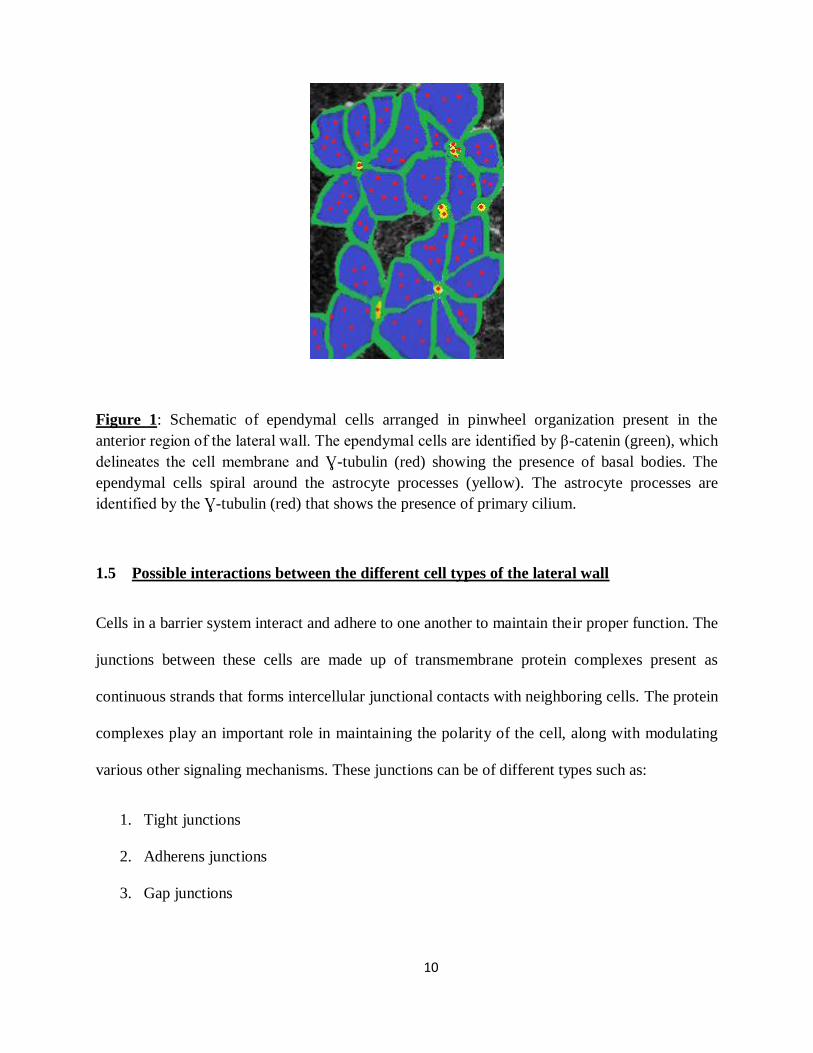
10
Figure 1: Schematic of ependymal cells arranged in pinwheel organization present in the
anterior region of the lateral wall. The ependymal cells are identified by β-catenin (green), which
delineates the cell membrane and Ɣ-tubulin (red) showing the presence of basal bodies. The
ependymal cells spiral around the astrocyte processes (yellow). The astrocyte processes are
identified by the Ɣ-tubulin (red) that shows the presence of primary cilium.
1.5 Possible interactions between the different cell types of the lateral wall
Cells in a barrier system interact and adhere to one another to maintain their proper function. The
junctions between these cells are made up of transmembrane protein complexes present as
continuous strands that forms intercellular junctional contacts with neighboring cells. The protein
complexes play an important role in maintaining the polarity of the cell, along with modulating
various other signaling mechanisms. These junctions can be of different types such as:
1. Tight junctions
2. Adherens junctions
3. Gap junctions

11
Of the different junctions, tight junctions play an important role in maintaining a selectively
permeable barrier wall.
1.5.1 Tight Junctions:
Tight junctions are intercellular protein complexes that help in adhering the neighboring cells
together.
They provide various advantages to the tissue such as:
1. Formation of a semi-permeable barrier: The membrane-bound protein complexes of one
cell form a tight seal with the extracellular domain of the protein molecules, from the
adjacent cell. This interaction facilitates the formation of a semipermeable barrier for the
movement of molecules that has both ion-specific and size-specific restrictions.
2. Demarcate the polarity of the cells: The formation of tight junction complexes precedes
asymmetric distribution of both the apical and basolateral composition of the proteins and
phospholipids. It also plays a very important role in restricting the movement of the different
lipids to their distinct domains, thereby forming a distinction between the apical and
basolateral membrane and giving the cell polarity (Mandel et al., 1993).
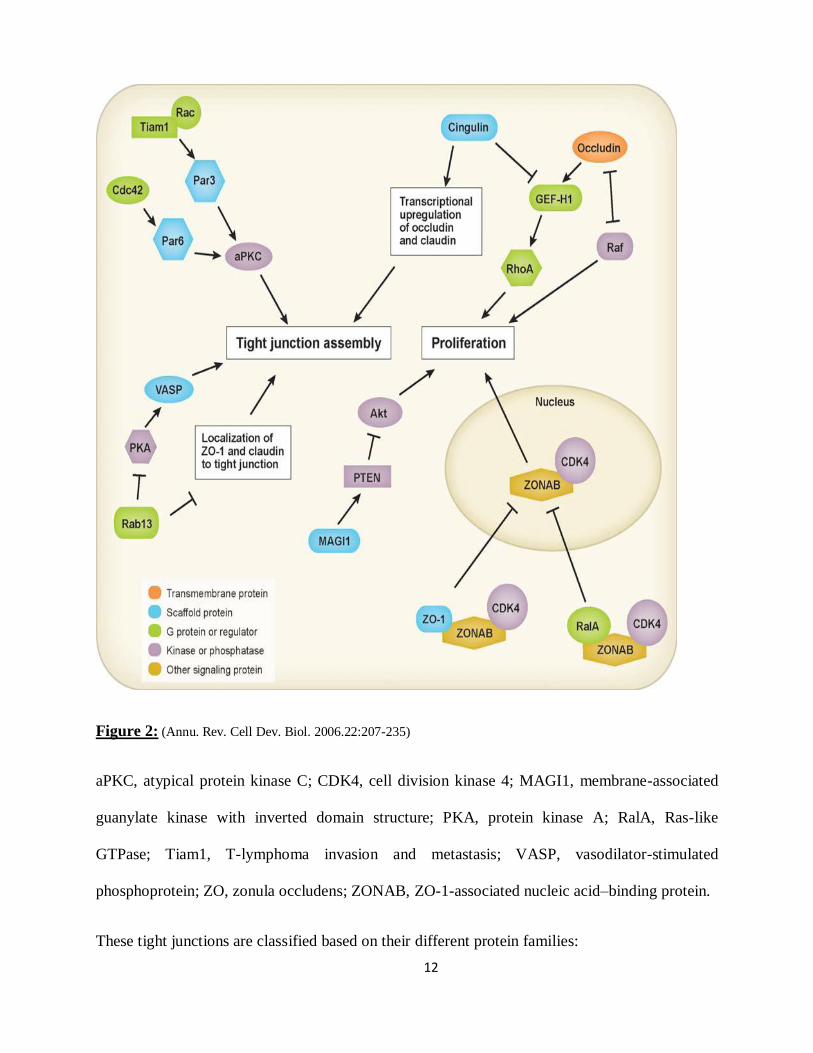
3. Role in cell proliferation and differentiation: Tight junction proteins regulate self-
assembly by controlling transcription and localization of tight junction proteins. In addition,
the proteins at tight junctions are involved in proliferation and differentiation as shown in the
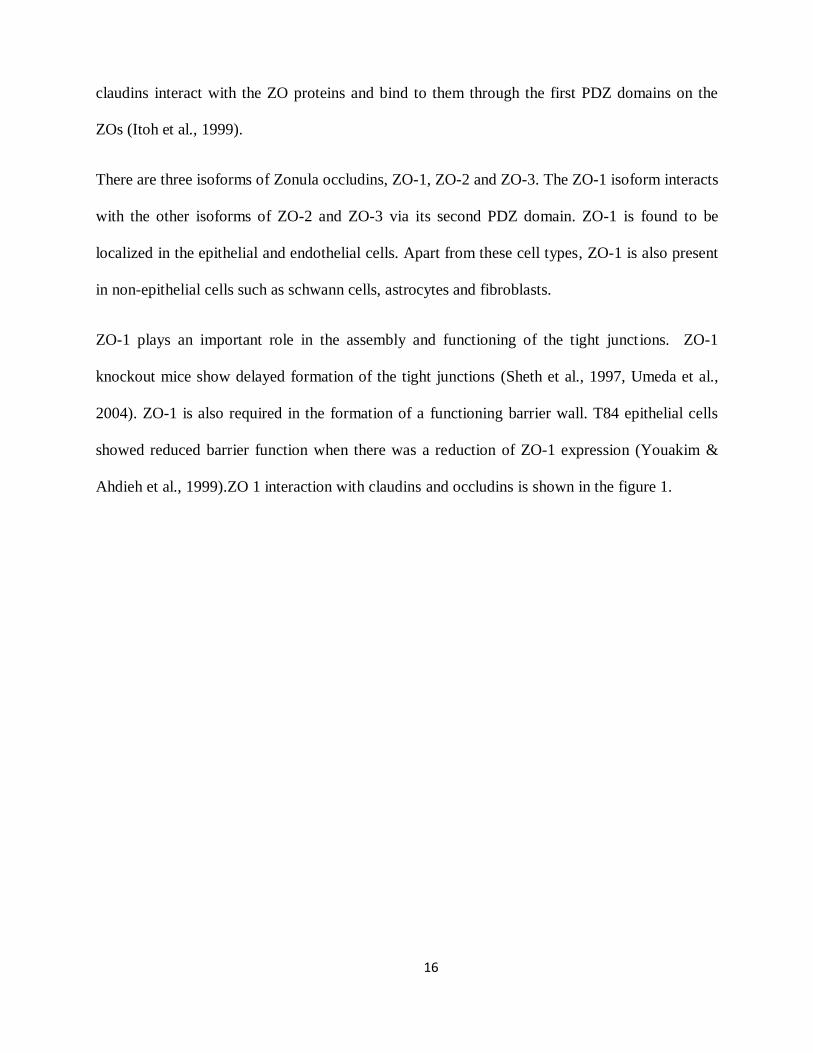
figure 2 (Annu. Rev. Cell Dev. Biol. 2006.22:207-235).
12
Figure 2: (Annu. Rev. Cell Dev. Biol. 2006.22:207-235)
aPKC, atypical protein kinase C; CDK4, cell division kinase 4; MAGI1, membrane-associated
guanylate kinase with inverted domain structure; PKA, protein kinase A; RalA, Ras-like
GTPase; Tiam1, T-lymphoma invasion and metastasis; VASP, vasodilator-stimulated
phosphoprotein; ZO, zonula occludens; ZONAB, ZO-1-associated nucleic acid–binding protein.
These tight junctions are classified based on their different protein families:

13
1. Transmembrane proteins
2. Peripheral membrane proteins
3. Integral membrane proteins
1.5.1.1 Transmembrane proteins:
Transmembrane proteins form a tight seal by reaching across the junction and connect the
membranes of the adjacent cells. Three families of the protein complexes make up the
transmembrane proteins, they are:
a) Occludins
b) Claudins
c) Junctional adhesion molecules.
a) Occludins: Occludins form one of the constituents of the intermembrane strands (Furuse
et al., 1993). Occludin consists of four transmembrane domains, two extracellular loops, and two
intracellular domains (Feldman et al., 2005).
Occludin interacts with peripheral membrane proteins such as zonula occludens ZO-1, ZO-2 and
ZO-3, which in turn interact with the actin cytoskeleton for the localization of the occludin at the
tight junction.
These occluding protein complexes have a lot of functional significance with paracellular
permeability. This was observed by expressing the C-terminal truncated form of occludin in
MDCK11 cell lines which showed increased paracellular permeability (Balda et al., 1996).
By expressing occludins in occludin-null fibroblasts, increased cell adhesion was also observed
(Itallie & Anderson et al., 1997). From these studies, we understand the role of occludins in
maintaining the paracellular permeability and cell adhesion.
14
Apart from this, occludins also take part in various signaling mechanisms of cell proliferation as
shown in the Figure 2. Overexpressions of occludins have shown to suppress the Raf-1-induced
tumor growth (Li & Mrsny et al., 2000; Wang et al., 2005). Occludins are also involved in RhoA
activation through GEF-H1/Lfc, a guanine nucleotide exchange factor associated with the tight
junction protein complexes.
b) Claudins: Claudins also belong to the transmembrane protein family with four
transmembrane domains, two extracellular and two intracellular domains. Despite the topological
similarities seen between the occludins and claudins, there exist no sequence similarities between
them.
Claudins family consists of 24 members. Mostly two of these members are expressed together in
a tissue specific manner. They play an important role in inducing cell-cell adhesion, which is
observed by expressing claudin-1 and claudin-2 in L-fibroblasts lacking tight junction. These
claudins when overexpressed in fibroblasts showed increased cell adhesion and made cell contact
at the regions where claudins were expressed (Furuse et al., 1998). Also when occludins and
claudins were coexpressed in L fibroblasts, occludins were recruited by the claudins in the tight
junction strands, showing the importance of the claudins.
Claudins form the tight seal, by heterotypically interacting with the adjacent cells. They also
interact homotypically with the claudin proteins of the same cell (Furuse et al., 1999). They have
conserved PDZ domain in the C terminus through which they interact with peripheral membrane
containing proteins, ZO-1, ZO-2 and ZO-3 (Roh et al., 2002; Tsukita et al., 2001).
Claudins have tissue-specific expression pattern i.e. different members of claudins are expressed
in different tissues. To parallel this, different members of the claudin family also have different
15
functional significance. This is observed through the mutations in claudin-16 in a human disease
syndrome, where the normal paracellular permeability of calcium and magnesium were affected
(Hou et al., 2005; Simon et al., 1999). Ectopic claudin 4 expression in the epithelia causes a
decreased paracellular conductance by reducing sodium permeability (Itallie et al., 2001).
Claudins 1, 3, 5 are expressed in the ependymal cells of the third ventricle (Mullier et al., 2010) .
It will be interesting to know if they are also expressed by the ependymal cells along the lateral
ventricles and what their contribution to barrier function is at the lateral ventricular surface..
c) Junctional Adhesion Molecules (JAM): JAMs belong to the immunoglobulin
superfamily and have a single transmembrane domain. They interact with the scaffolding
proteins such as ZO-1 and stabilize the tight junctions at the periphery. These form cell-cell
contact and help in maintaining the barrier wall.
1.5.1.2 Peripheral membrane protein:
Peripheral membrane proteins bind the transmembrane protein to the actin cytoskeleton through
their intracellular domain. The peripheral membrane proteins allow the transmembrane proteins
to organize in the membrane and initiate signaling processes. They also stabilize their junctional
interaction with their neighboring cell through the cytoskeletal attachment provided by the
peripheral membrane proteins.
Zonula Occludin (ZO): Zonula Occludins belong to the family of peripheral membrane
proteins. ZO proteins thus act as scaffolding proteins. They interact and bind to their binding
partners, such as the transmembrane proteins and also the cytoskeletal proteins. The ZO proteins
bind to the c-terminus of the occludins as well as the claudins. The C-terminus regions of the
16
claudins interact with the ZO proteins and bind to them through the first PDZ domains on the
ZOs (Itoh et al., 1999).
There are three isoforms of Zonula occludins, ZO-1, ZO-2 and ZO-3. The ZO-1 isoform interacts
with the other isoforms of ZO-2 and ZO-3 via its second PDZ domain. ZO-1 is found to be
localized in the epithelial and endothelial cells. Apart from these cell types, ZO-1 is also present
in non-epithelial cells such as schwann cells, astrocytes and fibroblasts.
ZO-1 plays an important role in the assembly and functioning of the tight junctions. ZO-1
knockout mice show delayed formation of the tight junctions (Sheth et al., 1997, Umeda et al.,
2004). ZO-1 is also required in the formation of a functioning barrier wall. T84 epithelial cells
showed reduced barrier function when there was a reduction of ZO-1 expression (Youakim &
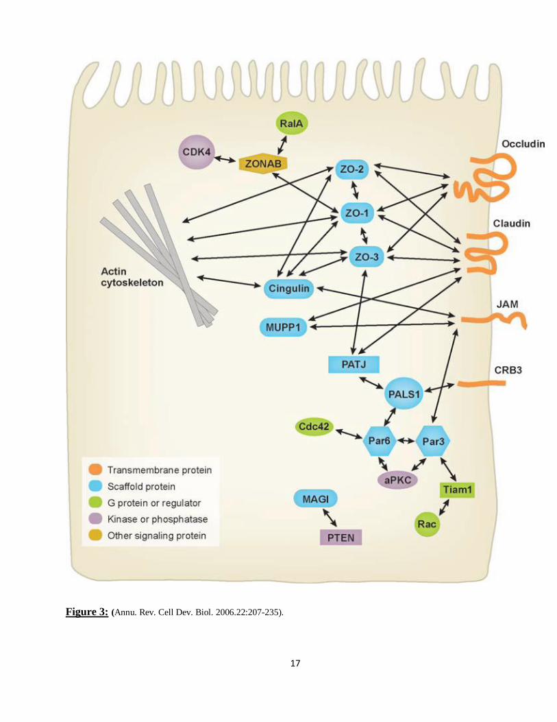
Ahdieh et al., 1999).ZO 1 interaction with claudins and occludins is shown in the figure 1.
17
Figure 3: (Annu. Rev. Cell Dev. Biol. 2006.22:207-235).
18
1.6 Tracers to study the permeability:
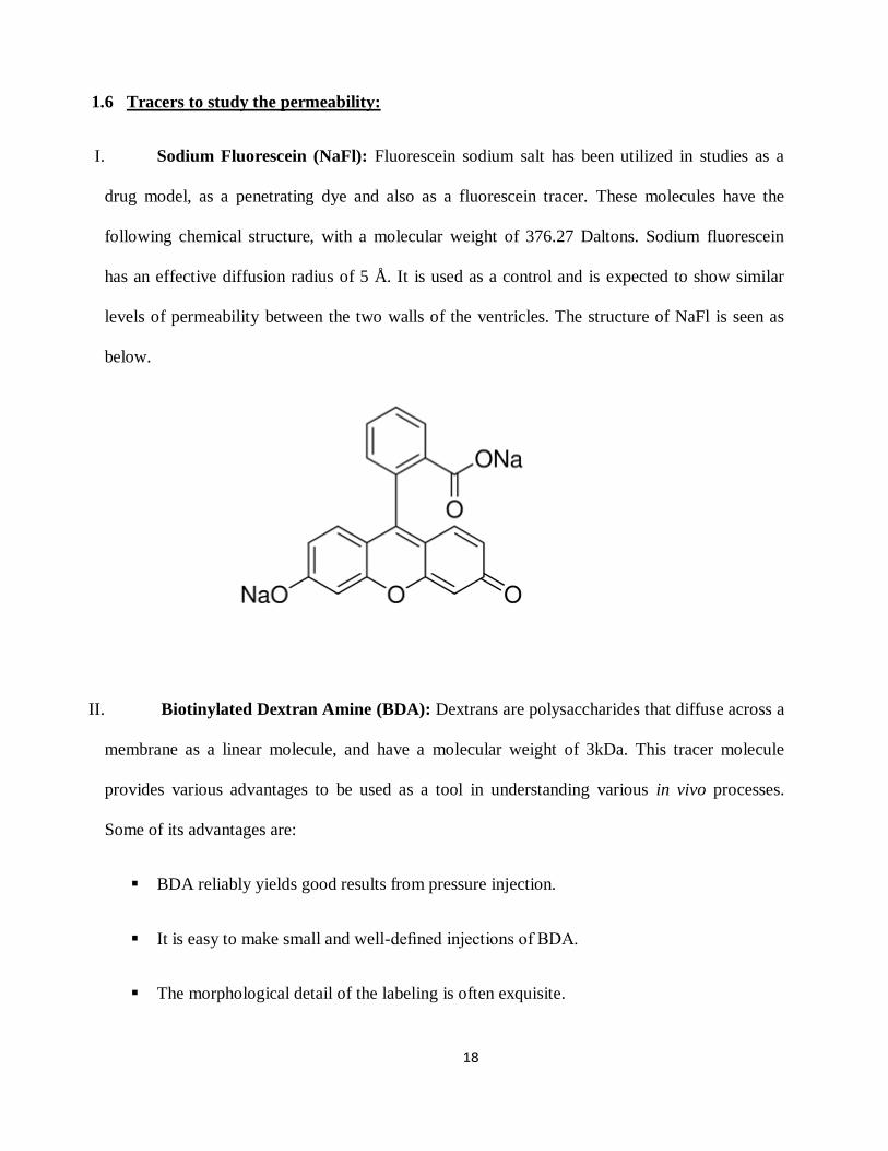
I. Sodium Fluorescein (NaFl): Fluorescein sodium salt has been utilized in studies as a
drug model, as a penetrating dye and also as a fluorescein tracer. These molecules have the
following chemical structure, with a molecular weight of 376.27 Daltons. Sodium fluorescein
has an effective diffusion radius of 5 Å. It is used as a control and is expected to show similar
levels of permeability between the two walls of the ventricles. The structure of NaFl is seen as
below.
II. Biotinylated Dextran Amine (BDA): Dextrans are polysaccharides that diffuse across a
membrane as a linear molecule, and have a molecular weight of 3kDa. This tracer molecule
provides various advantages to be used as a tool in understanding various in vivo processes.
Some of its advantages are:
BDA reliably yields good results from pressure injection.
It is easy to make small and well-defined injections of BDA.
The morphological detail of the labeling is often exquisite.
19
Availability of wide range of molecular size.
The simplicity of the visualization procedure.
Of all the above advantages, the availability of this tracer in different size ranges helps us to
choose a tracer, larger than the control tracer that freely diffuses between the junction proteins.
Thus, we choose 3kDa BDA that has the effective diffusion radius (EDR) of 12 Å. It is a Stokes
radius calculated from the average of hydrodynamic radius of a molecule, obtained from
viscosity and translation diffusion coefficients of the molecule in solution. This radius is closest
to the true molecular radius if the molecule takes up the shape of a sphere in the solution. Since
our tracer diffuses as a linear molecule comparing the permeability as a function of the Stoke’s
radius would not be accurate. Still, we choose this tracer because it is one of the prominent
tracers used in many in vivo diffusion studies.In vivo diffusion studies have shown prominent
occlusion of the tracer, 3kDa BDA during the formation of tight junction in the developing
choroid plexus of Monodelphis domestica (Liddelow et al., 2009).
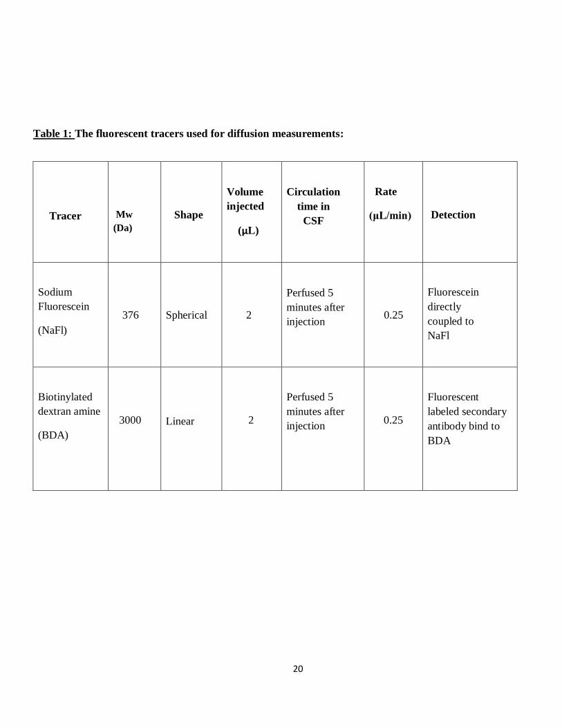
20
Table 1: The fluorescent tracers used for diffusion measurements:
Tracer
Mw
(Da)
Shape
Volume
injected
(μL)
Circulation
time in
CSF
Rate
(μL/min)
Detection
Sodium
Fluorescein
(NaFl)
376
Spherical
2
Perfused 5
minutes after
injection
0.25
Fluorescein
directly
coupled to
NaFl
Biotinylated
dextran amine
(BDA)
3000
Linear
2
Perfused 5
minutes after
injection
0.25
Fluorescent
labeled secondary
antibody bind to
BDA
21
1.7 Experimental Focus:
The neural stem cells in the sub-ventricular zone, SVZ are required in maintaining the adult
neurogenesis. Hence, it becomes important to understand how this stem cell niche, is maintained.
One of the possibilities through which these an environment could be maintained through
signaling molecules from the cerebro-spinal fluid (CSF) that circulates right next to the SVZ. We
know that some factors that maintain this niche are actively transported across the barrier wall
from the CSF to the SVZ niche (Riquelme et al., 2008). Although there is high degree of active
transport taking place through the lateral wall into the SVZ, it is important to note that the lateral
wall itself is very unique. The astrocyte processes of the B1 cells contact the lateral ventricular
wall unlike any other ventricular barrier walls. The presence of different cell types in the lateral
wall of the lateral ventricles, suggests various junctional interactions (as discussed before) to be
present between them. This led us to believe that there could be a different permeability in the
lateral ventricles that may contribute to maintaining the SVZ niche.
To test this hypothesis, we used 3-month old mice of the CD1 strain and injected two different
tracer- BDA and NaFl, into the right lateral ventricle and immediately sacrificed the mice to
study the distance of permeability of the tracer in the lateral and medial walls of the lateral
ventricle using immunolabelling and other imaging techniques. The results obtained from this
experiment were further validated using immunolabelling of the junctional protein ZO-1.
Further, tight junction structures were also examined using electron microscopy.
2. MATERIALS AND METHODS:
22
2.1 Animals and Tissue preparation:
Nine male CD1 mice, purchased from Charles River, were used in this study. All the
experiments were carried out in accordance with the protocol directed by the Institutional
Animal Care and Use Committees (IACUC).
2.2 Stereotaxic Injection:
NaFl was used as the control tracer and 3% 3kDa BDA was used as an experimental tracer for
this study. The intracerebroventicular injection (i.c.v) of the tracer molecules involved two sets
of 3-month old CD1 mice (i.c.v; n=3) for each tracer.
These adult mice were placed in a stereotactic frame after being anesthetized with isoflurane in
2% oxygen. Throughout the surgery the mice were constantly under anesthesia using isoflurane
in 2% oxygen. A burr hole was drilled 0.85 mm lateral to the bregma and 2.5 mm deep relative
to dura, according to the mouse brain atlas (Paxinos and Franklin, 2001).
A hamilton syringe filled with 2µL of NaFl or 2µL of 3 % 3kDA BDA was slowly inserted into
the right lateral ventricle of the brain. Using an infusion pump, the tracers were injected into the
mice at the rate of 0.25µL/min. The needle from the Hamilton syringe was removed 1 minute
after the entire amount of tracer was injected. The mice were perfused immediately following the
injection.
2.3 Gravity-Facilitated Perfusion:
Immediately after the intracerebroventricular (i.c.v) injection the animals were maintained in the
anesthetized state with an intraperitoneal (i.p.) injection of avertin (2.5%; 0.025mL per gram
mouse mass) followed by transcardial perfusion with 25mL of 0.9% saline, which flushed the
23
vascular system. Following this, the fixation process was carried out using 25mL of 4%
Paraformaldehyde, PFA.
The brains were quickly removed removed from the skull and post-fixed with 4% PFA at 4°C
overnight to prepare them for sectioning using the Leica vibratome. PFA-fixed brains were
washed 3 times for 20 minutes in PBS and sectioned (A/P coordinates 0.5-1.54mm, relative to
bregma) to obtain 50µm coronal sections. All the brain sections were collected in the 24 well
plates.
2.4 Immunohistochemistry:
Free-floating brain sections were permeabilized with 0.1% Triton X-100 in PBS for 10 min.
Following this the tissue were blocked with 10% horse serum in PBS/1% Triton X-100 for 1
hour at room temperature. Sections were incubated overnight with the following primary
antibodies: rabbit anti-β catenin, (1:100); rabbit anti-γ tubulin, (1:500); mouse anti-GFAP,
(1:500); rabbit anti-s100β (1:1000) and mouse anti-ZO-1 (1:10).
The next day, sections were washed three times in PBS and incubated for 1 hour at room
temperature with fluorescent-labeled secondary antibodies: Alex Fluor 488 donkey anti-mouse
(1:500), Alexa Fluor 568 donkey anti-rabbit (1:500) and Alex Fluor 647 donkey anti-mouse
(1:500). Secondary antibody staining alone was used as a control for BDA injections. The
sections were mounted on gelatin-coated slides and cover slips were placed over the sections.
The slides were allowed to dry overnight. All slides were imaged using a Leica TCS SP2
confocal laserscan microscope or Zeiss Axio imager M2 microscope, using HAMAMATSU
ORCA-R2 digital camera C10600.
2.5 Whole mount dissections:
24
The whole mount dissection protocol was adapted from Mirzadeh et al. (2008 and 2010). The
sections were immunostained with rabbit anti-β catenin (1:100), rabbit anti-γ tubulin (1:500),
mouse anti-ZO1 (1:10). The coverslip was placed over the whole mount with aquapolymount
and imaged on a Leica TCS SP2 confocal laserscan microscope.
2.6 Electron microscopy:
The electron microscopy images were processed using the protocol adapted from Luo et al.
(2003). The images were analyzed using Adobe Photoshop CS2.
2.7 Quantification of signal intensity:
All the images obtained from injected tracer NaFl and BDA were analyzed using Image J
software.
2.8 Statistical significance:
Statistical analysis was performed using a two-tailed unpaired Student’s t test. The sample size
used for this analyses was n=3. The level of significance was set at p < 0.05.
3. RESULTS:
25
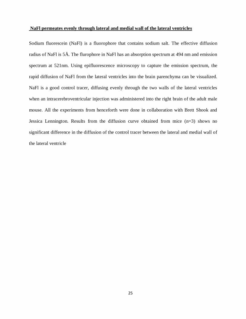
NaFl permeates evenly through lateral and medial wall of the lateral ventricles
Sodium fluorescein (NaFl) is a fluorophore that contains sodium salt. The effective diffusion
radius of NaFl is 5Å. The flurophore in NaFl has an absorption spectrum at 494 nm and emission
spectrum at 521nm. Using epifluorescence microscopy to capture the emission spectrum, the
rapid diffusion of NaFl from the lateral ventricles into the brain parenchyma can be visualized.
NaFl is a good control tracer, diffusing evenly through the two walls of the lateral ventricles
when an intracerebroventricular injection was administered into the right brain of the adult male
mouse. All the experiments from henceforth were done in collaboration with Brett Shook and
Jessica Lennington. Results from the diffusion curve obtained from mice (n=3) shows no
significant difference in the diffusion of the control tracer between the lateral and medial wall of
the lateral ventricle
26
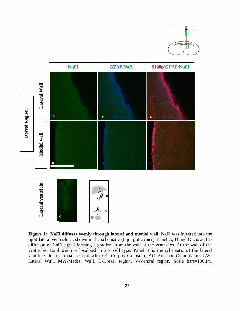
Figure 1: NaFl diffuses evenly through lateral and medial wall. NaFl was injected into the
right lateral ventricle as shown in the schematic (top right corner). Panel A, D and G shows the
diffusion of NaFl signal forming a gradient from the wall of the ventricles. At the wall of the
ventricles, NaFl was not localized in any cell type. Panel H is the schematic of the lateral
ventricles in a coronal section with CC–Corpus Callosum, AC–Anterior Commissure, LW-
Lateral Wall, MW-Medial Wall, D-Dorsal region, V-Ventral region. Scale bars=100µm.
D
Late
ral
ven
tric
le
G
V
H
A
E
F
2µL
B C A
D E F
Med
ial
wall
L
ate
ral
Wall
Dors
al
Reg
ion
NaFl S100β/GFAP/NaFl GFAP/NaFl
27
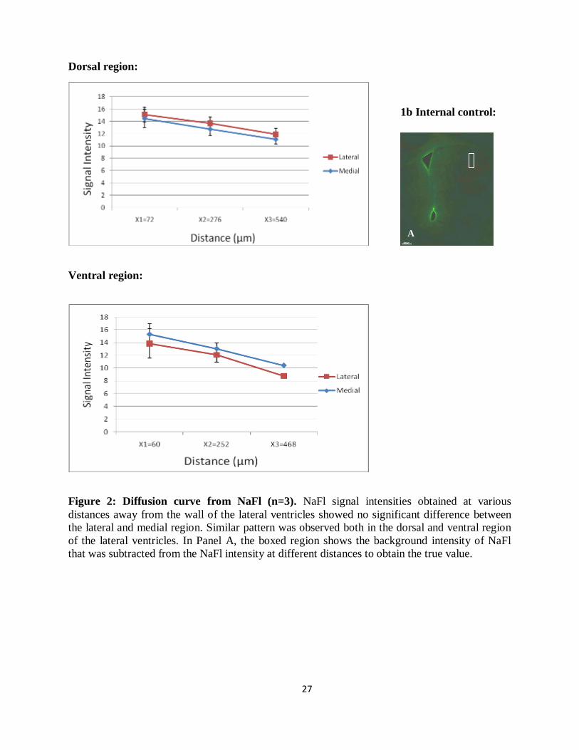
Dorsal region:
Ventral region:
Figure 2: Diffusion curve from NaFl (n=3). NaFl signal intensities obtained at various
distances away from the wall of the lateral ventricles showed no significant difference between
the lateral and medial region. Similar pattern was observed both in the dorsal and ventral region
of the lateral ventricles. In Panel A, the boxed region shows the background intensity of NaFl
that was subtracted from the NaFl intensity at different distances to obtain the true value.
1b Internal control:
A
A
28
3KDa BDA diffuses evenly through lateral and ventral wall
Biotinylated dextran amine (BDA) is a large molecule made up of linear polysaccharide chains.
BDA is occluded by the tight junctions found between the cells of the choroid plexus (Liddelow
et al., 2009). Hence, BDA was used as an experimental tracer to observe if there exists a
differential permeability between the two walls, which may be a result of differential junctional
protein expression. BDA was detected through streptavidin coupled with FITC (fluorescein
isothiocyanate). Streptavidin binds specifically to biotin molecules of BDA. FITC has an
excitation spectrum at 495nm and emission spectrum at 521 nm. Epifluorescence microscopy
captures the signal from FITC and was used to detect streptavidin-bound BDA. The results from
this experiment show that BDA is passively and actively transported at the wall of the ventricles.
The number of cells that actively take up BDA in the lateral wall is comparable to that of the
medial wall. Results from the diffusion curve (n=3) show no significant difference in the signal
intensity of BDA between the lateral and medial walls. Hence we conclude that there is no
significant permeability difference for BDA between the two walls
29
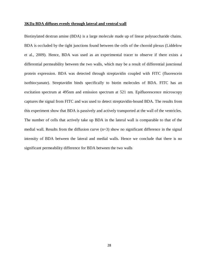
Figure 3: BDA is localized within the cell and at the periphery of the cell. Panel A, D, I and
L shows the BDA (green) signal intensity. White arrows indicate BDA signal, at the periphery of
the cells showing that it could possibly diffuse in between cells. Red arrows indicate BDA
signal, localized within cells showing the uptake of the experimental tracer. Yellow arrows
indicate the GFAP+ processes (blue) associated with the ependymal cells (red).Scale bars=50µm.
2µL
BDA GFAP/BDA Merge GFAP/BDA BDA Merge
Late
ral
wall
M
edia
l w
all
Med
ial
wall
L
ate
ral
wall
Dors
al
Reg
ion
Ven
tral
Reg
ion
B
A
C A
D E F
I J K
L M N
30
Dorsal region:
Ventral region:
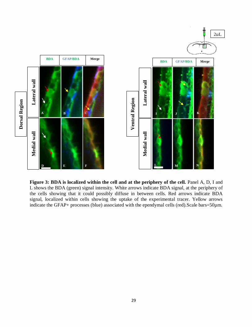
Diffusion curve for BDA (n=3). BDA signal obtained from the FITC–labeled streptavidin
which was bound to BDA was used to generate the diffusion curve. The intensity of the BDA
signal at different distances away from the wall was plotted on the curve. Panel A, shows the
signal intensity from FITC-labeled streptavidin only. The intensity of the background at x=72,
276 and 540 µm was obtained. This intensity was subtracted from the raw signal intensity (BDA
+ streptavidin), which was higher than the background was used to obtain the true signal. True
signal intensity was used to plot the diffusion curve shown above. No significant difference was
seen from the results obtained from both the walls. Scale bar=50 μm.
2b Internal control:
Strep only
A
Lateral wall Medial wall
31
Characteristics and coding of the cell types in the ventricular wall
Detailed examination of the cytoarchitectural arrangement of the cells in the lateral and medial
wall was conducted using the transmission electron microscopy images. The coronal sections of
ultra thin EM micrographs are presented in figure 5. The lateral wall consists of different cell
types and they were identified based on reports from Luo et al., 2006 and Doetsch et al., 1997.
The astrocytes interpolated through the lateral ventricular wall were found to establish both tight
and adherens junctions with the adjacent ependymal cells and other neighboring astrocytes,
Figure 5A.Tight and adherens junctions were also identified at the medial wall between
neighboring ependymal cells. These observations show no difference in the tight junction protein
interaction between the cells of the two walls. This thereby supports our findings of similar
diffusion patterns for different tracers throughout the ventricular walls.

32
Figure 5: Electron microscopic view of the lateral ventricular walls. Panel A, shows the lateral
ventricular wall with astrocytes, interposed within the ependyma (As-Astrocyte, E-Ependyma, N-
Neuroblast). Panel B, shows a single monolayer of ependymal cells with no other cell type present in the
environment. Panel C shows the ependymal cells (E) with many mitochondria, lipid droplet (black
arrow), basal bodies of cilia (black arrow head), microvilli, motile cilia (9+2 organization) and dense
cytoplasm in coronal view. Panel D shows astrocytes with irregular nucleus and light cytoplasm. The
boxed region shows the adherens junction formed by astrocytes with its neighboring cells. Panels E, F
and G show E-E cells, E-As cells and As-As cells interactions respectively. All the cell types forms both
adherens junctions (brackets) and tight junctions (arrow) as shown in the above figures
E
A
V
E
E
E
As
E
G F
As
As
2 µm
2 µm
E
E
D B
As
V
2 µm
C
E
As
N
5 µm
2 µm
2 µm
33
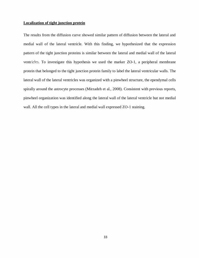
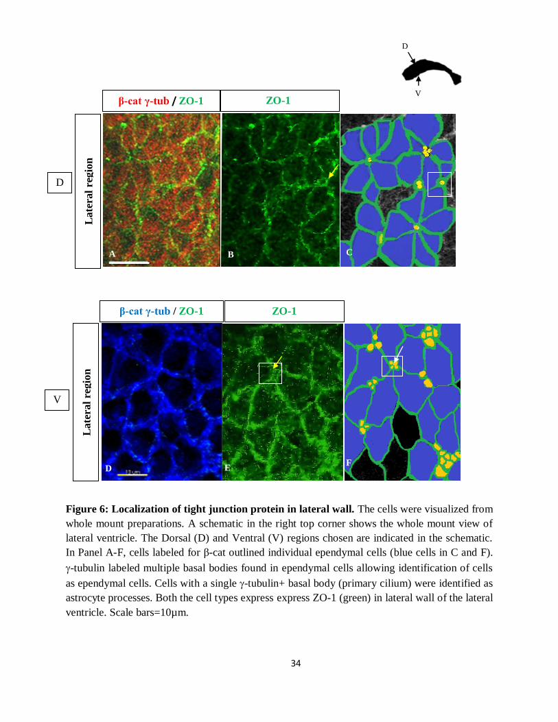
Localization of tight junction protein
The results from the diffusion curve showed similar pattern of diffusion between the lateral and
medial wall of the lateral ventricle. With this finding, we hypothesized that the expression
pattern of the tight junction proteins is similar between the lateral and medial wall of the lateral
ventricles. To investigate this hypothesis we used the marker ZO-1, a peripheral membrane
protein that belonged to the tight junction protein family to label the lateral ventricular walls. The
lateral wall of the lateral ventricles was organized with a pinwheel structure, the ependymal cells
spirally around the astrocyte processes (Mirzadeh et al., 2008). Consistent with previous reports,
pinwheel organization was identified along the lateral wall of the lateral ventricle but not medial
wall. All the cell types in the lateral and medial wall expressed ZO-1 staining.
2 µm
2 µm
2 µm
34
Figure 6: Localization of tight junction protein in lateral wall. The cells were visualized from
whole mount preparations. A schematic in the right top corner shows the whole mount view of
lateral ventricle. The Dorsal (D) and Ventral (V) regions chosen are indicated in the schematic.
In Panel A-F, cells labeled for β-cat outlined individual ependymal cells (blue cells in C and F).
-tubulin labeled multiple basal bodies found in ependymal cells allowing identification of cells
as ependymal cells. Cells with a single -tubulin+ basal body (primary cilium) were identified as
astrocyte processes. Both the cell types express express ZO-1 (green) in lateral wall of the lateral
ventricle. Scale bars=10µm.
D F
D
V
D
V
β-cat γ-tub / ZO-1 ZO-1
Late
ral
regio
n
β-cat γ-tub / ZO-1 ZO-1
Late
ral
regio
n
B C
E D F
A
35
Figure 7: Localization of tight junction protein in medial wall. The cells are visualized from
whole mount prep. A schematic in the right top corner shows the whole mount view of lateral
ventricle. The dorsal(D) and ventral(V) regions chosen are indicated in the schematic. Panel A,
B, C and D shows the β-cat/Ɣ-tub+ ependymal cells in the medial wall express ZO-1 (green).
β-cat γ-tub/ ZO-1 ZO-1
D
V
β-cat γ-tub / ZO-1 ZO-1
D
V
Late
ral
regio
n
Med
ial
regio
n
A
C D
B
36
DISCUSSION:
In this study, we used immunocytochemistry to systematically examine the distribution of the
tight junction protein, ZO-1, on the walls of the lateral ventricles. Our results revealed the
expression of ZO-1 protein by all the cell types present in lateral and medial wall. The
localization of the tight junction protein parallels the results from the diffusion curves that
exhibit similar diffusion pattern between the two walls, as shown by intracerebroventricular
injections of BDA and NaFl. Our findings reveal the barrier role of the SVZ, which is unique
with the presence of pinwheel organization, encompassing different cell types interacting with
one another as opposed to the other ventricular walls of the brain (Mirzadeh et al., 2008). The
similar diffusion pattern observed between the two walls permits us to understand the barrier role
of the lateral wall, which is maintained despite the interposed NSCs within the lateral wall.
The transport of substances from the cerebrospinal fluid to the brain parenchyma has been of
interest among many researchers. Detailed examination of the diffusion barriers has been
established at choroid plexus and the third ventricles (Muller et al., 2010; Liddellow et al.,
2009).The analyses of these diffusional barriers reveal differentially permeable domains that are
unique to their function. Along with this, now we also know the size range exclusion of
molecules that are not transported through the ventricular wall. This knowledge allows us to
predict the possible molecules that could be transported from the CSF into the brain parenchyma.
Our present study establishes the first characterization of the diffusion barrier along the lateral
ventricular walls, that ensheaths the proliferative SVZ niche. Along with the diffusion pattern
through the ventricle walls, our results indicate the presence of the honeycomb-like pattern of
immunoreactivity for ZO-1 by the ependymal cells. This pattern of immunoreactivity is typical
37
of tight junction proteins (Tsukita and Furuse, 2002) and has also been identified in the cells of
the choroid plexus and third ventricle (Wolburg et al., 2001; Petrov et al., 1994).
The tight junctions are present in the apical region of the lateral membrane. The proteins
expressed at these junctions form sealing strands fusing the plasma membrane together (Tsukita
et al., 2001; Tsukita and Furuse, 2002). The junction proteins that make up the paracellular
barrier, forms a belt-like anchoring junction encircling the cells through their interactions with
their immediate neighbors, giving rise to a honeycomb staining pattern (Coisne et al., 2005;
Wolburg et al., 2001). The results from the intracerebroventricular injection of BDA and NaFl
show similar diffusion patterns between the walls. The diffusion pattern, demonstrates the cells
of the wall are held together at the apical membrane by similar tight junction proteins. One such
protein that contributes to forming the tight junction is ZO-1 which is present in both the lateral
and medial wall, Figure 4. These results are also consistent with the findings from the TEM
images that show the presence of tight junctions between all the cell types of lateral and medial
wall.
The intracerebroventricular injections of NaFl demonstrate that the barrier layer of the
lateral and medial wall is equally permeable and uncontrolled exchange of solutes of the size
range of NaFl may occur between the CSF and the brain parenchyma at both the walls. Particles
of the si e of aFl with the ionic radius 4.5 can diffuse through the tight junction, which
occludes hydrophilic molecules of the si e greater than 20 (Anderson et al. 2001). This size
correlated to the size range of water, oxygen, carbon dioxide. In contrast, BDA is effectively
occluded by the tight junction protein complexes between the cells of choroid plexus in the
lateral ventricles and was expected to be occluded by the tight junctions of ependymal cells in
the lateral ventricles that had similar protein complexes to that of the cells of choroid plexus
38
(Liddelow et al., 2009). The injection of BDA into the lateral ventricular system showed both
localization of BDA within the cells and transport of BDA at the periphery of the cells. This
indicates that molecules of a bigger size range than NaFl are more readily taken up by the cell
(possibly through transcytosis), than through diffusion between cells, as would be expected.
Despite the similar degree of active transport that takes place along the lateral and medial wall
(as shown in Figure 2) a similar amount of BDA is shown to diffuse in between the cells of the
walls and get to the brain parenchyma (results from the diffusion curve of BDA; Figure 2).
The diffusion pattern along with the tight junction localization and characterization was
consistent at different domains (dorsal and ventral region) along the ventricular wall. This
finding at the walls of the lateral ventricular is in contrast with the third ventricular wall that
shows differential permeability at distinct domains (Muller et al., 2010).
Others have reported that, the regions lacking cilia along with disrupted tight junction protein
expression in the third ventricular wall show increased diffusion (Muller et al., 2010). Beating of
the cilia sets in motion of the CSF flow (Sawamoto et al., 2006) and thus limits the diffusion of
CSF, from the ventricles to the brain parenchyma through the ependymal sheet. This may result
in the exclusion of substances near the wall which might have, otherwise, diffused through it. In
the lateral ventricular region both the lateral and medial walls are uniformly ciliated and hence
we do not see a differential permeability pattern at different domains as seen in the third
ventricles. With aging, reports show cilia balding of ependymal cells in 22 months old mice (Luo
et al., 2006). Along with the loss of the cilia, the presences of ependymal like cells are also
observed at the lateral wall of the lateral ventricles (review by Conover et al., 2011). The
ependymal like cells have been observed as an intermediate cell type in aged mice (22 months
old) where in the loss of ependymal cells in lateral wall of lateral ventricles is partially
39
compensated by the SVZ repair mechanism mediated by the SVZ astrocytes that acquire the
attributes of ependymal cells and finally become the ependymal cell itself (Luo et al., 2006).
These factors can contribute to an increased permeability with aging and provide a potential
route of entry for toxins, generally small molecules (M.W. range of 4kDa) that can diffuse in
between cells, from the CSF to the brain parenchyma. Our present study on diffusion barriers
will provide a proper model for comparison on such future studies.
To our knowledge, this is the first study on the diffusion barriers of the lateral and medial walls
of the lateral ventricles of the adult mouse brain. The results of our immunofluorescence studies,
together with dye permeability experiments and TEM image analyses, provide detailed
information about the diffusion barriers between cerebrospinal fluid and brain compartment. This
result did not hold true with our initial hypothesis that predicted a differential permeability
between the lateral and medial wall. With these results that show similar diffusion pattern
between lateral and medial ventricle walls it is interesting to note that the wall of the SVZ
maintains a proper barrier.
40
4. REFERENCE:
Aimone JB, Wiles J, Gage FH (2009). Computational influence of adult neurogenesis on
memory encoding. Neuron, 61: 187-202
Alvarez-Buylla A, Lim DA (2004). For the long run: maintaining germinal niches in the adult
brain. Neuron, 41: 683–686
Alvarez-Buylla A, Garcia-Verdugo JM, Tramontin AD (2001). A unified hypothesis on the
lineage of neural stem cells. Nat Rev Neurosci, 2:287–293
Alvarez-Buylla A, Nottebohm F (1988). Migration of young neurons in adult avian brain.
Nature, 335:353–354
Altman J, Das GD (1965). Autoradiographic and histological evidence of postnatal
hippocampal neurogenesis in rats. J Comp Neurol, 124:319–335.
Altman J, Das GD (1966). Autoradiographic and histological studies of postnatal neurogenesis. I.
A longitudinal investigation of the kinetics, migration and transformation of cells incorporating
tritiated thymidine in neonate rats, with special reference to postnatal neurogenesis in some
brain regions. J Comp Neurol, 126:337–390.
Anderson (2001). Molecular Structure of Tight Junctions and Their Role in Epithelial Transport.
Physiology, 16: 126-130
Balda MS, Whitney JA, Flores C, Gonzalez S, Cereijido M, Matter K (1996). Functional
dissociation of paracellular permeability and transepithelial electrical resistance and disruption
of the apical-basolateral intramembrane diffusion barrier by expression of a mutant tight
junction membrane protein. J. Cell Biol, 134:1031–49
41
Bruni JE, Del Bigio MR, Clattenburg RE (1985). Ependyma: normal and pathological. A review
of the literature. Brain Res, 356:1–19.
Clelland CD, Choi M, Romberg C, Clem enson GD, Jr., Fragniere A, Tyers P, Jessberger S,
Saksida LM, Barker RA, Gage FH, Bussey TJ (2009). A functional role for adult hippocampal
neurogenesis in spatial pattern separation. Science, 325: 210-213
Conover JC, Shook BA (2011). Aging of the Subventricular Zone Neural Stem Cell Niche. Aging
and Disease, 2: 49-63
Coisne C, Dehouck L, Faveeuw C, Delplace Y, Miller F, Landry C, Morissette C, Fenart L,
Cecchelli R, Tremblay P, Dehouck B (2005). Mouse syngenic in vitro blood-brain barrier
model: a new tool to examine inflammatory events in cerebral endothelium. Lab Invest,
85:734–746.
Deng W, Saxe MD, Gallina IS, Gage FH (2009). Adult-born hippocampal dentate granule cells
undergoing maturation modulate learning and memory in the brain. J Neurosci, 29: 13532-
13542
Doetsch F (2003). The glial identity of neural stem cells. Nat. Neurosci., 6:1127–1134.
Doetsch F, Garcia-Verdugo JM, Alvarez-Buylla A (1997). Cellular composition and three-
dimensional organization of the subventricular germinal zone in the adult mammalian brain. J.
Neurosci., 17:5046 –5061.
Falk A, Frisen J (2002). Amphiregulin is a mitogen for adult neural stem cells. J. Neurosci. Res.,
69:757–762.

42
Feldman GJ, Mullin JM, Ryan MP (2005). Occludin: structure, function and regulation. Adv.
Drug Deliv. Rev., 57:883–917
Furuse M, Sasaki H, Tsukita S (1999). Manner of interaction of heterogeneous claudin
specieswithin and between tight junction strands. J. Cell Biol., 147:891–903
Furuse M, Sasaki H, Fujimoto K, Tsukita S (1998b). A single gene product, claudin-1 or-2,
reconstitutes tight junction strands and recruits occludin in fibroblasts. J. Cell Biol., 143:391–
401
Furuse M, Hirase T, Itoh M, Nagafuchi A, Yonemura S (1993). Occludin: a novel integral
membrane protein localizing at tight junctions. J. Cell Biol., 123:1777–88
Garcia-Verdugo JM, Doetsch F, Wichterle H, Lim DA, Alvarez-Buylla A (1998). Architecture
and cell types of the adult subventricular zone: in search of the stem cells. J. Neurobiol., 36:
234–248
Garthe A, Behr J, Kempermann G (2009). Adult-generated hippocampal neurons allow the
flexible use of spatially precise learning strategies. PLoS One, 4: 54-64
Gross CG (2000). Neurogenesis in the adult brain: death of a dogma. Nat Rev Neurosci., 1:67–7
Goldman SA, Nottebohm F (1983) Neuronal production, migration and differentiation in a vocal
control nucleus of the adult female canary brain. Proc Natl Acad Sci., 80:2390–2394
Hamazaki Y, Itoh M, Sasaki H, Furuse M, Tsukita S (2002). Multi-PDZ domain protein 1
(MUPP1) is concentrated at tight junctions through its possible interaction with claudin-1 and
junctional adhesion molecule. J. Biol. Chem., 277:455–61
43
Hou J, Paul DL, Goodenough DA (2005). Paracellin-1 and the modulation of ion selectivity of
tight junctions. J. Cell Sci., 118:5109–18
Imura T, Kornblum HI, Sofroniew MV (2003). The predominant neural stem cell isolated from
postnatal and adult forebrain but not early embryonic forebrain expresses GFAP. J Neurosci.,
23:2824–2832
Itoh M, Furuse M, Morita K, Kubota K, Saitou M, Tsukita S (1999). Direct binding of three tight
junction-associated MAGUKs, ZO-1, ZO-2, and ZO-3, with the COOH termini of claudins. J.
Cell Biol., 147:1351–63
Kempermann G, Jessberger S, Steiner B, Kronenberg G (2004) Milestones of neuronal
development in the adult hippocampus. Trends Neurosci., 27: 447–452
Kokovay, E., Shin, Q., and Temple, S. (2008). Neuron, 60: 420–42
Kriegstein A, Alvarez-Buylla A (2009). The glial nature of embryonic and adult neural stem cells.
Annu Rev Neurosci., 32: 149-184
Liddelow AS, Bouret SG (2009). Cellular transfer of macromolecules across the developing
choroid plexus of Monodelphis domestica. Euro. J. Neurosc., 29: 253–266
Li D, Mrsny RJ (2000). Oncogenic Raf-1 disrupts epithelial tight junctions via downregulation of
occludin. J. Cell Biol., 148:791–800
Luo J, Daniels SB, Lennington JB, Notti RQ, Conover JC (2006). The aging neurogenic
subventricular zone. Aging Cell, 5: 139-152
44
Mandel JS, Bond JH, Church TR, Snover DC, Bradley GM, Schuman LM, Ederer F (1993).
Reducing mortality from colorectal cancer by screening for fecal occult blood. Minnesota
Colon Cancer Control Study. N. Engl. J. Med., 328: 1365–1371
Mirzadeh, Z., Merkle, F.T., Soriano-Navarro, M., Garcia-Verdugo, J.M., and Alvarez-Buylla, A.
(2008). Neural stem cells confer unique pinwheel architecture to the ventricular surface in
neurogenic regions of the adult brain. Cell Stem Cell, 3: 265–278
Muller A, Bouret SG, Prevot V, Dehouck B (2010). Differential distribution of tight junction
proteins suggests a role for tanycytes in blood-hypothalamus barrier regulation in the adult
mouse brain. J Comp Neurol., 518: 943-62
Peterson DA (2002). Stem cells in brain plasticity and repair. Curr. Opin.Pharmacol. 2: 34– 4
Paxinos KBJ, Paxinos G (2001). The mouse brain in stereotaxic coordinates: Second Edition.
Academic Press, New York
Petrov T, Howarth AG, Krukoff TL, Stevenson BR (1994). Distribution of the tight junction-
associated protein ZO-1 in circumventricular organs of the CNS. Brain Res Mol Brain Res.,
21:235–246.
Roh MH, Liu CJ, Laurinec S, Margolis B (2002a). The carboxyl terminus of zona occludens-3
binds and recruits a mammalian homologue of discs lost to tight junctions. J. Biol. Chem., 277:
27501–9
Sheth B, Fesenko I, Collins JE, Moran B, Wild AE (1997). Tight junction assembly during mouse
blastocyst formation is regulated by late expression of ZO-1 α+ isoform. Development,
124:2027–37
45
Seroogy K.B, Lundgren K.H, Lee D.C, Guthrie K.M, Gall C.M (1993). Cellular localization of
transforming growth factor-a mRNA in rat forebrain. J. Neurochem., 60: 1777–1782
Simon DB, Lu Y, Choate KA, Velazquez H, Al-Sabban E (1999). Paracellin-1, a renal tight
junction protein required for paracellular Mg2+ resorption. Science, 285:103–6
Shors TJ, Miesegaes G, Beylin A, Zhao M, Rydel T, Gould E (2001). Neurogenesis in the adult is
involved in the formation of trace memories. Nature, 410: 372-376
Sawamoto K, Wichterle H, Gonzalez-Perez O, Cholfin JA, Yamada M, Spassky N, Murcia NS,
Garcia-Verdugo JM, Marin O, Rubenstein JL, Tessier-Lavigne M, Okano H, Alvarez-Buylla A
(2006). New neurons follow the flow of cerebrospinal fluid in the adult brain. Science,
311:629–632
Tsukita S, Furuse M (2002). Claudin-based barrier in simple and stratified cellular sheets. Curr
Opin. Cell Biol., 14:531–536
Tsukita S, Furuse M, Itoh M (2001). Multifunctional strands in tight junctions. Nat. Rev. Mol. Cell
Biol., 2:285–93
Umeda K, Matsui T, Nakayama M, Furuse K, Sasaki H (2004). Establishment and characterization
of cultured epithelial cells lacking expression of ZO-1. J. Biol. Chem., 279:44785–94
Van Itallie C, Rahner C, Anderson JM (2001). Regulated expression of claudin-4 decreases
paracellular conductance through a selective decrease in sodium permeability. J. Clin. Invest.,
107:1319–27
Van Itallie CM, Anderson JM. (1997). Occludin confers adhesiveness when expressed in
fibroblasts. J. Cell Sci., 110:1113–21
46
Wang Z, Mandell KJ, Parkos CA, Mrsny RJ, Nusrat A (2005). The second loop of occludin is
required for suppression of Raf1-induced tumor growth. Oncogene, 24:4412–20
Wolburg H, Wolburg-Buchholz K, Liebner S, Engelhardt B (2001).Claudin-1, claudin-2 and
claudin-11 are present in tight junctions of choroid plexus epithelium of the mouse. Neurosci.
Letter, 307:77–80.
Wurmser AE, Palmer TD, Gage FH (2004). Cellular interactions in the stem cell niche. Science,
304:1253–1255.
Youakim A, Ahdieh M (1999). Interferon-γ decreases barrier function in T84 cells by reducing
ZO-1 levels and disrupting apical actin. Am. J. Physiol., 276:1279–88
Zhao C, Deng W, Gage FH (2008). Cell, 132: 645–660