Embed Size (px)
Citation preview
[CANCER RESEARCH 42, 4348-4352, November 1982]0008-5472/82/0042-0000$02.00
Differentiation of Platelet-aggregating Effects of Human Tumor Cell
Lines Based on Inhibition Studies with Apyrase, Hirudin, andPhospholipase1
Eva Bastida,2 Antonio Ordinas,2 Steven L Giardina, and G. A. Jamieson3
American Red Cross Blood Services Laboratories, Bethesda. Maryland 20814
ABSTRACT
Three different mechanisms have been detected for theaggregation of platelets by tumor cells in a homologous humansystem based on inhibition studies with apyrase, hirudin, andphospholipase D. In the major group, platelet aggregationinduced by the SKBR3 (adenocarcinoma), SKNMC (neuroblastoma), HT29 (adenocarcinoma), and HT144 (melanoma) celllines was inhibited by apyrase and phospholipase D but not byhirudin, suggesting that adenosine 5'-diphosphate is involved
in the first step. However, since the reaction occurs only inheparinized plasma, the mechanism must differ from that ofplatelet aggregation which can be induced in citrated platelet-rich plasma by endogenous or exogenous adenosine 5'-di-
phosphate. In contrast, the Hut28 (mesothelioma) line wasinhibited by hirudin and phospholipase D but not by apyrase,suggesting that the mechanism in this system involves theactivation of the clotting system in the early stages. However,the coagulant-dependent mechanism observed with Hut28 can
be differentiated from the similar mechanism we have observedpreviously with the U87MG (glioblastoma) cell line since thelatter is unaffected by phospholipase D (Am J. Hematol., 11:367-378, 1981). Phospholipase C had no effect on platelet
aggregation induced by any of the human cell lines examinedwhile both phospholipase A and lysolecithin inhibited aggregation in every case. These results suggest that two categoriesof human tumor cells can be defined based on whether theyinitiate platelet aggregation by adenosine 5'-diphosphate or
coagulant-dependent mechanisms. However, within this latter
category, subclassification is possible based on the inhibitoryeffects of phospholipase D.
INTRODUCTION
Platelets in plasma can be aggregated by a wide variety oflow-molecular-weight agonists such as ADP and epinephrine
or by protein agonists such as thrombin and collagen. Themechanisms of these reactions are not clearly understood. Athird class of interaction involves platelet aggregation inducedby tumor cells (for review, see Ref. 7). Qualitative observationsof interactions between platelets and tumor cells were firstmade during postmortem examination over a century ago andhave been confirmed numerous times in a wide variety of modelsystems. In 1962, Gasic and Gasic (9) observed that the
1This work was supported, in part, by USPHS Grants HL 20971, HL 14697,
ROICA, 30538 and Biomédical Research Support Grant RR0575. Contribution515 from the American Red Cross.
2 Permanent address: Hospital ClÃnicoy Provincial, Servicio Hemoterapia y
Hemostasia, Universidad de Barcelona, Barcelona, Spain.3 To whom requests for reprints should be addressed.
Received April 22, 1981 : accepted August 4, 1982.
incidence of tumor métastases in mice given injections ofmammary adenocarcinoma TA3 tumor cells was decreased ifthere was a prior injection of neuraminidase, an effect theyascribed to increased "stickiness" resulting from the action of
neuraminidase on the membrane glycoproteins of the tumorcells or of host endothelial cells. Subsequently, they made theimportant quantitative observation that this effect was due toneuraminidase-induced thrombocytopenia and that the inci
dence of lung tumors following injection of TA3 ascites cellswas inversely proportional to the platelet count (11 ). They thenextended these observations to show that there was a roughcorrelation between the ability of various tumor cells to aggregate platelets in vitro, the number of lung métastasesproducedin mice, and the beneficial effect of thrombocytopenia in reducing metastasis (10).
Because of the difficulty of using in vivo assays for thequantitative assessment of platelet-tumor cell interactions,
most subsequent studies have utilized aggregometry in attempts to elucidate the basic mechanisms involved (12-14,
17). Surface components of the tumor cell are thought to playa major role in initiating platelet activation. Rous virus-trans
formed rat kidney cells produce membrane vesicles in largeramounts than do untransformed cells, and these vesicles cancause platelet aggregation and release (8). A platelet-aggregating material has been extracted with 1 M urea from SV40-transformed mouse 3T3 fibroblasts (17), and in variants of thePW20 rat renal sarcoma line, correlations have been madebetween the production of the platelet-aggregating material, its
sialic acid content, the ability of the tumor cells to induceplatelet aggregation, and their content of cell surface sialic acid(18). The mechanism by which these cell surface componentseffect platelet aggregation is not known but has generally beenascribed to the release of ADP from the platelets, based on theinhibitory effects of apyrase, with little contribution from theactivation of the coagulation system.
The relative contribution of these 2 systems in tumor cell-induced platelet aggregation may be evaluated by using specific enzymes. Apyrase removes ADP from solution by converting it to AMP. Thus, inhibition of aggregation by apyraseimplies the involvement of ADP. A similar conclusion can bedrawn if inhibition by a mixture of phosphoenolpyruvate-pyru-
vate kinase is observed. This mixture converts ADP to ATPand, because of the speed of the reaction in comparison toapyrase, can inhibit processes in which considerable amountsof ADP are being produced. Hirudin is a polypeptide which isa specific inhibitor of thrombin, and hence, inhibition by hirudinis taken to indicate the inhibition of procoagulant-dependent
reactions.Most of the previous work has utilized animal tumor cell
lines, mainly of rat or mouse origin, and human or rabbit
4348 CANCER RESEARCH VOL. 42
on April 22, 2020. © 1982 American Association for Cancer Research. cancerres.aacrjournals.org Downloaded from
Platelet Aggregation by Tumor Cells
platelets. We have recently begun to reexamine the interactionof platelets and tumor cells (3), their attachment as mixedthrombi at the vessel wall (16), and idiosyncratic aggregationresponses to tumor cells by platelets from different donors (2)utilizing homologous systems of well-characterized cell linesderived from human tumors and heparinized human PRP.4 Two
different mechanisms of platelet aggregation have been identified in these studies. With the Hut20 line, derived from ananaplastic murine tumor, the onset of aggregation appeared tobe dependent on ADP derived from the tumor cells but not fromthe platelets, since it occurred prior to the onset of the plateletrelease reaction. Aggregation occurred using platelets fromdonors who had ingested aspirin but was completely inhibitedin the presence of apyrase and was unaffected by hirudin. Onthe other hand, aggregation by the U87MG (human glioblas-
toma) line appeared to be due to a procoagulant activity released from the tumor cells since it was completely inhibitedby hirudin but was unaffected by apyrase. Furthermore, phos-
pholipase D, which cleaves phosphatidylcholine to choline andphosphatidic acid, inhibited platelet aggregation induced byHut20 tumor cells while aggregation induced by U87MG cellswas unaffected by the enzyme (3).
In the present studies with a further 6 human tumor cell lines,we have found that thrombin-mediated systems in the 2 cell
lines can be differentiated on the basis of their sensitivities tophospholipase D. ADP-dependent processes appear to be themajor mechanism modulating tumor cell-induced platelet ag
gregation in the homologous human systems examined thusfar.
MATERIALS AND METHODS
Materials. All chemicals were reagent grade. Heparin sodium wasobtained from Fisher Scientific Co., Pittsburgh, Pa., while the followingproducts were obtained from Sigma Chemical Co., St. Louis, Mo.:hirudin, Grade IV, from leeches (activity, 1000 units/mg protein,);phospholipase A2 from bee venom (1500 units/mg); phospholipase Ctype III from Bacillus cereus (80 units/mg); phospholipase D type Ifrom cabbage (100 units/mg); phosphoenolpyruvate trisodium salt;pyruvate kinase type II from rabbit muscle; bovine tendon collagen;ADP (Grade I); L-/8-lysophosphatidylcholine (L-ß-lysolecithin; type III,
from bovine liver). Apyrase, Grade 1, from potato (ADP activity, 500milliunits/mg) was obtained from Sigma and was shown to be free ofdetectable protease activity using agar plates containing either albuminor casein (5).
Tumor Cells and Cell Cultures. The HT29 (adenocarcinoma),SKNMC (neuroblastoma), SKBR3 (adenocarcinoma), and HT144 (melanoma) lines were provided by Dr. Jörgen Fogh, Sloan-Kettering Institute, Rye, N. Y., while the Hut23 (adenocarcinoma) and Hut28 (meso-
thelioma) lines were provided by Dr. Adi Gazdar, Veterans Administration Hospital, Washington, D. C.
Cells were grown to confluency in 150-ml polycarbonate flasks
(Falcon Plastics, Oxnard, Calif.) in a tissue culture incubator in anatmosphere of 95% O2-5% CO2. Hut23 and Hut28 lines were main
tained in Roswell Park Memorial Institute Tissue Culture Medium 1640supplemented with 10% fetal calf serum. The SKNMC line was maintained in minimum essential medium with Earle's salts supplemented
with 1% nonessential amino acids and 15% fetal calf serum. SKBR3,HT144, and HT29 cell lines were maintained in McCoy's Medium 5A
supplemented with 15% of fetal calf serum. Fifty units penicillin per mland 50 mg streptomycin per ml were included in all media used.
4 The abbreviations used are: PRP, platelet-rich plasma: HBSS, Hanks' bal
anced salt solution.
Cells were harvested without exposure to proteases by decantingthe culture medium, washing the monolayers twice with HBSS, andthen treating them for 5 min with HBSS free of Ca2* and Mg2+ andcontaining 5 mM ethylene glycol bis(/î-aminoetnyl ether)-W,A/,A/',W-
tetraacetic acid. The cell suspension was centrifugea at 800 x g for10 min, the supernatant solution was removed, and the cell pelletswere washed twice with a solution of HBSS free of Ca2+ and Mg2+ but
containing 0.2% bovine serum albumin and were finally resuspendedin the same solution but without apyrase. Cells were counted in ahemocytometer, and viability was determined by exclusion of trypanblue. The range of viable cells was 90 to 97%. None of the cell linesshowed contamination with fibroblasts, and the effects observed werenot affected by treatment of the cultures with collagenase.
Aggregometry. Blood from healthy laboratory staff, who had nottaken aspirin or related drugs in the previous 7 days, was collectedusing heparin (5 units/ml) as anticoagulant. Centrifugation of wholeblood was performed at 150 x g for 10 min in plastic tubes and PRPwas removed by aspiration. Platelet counts were determined and werein the normal range (250,000 to 350,000/cu mm) for all samplesexamined. Platelet aggregation was measured in an aggregometer(Chronolog Corp., Broomall, Pa.) at 37°with constant stirring at 1000
rpm using 450 /nl of PRP and 50 fil of the tumor cell suspension.Modification of Aggregation Response. To study the role of the
development of procoagulant activity, hirudin (100 units/ml) was incubated with PRP for 30 min at room temperature prior to the additionof tumor cells to the system. For studying the effects of apyrase, theenzyme was added at a level of 125 ¿ig/mlto PRP immediately prior tothe addition of tumor cells. For studies with the phospholipases, theenzymes were added to PRP in the aggregometer cuvet immediatelyprior to the aggregating dose of tumor cells. The phospholipases wereall used at a concentration of 10 units/ml (A2, 7 /¿g/ml;C, 125 jug/ml;D, 100 /ig/ml). None of these agents themselves caused aggregationon prolonged incubation with heparinized PRP in the absence of tumorcells.
RESULTS
Aggregation by Tumor Cells. Each of the cell lines examinedproduced different aggregation patterns and required differentamounts of tumor cells to effect aggregation. Aggregation wasdependent only on cell number and not on the degree ofconfluency when cells were harvested at earlier points in thegrowth cycle. The HT29 line aggregated platelets at a finalconcentration in the cuvet of 106 cells/ml. The Hut28, HT144,SKBR3, and SKNMC lines required 5 x 10⁶ cells/ml while the
Hut23 line did not cause platelet aggregation at tumor cellconcentrations as high as 107/ml. Differences were also ob
served between the different cell lines in the effects of apyrase,hirudin, and phospholipase D on the course of aggregation.These results are described below for each of the cell linesexamined.
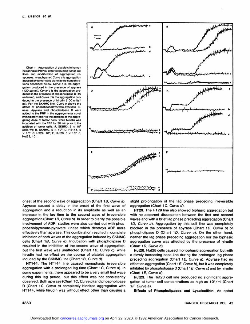
SKBR3. The aggregation profile of platelets exposed toSKBR3 cells consisted of a brief phase of reversible aggregation followed immediately by a larger irreversible phase (ChartÃŒA,Curve a). This profile is similar to that observed previouslywith the Hut20 line (3). Aggregation by this cell line wascompletely blocked by apyrase (250 /ig/ml) (Chart 1/1, Curveb). The second wave of aggregation, but not the first, wasinhibited by phospholipase D (Chart 1A, Curve c). Hirudin (100units/ml) had no effect on the aggregation profile (Chart 1/\,Curve d).
SKNMC. A similar pattern of biphasic aggregation was observed with the SKNMC cell line although in this case therewas a prolonged delay between the reversible wave and the
NOVEMBER 1982 4349
on April 22, 2020. © 1982 American Association for Cancer Research. cancerres.aacrjournals.org Downloaded from
E. Bastida et al.
Chart 1. Aggregation of platelets in humanheparinized PRP by different human tumor celllines and modification of aggregation responses. In each panel. Curve a is aggregationInduced by tumor cells alone at the concentrations described below, Curve b is the aggregation produced in the presence of apyrase(120 /ig/ml). Curve c is the aggregation produced in the presence of phospholipase D (10units/ml), and Curve d is the aggregation produced in the presence of hirudin (100 units/ml). For the SKNMC line. Curve e shows theeffect of phosphoenolpyruvate-pyruvate ki-nase. Apyrase and phospholipase D wereadded to the PRP in the aggregometer cuvetimmediately prior to the addition of the aggregating dose of tumor cells, while hirudin wasincubated with the PRP for 30 min prior to theaddition of tumor cells. A. SKBR3, 5 x 10⁶cells/ml; B, SKNMC, 5 X 10⁶; C. HT144, 5X 10e; D. HT29, 10"; E. Hut28, 5 X 10e; F.Hut23, IO7.
1 min
^ •¿�^HVU^^vWvVA^L, e
onset of the second wave of aggregation (Chart 16, Curve a).Apyrase caused a delay in the onset of the first wave ofaggregation and a reduction in its amplitude as well as anincrease in the lag time to the second wave of irreversibleaggregation (Chart 1B, Curve b). In order to clarify the possibleinvolvement of ADP, studies were also carried out with phosphoenolpyruvate-pyruvate kinase which destroys ADP more
effectively than apyrase. This combination resulted in completeinhibition of both waves of the aggregation induced by SKNMCcells (Chart 18, Curve e). Incubation with phospholipase Dresulted in the inhibition of the second wave of aggregation,but the first wave was unaffected (Chart 16, Curve c), whilehirudin had no effect on the course of platelet aggregationinduced by the SKNMC line (Chart 16, Curve cO.
HT144. The HT144 line caused monophasic irreversibleaggregation with a prolonged lag time (Chart 1C, Curve a). Insome experiments, there appeared to be a very small first waveduring this lag period, but this effect was not consistentlyobserved. Both apyrase (Chart 1C, Curve b) and phospholipaseD (Chart 1C, Curve c) completely blocked aggregation withHT144, while hirudin was without effect other than causing a
slight prolongation of the lag phase preceding irreversibleaggregation (Chart 1C, Curve d).
HT29. The HT29 line also showed biphasic aggregation butwith no apparent dissociation between the first and secondwaves and with a brief lag phase preceding aggregation (Chart1D, Curve a). Aggregation by this cell line was completelyblocked in the presence of apyrase (Chart 1D, Curve b) orphospholipase D (Chart 1D, Curve c). On the other hand,neither the lag phase preceding aggregation nor the biphasicaggregation curve was affected by the presence of hirudin(Chart 1D, Curve d).
Hut28. Hut28 cells caused monophasic aggregation but witha slowly increasing base line during the prolonged lag phasepreceding aggregation (Chart 1f, Curve a). Apyrase had noeffect on aggregation (Chart 1E, Curve b), but it was completelyinhibited by phospholipase D (Chart 1E, Curve c) and by hirudin(Chart 1E, Curve d).
Hut23. The Hut23 cell line produced no significant aggregation at tumor cell concentrations as high as 107/ml (Chart
1F, Curve a).Effects of Phospholipases and Lysolecithin. As noted
4350 CANCER RESEARCH VOL. 42
on April 22, 2020. © 1982 American Association for Cancer Research. cancerres.aacrjournals.org Downloaded from
Platelet Aggregation by Tumor Cells
above, phospholipase D showed differential effects on theaggregation produced by the various cell lines. PhospholipaseC (10 units/ml) had no effect on platelet aggregation producedby any of the cell lines examined, while phospholipase A2,added at the same time as the tumor cells, completely blockedaggregation by all of the human tumor cell lines. Lysolecithin,the product of the action of phospholipase A2 on phosphati-
dylcholine, at a concentration of 50 jug/ml, also caused complete inhibition of aggregation with all of the tumor cell linesexamined (data not shown).
DISCUSSION
In previous studies, we have examined platelet aggregationinduced by 2 tumor cell lines, Hut20 from an undifferentiatedmurine tumor and U87MG derived from a human glioblastoma(3). Two different mechanisms of induction of platelet aggregation were apparent. Platelet aggregation induced by theHut20 line appeared to be primarily dependent on ADP released from the tumor cells themselves, while aggregationinduced by the U87MG line was dependent on a procoagulantactivity elaborated by the tumor cell surface.
The present examination of a further 6 human tumor celllines shows that a third mechanism of platelet aggregation bycultured human tumor cells can be recognized based on aggregation responses and the inhibitory effects of apyrase,hirudin, and phospholipase D. The results are summarized inTable 1 together with the results for the Hut20, A549, andU87MG lines examined previously (3, 16). Some groupingsamong the various lines appear to be possible. In all cases,aggregation occurred only with heparinized PRP and not whencitrate was used as anticoagulant.
SKBR3 induces biphasic platelet aggregation that is inhibitedby apyrase but not by hirudin, and phospholipase D eliminatesthe second wave of aggregation. This pattern is similar to thatobserved previously with the Hut20 line. In this case, secretionof ADP from the tumor cells initiates the first wave of aggregation, which leads to platelet activation and to a second waveof aggregation. This second wave is associated with plateletsecretion which must be independent of thrombin productionsince it is not blocked by hirudin. SKNMC, HT144, and HT29also probably belong in this class. With SKNMC, the lag phasewas prolonged with apyrase and aggregation was completelyinhibited by phosphoenolpyruvate-pyruvate kinase. With
HT144 and Hut29, there was no clearly marked reversible firstwave, but platelet aggregation was completely inhibited by
apyrase. No effect was observed with hirudin for any of these3 lines.
Aggregation induced by the Hut28 lines shows a rising baseline prior to the onset of aggregation and is unaffected byapyrase but is completely blocked by phospholipase D andhirudin. The effects of apyrase and hirudin are identical tothose seen with the U87MG line examined previously (3),suggesting that the aggregation effects of these 2 lines involvesactivation of the coagulation system. However, platelet aggregation by these 2 lines may be differentiated since only withthe Hut28 line is aggregation inhibited by phospholipase D.
The various phospholipases have been of value in characterizing the platelet-aggregating effects of the tumor cell lines.The effects of phospholipase D on tumor cell-induced platelet
aggregation have not been investigated previously, but thisenzyme was able to differentiate the platelet-aggregating ef
fects of Hut28 from those of U87MG although activation of thecoagulation system appeared to be involved in each case. Forthose lines showing a clearly marked phase of reversible aggregation (SKBR3, SKNMC, and HT29), it may be noted thatphospholipase D inhibited the second, major wave. For HT29and Hut28, where a reversible first wave was not detectable,the aggregation response was also inhibited by phospholipaseD. Little is known about the effects of phospholipase D onmembranes, but the enzyme can alter calcium translocation insarcoplasmic reticulum (6), and this may explain its differentialeffects on the first and second waves of tumor cell-induced
platelet aggregation.Phospholipase A2 has been reported to completely inhibit
platelet aggregation induced by 2 mouse tumor cell lines (13,17) as well as by the 5 human tumor lines which causedaggregation in the present study. We have also found thattumor cell-induced platelet aggregation is inhibited by lysoleci-thin, the product of the action of phospholipase A2 on phos-
phatidylcholine. Lysolecithin also inhibits platelet aggregationinduced by ADP, epinephrine, collagen, and thrombin (15). Itis known to inhibit prostaglandin synthesis (19), and this maybe the basis for its antiaggregating effects, although it can alsoaffect membrane fluidity (21) and the levels of nucleotidecyclases (1, 20).
In addition to the usual aggregating agents, platelet aggregation can be induced by the platelet-aggregating factor elaborated by IgE-sensitized basophils. Platelet-aggregating factor
is lipidie in nature and is destroyed by phospholipases A2, C,and D (4). Since phospholipase C had no effect on plateletaggregation induced by any of the human tumor cell lines
Table 1Summary of cell lines examined for platelet-aggregating ability, aggregation profiles, and inhibitor effects
InhibitionbyCell
con-CelllineSKBR3Hut20aSKNMCHT144HT29Hut28U87MGaHut23A549aOriginAdenocarcinoma
ofbreastAnaplastiamurinetumorNeuroblastomaMelanomaAdenocarcinoma
ofcolonMesotheliomaGlioblastomaPoorly
differentiatedad-enocarcinomaSmall-cell
lung carcinomacentration51055105101010X6XXeX5r710610e10e10eAggregationBiphasicBiphasicBiphasicMonophasicBiphasicMonophasicMonophasicNoneNoneApyraseInhibitionInhibitionInhibition"InhibitionInhibitionNo
inhibitionNoinhibitionHirudinNo
inhibitionNoinhibitionNoinhibitionNoinhibitionNoinhibitionInhibitionInhibitionPhospholipase02nd
waveonly2ndwaveonly2ndwaveonlyInhibitionInhibitionInhibitionNo
inhibition
'' Lines examined previously (3, 17) are included for comparison.' Partial inhibition with apyrase. complete inhibition with phosphoenolpyruvate-pyruvate kinase.
NOVEMBER1982 4351
on April 22, 2020. © 1982 American Association for Cancer Research. cancerres.aacrjournals.org Downloaded from
E. Bastida et al.
examined, it is unlikely that platelet-aggregating factor-like
material is involved in this reaction.In summary, our results suggest that there are 2 major
mechanisms by which cultured human tumor cells initiate platelet aggregation. The major mechanism, in 4 of the 6 aggregating human lines so far examined, appears to involve the secretion of ADP from the tumor cells resulting in platelet stimulationand then irreversible aggregation. This secretion may reflectdamage to the tumor cells during harvesting, but the secretionof ADP from tumor cells in this group could be of physiologicalsignificance, and since aggregation does not occur in thepresence of citrate, it differs from the usual mechanisms ofADP-induced platelet aggregation. The second mechanisminvolves the initial activation of the coagulation system and thegeneration of thrombin as the mediator of aggregation. Withinthis second group, 2 subgroups can be identified based onwhether or not aggregation can be inhibited by phospholipaseD. However, the different patterns of aggregation and inhibitionwithin different groups of tumor cells, considerations of therelative importance of ADP and the coagulation system, andthe observation of different patterns of inhibition with phospholipase D all suggest that no single mechanism will explain thenature of the interaction between platelets and tumor cellsunder all circumstances. These preliminary studies also suggest that significant differences exist between mechanisms oftumor cell-induced platelet aggregation seen previously in het-erologous animal systems (8, 10, 12-14, 17, 18) and those
seen in the homologous human systems studied here.
ACKNOWLEDGMENTS
We are Indebted to Dr. Jergen Fogh of Sloan-Kettering Institute for CancerResearch, Rye, N. Y., and Dr. Adi Gazdar of the Veterans Administration Hospital,Washington, D. C.. for providing the cell lines examined. Invaluable guidance incell culture was provided by Terri Gouaux.
REFERENCES
1. Aunis. D., Pescheloche. M , Zwiller. J., and Mandel. P. Effects of lysolecithinon adenylate cyclase and guanylate cyclase in bovine adrenal medullaryplasma membranes. J. Neurochem., 3Õ: 355-357. 1978.
2. Bastida, E., Ordlnas, A., and Jamieson, G. A. Idiosyncratic platelet responses to human tumor cells. Nature (Lond.). 291. 661-662, 1981.
3. Bastida. E., Ordinas, A., and Jamieson, G. A. Differing platelet aggregating
effects by two tumor cell lines: absence of role for platelet-derived ADP.Am. J. Hematol., 11: 367-378, 1981.
4. Benveniste. J., Le Couedic, J. P., Polonsky, J., and Tence, M. Structuralanalysis of purified platelet activating factor by upases. Nature (Lond.), 269.170-171, 1977.
5. Bjerrum. O. J., Ramlau, J.. Clemmesen. I.. Ingild, A., and Boq-Hansen, T. C.An artifact in quantitative immunoelectrophoresis of spectrin caused byproteolytic activity in antibody preparations. Scand. J. Immunol. Suppl., 2.81-88, 1975.
6. Fiehn, W. The effect of phospholipase D on the function of fragmentedsarcoplasmic reticulum. Lipids, 73. 264-266, 1978.
7. de Gaetano. G., and Garattini, S. (eds.) Platelets: A Multidisciplinary Approach. New York: Raven Press. 1978.
8. Gasic, G. J., Boettiger, D., Catalfamo, J. L., Gasic, T. B., and Stewart, C. C.Aggregation of platelets and cell membrane vesiculation by rat cells transformed in vitro by Rous sarcoma virus. Cancer Res., 38:2950-2955, 1978.
9. Gasic, G. J.. and Gasic. T. B. Removal of sialic acid from the cell coat intumor cells and vascular endothelium and its effects on metastasis. Proc.Nati. Acad. Sei. U. S. A., 48: 1172-1177, 1962.
10. Gasic, G. J., Gasic. T. B., Galanti, N.. Johnson, T., and Murphy, S. Platelettumor-cell interactions in mice. The role of platelets in the spread of malignant disease. Int. J. Cancer, Õ1: 704-718, 1973.
11. Gasic, G. J., Gasic. T. 6., and Stewart, C. C. Antimetastatic effects associated with platelet reduction. Proc. Nati. Acad. Sei. U. S. A., 67: 46-52,1968.
12. Gasic, G. J.. Koch, P. A. G., Hsu, B., Gasic, T. B., and Niewarowski, S.Thrombogenic activity of mouse and human tumors: effects on platelets,coagulation and fibrinolysis, and possible significance for métastases. Z.Krebsforsch.. 86: 263-277, 1976.
13. Hará, Y., Steiner, S., and Baldini, M. G. Characterization of the platelet-aggregating activity of tumor cells. Cancer Res., 40: 1217-1222, 1980.
14. Holme, R., Ofteboro, R., and Hovig, T. In vitro interaction between culturedcells and human blood platelets. Thrombos. Haemostasis, 40: 89-102.1978.
15. Joist. J. H.. Dolezel, G., Cucuianu, M. P.. Nishizawa. E. E., and Mustard, J.F. Inhibition and potentiation of platelet function by lysolecithin. Blood, 43:101-112, 1977.
16. Marcum. J. M., McGill, M., Bastida, E.. Ordinas, A., and Jamieson. G. A.The interaction of platelets, tumor cells, and vascular subendothelium. J.Lab. Clin. Med.. 96: 1046-1053, 1981.
17. Pearlstein, E., Cooper, L. B., and Karpatkin, S. Extraction and characterization of platelet-aggregating material from SV40-transformed mouse 3T3fibroblasts. J. Lab. Clin. Med., 93. 332-344, 1979.
18. Pearlstein. E., Salk, P. L., Yogeeswaran, G., and Karpatkin, S. Correlationbetween spontaneous metastatic potential, platelet aggregating activity ofcell surface extracts and cell surface sialylation in 10 metastatic variantderivatives of a rat renal sarcoma line. Proc. Nati. Acad. Sei. U. S. A., 77:4336-4339, 1980.
19. Shier, W. T. Inhibition of prostaglandin biosynthesis by lysolecithin. Biochem.Biophys. Res. Commun., 78: 1168-1174. 1977.
20. Shier. W. T., Baldwin. J. H., Nilsen-Hamilton, M., Hamilton, R. T., andThanassi. N. M. Regulation of guanylate and adenylate cyclase activities bylysolecithin. Proc. Nati. Acad. Sei. U. S. A., 73: 1587-1590, 1976.
21. Utsumi, H., Inoue, K., Nojima, S.. and Kwan, T. Interaction of spin labeledlysophosphatidylcholine with rabbit erythrocytes. Biochemistry, 17: 1990-1996, 1978.
4352 CANCER RESEARCH VOL. 42
on April 22, 2020. © 1982 American Association for Cancer Research. cancerres.aacrjournals.org Downloaded from
1982;42:4348-4352. Cancer Res Eva Bastida, Antonio Ordinas, Steven L Giardina, et al. and PhospholipaseCell Lines Based on Inhibition Studies with Apyrase, Hirudin, Differentiation of Platelet-aggregating Effects of Human Tumor
Updated version
http://cancerres.aacrjournals.org/content/42/11/4348
Access the most recent version of this article at:
E-mail alerts related to this article or journal.Sign up to receive free email-alerts
Subscriptions
Reprints and
To order reprints of this article or to subscribe to the journal, contact the AACR Publications
Permissions
Rightslink site. Click on "Request Permissions" which will take you to the Copyright Clearance Center's (CCC)
.http://cancerres.aacrjournals.org/content/42/11/4348To request permission to re-use all or part of this article, use this link
on April 22, 2020. © 1982 American Association for Cancer Research. cancerres.aacrjournals.org Downloaded from