Embed Size (px)
Citation preview
Differentiation and morphogenesis of Triatoma infestans (Klug 1834)female gonads. I – Post embryonic development
CECILIA I. IBAÑEZ DE BARRETT, JUAN PABLO BOZZINI, AND MARTA MARIANO DE BOZZINI*
A.N.L.I.S. Dr. Carlos G. Malbrán. Avda. Velez Sarsfield 563, (1281) Buenos Aires, Argentina.*Member of the Research Career. National Research Council, Argentina.
Key words: Ovary, Development, Hemiptera, Insect Nymph
A B S T R A C T: The post-embryonic development of the female gonads in Triatoma infestans (Hemiptera,Heteroptera), insects of importance in health affairs as harbors and vectors of different tripanosomatideaflagellates, is presented in a complete follow-up since insect hatches from the egg up to the last molt in thefifth instar stage. The detailed description of the morphological changes which occur in each instar as well ascareful measurements evaluating its size increase have been analyzed by stereomicroscopy, phase contrast,dark field, and oblique illumination, in order to optimize the observations as well the photographic registerof gonad morphology and structure.
The analysis was performed on gonad specimens obtained from broods no less than twenty (20) nymphbugs, reared at constant temperature and fed-up regularly.
According to the results of our study we can assert that gonad differentiation takes place in early phasesof the insect development. Such is the case that first instars nymph’s present absolutely and easily recogniz-ably male and female gonads.
From the third instar on beside the filament region, the three zones in each ovariole body is distin-guished, a differentiation that is more noticeable during the fourth instar where a definite organization ispresent at the vitelarium. Such a clear cut zone development continues intensively during the fifth instar.
Finally at the end of such fifth nymph stage and when the last molt toward adults is prepared, clear signsof ovariole maturation take place since oocytes in early vitelogenesis are found.
Abbreviations used in the illustrations:est: stroma; f: suspensor filament; ft: terminal filament; ov: oocytes; ovd: oviduct; ovl: ovariole;pd: pedicel; tr: trachea (s); tro: tropharium; tz: transition zone; vt: vitelarium;
BIOCELL2004, 28(3): 259-269
ISSN 0327 - 9545PRINTED IN ARGENTINA
Introduction
There are few previous reports in the literature re-garding the study of the morphogenesis and embryonicdevelopment of the female gonad or ovary in Hemi-ptera. In this sense the work of Bonhag (1955) onOncopeltus fasciatus, of Case (1970), Huebner andAnderson (1972a) on Rhodnius prolixus, should bementioned.
Little is known about the morphogenesis of gonads,not only in Hemiptera but also in all the taxa with
telotrophic ovaries. The complexity of the structure ofthis type of ovary is probably responsible for the lim-ited number of studies available on differentiation, em-bryonic growth and morphologic variation of the ovaryduring the instar stages (Barth, 1973; Richard andDavies, 1983; Wigglesworth, 1951).
In order to gain a deeper knowledge of the mor-phogenesis of the gonad, we have carried out a thor-ough sequential study in Triatoma infestans, since wehad previously studied the maturation of the femalegamete in the adult specimen (Ibañez et al., 1999).
With regard to Hemiptera Reduvidae, group includ-ing among others Triatoma infestans and Rhodniusspecies, (Galliard, 1935; Gillet, 1935; Lutz and Huebner,1978); the data reported by these authors remain un-
Address correspondence to: Dra. Marta Mariano de Bozzini.A.N.L.I.S. «Carlos Malbrán», Virrey Cevallos 385 - 7º 40,(1077) Buenos Aires, ARGENTINA. FAX: (+54-11) 4303-1433.E-mail: [email protected] on June 13, 2003. Accepted on April 27, 2004.
CECILIA I. IBAÑEZ et al.260
clear. Added to this fact, each species may have uniquefeatures, so it is not feasible to extrapolate the data ob-tained to other species.
Ever since the pioneer work of Del Ponte (1921) inTriatoma, the main interest with regards to the differ-ences between male and female insects, was focusedon the observation and description of the external sexualcharacteristics both in adult and instar stage insects(Canale and Carcavallo, 1985; Espínola, 1966;Gonçalves et al., 1985; Lent and Wygodzinsky, 1979).
This article presents a study of the morphogenesischanges of T. infestans´ female gonad during each of thefive nymphal stages. The anatomic features and a briefalbeit complete view of the histological structures areshown; a more detailed analysis, which includes the sub-microscopic structure, is part of another study in progress.
Material and Methods
Biologic material
The nymphal gonads were obtained by dissectionperformed immediately after hatching or molting in eachstage with no food intake between molting and dissec-
tion. In the 4th instar stage dissections were performedimmediately after molting from the third, and few hoursprior to the next instar molt taking into account the pres-ence of a new cuticle under the old one to be abandonedby the insect reaching the new instar stage.
Before dissecting the insects were anesthetized fora few seconds with chloroform, then placed with theirback upwards on a paraffin bottom Petri dish and im-mobilized with entomology pins.
Fixation
Once the chitin cuticle of the back had been re-moved (under stereoscopic binocular microscope) andthe internal organs were exposed, an in situ fixation wasperformed with freshly prepared Karnovsky´s fixative,at a temperature below 10ºC. The specimen was thenplaced in a small flask containing the same fixative forno less than 30 minutes, in a bath at approximately 8ºC,so that immersion fixation may proceed. Once the fixa-tion stage was completed the gonads were separated ona Petri dish and the necessary incisions performed,mainly at the level of the suspending filament and/orterminal duct (the dissection should be done with greatcare in order to avoid damaging the extremely fragile
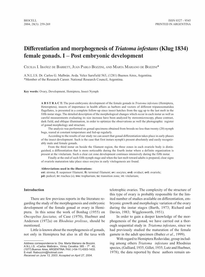
FIGURE 1.
261OVARY POST EMBRYONIC DEVELOPMENT IN Triatoma infestans
gonads). The separated gonads were placed in smallflasks with the same fixative, and kept in a cold bathfor two hours. Post fixation with 1% osmium tetra-ox-ide also achieves a contrasting effect on the sample.
Taking advantage of the experience we have accu-mulated in the recognition of gonads in adult specimensand considering that the size of the insect and the organfacilitates its identification, we were able to perform asequential study, in a decreasing order, of each instarstage and recognize the corresponding structure.
Inclusion, sectioning, staining and embeding
The specimens were dehydrated using a series ofgraded alcohols (ethanol), then rinsed twice in propy-lene oxide. Samples of each instar stage were embeddedin Poly-Bed Araldite resins, according to Mollenhauert(1964).
Semi-thin sections (2µm) obtained by the ultrami-crotome were stained with a mixture of Azure II- meth-ylene blue and mounted with the same inclusion resinfor optic microscope observation.
Results
The procedure used to recognize the gonads con-sisted in a comparative study of these organs in a de-creasing order, each instar stage with those of the pre-vious one, that is, from the last stage (5th instar) to thefirst one.
Although the gonad is a structure that can easily bemistaken with the surrounding tissue, particularly in thefirst stages, it is possible to identify it as an indepen-dent structure if certain parameters are taken into ac-count: location, shape and relative size.
Located between the digestive tract wall and thetegument, the gonad is found associated to the fatty bodyand components of the respiratory system, both presentin all of the insects’ body cavity. On the one hand, thetexture of the fatty body seen as a loose web of fattylobes and, on the other, the bigger or smaller trachealtubes, are criteria that help us to recognize a differentstructure, somewhat compact although very fragile, thatlooks like a flattened sac and has a different light re-fraction index when compared to the surrounding tis-sues. When the dissected specimen is observed under astereoscopic microscope - with a magnification higherthan 30x - the gonads can be identified as a pair of struc-tures located symetricaly in the abdomen. The exami-nation of these structures under phase contrast, dark
field and oblique illumination optical microscopy, willshow the following images:
1.- The structures that belong to the same speci-men have seven equivalent formations, of the same sizeand characteristic general morphology.
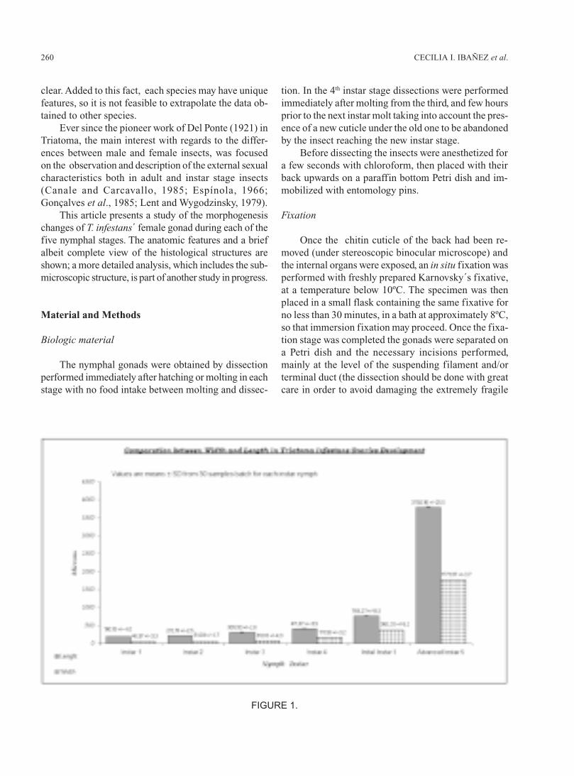
FIGURE 2. T. infestans (Klug 1834) Photomicrographexposed at the same magnification giving a relationshipof male (A) and female (B) gonads size at first nymphinstar few hours after hatching. (A) The arrow pointsone of the seven testicular follicles conforming the malegonad Phase contrast. Female gonad (B) shown underdark field illumination with a superimposed negativeobject micrometer (1mm ÷ 100sections). The seven pri-mordial ovarioles are easily distinguished. Each mi-crometer division match up 10 mm. Male gonad has al-ways a little larger size.
CECILIA I. IBAÑEZ et al.262
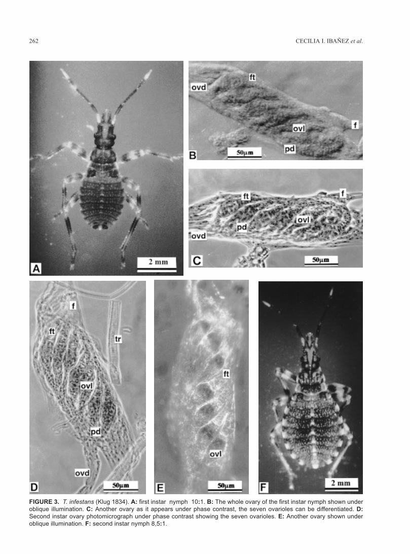
FIGURE 3. T. infestans (Klug 1834). A: first instar nymph 10:1. B: The whole ovary of the first instar nymph shown underoblique illumination. C: Another ovary as it appears under phase contrast, the seven ovarioles can be differentiated. D:Second instar ovary photomicrograph under phase contrast showing the seven ovarioles. E: Another ovary shown underoblique illumination. F: second instar nymph 8,5:1.
263OVARY POST EMBRYONIC DEVELOPMENT IN Triatoma infestans
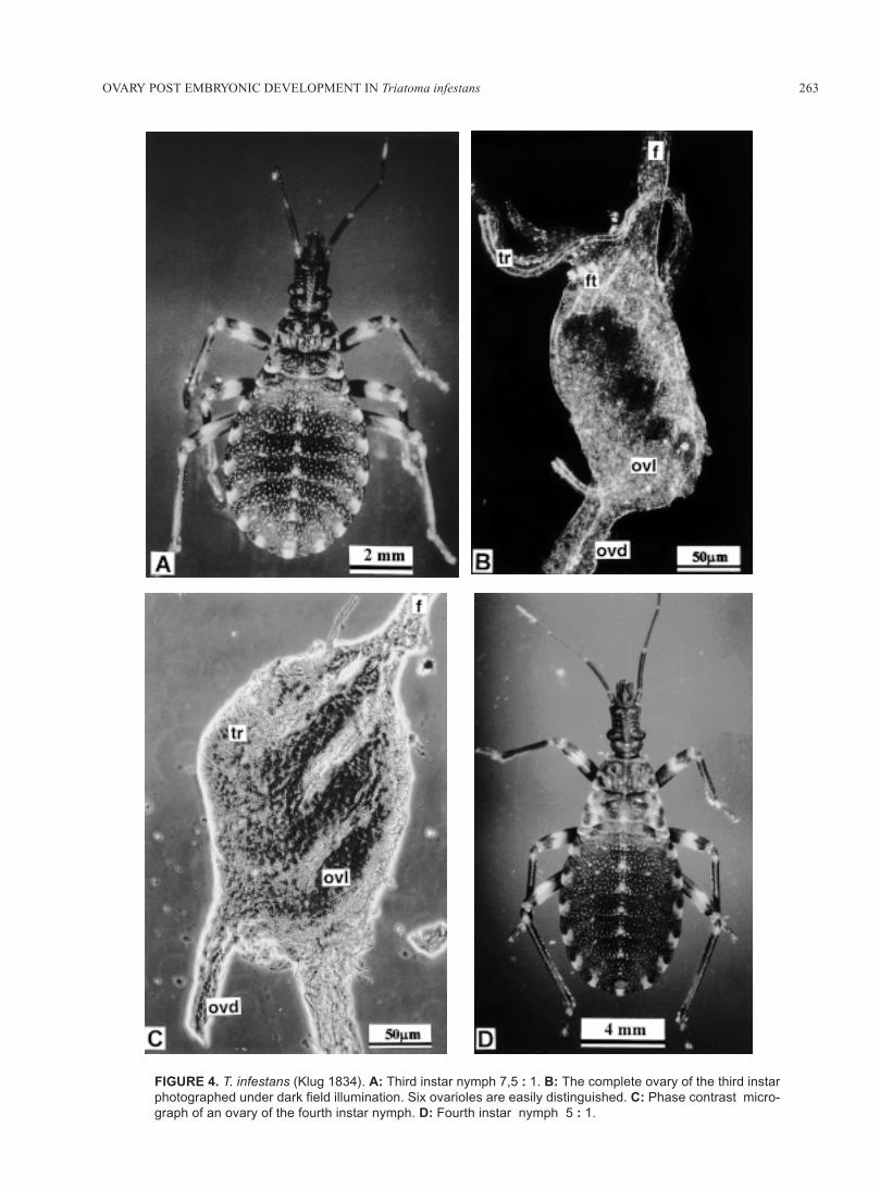
FIGURE 4. T. infestans (Klug 1834). A: Third instar nymph 7,5 : 1. B: The complete ovary of the third instarphotographed under dark field illumination. Six ovarioles are easily distinguished. C: Phase contrast micro-graph of an ovary of the fourth instar nymph. D: Fourth instar nymph 5 : 1.
CECILIA I. IBAÑEZ et al.264
2.- The structures from distinct specimens can beseparated into two groups, according to the morpho-logic differences found in their components. Female andmale gonads are thus recognized. In Triatoma we haveestablished such differences from the 1st instar stage on(Fig. 2A, 2B).
The size variations of each component (ovariole)of the female gonad (ovary) in each of the five nymphalstages are shown in the graphic (Fig. 1).
First instar stage – (Fig 3A)
An elongated gonad is observed, its size is shownin Figure 1. The body of the ovary is formed by sevenovarioles, laid out in an almost parallel way, clearly sepa-rated from one another. All the ovariole terminal fila-ments converge at the front end to form the suspensorfilament. The caudal extremity of the ovarioles is lessdefined; nevertheless, each of them is oriented towardsthe back and extend to form a primary duct (oviduct)(Fig. 3B, 3C) In the space between ovarioles the cellsthat make up the stroma of the gonad are found.
At the histological level, we can distinguish in eachovariole three regions: the filament, which is quite big,a middle region or body of the ovariole and a terminalregion or pedicel (Fig. 6A).
As already mentioned, in this stage we can differ-entiate the male and female gonads and the first ob-servable difference, as shown by the stage micrometer,is the slightly bigger size of the former (Fig. 2A, 2B).
Second instar stage – (Fig. 3F)
The form of the gonad is slightly oval and elon-gated, becoming thinner at both extremes. As in thefirst stage, the body of the seven ovarioles, each of themsurrounded by their basal lamina, is perfectly outlined.Each group of seven ovarioles is oriented and convergetowards the genital duct. The separation between themhas decreased (Fig. 3D, 3E).
Under a phase contrast microscope we can observethat the ovarioles present a thinner filament region. Ineach ovariole body two regions can already be distin-guished: the tropharium and a not yet differentiated re-gion; the pedicel has approximately the same size asthat of the 1st stage The stroma of the gonad isbest seen at the level of the ovariole tropharium. (Fig.6B) The whole organ is surrounded and encircled bybranched small tracheas (tracheoles). The quantity oftracheoles is moderate and their relationship to the or-gan is modified during the different stages.
Third instar stage – (Fig. 4A)
The shape of the ovary is similar to that of the pre-vious one. The well outlined ovarioles are no longerseparated and their convergence at the cephalic and cau-dal ends is more evident and increases the compact as-pect of the ovary (Fig. 4B), with a well defined stroma.At the histological level the body of the ovariole haschanged considerably and can be divided in regions:
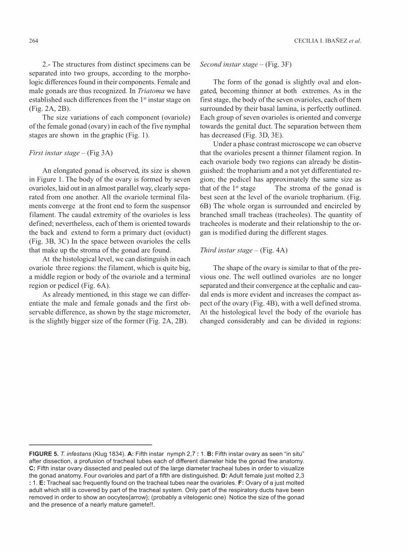
FIGURE 5. T. infestans (Klug 1834). A: Fifth instar nymph 2,7 : 1. B: Fifth instar ovary as seen “in situ”after dissection, a profusion of tracheal tubes each of different diameter hide the gonad fine anatomy.C: Fifth instar ovary dissected and pealed out of the large diameter tracheal tubes in order to visualizethe gonad anatomy. Four ovarioles and part of a fifth are distinguished. D: Adult female just molted 2,3: 1. E: Tracheal sac frequently found on the tracheal tubes near the ovarioles. F: Ovary of a just moltedadult which still is covered by part of the tracheal system. Only part of the respiratory ducts have beenremoved in order to show an oocytes{arrow}; (probably a vitelogenic one) Notice the size of the gonadand the presence of a nearly mature gamete!!.
265OVARY POST EMBRYONIC DEVELOPMENT IN Triatoma infestans
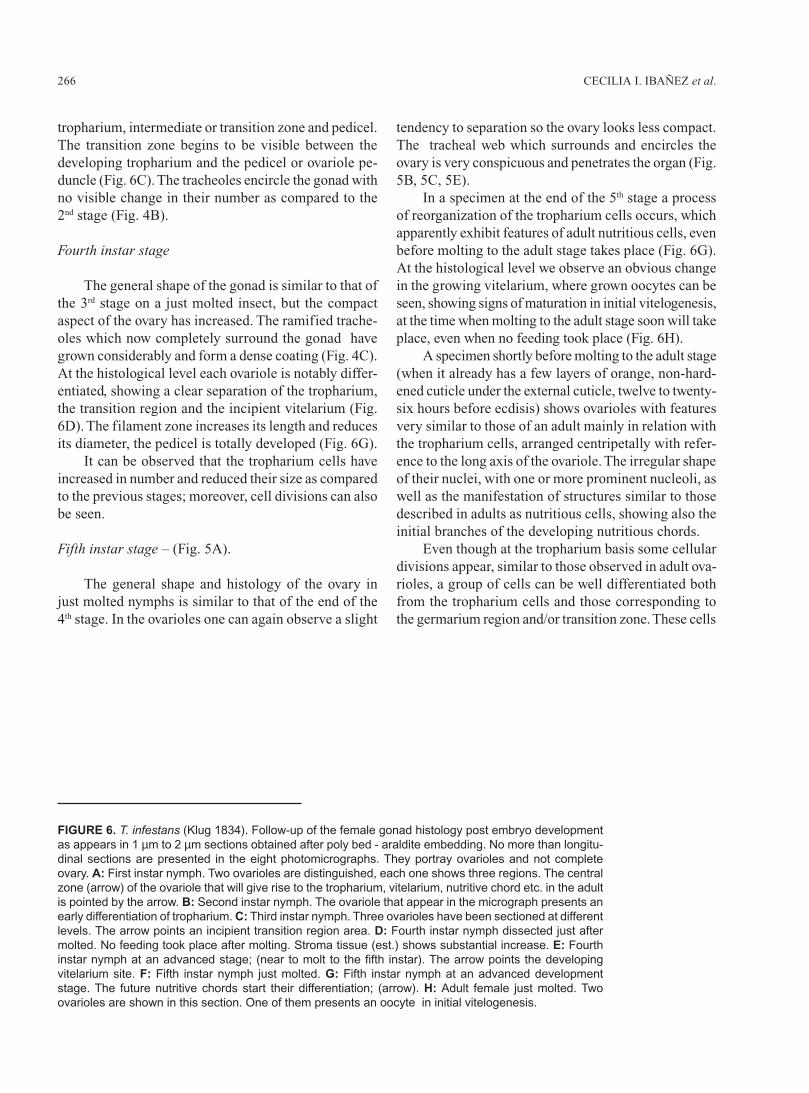
CECILIA I. IBAÑEZ et al.266
tropharium, intermediate or transition zone and pedicel.The transition zone begins to be visible between thedeveloping tropharium and the pedicel or ovariole pe-duncle (Fig. 6C). The tracheoles encircle the gonad withno visible change in their number as compared to the2nd stage (Fig. 4B).
Fourth instar stage
The general shape of the gonad is similar to that ofthe 3rd stage on a just molted insect, but the compactaspect of the ovary has increased. The ramified trache-oles which now completely surround the gonad havegrown considerably and form a dense coating (Fig. 4C).At the histological level each ovariole is notably differ-entiated, showing a clear separation of the tropharium,the transition region and the incipient vitelarium (Fig.6D). The filament zone increases its length and reducesits diameter, the pedicel is totally developed (Fig. 6G).
It can be observed that the tropharium cells haveincreased in number and reduced their size as comparedto the previous stages; moreover, cell divisions can alsobe seen.
Fifth instar stage – (Fig. 5A).
The general shape and histology of the ovary injust molted nymphs is similar to that of the end of the4th stage. In the ovarioles one can again observe a slight
tendency to separation so the ovary looks less compact.The tracheal web which surrounds and encircles theovary is very conspicuous and penetrates the organ (Fig.5B, 5C, 5E).
In a specimen at the end of the 5th stage a processof reorganization of the tropharium cells occurs, whichapparently exhibit features of adult nutritious cells, evenbefore molting to the adult stage takes place (Fig. 6G).At the histological level we observe an obvious changein the growing vitelarium, where grown oocytes can beseen, showing signs of maturation in initial vitelogenesis,at the time when molting to the adult stage soon will takeplace, even when no feeding took place (Fig. 6H).
A specimen shortly before molting to the adult stage(when it already has a few layers of orange, non-hard-ened cuticle under the external cuticle, twelve to twenty-six hours before ecdisis) shows ovarioles with featuresvery similar to those of an adult mainly in relation withthe tropharium cells, arranged centripetally with refer-ence to the long axis of the ovariole. The irregular shapeof their nuclei, with one or more prominent nucleoli, aswell as the manifestation of structures similar to thosedescribed in adults as nutritious cells, showing also theinitial branches of the developing nutritious chords.
Even though at the tropharium basis some cellulardivisions appear, similar to those observed in adult ova-rioles, a group of cells can be well differentiated bothfrom the tropharium cells and those corresponding tothe germarium region and/or transition zone. These cells
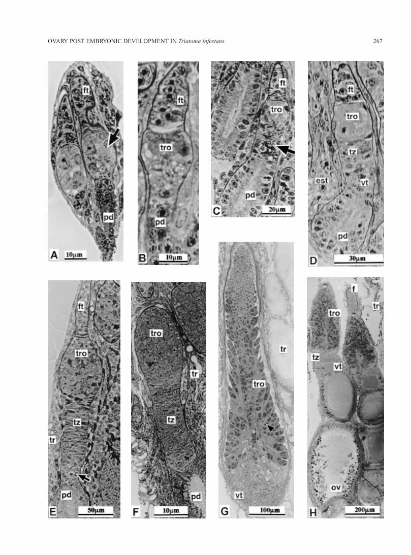
FIGURE 6. T. infestans (Klug 1834). Follow-up of the female gonad histology post embryo developmentas appears in 1 µm to 2 µm sections obtained after poly bed - araldite embedding. No more than longitu-dinal sections are presented in the eight photomicrographs. They portray ovarioles and not completeovary. A: First instar nymph. Two ovarioles are distinguished, each one shows three regions. The centralzone (arrow) of the ovariole that will give rise to the tropharium, vitelarium, nutritive chord etc. in the adultis pointed by the arrow. B: Second instar nymph. The ovariole that appear in the micrograph presents anearly differentiation of tropharium. C: Third instar nymph. Three ovarioles have been sectioned at differentlevels. The arrow points an incipient transition region area. D: Fourth instar nymph dissected just aftermolted. No feeding took place after molting. Stroma tissue (est.) shows substantial increase. E: Fourthinstar nymph at an advanced stage; (near to molt to the fifth instar). The arrow points the developingvitelarium site. F: Fifth instar nymph just molted. G: Fifth instar nymph at an advanced developmentstage. The future nutritive chords start their differentiation; (arrow). H: Adult female just molted. Twoovarioles are shown in this section. One of them presents an oocyte in initial vitelogenesis.
267OVARY POST EMBRYONIC DEVELOPMENT IN Triatoma infestans
CECILIA I. IBAÑEZ et al.268
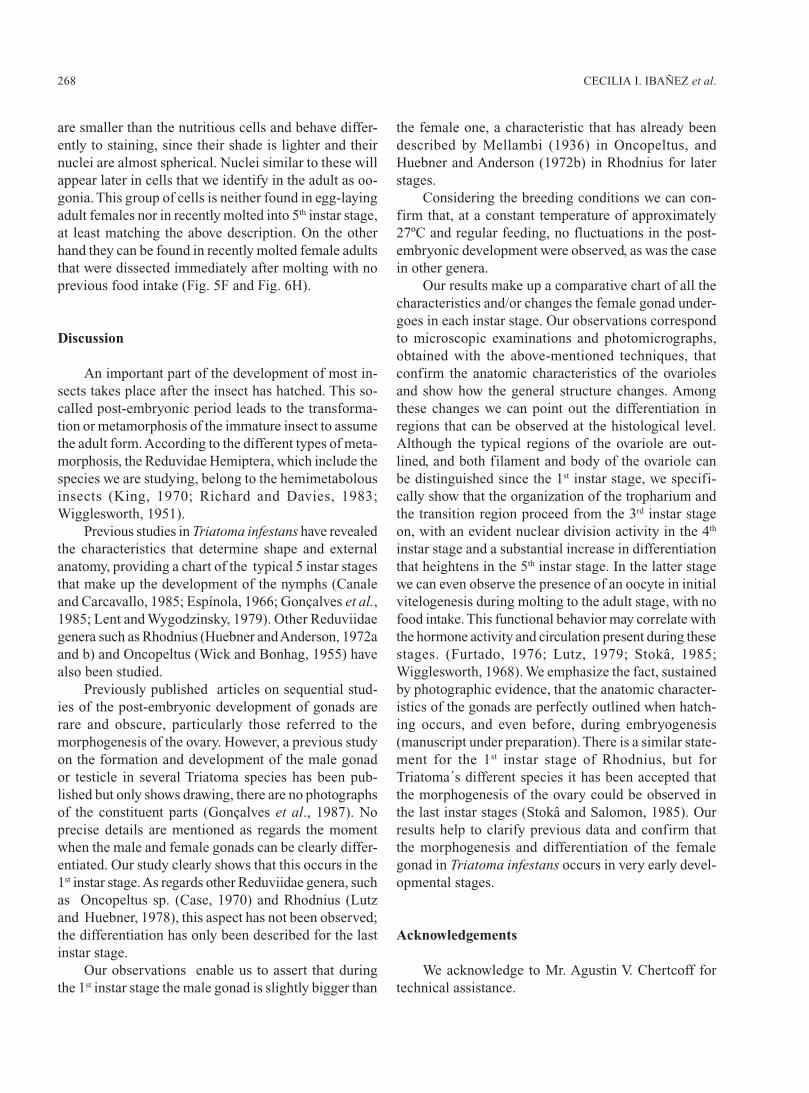
are smaller than the nutritious cells and behave differ-ently to staining, since their shade is lighter and theirnuclei are almost spherical. Nuclei similar to these willappear later in cells that we identify in the adult as oo-gonia. This group of cells is neither found in egg-layingadult females nor in recently molted into 5th instar stage,at least matching the above description. On the otherhand they can be found in recently molted female adultsthat were dissected immediately after molting with noprevious food intake (Fig. 5F and Fig. 6H).
Discussion
An important part of the development of most in-sects takes place after the insect has hatched. This so-called post-embryonic period leads to the transforma-tion or metamorphosis of the immature insect to assumethe adult form. According to the different types of meta-morphosis, the Reduvidae Hemiptera, which include thespecies we are studying, belong to the hemimetabolousinsects (King, 1970; Richard and Davies, 1983;Wigglesworth, 1951).
Previous studies in Triatoma infestans have revealedthe characteristics that determine shape and externalanatomy, providing a chart of the typical 5 instar stagesthat make up the development of the nymphs (Canaleand Carcavallo, 1985; Espínola, 1966; Gonçalves et al.,1985; Lent and Wygodzinsky, 1979). Other Reduviidaegenera such as Rhodnius (Huebner and Anderson, 1972aand b) and Oncopeltus (Wick and Bonhag, 1955) havealso been studied.
Previously published articles on sequential stud-ies of the post-embryonic development of gonads arerare and obscure, particularly those referred to themorphogenesis of the ovary. However, a previous studyon the formation and development of the male gonador testicle in several Triatoma species has been pub-lished but only shows drawing, there are no photographsof the constituent parts (Gonçalves et al., 1987). Noprecise details are mentioned as regards the momentwhen the male and female gonads can be clearly differ-entiated. Our study clearly shows that this occurs in the1st instar stage. As regards other Reduviidae genera, suchas Oncopeltus sp. (Case, 1970) and Rhodnius (Lutzand Huebner, 1978), this aspect has not been observed;the differentiation has only been described for the lastinstar stage.
Our observations enable us to assert that duringthe 1st instar stage the male gonad is slightly bigger than
the female one, a characteristic that has already beendescribed by Mellambi (1936) in Oncopeltus, andHuebner and Anderson (1972b) in Rhodnius for laterstages.
Considering the breeding conditions we can con-firm that, at a constant temperature of approximately27ºC and regular feeding, no fluctuations in the post-embryonic development were observed, as was the casein other genera.
Our results make up a comparative chart of all thecharacteristics and/or changes the female gonad under-goes in each instar stage. Our observations correspondto microscopic examinations and photomicrographs,obtained with the above-mentioned techniques, thatconfirm the anatomic characteristics of the ovariolesand show how the general structure changes. Amongthese changes we can point out the differentiation inregions that can be observed at the histological level.Although the typical regions of the ovariole are out-lined, and both filament and body of the ovariole canbe distinguished since the 1st instar stage, we specifi-cally show that the organization of the tropharium andthe transition region proceed from the 3rd instar stageon, with an evident nuclear division activity in the 4th
instar stage and a substantial increase in differentiationthat heightens in the 5th instar stage. In the latter stagewe can even observe the presence of an oocyte in initialvitelogenesis during molting to the adult stage, with nofood intake. This functional behavior may correlate withthe hormone activity and circulation present during thesestages. (Furtado, 1976; Lutz, 1979; Stokâ, 1985;Wigglesworth, 1968). We emphasize the fact, sustainedby photographic evidence, that the anatomic character-istics of the gonads are perfectly outlined when hatch-ing occurs, and even before, during embryogenesis(manuscript under preparation). There is a similar state-ment for the 1st instar stage of Rhodnius, but forTriatoma´s different species it has been accepted thatthe morphogenesis of the ovary could be observed inthe last instar stages (Stokâ and Salomon, 1985). Ourresults help to clarify previous data and confirm thatthe morphogenesis and differentiation of the femalegonad in Triatoma infestans occurs in very early devel-opmental stages.
Acknowledgements
We acknowledge to Mr. Agustin V. Chertcoff fortechnical assistance.
269OVARY POST EMBRYONIC DEVELOPMENT IN Triatoma infestans
References
Barth R (1973). Estuds anatômicos e histológicos sobre a subfamília Triatominae (Heteroptera Reduvidae). Parte XIII. O ovario deTriatoma infestans. Mem Inst Oswaldo Cruz 71-137.
Bonhag PF (1955). Histochemical studies of the ovarian nurse tissue and oocytes of the Milkweed Bug Oncopeltus fasciatus (Dallas). JMorphol Vol 96(3): 381-421.
Canale DM, Carcavallo RV (1985). Factores Biológicos y Ecológicos En la Enfermedad de Chagas Cap. XVIII Triatoma infestans(Klug). Ed. Centro Panamericano de Ecología Humana y Salud de la OMS. pp. 237-250.
Case DC (1970). Post-embryonic development of the ovary of Rhodnius prolixus (Stahl). Master MSc Thesis Department of Zoology McGill University, Montreal, Quebec, Canada.
Del Ponte E (1921). Contribución al estudio del Género Triatoma (Lap). Revista del Instituto Bacteriológico Vol. II (6): 1-64, Lam.I-XV.Espinola HN (1966). Nota sobre diferencas sexuais em formas inmaturas de Triatominae (Hemiptera, Reduvidae). Rev Brasil Biol. Vol.
26(3): 263-267.Furtado A (1976). Controle endocrine de l’ovogenése au cours de cinquieme stado nymphal de Panstrongylus megistus (Hemiptera
Heteroptera Reduvidae). C. R. Acad. Sc. Paris, 561-564.Galliard L (1935). Recherches sur les Reduvides Hématophages Rhodnius et Triatoma. II Abdome et evacues d l’ armature génitale des
nimphes. Ann Parasit Hum Com, Vol XIII (4): 293-298.Gillet JP (1935). The genital sterna of the immature stages of Rhodnius prolixus (Hemiptera). Trans R. Ent. Soc. Lond. Vol. 83(1):
1-5.Gonçalves TC, Jurberg J, Costa MJ, de Souza W (1985). Estudo morfológico comparativo de ovos e ninfas de Triatoma maculata (Erichson
1848) e Triatoma pseudomaculata (Correa & Espínola 1964) (Hemiptera Reduvidea, Triatominae). Mem. Inst. Oswaldo Cruz RíoJ. Vol. 80(3): 263-276.
Gonçalves TC, Lent H, Ribeiro de Almeida J (1987). Estudo Anatómico e morfométrico dos folículos testiculares de algunas espécies deTriatominae (Hemiptera, Reduvidae). Mem Inst Oswaldo Cruz Rio de Janeiro Vol. 82(4): 543-550.
Huebner E, Anderson E (1972a). A cytological study of the ovary of Rhodnius prolixus. I The ontogeny of the follicular epithelium. JMorphol. 136: 459-563.
Huebner E, Anderson E (1972b). A cytological study of the ovary of Rhodnius prolixus. II Oocyte differentiation. J Morphol. 137:385-416.
Ibañez de Barret CI, Bozzini JP, Mariano de Bozzini MI (1999). Cellular Interactions During the Female Gametogenesis of Triatomainfestans (Klug 1834). Biocell 23(2): 103-112.
King RC (1970). Ovarian Development in Drosophila melanogaster. Ed. Academic Press N. York, 227pp.Lent H, Wygodzinsky P (1979). Revision of the Triatominae (Hemiptera Reduvidae) and their significance as vectors of Chagas’s dis-
ease. Bull Am Mus Nat Hist. 163: 127-520.Lutz D, Huebner E (1978). Structural and physiological aspects of post-embryonic ovarian development in Rhodnius prolixus. J Cell
Biol. 79 (2 part 2): G1026 (abstract).Lutz DA (1979). Structural and physiological aspects of 5th instar ovarian development in Rhodnius prolixus (Insecta, Hemiptera). M Sch
Thesis. Dept. Zoll. Univ. Manitoba. Canada.Mellanby H (1936). The later embryology of Rhodnius prolixus. Q J Micros Sci. 79: 1-42.Mollenhauer HH (1964). Plastic embedding mixtures for use in electron microscopy. Stain Technology 39: 111.Richard OW, Davies RG (1983). El Sistema Reproductor. Cap. 17. In: Tratado de Entomología. Ed. Omega Madrid, Vol. 1: 305–336.Stokâ AM, Salomón OD (1985). Desarrollo y maduración de los ovocitos en Triatominæ. Cap IX. In : Factores Biológicos y Ecológicos
en la Enfermedad de Chagas Vol. I. Epidemiología y Vectores. Ed. Carcavallo R., Rabinovich J L, John B.Stokâ AM (1985). Mecanismos endocrinos en la regulación del desarrollo de triatominos. Cap X: 103 – 111 in: Factores biológicos y
ecológicos en la enfermedad de Chagas. Centro Panam. de Ecol. Humana y Salud de la OMS. Editores Canals D.A., Carcavalo R.Wick JP, Bonhag PF (1955). Postembrionic development of the ovaries of Oncopeltus fasciatus (Dallas). J Morphol. 96: 31–66.Wigglesworth UB (1951). Metamorphosis in Insects. Review. Proc. Roy. Ent. Soc. London Ser. C Vol. 15: 78-81.Wigglesworth UB (1968). Summing up Growth Hormons and the Gene System in the Insect Rhodnius. Mem Soc Endoc. 15: 77-85.