Embed Size (px)
Citation preview
1
Differential suppression of spontaneous and noxious-evoked
somatosensory cortical activity by isoflurane in the neonatal rat
Pi-shan Chang, PhD, Department of Neuroscience, Physiology & Pharmacology, University College
London (UCL), London WC1E6BT, United Kingdom
Suellen M Walker, MBBS, PhD, FANZCA, Pain Research (Respiratory Critical Care and Anaesthesia),
UCL Institute of Child Health and Department of Anaesthesia and Pain Medicine, Great Ormond
Street Hospital for Children NHS Foundation Trust, London WC1N1EH, United Kingdom
Maria Fitzgerald, PhD, Department of Neuroscience, Physiology & Pharmacology, University College
London (UCL), London WC1E6BT, United Kingdom
Corresponding Author: Professor Maria Fitzgerald, Department of Neuroscience, Physiology &
Pharmacology, Medawar Building, University College London, Gower Street, London WC1E6BT, United
Kingdom. Phone: (+44) 02076791303 Email: [email protected]
Disclosure of funding: This research was supported by the Medical Research Council, London, United
Kingdom, Grants G0901269 (MF) and MR/K022636/1 (SMW). The authors declare no competing
interests.
Acknowledgements: The authors thank Mr Tom Carson, Department of Neuroscience, Physiology &
Pharmacology, UCL for his technical support. The authors declare no competing interests.
Abbreviated Title: Neonatal cortical pain activity and isoflurane
2
Abstract:
Background: The effect of neonatal anesthesia and pain on the developing brain is of considerable
clinical importance but few studies have evaluated noxious surgical input to the infant brain under
anesthesia. Here we tested the effect of increasing isoflurane concentration upon spontaneous and
evoked nociceptive activity in the somatosensory cortex of rats at different postnatal ages.
Methods: Intracortical extracellular field potentials evoked by hindpaw C-fiber electrical stimulation
were recorded in the rat somatosensory cortex at postnatal day (P)7, 14, 21 and 30 during isoflurane
anesthesia (n=7 per group). The amplitudes of evoked potentials and the energies of evoked
oscillations (1-100Hz over 3 sec) were measured following equilibration at 1.5% isoflurane and during
step increases in inspired isoflurane. Responses during and after plantar hindpaw incision were
compared at P7 and P30 (n=6 per group).
Results: At P7, cortical activity was silent at isoflurane 1.5% but noxious evoked potentials decreased
only gradually in amplitude and energy with step increases in isoflurane. The resistance of noxious
evoked potentials to isoflurane at P7 was significantly enhanced following surgical hindpaw incision
(69±16% vs 6±1% in non-incised animals at maximum inspired isoflurane). This resistance was age
dependent; at P14-30, noxious evoked responses decreased sharply with increasing isoflurane (step 3
(4%) P7: 50±9%, P30: 4±1% of baseline). Hindpaw incision at P30 sensitized noxious evoked potentials,
but this was suppressed by higher isoflurane concentrations.
Conclusions: Despite suppression of spontaneous activity, cortical evoked potentials are more
resistant to isoflurane in young rats and are further sensitized by surgical injury.
3
Introduction
An optimal level of neonatal anesthesia achieves both hypnosis and anti-nociception while
maintaining physiological stability and minimising potential neurotoxicity 1,2. As both anesthesia and
uncontrolled pain may alter cortical activity and impair neurodevelopmental outcomes 3–5, the impact
of anesthetic agents upon both spontaneous and noxious-evoked neural activity in the developing
brain requires further evaluation. An important aspect of neonatal anesthesia research is the effect
of nociceptive sensory input on activity within cortical sensory circuits and the degree to which central
nociceptive activity is modulated by anesthesia and analgesia. Both animal and clinical evidence point
to long term consequences of early life procedural and surgical tissue injury upon somatosensory and
nociceptive systems6,7, highlighting the need to consider the impact of postnatal age on changes in
both spontaneous and noxious-evoked cortical activity during surgery and anesthesia.
Extracellular field recording, including electroencephalogram (EEG), electrocorticogram
(ECoG) and local field potentials (intracortical activity) are commonly used to monitor ongoing
spontaneous brain activity and levels of anesthesia in human and rodent neonates but are also used
to record specific potentials evoked by a sensory stimulus. Somatosensory potentials evoked by
experimental noxious cutaneous stimulation 8–11 are commonly used to measure pain activity in the
adult human and rodent brain 12. Specific nociceptive potentials are also evoked by single, clinically
required skin breaking procedures in the human infant brain13,14 and have been used in this age group
to measure the postnatal development of cortical pain processing10. In adult humans and rodents,
nociceptive evoked potential amplitudes decrease with increasing concentrations of isoflurane 9,15 but,
to date, the sensitivity of nociceptive potentials to anesthesia in infants has not been studied. In a
study of the whisker barrel cortex of neonatal rats, sensory potentials evoked by whisker deflection
persisted at surgical isoflurane levels (1.5-2%) that completely suppressed the EEG and silenced
4
spontaneous neuronal firing16. This suggests that noxious evoked potentials and spontaneous
intracortical activity might be differentially sensitive to anesthesia in infants. Furthermore, since
nociceptive potential amplitudes can be increased by peripheral C fiber sensitization in both humans
and rat cortex 8,11, surgical injury may enhance evoked activity and add to this differential response to
anesthesia in infancy.
The primary aim of this experimental laboratory study was to test the impact of increasing
isoflurane concentration upon the pattern of spontaneous and evoked nociceptive activity in the
somatosensory cortex of infant rats undergoing hindpaw incision. In addition, we compared effects
of isoflurane on cortical activity at different postnatal ages. We hypothesised that, in infant rats,
noxious evoked brain activity is more resistant than spontaneous brain activity to isoflurane anesthesia
and that this difference declines with postnatal age.
5
Materials and Methods
Animals
All experiments were performed under personal and project licences approved by the Home
Office, London, United Kingdom under regulations of the United Kingdom Animal (Scientific
Procedures) Act, 1986. Male Sprague-Dawley rats aged postnatal day (P) 7, 14, 21 and 30, were
obtained from the Biological Services Unit, University College London. All animals were from the
same colony, bred and maintained in-house, and exposed to the same caging, diet and handling
throughout development. Rats were housed in cages of six age-matched animals (P30) or with the
dam and littermates (P7, 14 and 21) under controlled environmental conditions (24–25°C; 50–60%
humidity; 12 h light/dark cycle) with free access to food and water. Animals were randomly picked
from litters by hand for recording and alternately assigned to an incision or no incision group.
Treatment groups were distributed across multiple litters and/or adult cage groups. At the end of an
experiment, the isoflurane was increased to 5% until there was no heart beat and the neck dislocated.
Cortical recordings
Rats were anaesthetised with 4% isoflurane (Abbot, AbbVie Ltd., Maidenhead, United
Kingdom) in 100% oxygen via a nose cone, and following insertion of a tracheal cannula were
mechanically ventilated (Small Animal Ventilator, Bioscience, Kent, United Kingdom) with 100% oxygen
and isoflurane delivered from a recently calibrated vaporizer (Harvard Apparatus, Cambridge, MA).
The adjustments for ventilation of rats across the ages were based on the breath rates and tidal
volume. The intermittent positive pressure ventilation was achieved by using a T-type system in
conjunction with a small animal lung ventilator. This system affords control of inspired gas mixture
(isoflurane concentration and oxygen) inspiratory flow rate, respiratory rate and peak inspiratory
pressure. A simple water manometer placed in the inspiratory limb provided a monitoring and
6
pressure limiting device with visual display. Using an inspiratory flow rate between 1-1.5 litres per
minute and adjusting the peak inspiratory pressure between 12-15 cm water 17 allowed the tidal
volume to be adjusted according to the size of animals. This mode was confirmed to be adequate in
our lab by using a transcutaneous combination probe to monitor transcutaneous oxygen and carbon
dioxide. Body temperature was maintained with a thermostatically controlled heated blanket and
the electrocardiogram was monitored throughout (Neurolog, Digitimer, Welwyn Garden City, United
Kingdom).
During anesthesia with 2.5% isoflurane, animals were placed in a stereotaxic frame and a
craniotomy was performed to expose the surface of the cerebral cortex. A recording electrode
(stainless steel, E363/1, Plastic One Inc. Roanoke, Virginia, USA) was inserted into the primary
somatosensory cortex in the somatotopic region for the hindpaw. Coordinates for P7 and P14 rats
were lateral 2.0 mm from midline and posterior 0.5 mm from the bregma; and for P21 and P30 rats
were lateral 2.5 mm from midline, and posterior 1 mm from the bregma 18,19. The reference electrode
was placed subcutaneously on the surface of the skull anterior to the bregma, and the ground
electrode was placed subcutaneously in the back. The inspired isoflurane concentration was then
reduced to 1.5% and allowed to equilibrate for at least 40 minutes.
Continuous intracortical activity recordings were performed using a Neurolog NL100
headstage connected to a NL104 amplifier and a NL125 filter (100 Hz). The signal was digitized at 16K
Hz using Axon Instruments (Digidata 1400A, Molecular Devices, Sunnyvale, CA). Data was acquired
and stored using a Windows PC based programme, WinEDR v3.3.6 (John Dempster, University of
Strathclyde, United Kingdom) for later analysis. The depth of the recording electrode was adjusted to
optimise the somatosensory evoked potential amplitude. At the end of an experiment, the brain was
removed and immediately immersed in 4% paraformaldehyde for over 24 hours then transferred to
30% sucrose post-fixation solution. Brain sections (40-μm thick thickness) were cut using a micotome
7
(Leica SM2000R, Leica Microsystems (UK) Ltd, United Kingdom) and stained with cresyl violet for
histological location of the electrode track. This procedure verified that recordings were in layer 5-6
of the somatosensory cortex (Fig. 1). All the data recorded were included in this study and no animals
were excluded.
Electrical stimulation of the hindpaw
Two stainless steel pin electrodes were placed subcutaneously 3-5 mm apart on the plantar
hindpaw, contralateral to the cortical recording site. A train of 10 stimuli of 3.2 mA, 500 µs were
applied at 10 sec interstimulus intervals, using a constant current stimulator (Neurolog, Digitimer,
Welwyn Garden City, UK). These stimulation parameters are sufficient to recruit both A and C fibers20
(here called ‘C fiber’ stimulation) and were established in pilot experiments to evoke clear potentials
restricted to the somatosensory cortex. At all ages, electrical hindpaw stimulation failed to evoke
visible hindlimb reflex responses at 1.5% or higher inspired isoflurane concentrations. In a separate
group of P7 animals, a train of lower intensity stimuli of 0.32mA, 50 µs was also applied, sufficient to
recruit only A beta fibres20 (here called ‘A fiber’ stimulation), for comparison.
Plantar hindpaw incision
Plantar hindpaw incision was performed in P7 and P30 animals (n=6 per age group) following
equilibration at 1.5% isoflurane in oxygen. A midline longitudinal incision through the skin and fascia
extended from the midpoint of the heel to the proximal border of the first footpad to incise a similar
relative length of paw at different ages and the underlying plantar muscle was elevated and incised
21,22. Skin edges were closed with 5-0 nylon suture (Ethicon, Edinburgh, United Kingdom), and the
procedure took 2.5 to 3 minutes. The initial skin incision evoked a brief visible muscle reflex in young
animals (P7), but not in older animals (P30).
Experimental timeline
8
The experimental timeline is illustrated in Figure 1. Following surgical preparation and
equilibration at 1.5% inspired isoflurane concentration, spontaneous intracortical activity was
recorded for 100 s, followed by evoked activity during hindpaw electrical stimulation in P7, P14, P21
and P30 rats (Fig. 1, Group 1). In pilot studies, and subsequent experimental recordings, it was clear
that increasing the inspired isoflurane concentration rapidly altered spontaneous activity. Therefore,
the inspired isoflurane concentration was increased at 5 minute intervals (step 1 = 2%, step 2 = 3%,
step 3 = 4%, and step 4 = 5%), and spontaneous and evoked potentials were measured in the 3-5
minute period after each step. The step terminology was adopted because this frequency of change
would not have allowed sufficient time for equilibration at each inspired concentration. However,
this protocol allowed us to assess the response to increasing isoflurane exposure without the
cardiovascular compromise associated with more prolonged exposure to high concentrations.
In additional groups at P7 or P30 a plantar hindpaw incision was performed following
equilibration at 1.5% isoflurane (Fig. 1, Group 2). In these experiments, spontaneous activity was
measured (sampled from a 100s epoch) in the 2 minute period before, during, and 2 minutes after
plantar hindpaw incision with inspired isoflurane maintained at 1.5%. Ten minutes following plantar
hindpaw incision, recordings were performed during step increases in inspired isoflurane
concentration as described for Group 1.
Electrocardiography confirmed that the heart rate remained stable throughout the recording periods
at 1.5% isoflurane in both groups. At all ages, the heart rate was reduced by Step 4. When expressed
as percentage change from baseline (as heart rate differs with postnatal age in rodents), the degree of
change in heart rate was not significantly different at P7 and P30 (81±3% v. P30: 88±3%, p=0.157,
Student’s T test) (data not shown).
Statistical Analysis
9
Sample sizes (n=7 animals for each age) were based upon previously published group effects of volatile
anesthesia on cortical activity. Statistically significant dose-dependent effects of isoflurane on evoked
responses have been reported in the somatosensory cortex of adult rats (n=5)9 and in the barrel cortex
of neonatal (P2-7) rats (n=6)16.
Ongoing spontaneous intracortical activity was analysed in 100 second epochs at each
isoflurane step and before, during, and after plantar hindpaw incision. Frequency domain analysis
was performed by Fast Fourier Transformation (FFT) (Spectrum type: one-sided) using the ‘Welch
Window’ 23. Intracortical responses were converted from the time domain to the frequency domain
and the Fourier transform components amplitude (millivolts per Hertz, mV/Hz) was computed using
OriginPro 9 (OriginLab Corporation, Northampton, Massachusetts, USA). Brain waves have been
categorized into five frequency bands: delta (δ=2-4 Hz), theta (θ =4–8 Hz), alpha (α =8–12 Hz), beta (β=
13–30 Hz), and gamma (γ>30 Hz)24. Network oscillations with a range of frequencies are thought to
control the flow of information through anatomical pathways, and communicate among local
networks. Changing patterns of network oscillation are tightly correlated with behavior or features
of sensory stimuli25
Individual noxious evoked potentials were averaged (10 stimuli per animal) and peak
amplitudes were computed for each animal. Data are given as grand mean ± standard error of the
mean (SEM) (number of stimuli * number of animals). In addition evoked potentials at Steps 2-5 were
normalised to baseline (1.5 % of inspired isoflurane) and presented as % of baseline. Statistical
analysis of peak amplitudes was performed using one way repeated measures ANOVA (RM ANOVA)
followed by Dunnett’s post-hoc comparison tests.
Time frequency (TF) analysis was also used to detect the energy of the oscillations in the brain
evoked by the noxious electrical stimulus. Time frequency analysis has been extensively used in
functional studies of brain activity in humans and in animal models 10,26,27. First, continuous
10
intracortical activity was high-pass filtered at 0.5 Hz with a zero phase 2nd order Butterworth filter.
TF analysis was then performed using a complex Morse wavelet transform 26,28. This allowed us to
calculate a complex time-frequency spectral estimate W(a,b) of the intracortical activity at each point
(a,b) of the time-frequency plane from 3s before the stimulus to 3s after stimulus in the time domain,
and between 0.5 and 100 Hz (in logarithmic steps) in the frequency domain. The changes in time-
frequency spectral energy (i.e., modulus square) in the intracortical activity in response to the
stimulation were estimated by normalizing to the mean energy content of the baseline period (a period
of quiescence of 1 sec) at each frequency 10. The time-frequency energy, time-locked to the
stimulation in each group (anesthetic step, age, incision), was presented as a group median. The
stimulus-induced energy changes, time-locked to each stimulus, were estimated separately at each
postnatal age and anesthetic step.

11
Results
Noxious evoked somatosensory cortical activity is resistant to isoflurane anesthesia in neonatal rats
We first recorded intracortical spontaneous activity in the primary somatosensory cortex of
P7 rats following 40 minutes equilibration at 1.5% isoflurane and through subsequent step increases
in inspired concentration (n=7 animals). Figure 2A shows typical traces of S1 intracortical activity at
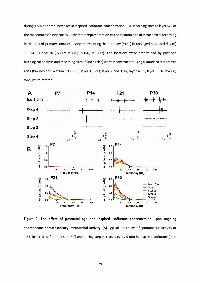
each inspired isoflurane concentration. No spontaneous activity was recorded in any frequency band
in the P7 primary somatosensory cortex at 1.5% inspired isoflurane or at higher concentrations (Figure
2B)
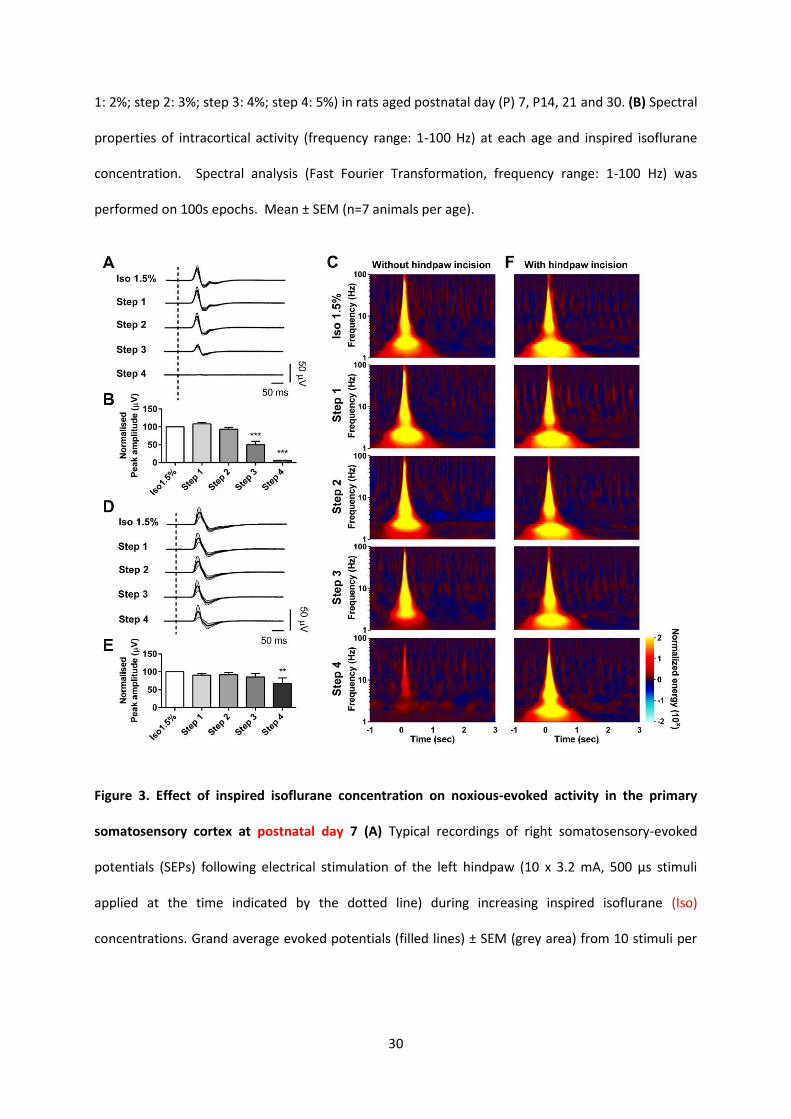
Despite the lack of spontaneous activity, hindpaw electrical stimulation evoked clear low
amplitude evoked potentials, with simple positive-negative voltage waveforms, in the P7
somatosensory cortex (Figure 3). A significant decrease in the mean evoked potential amplitude did
not occur until step 3, where it dropped to half the amplitude recorded at 1.5% inspired isoflurane
(50±9%) (Figure 3A & B). At the maximum inspired isoflurane concentration (step 4), evoked
potentials were greatly diminished (6±1%) but importantly, were still clearly detectable in five of the
seven animals studied. A time frequency analysis of this evoked activity is shown in (Figure 3C) to
demonstrate the mean energy across different frequencies (1-100Hz) evoked in the 3 seconds
following noxious electrical stimulation. At 1.5% inspired isoflurane, a strong increase in energy at all
frequencies is observed in the first 500 milliseconds (ms) post stimulus, but this declines with
increasing levels of inspired isoflurane, notably at Step 3 and 4 (Figure 3C). At Step 4 the oscillation
energy is greatly reduced, but still present.
We next tested whether hindpaw incision influenced the sensitivity of plantar C-fiber evoked
potentials to step increases in anesthesia. The immediate effect of the plantar incision is shown in
Figure 4. In P7 rats, spontaneous activity was increased during, and immediately after, the incision,
12
(delta band activity (0.02±0.01 µV/Hz to 0.10±0.02 µV/Hz; RM ANOVA F(3, 15) = 13.5, P=0.0002; post hoc
Dunnetts P<0.001), and beta band activity (0.01±0.00 µV/Hz to 0.02±0.01 µV/Hz, RM ANOVA, F(3, 15) =
6.1, P=0.0062; post hoc Dunnetts P<0.05), which returned to baseline at 10 mins (Figure 4A and B). In
addition Figure 3 shows that hindpaw incision had a more persistent effect on the response to
subsequent noxious electrical stimuli. In animals with prior hindpaw incision, the mean peak
amplitude of the electrical evoked potential was not significantly altered by step increase in isoflurane
level, except at step 4 (RM ANOVA F (4, 20) = 3.7, P=0.0210; post hoc Dunnetts P<0.01 between baseline
1.5% and step 4), and even at the maximal inspired isoflurane concentration, the mean response
remained at 67±16% of the peak amplitude evoked at the initial 1.5% isoflurane concentration (Figure
3D and E). This is further confirmed by the time frequency analysis (Figure 3F). At baseline 1.5%
isoflurane and at step 1, the strong increase in energy at all frequencies in the first 500 ms post stimulus
does not differ in animals with and without skin incision, but this is not so at higher levels of isoflurane.
Thus, following hindpaw incision, the energy of cortical activity does not diminish with increasing
inspired concentration of isoflurane. Comparison of oscillation energies with and without plantar
hindpaw incision at Step 4, shown in Figure 3, shows that the presence of surgical injury increases the
resistance of infant S1 cortex nociceptive activity to isoflurane (Figure 3C and Figure 3F, bottom
panels).
To test whether this effect of skin incision was nociceptive specific, we compared the effects upon
potentials evoked by low intensity innocuous A fiber stimulation versus those evoked by high intensity
noxious C fiber stimulation in the same animal. Figure 5A and B show that, while in intact skin A fiber
stimulation evokes distinct potentials which slowly diminish with increasing levels of isoflurane,
following skin incision the A fiber evoked response is completely absent at all isoflurane levels (Fig. 5C
and D). This is in marked contrast to the C fiber evoked potentials which persist unchanged following
skin incision at all but the highest step level of isoflurane (Fig.5E and F).
13
Effects of isoflurane on spontaneous intracortical activity are influenced by postnatal age
We next examined the effect of postnatal age upon the relationship between primary
somatosensory cortical spontaneous and evoked activity at increasing inspired isoflurane
concentrations. To do this, the same experiment was performed at P14, P21 and P30 and the results
compared with those obtained at P7.
Figure 2 shows the intracortical spontaneous activity in the primary somatosensory cortex of
the rat, at the four postnatal ages following equilibration at 1.5% isoflurane and through subsequent
step increases in inspired isoflurane concentration (n=7 animals at each age). In contrast to the lack
of spontaneous activity at P7, Figure 2A shows that at P14, P21 and P30, an inspired isoflurane
concentration of 1.5% induced burst-suppression activity in the somatosensory cortex, characterized
by intermittent, highly synchronized neuronal discharges (bursts) separated by silent periods
(suppression), as described elsewhere 29. The burst-suppression activity disappears as inspired
isoflurane concentration increases. Figure 2B shows the distribution of frequency components of
spontaneous somatosensory cortical activity at each age. At P14-30, activity across all frequencies
decreases steadily as the inspired isoflurane concentration increases, and there is no spontaneous
activity and an iso-electric intracortical activity by step 4.
Noxious-evoked activity in the somatosensory cortex is more sensitive to increasing isoflurane
concentration in older rats
We next compared the primary somatosensory (S1) cortical potentials evoked by noxious, C
fiber intensity electrical stimuli in the contralateral hindpaw (10 stimuli/step/animal, n=7 animals) in
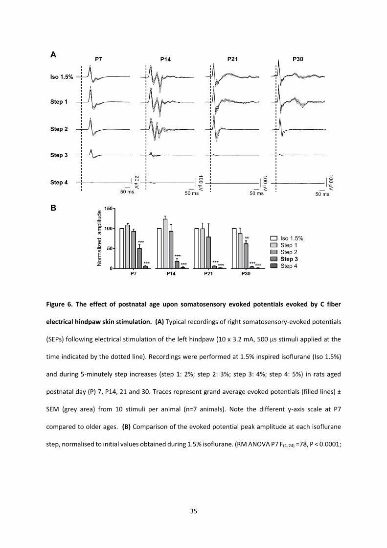
P7, P14, P21 and P30 rats during increasing inspired isoflurane concentrations (Figure 6). Sample
traces are shown in Figure 6A and the mean peak amplitude of the evoked potential is plotted in Figure
14
6B, as the percentage decrease with each step increase in isoflurane, normalized to the initial values
recorded following equilibration at 1.5% isoflurane. The relative decrease in evoked potential peak
amplitude with each step increase in isoflurane is more marked in older animals (P14, P21, P30) than
at P7. At step 3, the mean peak amplitude has reduced to half in P7 animals (50 ± 9% of initial 1.5%
isoflurane values), in contrast to P21 and 30 where it drops below 10% (P21: 6 ±1 %; P30: 4 ±1 %). At
Step 4, no discernible evoked potentials were recorded from any P21 or P30 animals but were recorded
from three out of seven P14 animals, and five out of seven P7 animals.
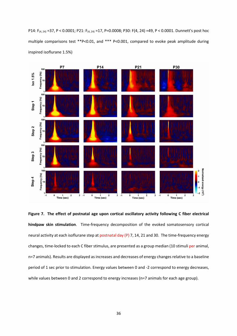
Figure 7 shows time frequency analysis of evoked activity at each age. The mean energy of
cortical activity across different frequencies (1-100Hz) evoked in the 3 seconds following noxious
electrical stimulation is shown at the four steps of increasing inspired isoflurane (10
stimuli/step/animal, n=7 animals for each age group). At all ages, the evoked response energy
decreases with increasing inspired isoflurane. Whereas noxious evoked cortical activity is relatively
resistant to increasing isoflurane concentration at P7, there is a gradual increase in the degree of
suppression by isoflurane at older ages (Figure 7).
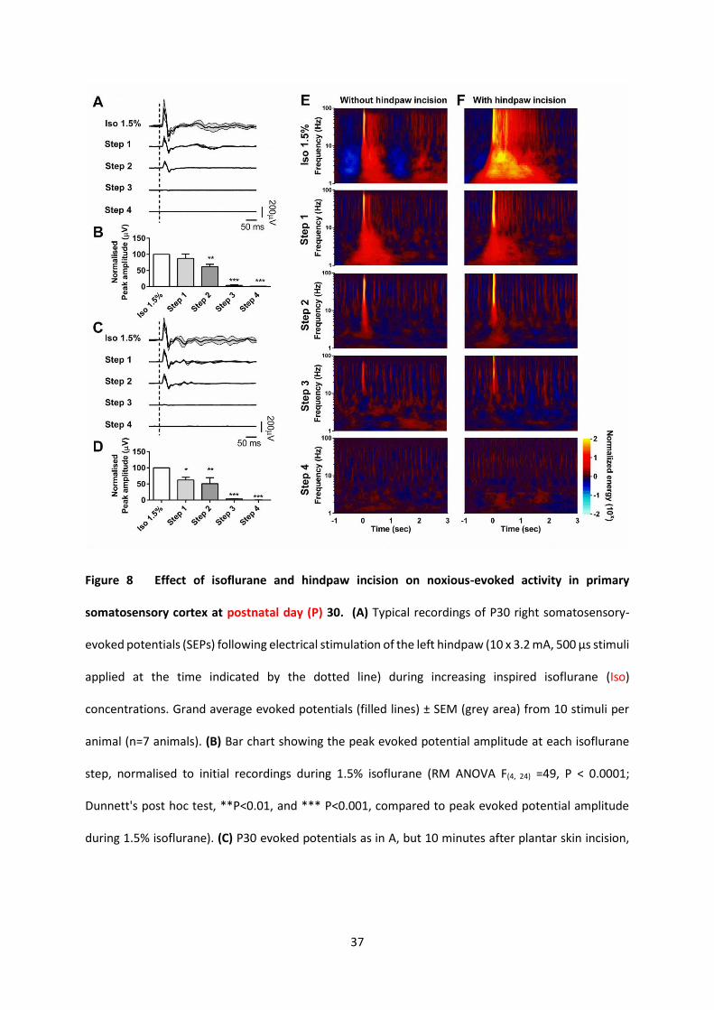
The effect of hindpaw incision upon cortical nociceptive activity at P30 differs from that at P7
As surgical incision had a profound effect upon the subsequent cortical response to noxious
electrical stimulation at P7, we tested whether this also occurred at an older age. Figure 4 shows that
there was no immediate effect of the plantar incision upon spontaneous activity during, and
immediately after, the incision at P30 (Figure 4C and D). Figure 8 illustrates the effect of plantar
incision upon nociceptive evoked potential at each anaesthetic step in P30 rats. Electrical evoked
nociceptive potentials are progressively suppressed by increasing inspired isoflurane concentration, in
the absence (Figure 8A and B) and presence (Figure 8C and D) of skin incision. The reduction in peak
evoked potential amplitude produced by step increases in inspired isoflurane (Figure 8B) was not
15
altered following hindpaw incision (Figure 8D). There was a steady fall in mean evoked potential
amplitude, expressed as a percentage change from baseline (1.5% isoflurane) in both non-incised (Step
1, 88±14; Step 2, 62±8; Step 3: 4±1; Step 4: 1.4±0.5, F (4, 20) = 49, P<0.0001) and incised groups (Step 1,
62±9; Step 2, 50±19; Step 3: 3.5±1; Step 4: 0.13±0.1, F (4, 20) = 25, P<0.0001) (Figure 8B and 8D). Time
frequency analysis (Figure 8E and 8F) shows that skin incision at 1.5% isoflurane causes an increased
energy and duration of evoked cortical oscillations at P30, not seen at P7, suggesting some underlying
sensitization of cortical pain circuits. Although clear at an inspired isoflurane concentration of 1.5%,
this increased cortical response is highly sensitive to subsequent increases in isoflurane, and the
difference in response between P30 animals with (Figure 8F) and without (Figure 8E) hindpaw incision
is largely lost at step 1, and evoked responses in both groups are significantly diminished at step 2 and
3 and effectively gone at step 4. These results differ significantly from those obtained at P7 (Figure
3), where cortical evoked potentials and oscillatory activity persisted and were resistant to increasing
inspired isoflurane concentration.
16
Discussion
The primary aim of this study was to test the impact of increasing isoflurane concentration upon
spontaneous and evoked nociceptive activity in the somatosensory cortex of infant rats undergoing
hindpaw incision. We hypothesised that, in infant rats, noxious evoked brain activity is more resistant
than spontaneous brain activity to isoflurane anesthesia. The results, obtained by recording intra
cortical neuronal activity from layer 5-6 of the somatosensory cortex in postnatal day (P)7 rats during
increasing inspired concentrations of isoflurane , support this hypothesis. The data show that
isoflurane influences spontaneous activity and evoked activity in the infant rat somatosensory cortex
quite differently.
At P7, all concentrations of inspired isoflurane (1.5-5%) silenced cortical neurons and suppressed
spontaneous bursts and oscillations. This effect is consistent with a previous study in the neonatal
somatosensory cortex, where cortical activity was completely suppressed in P7 animals by 1.5-2%
isoflurane16. This is in contrast to the spontaneous cortical activity observed in awake or very lightly
anesthetised animals at this age, which is characterized by intermittent bursts organized in oscillations
in alpha-beta (spindle bursts) and gamma frequency bands (early gamma oscillations, EGOs)16,30–33.
In contrast to the absence of spontaneous activity, noxious somatosensory evoked potentials and
evoked oscillatory activity persisted, even at high inspired concentrations of isoflurane. This result
highlights the differences in the neural mechanisms generating spontaneous activity and evoked
activity in the immature somatosensory cortex. Activity in the developing brain changes with age
and early cortical development is marked by unique patterns of activity as functional circuits
mature34,35. Early oscillatory bursts are generated in thalamocortical circuits 32,36 but can also be
triggered by sensory inputs and are likely to be generated by glutamatergic AMPA (α-amino-3-hydroxy-
5-methyl-4-isoxazolepropionic acid) and NMDA (N-methyl-D-aspartate) receptors while GABAergic
17
(gamma-aminobutyric acid releasing) interneurons compartmentalize the activated areas via surround
inhibition37,38. The transition from predominantly nonspecific neuronal bursts to specific cortical
sensory evoked potentials appears to be triggered by increasing sensory input 31 in the neonatal rodent
somatosensory cortex 16. A similar transition begins in the preterm human infant brain at around 35
weeks postconceptional age 26,39. Thus the nociceptive evoked activity in the newborn infant cortex
recorded at P7 is still relatively immature and may be less tightly coupled within specific networks,
rendering it less susceptible to suppression by volatile anesthetics than at older ages.
A further finding here is that the relative insensitivity of noxious evoked activity to isoflurane was
enhanced following plantar hindpaw incision. This well-established model of surgical incision pain21,
adapted for younger rat pups 40,41, rendered infant rat nociceptive evoked cortical potentials and
oscillatory network activity totally resistant to the maximum inspired isoflurane tested (5%). The
mechanism for this is not clear but may reflect widespread depolarization of central neurons by the
incoming nociceptive barrage. It is notable also that the incision itself increased cortical activity in
young animals, consistent with the reported spike activity and sensitization of receptive fields in infant
dorsal horn cells following hindpaw incision42. Importantly, the enhanced isoflurane resistance
following skin incision at P7 was selective for C fiber nociceptive evoked potentials. In contrast, A
fiber innocuous evoked potentials were completely abolished following skin incision at all isoflurane
levels, reflecting the different sensitivities of immature A and C fibre evoked activity to skin incision43.
A secondary outcome of this study was the significant age-dependence of the isoflurane effects upon
spontaneous and noxious-evoked activity in the somatosensory cortex. At P14 and older ages,
isoflurane 1.5% produced a typical pattern of burst suppression with high-amplitude low-frequency
activity, and a progressive decrease in spontaneous activity with step increases in inspired isoflurane
concentration, as reported elsewhere29,44,45. This contrasted with the suppression of spontaneous
18
spindle bursts and gamma oscillations even at the initial inspired concentration of 1.5% isoflurane at
P7. More important was the greater sensitivity of the noxious evoked potentials to isoflurane in
older animals. As reported in adult rats9 noxious evoked potentials at P14-30 decreased markedly
with increasing concentrations of inspired isoflurane and at P21 and P30 were completely absent at
5% isoflurane. Differences were especially clear using time frequency analysis, which reveals the
energy and power of oscillating signals in the cortex and thus the changes within different frequency
bands that are linked to specific sensory and motor functions25. This form of analysis has been used
during isoflurane anesthesia in adult rats to characterize concentration-dependent changes in both
low (30-50Hz) and high (70-140Hz) frequency gamma power in different brain regions 44.
A further secondary outcome was the failure of surgical incision to affect noxious evoked potential
sensitivity to isoflurane at the older age of P30. Unlike the P7 rat, surgical incision itself at P30 caused
no immediate cortical activity, but did have a sensitizing effect upon noxious cortical evoked activity
at 1.5% isoflurane. This is consistent with previous reports of increased C fibre input to the adult rat
somatosensory cortex in a UV-B irradiation model of hyperalgesia8. However, in contrast to P7 rats,
the reduction of evoked potentials and oscillatory activity by isoflurane were not altered by surgical
incision in rats at P30. These data suggest that the powerful effect at P7 is not due to the effects of
central sensitization, but a different, as yet unknown effect.
Our aim in this study was not to compare ‘hypnotic potency’ at the different ages, but rather to
evaluate within age-group changes in cortical activity during isoflurane anesthesia. Equipotent
concentrations of volatile anesthetics have been traditionally based upon the minimum alveolar
concentration (MAC) that prevents movement to a standardized noxious stimuli. However, these
values do not reflect hypnotic potency in the brain46. The MAC of isoflurane is higher in P7-P9
rodents,47,48 as spinal reflex excitability is greater at this age, but a clear stimulus-response relationship
that is sensitive to injury, analgesia and inspired volatile agent concentration is evident across all ages
19
22,49. While isoflurane actions in the spinal cord may reduce ascending somatosensory information
and indirectly alter cortical activity50, the level of cortical-evoked activity cannot be inferred from the
presence or absence of a visible reflex response. Here, hindpaw incision (but not electrical
stimulation) produced a brief visible reflex response in P7 but not older animals, consistent with the
reported greater MAC, but in older animals noxious stimuli produced clear cortical evoked responses
at 1.5% isoflurane, despite a lack of reflex response. The age dependence of cortical evoked response
sensitivity to isoflurane may have been influenced by alterations in physiological parameters. Heart
rate was monitored throughout the experiments and even in the more prolonged protocols including
hindpaw incision, decreases were only seen at the highest level of inspired isoflurane, and to the same
degree (approximately 20% reduction) in P7 and P30 groups. Animals were mechanically ventilated
in isoflurane and oxygen with age-adjusted settings, and although we cannot confirm that partial
pressures of carbon dioxide (pCO2) were the same across the different ages, changes with increasing
isoflurane concentration would not have been influenced by respiratory parameters.
These rat data obtained have considerable implications for clinical practice. The timing and sequence
of key events in brain development are exceptionally similar across mammals and a recent
neuroinformatic analysis shows that sensorimotor events in the cortex of the P7 rat translate to 1-2
months in the human 51. This study clearly demonstrates that spontaneous intracortical network
activity is more effectively silenced by isoflurane in the neonatal brain, but that isoflurane has less
effect upon cortical activity evoked by peripheral noxious sensory inputs. As the developing central
nervous system is vulnerable to changes in neural activity, maintaining appropriate anesthesia in
infants is likely to require avoidance of both excessive reductions in cortical activity that may enhance
neuronal apoptosis and excessive increases in activity due to uncontrolled noxious inputs1–4.
Anesthesia-induced developmental neurotoxicity has been clearly demonstrated in neonatal animals3,
and the associated long-lasting cognitive impairment has raised concern for young children undergoing
20
anesthesia4. The data here highlight the complexity of using intracortical activity parameters to define
or measure the level of anesthesia required to produce ‘hypnosis’ in neonates and infants. The
persistence of noxious sensory-evoked responses despite increasing isoflurane concentration in young
rats emphasizes the critical need to provide analgesia in neonates and infants. Surgery and tissue
injury in neonatal rodents can produce long-term changes in sensory processing,52 but this can be
modified by morphine53,54 or local anesthetic blockade40,55. Increased understanding of age- and
anesthesia-dependent changes in intracortical activity may improve algorithms for evaluating depth
of anesthesia in neonates and infants, and comparative studies of the ability of different types and
doses of analgesic to minimize noxious evoked responses are likely to improve acute clinical care and
long-term neurodevelopmental outcome following neonatal surgery.
21
References
1. Sanders RD, Hassell J, Davidson AJ, Robertson NJ, Ma D: Impact of anaesthetics and surgery on
neurodevelopment: an update. Br. J. Anaesth. 2013; 110:i53–72
2. Lin EP, Soriano SG, Loepke AW: Anesthetic neurotoxicity. Anesthesiol. Clin. 2014; 32:133–55
3. Liu J, Rossaint R, Sanders RD, Coburn M: Toxic and protective effects of inhaled anaesthetics on
the developing animal brain: systematic review and update of recent experimental work. Eur. J.
Anaesthesiol. 2014; 31:669–77
4. Warner DO, Flick RP: Effects of anesthesia and surgery on the developing brain: problem solved?
Pediatr. Anesth. 2015; 25:435–6
5. Brummelte S, Grunau RE, Chau V, Poskitt KJ, Brant R, Vinall J, Gover A, Synnes AR, Miller SP:
Procedural pain and brain development in premature newborns. Ann. Neurol. 2012; 71:385–96
6. Walker SM: Biological and neurodevelopmental implications of neonatal pain. Clin. Perinatol.
2013; 40:471–91
7. Schwaller F, Fitzgerald M: The consequences of pain in early life: injury-induced plasticity in
developing pain pathways. Eur. J. Neurosci. 2014; 39:344–52
8. Jensenl T, Granmo M, Schouenborg J: Altered nociceptive C fibre input to primary somatosensory
cortex in an animal model of hyperalgesia. Eur. J. Pain 2011; 15:368–75
9. Granmo M, Jensen T, Schouenborg J: Nociceptive transmission to rat primary somatosensory
cortex--comparison of sedative and analgesic effects. PloS One 2013; 8:e53966
22
10. Fabrizi L, Williams G, Lee A, Meek J, Slater R, Olhede S, Fitzgerald M: Cortical activity evoked by an
acute painful tissue-damaging stimulus in healthy adult volunteers. J. Neurophysiol. 2013;
109:2393-403.
11. Iannetti GD, Baumgärtner U, Tracey I, Treede R-D, Magerl W: Pinprick-evoked brain potentials
(PEPs): a novel tool to assess central sensitisation of nociceptive pathways in humans. J.
Neurophysiol. 2013; 110:1107-16
12. Baumgärtner U, Greffrath W, Treede R-D: Contact heat and cold, mechanical, electrical and
chemical stimuli to elicit small fiber-evoked potentials: merits and limitations for basic science and
clinical use. Neurophysiol. Clin. 2012; 42:267–80
13. Slater R, Worley A, Fabrizi L, Roberts S, Meek J, Boyd S, Fitzgerald M: Evoked potentials generated
by noxious stimulation in the human infant brain. Eur. J. Pain 2010; 14:321–6
14. Verriotis M, Fabrizi L, Lee A, Ledwidge S, Meek J, Fitzgerald M: Cortical activity evoked by
inoculation needle prick in infants up to one-year old. Pain 2015; 156:222–30
15. Roth D, Petersen-Felix S, Bak P, Arendt-Nielsen L, Fischer M, Bjerring P, Zbinden AM: Analgesic
effect in humans of subanaesthetic isoflurane concentrations evaluated by evoked potentials. Br.
J. Anaesth. 1996; 76:38–42
16. Sitdikova G, Zakharov A, Janackova S, Gerasimova E, Lebedeva J, Inacio AR, Zaynutdinova D,
Minlebaev M, Holmes GL, Khazipov R: Isoflurane suppresses early cortical activity. Ann. Clin.
Transl. Neurol. 2014; 1:15–26
23
17. Prost N de, Ricard J-D, Saumon G, Dreyfuss D: Ventilator-induced lung injury: historical
perspectives and clinical implications. Ann. Intensive Care 2011; 1:28
18. Paxinos, Watson: The Rat Brain in Stereotaxic Coordinates, 7th Edition, 7th edition. Elsevier, 2013
19. Paxinos G, Ashwell KW., Tork I: Atlas of the developing rat nervous system. London, Academic
Press, 1994
20. Jennings E, Fitzgerald M: Postnatal changes in responses of rat dorsal horn cells to afferent
stimulation: a fibre-induced sensitization. J. Physiol. 1998; 509:859–68
21. Brennan TJ, Vandermeulen EP, Gebhart GF: Characterization of a rat model of incisional pain. Pain
1996; 64:493–502
22. Walker SM, Tochiki KK, Fitzgerald M: Hindpaw incision in early life increases the hyperalgesic
response to repeat surgical injury: critical period and dependence on initial afferent activity. Pain
2009; 147:99–106
23. http://www.originlab.com/doc/Origin-Help/FFT1-Algorithm. Last accessed 10/28/2015
24. Rampil IJ: A primer for EEG signal processing in anesthesia. Anesthesiology 1998; 89:980–1002
25. Akam T, Kullmann DM: Oscillatory multiplexing of population codes for selective communication
in the mammalian brain. Nat. Rev. Neurosci. 2014; 15:111–22
26. Fabrizi L, Slater R, Worley A, Meek J, Boyd S, Olhede S, Fitzgerald M: A shift in sensory processing
that enables the developing human brain to discriminate touch from pain. Curr. Biol. 2011;
21:1552–8
24
27. Narayanan NS, Cavanagh JF, Frank MJ, Laubach M: Common medial frontal mechanisms of
adaptive control in humans and rodents. Nat. Neurosci. 2013; 16:1888–95
28. Olhede S., Walden A.: Generalized morse wavelets. IEEE Trans. SIGNAL Process. 2002; 50:2661–70
29. Ferron JF, Kroeger D, Chever O, Amzica F: Cortical Inhibition during Burst Suppression Induced
with Isoflurane Anesthesia. J. Neurosci. 2009; 29:9850–60
30. Khazipov R, Sirota A, Leinekugel X, Holmes GL, Ben-Ari Y, Buzsáki G: Early motor activity drives
spindle bursts in the developing somatosensory cortex. Nature 2004; 432:758–61
31. Colonnese MT, Kaminska A, Minlebaev M, Milh M, Bloem B, Lescure S, Moriette G, Chiron C, Ben-
Ari Y, Khazipov R: A conserved switch in sensory processing prepares developing neocortex for
vision. Neuron 2010; 67:480–98
32. Yang JW, An S, Sun JJ, Reyes-Puerta V, Kindler J, Berger T, Kilb W, Luhmann HJ: Thalamic Network
Oscillations Synchronize Ontogenetic Columns in the Newborn Rat Barrel Cortex. Cereb. Cortex
2013; 23:1299–316
33. Tiriac A, Uitermarkt BD, Fanning AS, Sokoloff G, Blumberg MS: Rapid whisker movements in
sleeping newborn rats. Curr. Biol. 2012; 22:2075–80
34. Blankenship A., Feller M.: Mechanisms underlying spontaneous patterned activity in developing
neural circuits. Nat Rev Neurosci 2010; 11:18–29
35. Khazipov R, Luhmann HJ: Early patterns of electrical activity in the developing cerebral cortex of
humans and rodents. Trends Neurosci. 2006; 29:414–8
25
36. Minlebaev M, Colonnese M, Tsintsadze T, Sirota A, Khazipov R: Early Gamma Oscillations
Synchronize Developing Thalamus and Cortex. Science 2011; 334:226–9
37. Minlebaev M, Ben-Ari Y, Khazipov R: Network Mechanisms of Spindle-Burst Oscillations in the
Neonatal Rat Barrel Cortex In Vivo. J. Neurophysiol. 2007; 97:692–700
38. Minlebaev M, Ben-Ari Y, Khazipov R: NMDA Receptors Pattern Early Activity in the Developing
Barrel Cortex In Vivo. Cereb. Cortex 2009; 19:688–96
39. Hrbek A, Karlberg P, Olsson T: Development of visual and somatosensory evoked responses in pre-
term newborn infants. Electroencephalogr. Clin. Neurophysiol. 1973; 34:225–32
40. Walker SM, Tochiki KK, Fitzgerald M: Hindpaw incision in early life increases the hyperalgesic
response to repeat surgical injury: critical period and dependence on initial afferent activity. Pain
2009; 147:99–106
41. Beggs S, Currie G, Salter MW, Fitzgerald M, Walker SM: Priming of adult pain responses by
neonatal pain experience: maintenance by central neuroimmune activity. Brain 2012; 135:404–17
42. Ririe DG, Bremner LR, Fitzgerald M: Comparison of the immediate effects of surgical incision on
dorsal horn neuronal receptive field size and responses during postnatal development.
Anesthesiology 2008; 109:698–706
43. Boada MD, Gutierrez S, Giffear K, Eisenach JC, Ririe DG: Skin incision-induced receptive field
responses of mechanosensitive peripheral neurons are developmentally regulated in the rat. J.
Neurophysiol. 2012; 108:1122–9
26
44. Hudetz AG, Vizuete JA, Pillay S: Differential effects of isoflurane on high-frequency and low-
frequency γ oscillations in the cerebral cortex and hippocampus in freely moving rats.
Anesthesiology 2011; 114:588–95
45. Kortelainen J, Jia X, Seppänen T, Thakor N: Increased electroencephalographic gamma activity
reveals awakening from isoflurane anaesthesia in rats. Br. J. Anaesth. 2012; 109:782–9
46. Palanca BJA, Mashour GA, Avidan MS: Processed electroencephalogram in depth of anesthesia
monitoring. Curr. Opin. Anaesthesiol. 2009; 22:553–9
47. Orliaguet G, Vivien B, Langeron O, Bouhemad B, Coriat P, Riou B: Minimum alveolar concentration
of volatile anesthetics in rats during postnatal maturation. Anesthesiology 2001; 95:734–9
48. Stratmann G, Sall JW, Eger EI, Laster MJ, Bell JS, May LDV, Eilers H, Krause M, Heusen F v d,
Gonzalez HE: Increasing the duration of isoflurane anesthesia decreases the minimum alveolar
anesthetic concentration in 7-day-old but not in 60-day-old rats. Anesth. Analg. 2009; 109:801–6
49. Walker SM, Fitzgerald M: Characterization of spinal alpha-adrenergic modulation of nociceptive
transmission and hyperalgesia throughout postnatal development in rats. Br. J. Pharmacol. 2007;
151:1334–42
50. Antognini JF, Jinks SL, Atherley R, Clayton C, Carstens E: Spinal anaesthesia indirectly depresses
cortical activity associated with electrical stimulation of the reticular formation. Br. J. Anaesth.
2003; 91:233–8
51. Workman AD, Charvet CJ, Clancy B, Darlington RB, Finlay BL: Modeling transformations of
neurodevelopmental sequences across mammalian species. J. Neurosci. 2013; 33:7368–83
27
52. Schwaller F, Fitzgerald M: The consequences of pain in early life: injury induced plasticity in
developing pain pathways. Eur. J. Neurosci. 2014;39:344-52
53. Laprairie JL, Johns ME, Murphy AZ: Preemptive morphine analgesia attenuates the long-term
consequences of neonatal inflammation in male and female rats. Pediatr. Res. 2008; 64:625–30
54. Sternberg WF, Scorr L, Smith LD, Ridgway CG, Stout M: Long-term effects of neonatal surgery on
adulthood pain behavior. Pain 2005; 113:347–53
55. Walker SM, Fitzgerald M, Hathway GJ: Surgical injury in the neonatal rat alters the adult pattern
of descending modulation from the rostroventral medulla. Anesthesiology 2015; 122:1391–400

28
Figure 1. (A) Diagram showing the experimental timelines and recording sites. Animals were initially
anesthetized with isoflurane 2.5-4% in oxygen for surgical preparation and electrode insertion, and
then maintained at 1.5% for 40 minutes to allow equilibration. The electrocardiogram (ECG) was
monitored throughout. Group 1: On postnatal day (P) 7, 14, 21 or 30, continuous intracortical
spontaneous activity in the right somatosensory cortex was recorded for 100s, followed by evoked
responses to electrical stimulation of the left hindpaw (10 stimuli of 3.2 mA for 500ms at 10s intervals).
Recordings were repeated at 5-minute intervals following step increases in inspired isoflurane
concentration: step 1 (2%), step 2 (3%) step 3 (4%) and step 4 (5%). Group 2: In P7 and P30 animals,
spontaneous activity was recorded 2 minutes before, during and 2 minutes following left plantar
hindpaw incision. Ten minutes following incision, spontaneous and electrical activity were recorded
29
during 1.5% and step increases in inspired isoflurane concentration. (B) Recording sites in layer 5/6 of
the rat somatosensory cortex. Schematic representation of the location site of intracortical recording
in the area of primary somatosensory representing the hindpaw (S1HL) in rats aged postnatal day (P)
7, P14, 21 and 30 (P7=12; P14=6; P21=6; P30=12). The locations were determined by post-hoc
histological analysis and recording sites (filled circles) were reconstructed using a standard stereotaxic
atlas (Paxinos and Watson 1998). L1, layer 1; L2/3, layer 2 and 3; L4, layer 4; L5, layer 5; L6, layer 6;
WM, white matter.
Figure 2. The effect of postnatal age and inspired isoflurane concentration upon ongoing
spontaneous somatosensory intracortical activity. (A) Typical 10s traces of spontaneous activity at
1.5% inspired isoflurane (iso 1.5%) and during step increases every 5 min in inspired isoflurane (step
30
1: 2%; step 2: 3%; step 3: 4%; step 4: 5%) in rats aged postnatal day (P) 7, P14, 21 and 30. (B) Spectral
properties of intracortical activity (frequency range: 1-100 Hz) at each age and inspired isoflurane
concentration. Spectral analysis (Fast Fourier Transformation, frequency range: 1-100 Hz) was
performed on 100s epochs. Mean ± SEM (n=7 animals per age).
Figure 3. Effect of inspired isoflurane concentration on noxious-evoked activity in the primary
somatosensory cortex at postnatal day 7 (A) Typical recordings of right somatosensory-evoked
potentials (SEPs) following electrical stimulation of the left hindpaw (10 x 3.2 mA, 500 µs stimuli
applied at the time indicated by the dotted line) during increasing inspired isoflurane (Iso)
concentrations. Grand average evoked potentials (filled lines) ± SEM (grey area) from 10 stimuli per
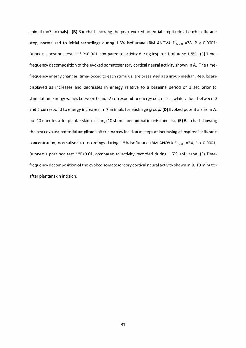
31
animal (n=7 animals). (B) Bar chart showing the peak evoked potential amplitude at each isoflurane
step, normalised to initial recordings during 1.5% isoflurane (RM ANOVA F(4, 24) =78, P < 0.0001;
Dunnett's post hoc test, *** P<0.001, compared to activity during inspired isoflurane 1.5%). (C) Time-
frequency decomposition of the evoked somatosensory cortical neural activity shown in A. The time-
frequency energy changes, time-locked to each stimulus, are presented as a group median. Results are
displayed as increases and decreases in energy relative to a baseline period of 1 sec prior to
stimulation. Energy values between 0 and -2 correspond to energy decreases, while values between 0
and 2 correspond to energy increases. n=7 animals for each age group. (D) Evoked potentials as in A,
but 10 minutes after plantar skin incision, (10 stimuli per animal in n=6 animals). (E) Bar chart showing
the peak evoked potential amplitude after hindpaw incision at steps of increasing of inspired isoflurane
concentration, normalised to recordings during 1.5% isoflurane (RM ANOVA F(4, 20) =24, P < 0.0001;
Dunnett's post hoc test **P<0.01, compared to activity recorded during 1.5% isoflurane. (F) Time-
frequency decomposition of the evoked somatosensory cortical neural activity shown in D, 10 minutes
after plantar skin incision.
32
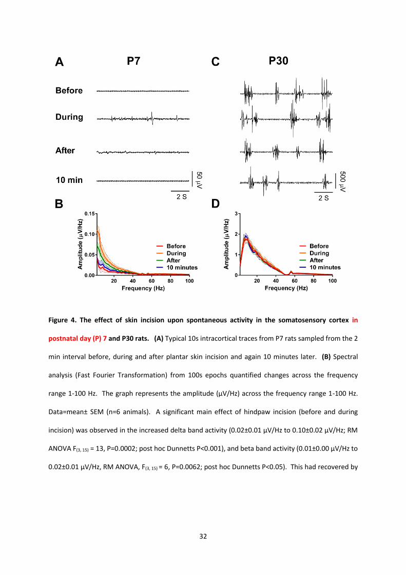
Figure 4. The effect of skin incision upon spontaneous activity in the somatosensory cortex in
postnatal day (P) 7 and P30 rats. (A) Typical 10s intracortical traces from P7 rats sampled from the 2
min interval before, during and after plantar skin incision and again 10 minutes later. (B) Spectral
analysis (Fast Fourier Transformation) from 100s epochs quantified changes across the frequency
range 1-100 Hz. The graph represents the amplitude (µV/Hz) across the frequency range 1-100 Hz.
Data=mean± SEM (n=6 animals). A significant main effect of hindpaw incision (before and during
incision) was observed in the increased delta band activity (0.02±0.01 µV/Hz to 0.10±0.02 µV/Hz; RM
ANOVA F(3, 15) = 13, P=0.0002; post hoc Dunnetts P<0.001), and beta band activity (0.01±0.00 µV/Hz to
0.02±0.01 µV/Hz, RM ANOVA, F(3, 15) = 6, P=0.0062; post hoc Dunnetts P<0.05). This had recovered by
33
10 mins post-surgery. (C) Typical 10s intracortical traces from P30 sampled from the 2 min interval
before, during and after plantar skin incision and again 10 minutes later. (D) Spectral analysis in P30
rats. In contrast to P7 rats, the frequency distribution of the mean spontaneous activity at P30 (n=6
animals at each age) in a 100s epoch before, during and after incision under 1.5 % isoflurane
anaesthesia was unchanged (δ band from 1.05±0.14 µV/Hz to 1.22±0.12 µV/Hz, RM ANOVA F (3, 15) =
0.8, P=0.4994, non-significant; β band from 0.01±0.00 µV/Hz to 0.02±0.01 µV/Hz, RM ANOVA F (3, 15) =
0.7, P=0.5688, non-significant.
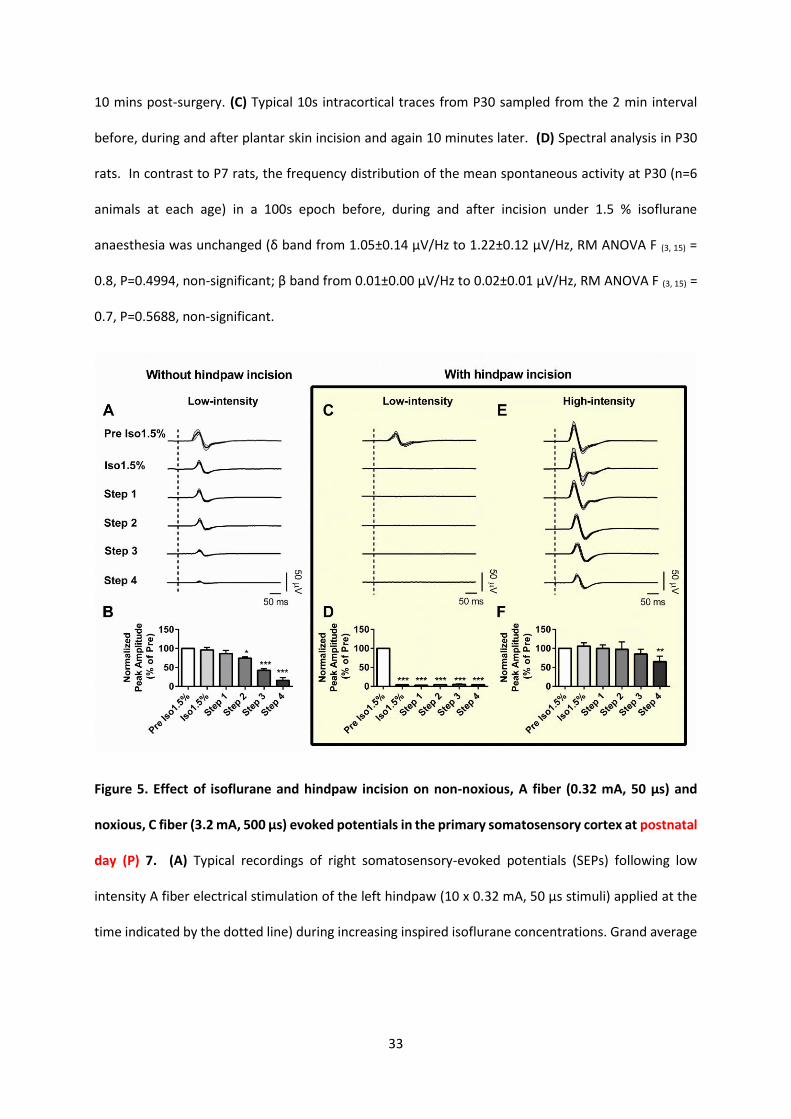
Figure 5. Effect of isoflurane and hindpaw incision on non-noxious, A fiber (0.32 mA, 50 µs) and
noxious, C fiber (3.2 mA, 500 µs) evoked potentials in the primary somatosensory cortex at postnatal
day (P) 7. (A) Typical recordings of right somatosensory-evoked potentials (SEPs) following low
intensity A fiber electrical stimulation of the left hindpaw (10 x 0.32 mA, 50 µs stimuli) applied at the
time indicated by the dotted line) during increasing inspired isoflurane concentrations. Grand average
34
evoked potentials (filled lines) ± SEM (grey area) from 10 stimuli per animal (n=5 animals). (B) Bar
chart showing the peak evoked potential amplitude at each isoflurane (Iso) step, normalised to initial
recordings during 1.5% isoflurane (Pre Iso 1.5%) (RM ANOVA F(5, 20) =40, P < 0.0001; Dunnett's post hoc
test, *P<0.05; *** P<0.001, compared to activity during Pre Iso 1.5%. (C) and (E) show typical
recordings of P7 right somatosensory-evoked potentials (SEPs in response to (C) low-intensity, A fiber
(C, 5 x 0.32 mA, 50 µs stimuli) and (E) high-intensity, C fiber (5 x 3.2 mA, 500 µs) electrical stimulation
of the left hindpaw following hindpaw incision at increasing inspired isoflurane steps. Traces represent
grand average evoked potentials (filled lines) ± SEM (grey area) from 5 stimuli (5 x low-intensity, and
5 x high-intensity) per animal (n=5 animals). The dotted line indicates the time of electrical stimulation.
(D) and (F) show bar charts of the peak evoked potential amplitude at each isoflurane step with low-
intensity electrical stimulation (D) and high-intensity electrical stimulation (F). The data were
normalised to initial recordings during 1.5% isoflurane (Pre Iso 1.5%) (For low intensity stimulation:
RM ANOVA F(5, 20) =1673, P < 0.0001; for high intensity stimulation: RM ANOVA F(5, 20) =6, P =0.0011,
Dunnett's post hoc test, *P<0.05; **P<0.01; *** P<0.001, compared to activity during Pre Iso 1.5%).
35
Figure 6. The effect of postnatal age upon somatosensory evoked potentials evoked by C fiber
electrical hindpaw skin stimulation. (A) Typical recordings of right somatosensory-evoked potentials
(SEPs) following electrical stimulation of the left hindpaw (10 x 3.2 mA, 500 µs stimuli applied at the
time indicated by the dotted line). Recordings were performed at 1.5% inspired isoflurane (Iso 1.5%)
and during 5-minutely step increases (step 1: 2%; step 2: 3%; step 3: 4%; step 4: 5%) in rats aged
postnatal day (P) 7, P14, 21 and 30. Traces represent grand average evoked potentials (filled lines) ±
SEM (grey area) from 10 stimuli per animal (n=7 animals). Note the different y-axis scale at P7
compared to older ages. (B) Comparison of the evoked potential peak amplitude at each isoflurane
step, normalised to initial values obtained during 1.5% isoflurane. (RM ANOVA P7 F(4, 24) =78, P < 0.0001;
36
P14: F(4, 24) =37, P < 0.0001; P21: F(4, 24) =17, P=0.0008; P30: F(4, 24) =49, P < 0.0001. Dunnett's post hoc
multiple comparisons test **P<0.01, and *** P<0.001, compared to evoke peak amplitude during
inspired isoflurane 1.5%)
Figure 7. The effect of postnatal age upon cortical oscillatory activity following C fiber electrical
hindpaw skin stimulation. Time-frequency decomposition of the evoked somatosensory cortical
neural activity at each isoflurane step at postnatal day (P) 7, 14, 21 and 30. The time-frequency energy
changes, time-locked to each C fiber stimulus, are presented as a group median (10 stimuli per animal,
n=7 animals). Results are displayed as increases and decreases of energy changes relative to a baseline
period of 1 sec prior to stimulation. Energy values between 0 and -2 correspond to energy decreases,
while values between 0 and 2 correspond to energy increases (n=7 animals for each age group).
37
Figure 8 Effect of isoflurane and hindpaw incision on noxious-evoked activity in primary
somatosensory cortex at postnatal day (P) 30. (A) Typical recordings of P30 right somatosensory-
evoked potentials (SEPs) following electrical stimulation of the left hindpaw (10 x 3.2 mA, 500 µs stimuli
applied at the time indicated by the dotted line) during increasing inspired isoflurane (Iso)
concentrations. Grand average evoked potentials (filled lines) ± SEM (grey area) from 10 stimuli per
animal (n=7 animals). (B) Bar chart showing the peak evoked potential amplitude at each isoflurane
step, normalised to initial recordings during 1.5% isoflurane (RM ANOVA F(4, 24) =49, P < 0.0001;
Dunnett's post hoc test, **P<0.01, and *** P<0.001, compared to peak evoked potential amplitude
during 1.5% isoflurane). (C) P30 evoked potentials as in A, but 10 minutes after plantar skin incision,
38
(10 stimuli per animal in n=6 animals). (D) Bar chart showing the peak evoked potential amplitude
after hindpaw incision at steps of increasing of inspired isoflurane concentration, normalised to
recordings during 1.5% isoflurane (RM ANOVA F(4, 20) =24, P < 0.0001; Dunnett's post hoc test, *P<0.05,
**P<0.01, *** P<0.001, compared to activity recorded during 1.5% isoflurane. (E) Time-frequency
decomposition of the evoked somatosensory cortical neural activity shown in A. The time-frequency
energy changes, time-locked to each stimulus, are presented as a group median. Results are displayed
as increases and decreases in energy relative to a baseline period of 1 sec prior to stimulation. Energy
values between 0 and -2 correspond to energy decreases, while values between 0 and 2 correspond
to energy increases. n=7 animals for each age group. (F) Time-frequency decomposition of the evoked
somatosensory cortical neural activity shown in C, 10 minutes after plantar skin incision.
39
Supplementary Figures
40
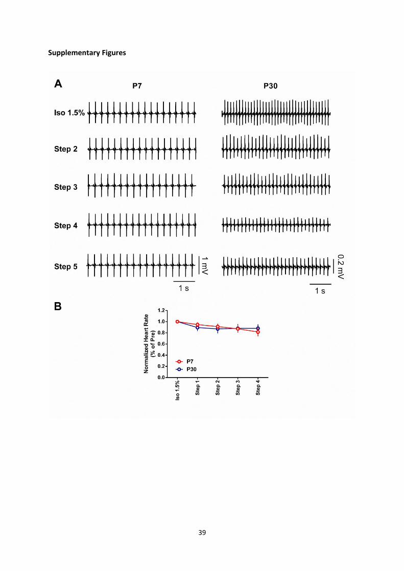
Supplemental Digital Content 1: The effect of isoflurane on the heart rate
(A) Example of recordings of electrocardiogram (ECG) at 1.5% inspired isoflurane (iso 1.5%) and during
step increases (step 1: 2%; step 2: 3%; step 3: 4%; step 4: 5%) in rats aged postnatal day (P) 7and 30.
(B) The effect of step increasing of isoflurane on the heart rate in different age of group (n=6 in P7; n=6
in P30). The normalized heart rates (normalized to Iso 1.5%) were plotted against the step increase in
isoflurane. The heart rate slightly decreases as isoflurane increases. However, there is no significant
difference between ages. Step 1: t 0.05(2),10=1.762, P= 0.1086 ; Step2: t 0.05(2),10=1.072, P=0.3091;
step 3: t 0.05(2),10=0.1818, P=0.8594; Step 4: t 0.05(2),10=1.530, P=0.1571, Student’s T test).
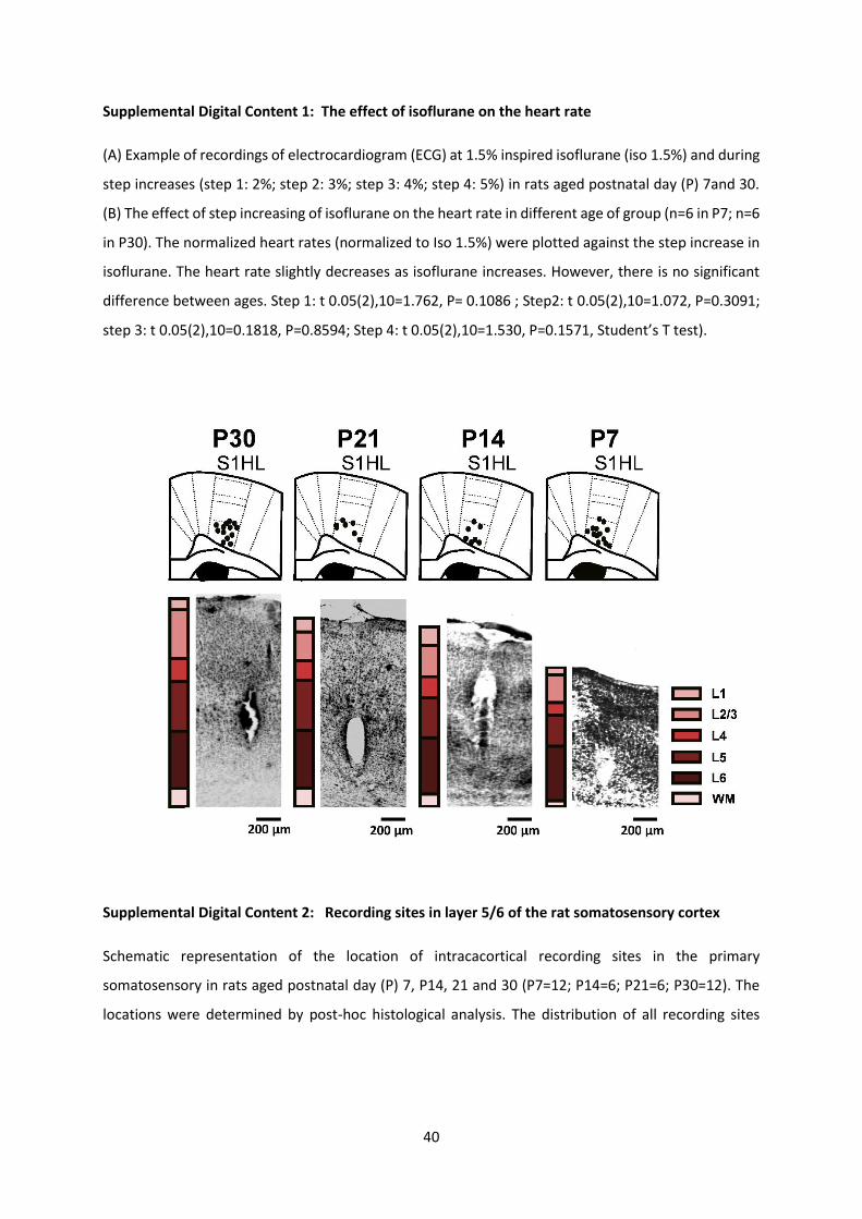
Supplemental Digital Content 2: Recording sites in layer 5/6 of the rat somatosensory cortex
Schematic representation of the location of intracacortical recording sites in the primary
somatosensory in rats aged postnatal day (P) 7, P14, 21 and 30 (P7=12; P14=6; P21=6; P30=12). The
locations were determined by post-hoc histological analysis. The distribution of all recording sites
41
(filled circles) was reconstructed onto maps of the primary somatosensory cortex corresponding to
hindpaw (S1HL) in a standard stereotaxic atlas 56(Paxinos and Watson 1998). L1, layer 1; L2/3, layer 2
and 3; L4, layer 4; L5, layer 5; L6, layer 6; WM, white matter.
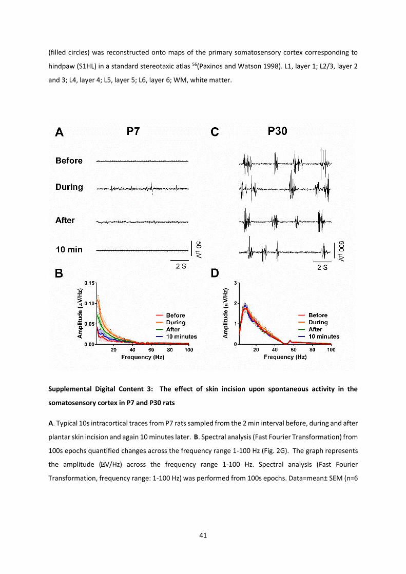
Supplemental Digital Content 3: The effect of skin incision upon spontaneous activity in the
somatosensory cortex in P7 and P30 rats
A. Typical 10s intracortical traces from P7 rats sampled from the 2 min interval before, during and after
plantar skin incision and again 10 minutes later. B. Spectral analysis (Fast Fourier Transformation) from
100s epochs quantified changes across the frequency range 1-100 Hz (Fig. 2G). The graph represents
the amplitude ( V/Hz) across the frequency range 1-100 Hz. Spectral analysis (Fast Fourier
Transformation, frequency range: 1-100 Hz) was performed from 100s epochs. Data=mean± SEM (n=6
42
animals). The significant main effect of hindpaw incision (before and during incision) was observed in
the increased delta band activity (0.02±0.01 µV/Hz to 0.10±0.02 µV/Hz; RM ANOVA F(3, 15) = 13.42,
P=0.0002; post hoc Dunnetts P<0.001), and beta band activity (0.01±0.00 µV/Hz to 0.02±0.01 µV/Hz,
RM ANOVA, F(3, 15) = 6.136, P=0.0062; post hoc Dunnetts P<0.05) This had recovered by 10 mins post-
surgery. C. Typical 10s intracortical traces from P30 sampled from the 2 min interval before, during
and after plantar skin incision and again 10 minutes later. D. Spectral analysis in P30 rats. In contrast
to P7 rats, the frequency distribution of the mean spontaneous activity at P30 (n=6 animals at each
age) in a 100s epoch before, during and after incision under 1.5 % isoflurane anesthesia was unchanged
(δ band from 1.05±0.14 µV/Hz to 1.22±0.12 µV/Hz, RM ANOVA F (3, 15) = 0.8270, P=0.4994, non-
significant; β band from 0.01±0.00 µV/Hz to 0.02±0.01 µV/Hz, RM ANOVA F (3, 15) = 0.6960, P=0.5688,
non-significant.


![A comparative study of evoked otoacoustic emissions in ...SOAEs - We also examined spontaneous otoacoustic emissions, which had been previously reported in this gecko species [5]](https://img.pdfslide.us/doc/110x75/60806f7e0c731c1c4f6b0c15/a-comparative-study-of-evoked-otoacoustic-emissions-in-soaes-we-also-examined.jpg)