Embed Size (px)
Citation preview
Differential Involvement of Cortical andCerebellar Areas Using Dominant andNondominant Hands: An FMRI Study
Adnan A.S. Alahmadi,1,2* Matteo Pardini,1,3 Rebecca S. Samson,1
Egidio D’Angelo,4,5 Karl J. Friston,6 Ahmed T. Toosy,1,7 andClaudia A.M. Gandini Wheeler-Kingshott1,5
1NMR Research Unit, Department of Neuroinflammation, Queen Square MS Centre, Univer-sity College London (UCL), Institute of Neurology, London, United Kingdom
2Department of Diagnostic Radiology, Faculty of Applied Medical Science, King AbdulazizUniversity (KAU), Jeddah, Saudi Arabia
3Department of Neurosciences, Rehabilitation, Ophthalmology, Genetics and Maternal andChild Health, University of Genoa, Genoa, Italy
4Department of Brain and Behavioral Sciences, University of Pavia, Pavia, Italy5Brain Connectivity Center, C. Mondino National Neurological Institute, Pavia, Italy
6Wellcome Centre for Imaging Neuroscience, UCL Institute of Neurology, University CollegeLondon, London, United Kingdom
7NMR Research Unit, Department of Brain Repair and Rehabilitation, Queen Square MSCentre, UCL Institute of Neurology, London, United Kingdom
r r
Abstract: Motor fMRI studies, comparing dominant (DH) and nondominant (NDH) hand activationshave reported mixed findings, especially for the extent of ipsilateral (IL) activations and their relation-ship with task complexity. To date, no study has directly compared DH and NDH activations using anevent-related visually guided dynamic power-grip paradigm with parametric (three) forces (GF) inhealthy right-handed subjects. We implemented a hierarchical statistical approach aimed to: (i) identifythe main effect networks engaged when using either hand; (ii) characterise DH/NDH responses at dif-ferent GFs; (iii) assess contralateral (CL)/IL-specific and hemisphere-specific activations. Beyond con-firming previously reported results, this study demonstrated that increasing GF has an effect on motorresponse that is contextualised also by the use of DH or NDH. Linear analysis revealed increased acti-vations in sensorimotor areas, with additional increased recruitments of subcortical and cerebellarareas when using the NDH. When looking at CL/IL-specific activations, CL sensorimotor areas and ILcerebellum were activated with both hands. When performing the task with the NDH, several areaswere also recruited including the CL cerebellum. Finally, there were hand-side-independent activationsof nonmotor-specific areas in the right and left hemispheres, with the right hemisphere being involved
Additional Supporting Information may be found in the onlineversion of this article.
Contract grant sponsors: MS society of the UK; National Institutefor Health Research, University College London; AA is supportedby KAU, UKSACB, MOE; MP thanks AKWO Association, Lava-gna (Italy); KJF is funded by a Wellcome Trust Principal ResearchFellowship (Ref: 088130/Z/09/Z)
*Correspondence to: Adnan Alahmadi, Department of Neuroin-flammation UCL Institute of Neurology Queen Square, University
College London, London WC1N 3BG, UK. E-mail: [email protected]
Received for publication 21 April 2015; Revised 9 August 2015;Accepted 6 September 2015.
DOI: 10.1002/hbm.22997Published online 29 September 2015 in Wiley Online Library(wileyonlinelibrary.com).
r Human Brain Mapping 36:5079–5100 (2015) r
VC 2015 The Authors. Human Brain Mapping Published by Wiley Periodicals, Inc.This is an open access article under the terms of the Creative Commons Attribution License, which permits use, distribution andreproduction in any medium, provided the original work is properly cited.
more extensively in sensori-motor integration through associative areas while the left hemisphereshowing greater activation at higher GF. This study shows that the functional networks subtendingDH/NDH power-grip visuomotor functions are qualitatively and quantitatively distinct and thisshould be taken into consideration when performing fMRI studies, particularly when planning inter-ventions in patients with specific impairments. Hum Brain Mapp 36:5079–5100, 2015. VC 2015 The Authors.
Human Brain Mapping Published by Wiley Periodicals, Inc.
Key words: FMRI; dominant; force; contra-lateral; squeeze-ball
r r
INTRODUCTION
The neural representation of hand movements is com-plex. Hand movements have been reliably associated withactivations of the contralateral (CL) primary motor cortex,which in turn is modulated during movement by a wide-spread yet only partially understood network of CL andipsilateral (IL) cortical areas—as well as by deep grey mat-ter and cerebellar structures. Indeed, the functional rolesof the aforementioned IL and CL activated regions duringmotor tasks, as well as their modulation by task context(and by inter-individual differences in handedness) repre-sents one of the least understood facets of the functionalarchitecture of the motor system.
Previous studies, mostly using functional magnetic res-onance imaging (fMRI), of motor network activity duringmotor tasks performed using the dominant (DH) andnondominant hand (NDH) have reported mixed resultsincluding: CL activations—especially localized in the pri-mary and secondary motor cortex—produced by move-ment of the dominant (DH) or nondominant (NDH)hands [Verstynen et al., 2005; Ward and Frackowiak2003]; bilateral activations using either hand [Laxmi,1998; Seong-Gi et al., 1993; Volkmann et al., 1998]; orstrong activations ipsilateral to the hand moved [Kawa-shima, 1997; Seong-Gi et al., 1993; Verstynen et al., 2005].This heterogeneity of results is difficult to interpret, espe-cially given methodological differences between differentstudies. For example, some studies have focused on per-forming a motor task using finger tapping [J€ancke et al.,1998; Verstynen et al., 2005], repetitive [Kuhtz-Buschbecket al., 2008] or static [Keisker et al., 2010] power griptasks. However, there are differences in the networksinvolved in controlling these movements [Keisker et al.,2010; Khorrami et al., 2011; King et al., 2014]. Addition-ally, task context has been variably defined by differentauthors, usually by appealing to the notion of “task com-plexity.” Several groups, for example, consider that morecomplex (difficult) tasks are implicitly performed withthe NDH compared to the DH [Ng et al., 2008; Verstynenet al., 2005]. Other groups instead associate “complexity”with the increasing demand needed to perform the motortask [e.g., Keisker et al., 2009; Verstynen et al., 2005]. Inthe study presented here, we will equate complexity withthe computational complexity of completing the task that
can depend upon a number of factors [i.e., the complexityof the task identified with reaching each grip force (GF)level and the complexity of executing the task identifiedwith the use of the DH versus NDH, in the ecologicalcontext of a power grip dynamic task that involves proc-essing visual and sensory inputs from the whole hand,mimicking everyday life].
Moreover, the lateralization of neural activations dur-ing complex motor tasks performed with DH or NDHremains poorly characterized—especially when using anevent-related dynamic power grip task, which, to ourknowledge, has not yet been investigated using fMRI.Characterizing neuronal responses when performingtasks with the DH or NDH is interesting, not only from aneuroscience point of view but also because neurologicaland neurodegenerative diseases may have selectiveeffects on these responses. Previous fMRI studies of thevisuomotor network have assumed that DH and NDHactivations mirror each other, such that flipping the acti-vations found using the NDH correspond to DH activa-tions [e.g., Ward and Frackowiak, 2003; Ward et al., 2004,2006, 2007].
In addition to putative asymmetries, one might ask howthe response depends on the complexity of the task andhow this depends on the complexity of executing it usingthe DH or NDH. In this study, in an attempt to contributeto this discussion, an event-related dynamic power gripfMRI paradigm was designed with three different GF lev-els to experimentally manipulate complexity, while usingeither the DH or NDH to perform the task. The acquireddata was then analysed with a succession of statisticaltests purposely devised to address to the best of ourknowledge the following key questions:
1. What are the functional networks engaged by themain effect of whole-hand visually guided sensori-motor processing (irrespective of the applied forces)using the DH or NDH?
2. What are the effects of different GF levels on thesefunctional responses?
3. Which regions show common activations in the rightand left hemispheres independently of the hand used(hemisphere-specific) and which show common CLor IL (laterality-specific) activations, independently ofthe hand used?
r Alahmadi et al. r
r 5080 r
METHODS
Participants
Fourteen [5F, 9M; mean age 31.0 (64.48) years] right-handed healthy volunteers were recruited for this study.The handedness of each subject was evaluated using theEdinburgh handedness-scaling questionnaire [Oldfield,1971]. The mean laterality index using the handednessquestionnaire was 93 (69). No subject had a history ofneurological or psychiatric disease. The local research andethics committee approved the study and all participantsgave written informed consent.
Paradigm
Subjects performed a power grip (repetitive dynamicsqueezing) task with an MR-compatible squeezeball usingboth hands unimanually. The squeezeball, used also in aprevious study [Alahmadi et al., 2015], is a pneumatic flex-ible pad. Compression of the ball results in an air pressuremeasurement proportional to the force exerted, which wassampled at a rate of 20 Hz.
Two consecutive fMRI scanning sessions were per-formed, comprising 75 three second trials, started using avisual cue and interspersed with 75 null events (baseline).The 75 active trials were divided equally into sets of 25 tri-als at GF levels of 20, 40, and 60% of the subject’s maxi-mum voluntary contraction (MVC). Four inter-trialintervals were used: 2, 4, 7.5, and 9 s. All trials were pre-sented in a randomized and counter-balanced order, opti-mised using the OptSeq software (http://www.surfer.nmr.mgh.harvard.edu/optseq). Before the fMRI session, sub-jects were trained using a similar but different 2-mindesign with GF levels ranging from 10 to 70% of theirMVC. The training session consisted of: observing the taskbeing performed and performing the task while lying inthe scanner bore. Half of the subjects started the fMRI ses-sion using their right (dominant) hand, the other halfstarted with their left hand. The visual cue was a blackstatic horizontal bar (presented for 3 s), indicating the tar-get level to reach. This cue was projected onto an MR-compatible white screen and show together with a col-oured bar indicating the actual force level reached, therebyproviding real-time feedback of the subject’s performance.
MRI Acquisition
A 3T Philips Achieva MR scanner (Philips Healthcare,Best, The Netherlands) with a 32-channel head coil wasused to perform a 3DT1-weighted anatomical scan andtwo T2*-weighted EPI fMRI scans. The 3DT1-weightedsequence acquisition parameters were as follows: 3Dinversion-recovery prepared gradient-echo (fast field echo)sequence with inversion time (TI) 5 824 ms, echo time(TE)/repetition time (TR) 5 3.1/6.9 ms, flip angle 5 88 andvoxel size 5 1 mm isotropic. The fMRI sequence acquisi-
tion parameters were: TR/TE 5 2,500/35 ms, 46 2.7 mmslices positioned to include the cerebellum, with 3 mm2 in-plane resolution, inter-slice gap of 0.3 mm,FOV 5 192 mm2, SENSE factor 5 2, flip angle 5 908 and 200repeated volumes.
Data Preprocessing
Image analysis was performed using SPM8 (www.fil.ion.ucl.ac.uk/spm), implemented in Matlab12b (Math-works, Sheborn, MA), using conventional preprocessingsteps: slice timing, realignment, coregistration, normaliza-tion [using a symmetrical MNI template (Fonov et al.,2011)] and smoothing with an 8-mm isotropic Gaussiankernel.
Statistical Analysis
The statistical analysis was specifically designed toexploit available SPM8 tools such as the general linearmodel statistics and conjunction analysis first for a qualita-tive assessment followed by quantitative comparisons toanswer specific questions. Details of the analysis are givenhere below.
Within-subject
A general linear model (GLM) was constructed by defin-ing the peak of the plateau of trial responses at the 3 GFlevels as separate conditions (regressors). These covariateswere convolved with a canonical hemodynamic responsefunction and used in a conventional whole brain (statisti-cal parametric mapping) analysis [Friston et al., 1995,1998]. The movement parameters from the realignmentstep were also included in the GLM as regressors of nointerest [Friston et al., 1996].
For the DH and NDH, t-statistic contrasts were used atwithin subject level to generate contrast images (or sum-mary statistics) for:
� Main effect of movement versus baseline (rest), inde-pendently of GF;� Main effect of movement versus rest at each GF level;� Positive linear effects of different GF levels on BOLD
signal.
To compare CL and IL activations (see below), the maineffect contrast images for each subject—and for the DHand NDH at each GF level—were flipped about the mid-sagittal line and resliced with respect to the original(unflipped) images to generate flipped contrast images(fDH and fNDH) [Callaert et al., 2011; Van Impe et al.,2009; Ward et al., 2006, 2007]. This effectively introduces afurther design factor; namely, laterality [Salmond et al.,2000]. This analysis shows true bilateral (as opposed tounilateral) activations when voxels overlap using the
r Effects of Forces on Motor Task Using Both Hands r
r 5081 r
conjunction statistical analyses reported below. This analy-sis reaffirmed lateralization (or true unilateral activations)identified using the lateralization method also described indetail below.
Between-subjects
The contrast images from the “within-subject” analysiswere entered into random effects analyses (RFXs), asdetailed below (main effect of movement, main effect ofGF, linear response analysis, and laterality). For all tests,the significance level was set at a corrected P< 0.05 (FWE)at cluster level with a minimum extent of 10 voxels(classes were defined using an uncorrected threshold ofP< 0.0001; T 5 5.11).
Conjunction analysis (CA) was performed to definecommon areas that were specific to the right and lefthemisphere responses as well as areas that were commonto CL and IL activations, independently of using the DHor NDH.
Anatomical designation of the regional effects werebased upon maxima or peaks in the ensuing statisticalparametric maps: cluster peaks were first extracted usingthe Peak_nii software (www.nitrc.org/projects/peak_nii)and then labelled using the cytoarchitectonic probabilitymaps provided by the SPM Anatomy toolbox [Eickhoffet al., 2005, 2007b]. The SPM anatomy toolbox includedindividual areas such as: primary motor cortex [Brodmannareas (BA) 4a and 4p] [Geyer et al., 1996], premotor cortex(BA 6) [Geyer, 2003], primary somatosensory cortex (BA3a, 3b, 1) [Geyer et al., 2000], somatosensory cortex (BA 2)[Grefkes et al., 2001], parietal operculum or S2 (OP 1-4)[Eickhoff et al., 2006a,b,2007a], intraparietal sulcus [Choiet al., 2006; Scheperjans et al., 2008a,b], superior parietalcortex [Scheperjans et al., 2008a,,b], inferior parietal cortex[Caspers et al., 2008], and visual areas (V4) [Rottschyet al., 2007] (V4) [Malikovic et al., 2007]. Some Figureswere generated using FIVE (www.nmr.mgh.harvard.edu/harvardagingbrain/People/AaronSchultz/FIVE) and MRI-cron (http://www.nitrc.org/projects/mricron).
In addition, in order to identify parietal and cerebellarregions, activations obtained at whole brain level asdescribed below in the various sections were mapped tospecific templates:
Parietal and premotor regions
The corresponding anatomical areas were extractedfrom the SPM anatomy toolbox and masked with the sig-nificant activations. Moreover, in order to investigate inmore detail the premotor subdivisions, the dorsal premo-tor cortex (PMd) and the ventral premotor cortex (PMv)were identified using the human motor area template(HMAT) (Mayka et al., 2006). Therefore, we used theresultant maps for the premotor and parietal cortices torepresent significant activations within key regions ofthese two cortical regions.
Cerebellar regions
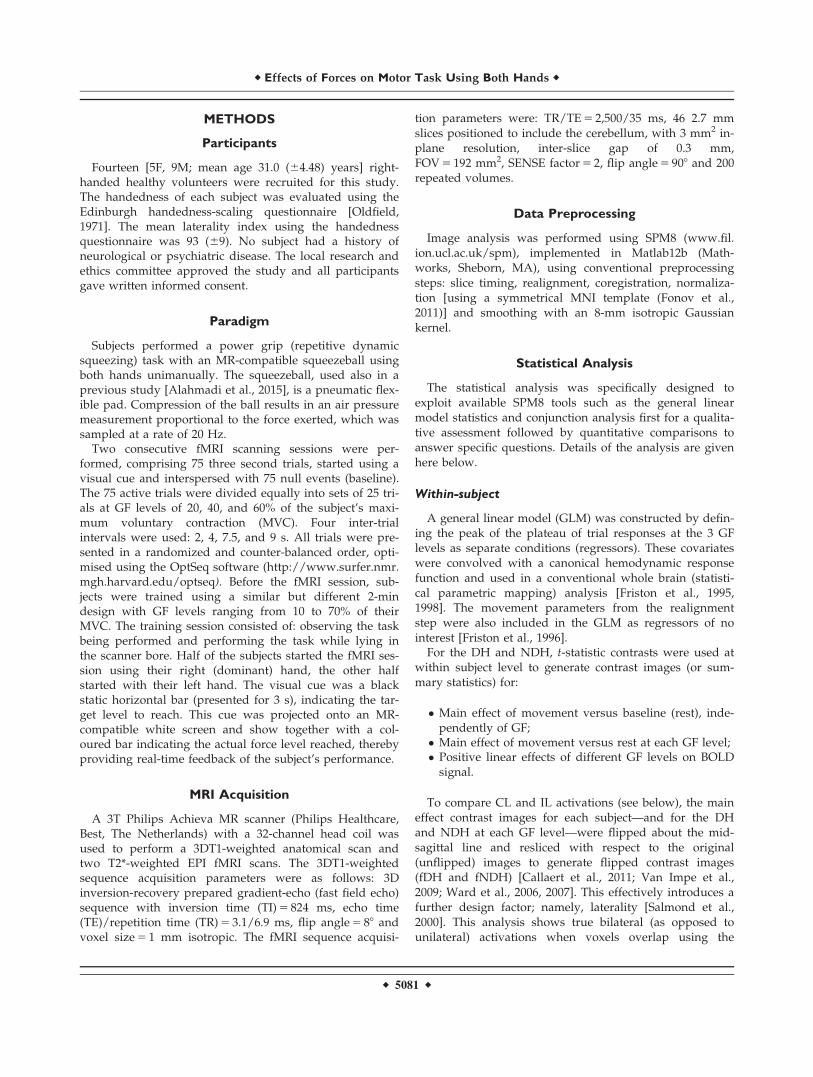
The spatially unbiased infratentorial flattened template(SUIT) for the cerebellum [Diedrichsen, 2006] was usedtogether with the Caret Human Connectome workbench(http://brainmap.wustl.edu/caret) [Van Essen et al., 2001]to map the activated volumes to the SUIT flattened map(Diedrichsen, J. & Zotow, E. (submitted); http://www.ic-n.ucl.ac.uk/motorcontrol/imaging/suit_flatmap.htm). Inaddition, the overlap between the cerebellar activationsand the deep cerebellar nuclei, and in particular the den-tate cerebellar nuclei (DCN), were assessed visually byselecting coronal slices from the mean EPI volume wherethe DCN can be identified as hypo-intense regions, due tothe high iron content that shortens T2* (Fig. 1B.a). Thiswas performed for the main effect of movement for eachhand at each GF level (see below RFX 1 and 2).
DETAILS OF THE PERFORMED ANALYSIS ARE
GIVEN BELOW
RFX1: Main Effect of Movement
Main group effects were identified using a one-sample ttest on contrast images obtained from the “within-subject”analysis testing for a main effect of movement. Compari-sons of all GFs against baseline were performed for DH orNDH separately.
RFX2: Main Effect of GF
Main group effects were identified using a one-sample t
test based on contrast images from the “within-subject”analysis at each GF level for DH, NDH, and fNDH. Thiscorresponds to a test for simple main effects of movementunder each GF or complexity level and contextualises theanalysis of flipped contrast images implicit in the conjunc-tion analyses below.
RFX3: Linear Response Analyses
Linear analysis of grip-related responses was performedto identify increased activations at the higher GF, using aone-sample t test on the within subject linear effect con-trast images for the DH or NDH.
RFX4: Specificity, Lateralisation, and Strength of
Activations
Three paired t tests were performed to assess (a) speci-ficity, (b) lateralisation, and (c) strength of activationswhen using the DH or the NDH:
a. A paired t test between the DH and NDH was per-formed at each GF to test the specificity of regionalactivations in relation to (right) hand dominance;
r Alahmadi et al. r
r 5082 r
b. Comparing contrast images with their correspondingflipped data (i.e., running a t test of DH with fDHand NDH with fNDH) enabled us to assess the later-alisation of hand-dependent responses, i.e. highlight-ing areas that showed an interaction between handdominance and hemisphere.
c. Comparing contrast images from DH with fNDH, itwas possible to compare the strength of activationwhen using the DH or NDH in corresponding CLand IL regions. This also constitutes an interactionwith hemisphere; however it can also be consideredas a (simple) main effect in terms of the ipsilateral orcontralateral relation to the hand used.
CA: Common Areas of Activations
Conjunction analyses (Supporting Information Fig. 1)were performed to identify common regions of activationat each GF level. Each of the group contrast images forDH, NDH, and fNDH obtained in RFX2 (at each GF level)were thresholded and used as conjoint masks to performconjunction analyses, testing for common regions in termsof:
a. Common right and left hemisphere activations, inde-pendently of handedness, identified by a conjunctionof DH and NDH effects (Supporting Information Fig.1a).
b. CL and IL hemisphere activations common to bothDH and NDH, assessed by a conjunction of fNDHand the DH effects (Supporting Information Fig. 1b).
Effect Size
In addition, the voxel by voxel (unstandardized) effectsize of the group level statistical analyses was calculatedand masked with the significant thresholded activations(for the purpose of illustrations—using a voxel levelthreshold of P< 0.001—corrected at the cluster level) togenerate significant effect size maps.
RESULTS
Task Performance
All subjects performed the task adequately using eitherhand (Table I, Supporting Information). Also there was noeffect of the lateralization scores as measured by the hand-edness tests on the group findings.
The main results of this study are reported in the firstinstance as an overview, based on main regional designa-tions, i.e. primary motor, premotor, parietal, visual, andcerebellar areas. Following this, the results of each analy-ses are described in detail.
Primary Motor Area (M1)
The primary motor area (M1) CL to each hand was acti-vated at each GF level regardless of the used hand. Thelinearity analyses revealed, using either hand, that the CLM1 was increasingly involved with increasing GF levels.Using the conjunction analyses, DH CL M1 was mirroringthe NDH CL M1 and the right M1 was also commonlyshared during both the NDH and DH task.
Premotor Areas and SMA
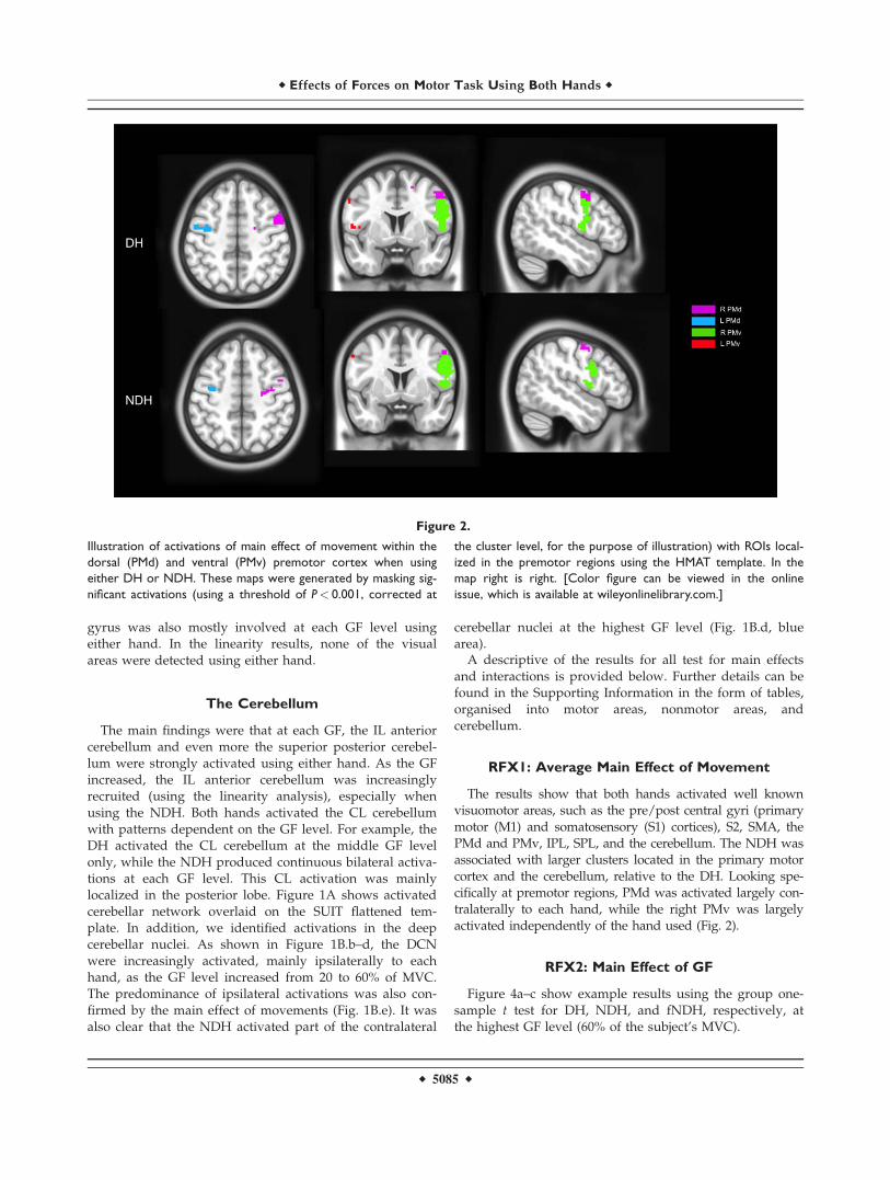
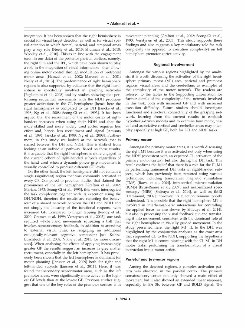
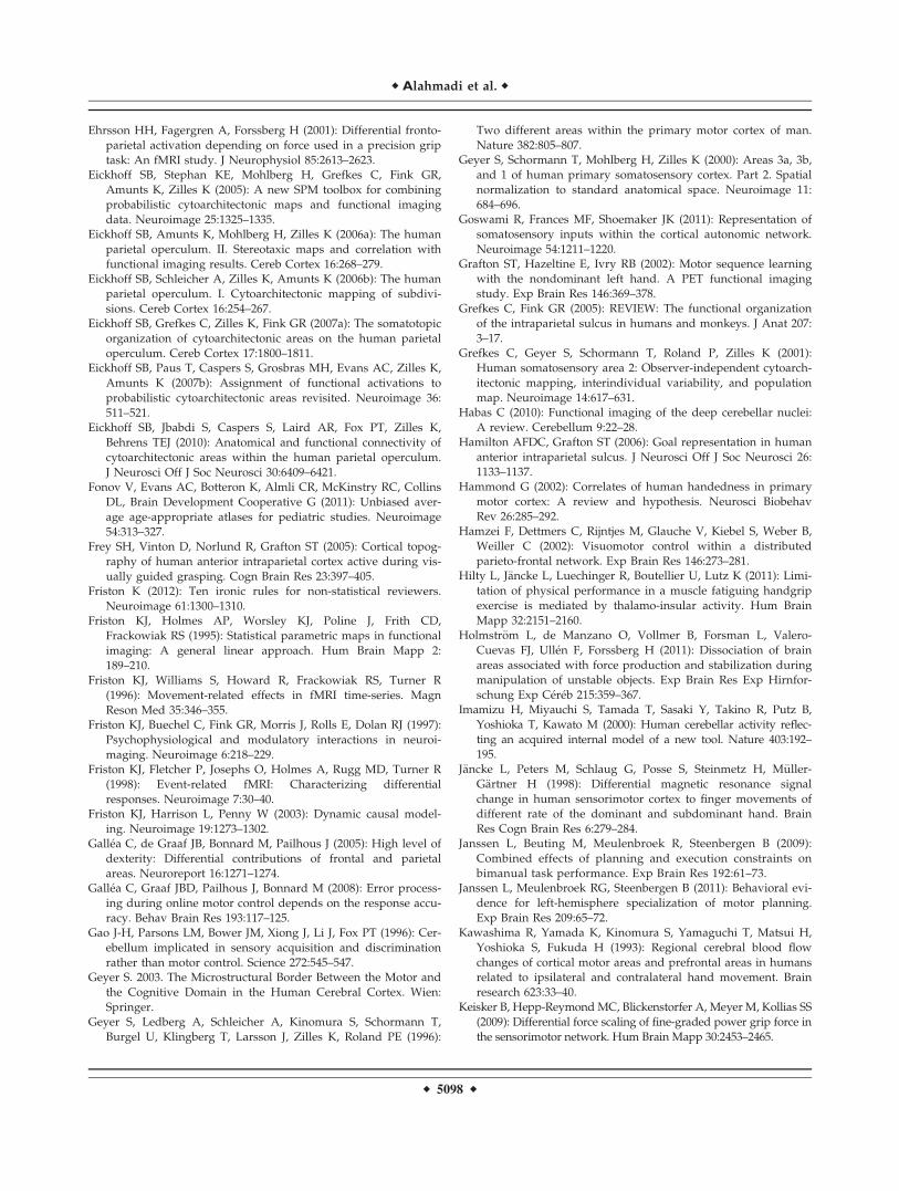
The premotor cortex was more lateralized to the righthemisphere at the highest GF levels using either hand.Using the NDH, as the GF increased, the right PMv andPMd activations increased too, as shown by the linearityanalysis. Using the conjunction analyses, the premotor cor-tex was commonly activated contralaterally to each hand.The conjunction analyses showed that overall the rightPMv was commonly activated between the two hands.Figure 2 shows examples of activations in PMv and PMdusing the DH and NDH, respectively. Moreover, the CLsupplementary motor area (SMA) was activated using theDH at each GF while the IL SMA was activated (signifi-cantly) at the highest GF level. Using the NDH, SMA wasactivated bilaterally especially at GF level of 40%, wherethere was a high anatomical probability detection of theSMA. There was no specificity or lateralization in SMAusing either hand. SMA was commonly shared betweenactivations produced by the two hands, especially at thehighest GF level.
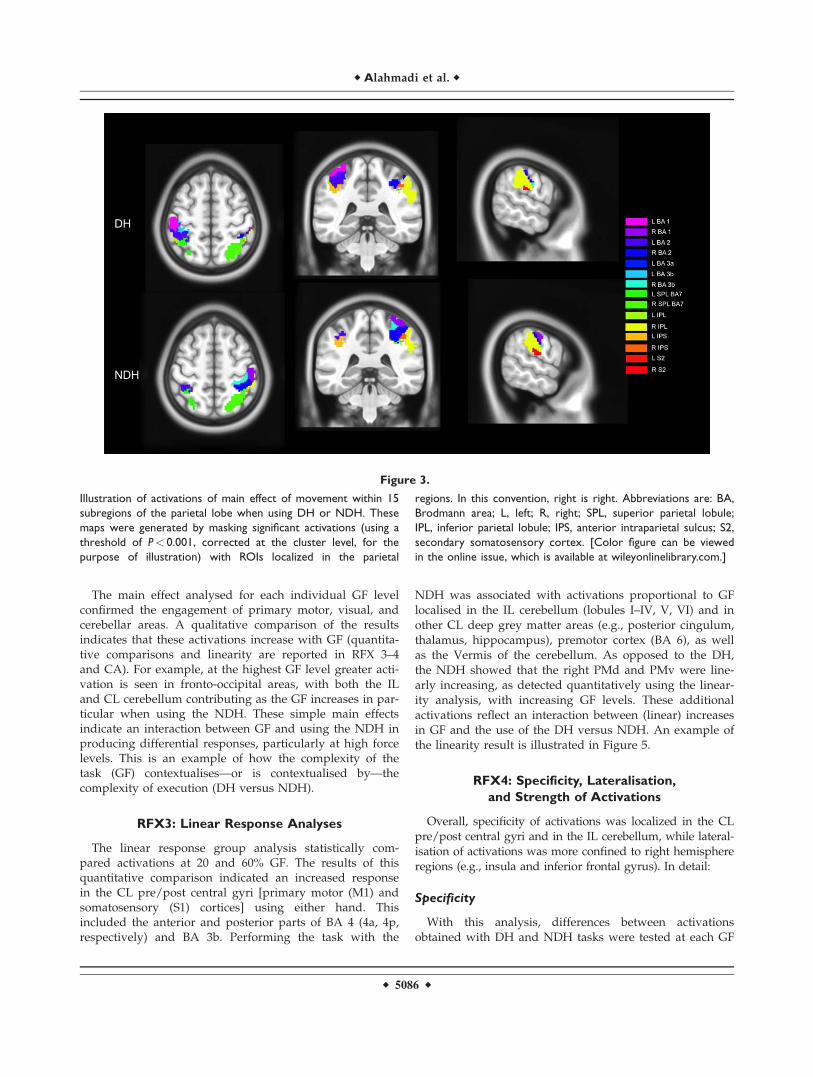
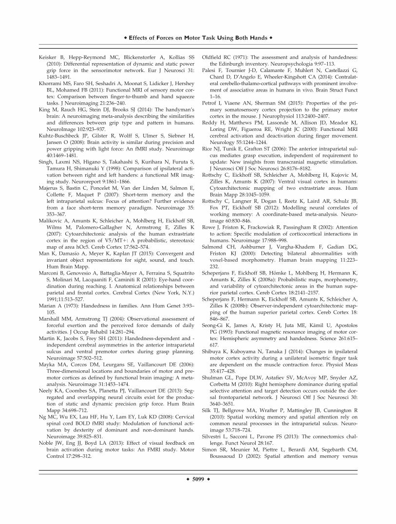
Parietal Cortex
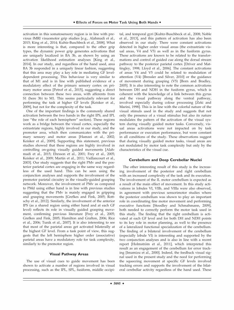
The primary somatosensory areas (BA 1, 2, 3) were later-alized to the CL hemisphere to each hand and at each GFlevels. The secondary somatosensory cortex (S2) wasdefined using the cytoarchitectonic software as areas corre-sponding to the parietal Operculum (OP1-OP4) (Eickhoffet al., 2006a,b,2007a,2010). Our result indicates that duringthe main effect analyses, the DH activated S2 bilaterally,while it was lateralized to the right hemisphere using theNDH. Qualitative analysis of this region showed thatstrong bilateral activation using the DH was observed atthe highest GF level. In addition, the linearity analysesshowed that both hands produced increased activationsmainly in BA 3b but also part of BA 2, CL to each hand.Moreover, there were no common regions (hemisphericconjunction analysis) between both hands in the primarysomatosensory cortex (i.e., those regions were mainly con-tralaterally activated). On the other hand, the right S2 wascommonly shared at each GF level between the DH andNDH. Looking at higher parietal cortical areas [intraparie-tal sulcus (IPS), the inferior (IPL) and superior parietallobule (SPL)] in the right hemisphere, the IPL was com-monly activated using both hands. As the GF levelincreased, the SPL (BA 7A) was commonly detected in the
r Effects of Forces on Motor Task Using Both Hands r
r 5083 r
right hemisphere, too. The DH activated bilaterally theIPS, especially at the lowest GF level. Also both handscommonly and constantly activated the IPS in the righthemisphere at each GF level. At the highest GF, the poste-rior parietal cortex was bilaterally engaged using eitherhand. Figure 3 shows examples of activations in 15 parie-tal areas using the DH and NDH, respectively.
Visual Pathway Areas
There were significant activations in the primary visualand extrastriate visual areas. The extrastriate visual areas(namely V4 and V5) were activated regardless of the dif-ferent GF levels or the hand used (Supporting Informa-tion—Table IX; common areas). In addition, the fusiform
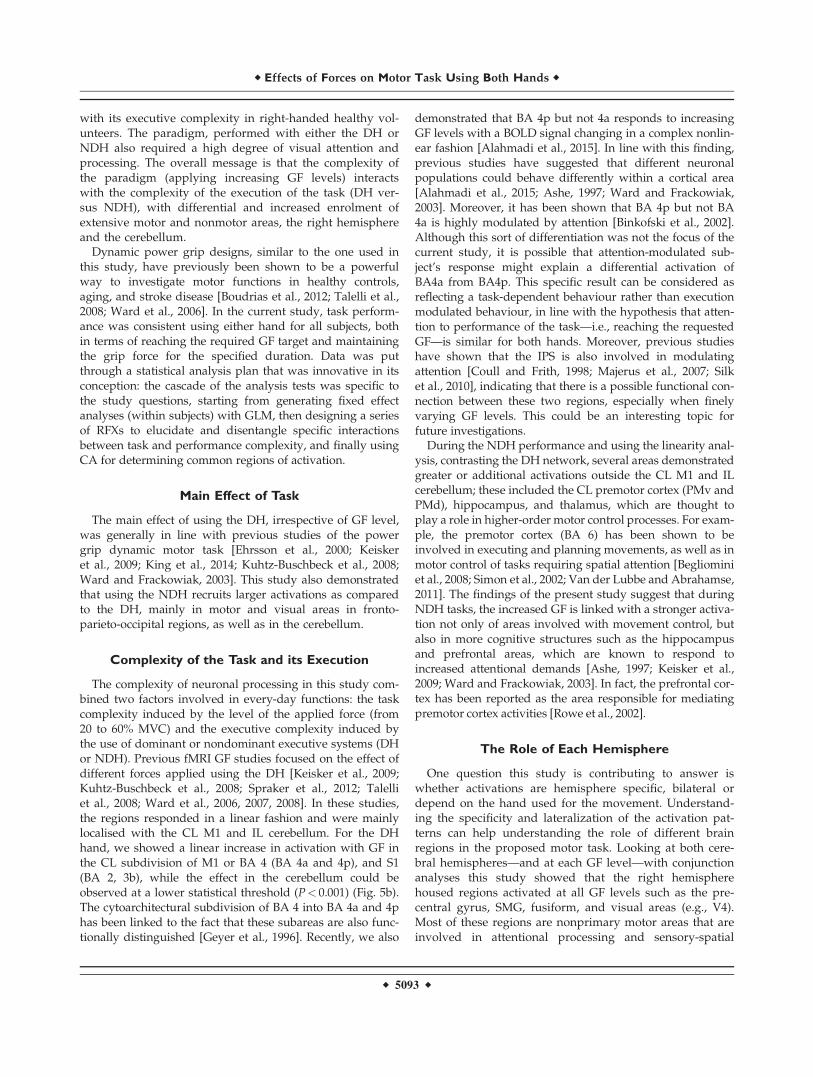
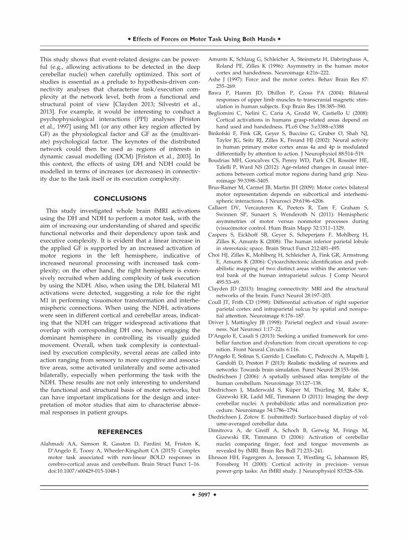
Figure 1.
(A) Illustration of activation maps for DH and NDH on the
SUIT flattened cerebellum. Clearly, the anterior and superior
posterior ipsilateral cerebellum are involved at each GF. The
contralateral cerebellum, on the other hand, is highly involved
when using the NDH. (B) Illustration of the involvement of the
deep cerebellar nuclei with the task. (a) The dentate cerebellar
nuclei (DCN) is the largest nucleus and has a high iron content,
therefore appearing hypointense on the EPI template, at either
side of the cerebellar midline (green arrow). (b–d) Activations
produced by either the DH (top row, red) or NDH (bottom
row, blue), at GF levels of b. 20%, c. 40%, d. 60% of maximum
voluntary contraction (MVC), (e) Main effect of grip, showing
that as the GF increases the DCNs are more engaged, hence
indicating an interaction with complexity of both the task and its
execution. [Color figure can be viewed in the online issue,
which is available at wileyonlinelibrary.com.]
r Alahmadi et al. r
r 5084 r
gyrus was also mostly involved at each GF level usingeither hand. In the linearity results, none of the visualareas were detected using either hand.
The Cerebellum
The main findings were that at each GF, the IL anteriorcerebellum and even more the superior posterior cerebel-lum were strongly activated using either hand. As the GFincreased, the IL anterior cerebellum was increasinglyrecruited (using the linearity analysis), especially whenusing the NDH. Both hands activated the CL cerebellumwith patterns dependent on the GF level. For example, theDH activated the CL cerebellum at the middle GF levelonly, while the NDH produced continuous bilateral activa-tions at each GF level. This CL activation was mainlylocalized in the posterior lobe. Figure 1A shows activatedcerebellar network overlaid on the SUIT flattened tem-plate. In addition, we identified activations in the deepcerebellar nuclei. As shown in Figure 1B.b–d, the DCNwere increasingly activated, mainly ipsilaterally to eachhand, as the GF level increased from 20 to 60% of MVC.The predominance of ipsilateral activations was also con-firmed by the main effect of movements (Fig. 1B.e). It wasalso clear that the NDH activated part of the contralateral
cerebellar nuclei at the highest GF level (Fig. 1B.d, bluearea).
A descriptive of the results for all test for main effectsand interactions is provided below. Further details can befound in the Supporting Information in the form of tables,organised into motor areas, nonmotor areas, andcerebellum.
RFX1: Average Main Effect of Movement
The results show that both hands activated well knownvisuomotor areas, such as the pre/post central gyri (primarymotor (M1) and somatosensory (S1) cortices), S2, SMA, thePMd and PMv, IPL, SPL, and the cerebellum. The NDH wasassociated with larger clusters located in the primary motorcortex and the cerebellum, relative to the DH. Looking spe-cifically at premotor regions, PMd was activated largely con-tralaterally to each hand, while the right PMv was largelyactivated independently of the hand used (Fig. 2).
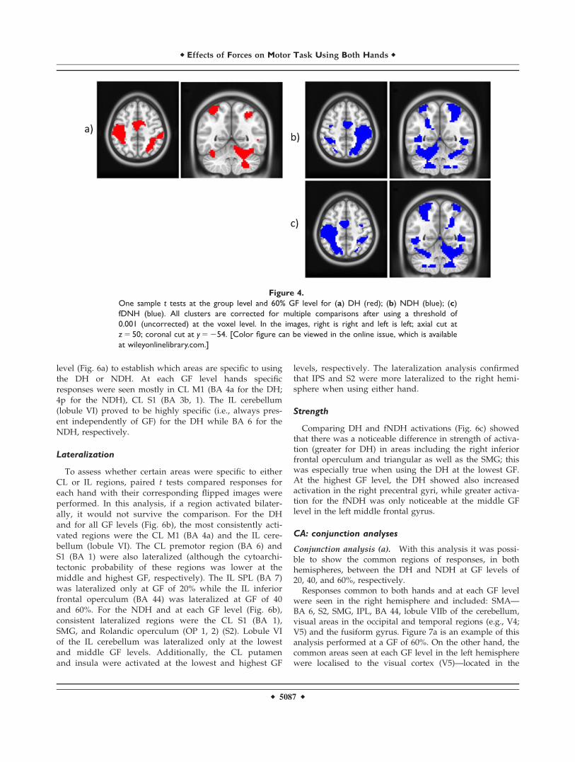
RFX2: Main Effect of GF
Figure 4a–c show example results using the group one-sample t test for DH, NDH, and fNDH, respectively, atthe highest GF level (60% of the subject’s MVC).
Figure 2.
Illustration of activations of main effect of movement within the
dorsal (PMd) and ventral (PMv) premotor cortex when using
either DH or NDH. These maps were generated by masking sig-
nificant activations (using a threshold of P< 0.001, corrected at
the cluster level, for the purpose of illustration) with ROIs local-
ized in the premotor regions using the HMAT template. In the
map right is right. [Color figure can be viewed in the online
issue, which is available at wileyonlinelibrary.com.]
r Effects of Forces on Motor Task Using Both Hands r
r 5085 r
The main effect analysed for each individual GF levelconfirmed the engagement of primary motor, visual, andcerebellar areas. A qualitative comparison of the resultsindicates that these activations increase with GF (quantita-tive comparisons and linearity are reported in RFX 3–4and CA). For example, at the highest GF level greater acti-vation is seen in fronto-occipital areas, with both the ILand CL cerebellum contributing as the GF increases in par-ticular when using the NDH. These simple main effectsindicate an interaction between GF and using the NDH inproducing differential responses, particularly at high forcelevels. This is an example of how the complexity of thetask (GF) contextualises—or is contextualised by—thecomplexity of execution (DH versus NDH).
RFX3: Linear Response Analyses
The linear response group analysis statistically com-pared activations at 20 and 60% GF. The results of thisquantitative comparison indicated an increased responsein the CL pre/post central gyri [primary motor (M1) andsomatosensory (S1) cortices] using either hand. Thisincluded the anterior and posterior parts of BA 4 (4a, 4p,respectively) and BA 3b. Performing the task with the
NDH was associated with activations proportional to GFlocalised in the IL cerebellum (lobules I–IV, V, VI) and inother CL deep grey matter areas (e.g., posterior cingulum,thalamus, hippocampus), premotor cortex (BA 6), as wellas the Vermis of the cerebellum. As opposed to the DH,the NDH showed that the right PMd and PMv were line-arly increasing, as detected quantitatively using the linear-ity analysis, with increasing GF levels. These additionalactivations reflect an interaction between (linear) increasesin GF and the use of the DH versus NDH. An example ofthe linearity result is illustrated in Figure 5.
RFX4: Specificity, Lateralisation,
and Strength of Activations
Overall, specificity of activations was localized in the CLpre/post central gyri and in the IL cerebellum, while lateral-isation of activations was more confined to right hemisphereregions (e.g., insula and inferior frontal gyrus). In detail:
Specificity
With this analysis, differences between activationsobtained with DH and NDH tasks were tested at each GF
Figure 3.
Illustration of activations of main effect of movement within 15
subregions of the parietal lobe when using DH or NDH. These
maps were generated by masking significant activations (using a
threshold of P< 0.001, corrected at the cluster level, for the
purpose of illustration) with ROIs localized in the parietal
regions. In this convention, right is right. Abbreviations are: BA,
Brodmann area; L, left; R, right; SPL, superior parietal lobule;
IPL, inferior parietal lobule; IPS, anterior intraparietal sulcus; S2,
secondary somatosensory cortex. [Color figure can be viewed
in the online issue, which is available at wileyonlinelibrary.com.]
r Alahmadi et al. r
r 5086 r
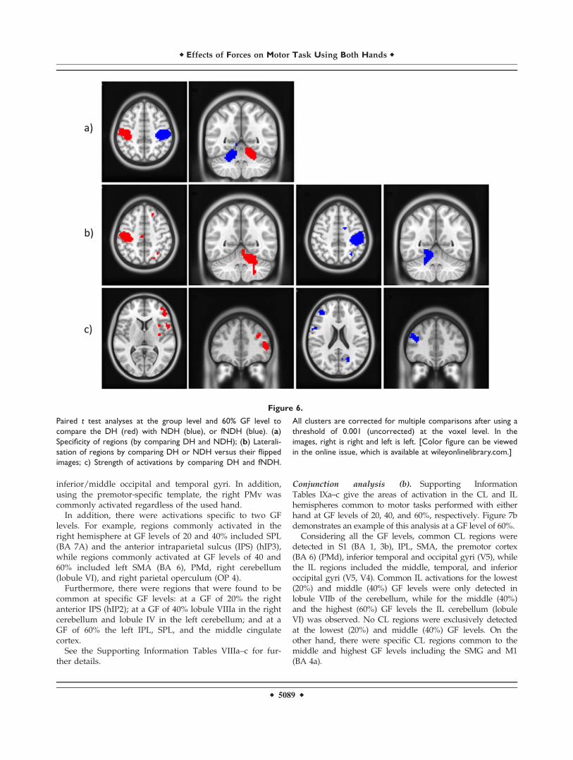
level (Fig. 6a) to establish which areas are specific to usingthe DH or NDH. At each GF level hands specificresponses were seen mostly in CL M1 (BA 4a for the DH;4p for the NDH), CL S1 (BA 3b, 1). The IL cerebellum(lobule VI) proved to be highly specific (i.e., always pres-ent independently of GF) for the DH while BA 6 for theNDH, respectively.
Lateralization
To assess whether certain areas were specific to eitherCL or IL regions, paired t tests compared responses foreach hand with their corresponding flipped images wereperformed. In this analysis, if a region activated bilater-ally, it would not survive the comparison. For the DHand for all GF levels (Fig. 6b), the most consistently acti-vated regions were the CL M1 (BA 4a) and the IL cere-bellum (lobule VI). The CL premotor region (BA 6) andS1 (BA 1) were also lateralized (although the cytoarchi-tectonic probability of these regions was lower at themiddle and highest GF, respectively). The IL SPL (BA 7)was lateralized only at GF of 20% while the IL inferiorfrontal operculum (BA 44) was lateralized at GF of 40and 60%. For the NDH and at each GF level (Fig. 6b),consistent lateralized regions were the CL S1 (BA 1),SMG, and Rolandic operculum (OP 1, 2) (S2). Lobule VIof the IL cerebellum was lateralized only at the lowestand middle GF levels. Additionally, the CL putamenand insula were activated at the lowest and highest GF
levels, respectively. The lateralization analysis confirmedthat IPS and S2 were more lateralized to the right hemi-sphere when using either hand.
Strength
Comparing DH and fNDH activations (Fig. 6c) showedthat there was a noticeable difference in strength of activa-tion (greater for DH) in areas including the right inferiorfrontal operculum and triangular as well as the SMG; thiswas especially true when using the DH at the lowest GF.At the highest GF level, the DH showed also increasedactivation in the right precentral gyri, while greater activa-tion for the fNDH was only noticeable at the middle GFlevel in the left middle frontal gyrus.
CA: conjunction analyses
Conjunction analysis (a). With this analysis it was possi-ble to show the common regions of responses, in bothhemispheres, between the DH and NDH at GF levels of20, 40, and 60%, respectively.
Responses common to both hands and at each GF levelwere seen in the right hemisphere and included: SMA—BA 6, S2, SMG, IPL, BA 44, lobule VIIb of the cerebellum,visual areas in the occipital and temporal regions (e.g., V4;V5) and the fusiform gyrus. Figure 7a is an example of thisanalysis performed at a GF of 60%. On the other hand, thecommon areas seen at each GF level in the left hemispherewere localised to the visual cortex (V5)—located in the
Figure 4.
One sample t tests at the group level and 60% GF level for (a) DH (red); (b) NDH (blue); (c)
fDNH (blue). All clusters are corrected for multiple comparisons after using a threshold of
0.001 (uncorrected) at the voxel level. In the images, right is right and left is left; axial cut at
z 5 50; coronal cut at y 5 254. [Color figure can be viewed in the online issue, which is available
at wileyonlinelibrary.com.]
r Effects of Forces on Motor Task Using Both Hands r
r 5087 r
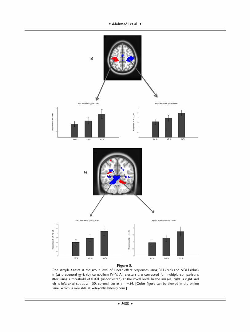
Figure 5.
One sample t tests at the group level of Linear effect responses using DH (red) and NDH (blue)
in (a) precentral gyri; (b) cerebellum IV–V. All clusters are corrected for multiple comparisons
after using a threshold of 0.001 (uncorrected) at the voxel level. In the images, right is right and
left is left; axial cut at z 5 50; coronal cut at y 5 254. [Color figure can be viewed in the online
issue, which is available at wileyonlinelibrary.com.]
r Alahmadi et al. r
r 5088 r
inferior/middle occipital and temporal gyri. In addition,using the premotor-specific template, the right PMv wascommonly activated regardless of the used hand.
In addition, there were activations specific to two GFlevels. For example, regions commonly activated in theright hemisphere at GF levels of 20 and 40% included SPL(BA 7A) and the anterior intraparietal sulcus (IPS) (hIP3),while regions commonly activated at GF levels of 40 and60% included left SMA (BA 6), PMd, right cerebellum(lobule VI), and right parietal operculum (OP 4).
Furthermore, there were regions that were found to becommon at specific GF levels: at a GF of 20% the rightanterior IPS (hIP2); at a GF of 40% lobule VIIIa in the rightcerebellum and lobule IV in the left cerebellum; and at aGF of 60% the left IPL, SPL, and the middle cingulatecortex.
See the Supporting Information Tables VIIIa–c for fur-ther details.
Conjunction analysis (b). Supporting InformationTables IXa–c give the areas of activation in the CL and ILhemispheres common to motor tasks performed with eitherhand at GF levels of 20, 40, and 60%, respectively. Figure 7bdemonstrates an example of this analysis at a GF level of 60%.
Considering all the GF levels, common CL regions weredetected in S1 (BA 1, 3b), IPL, SMA, the premotor cortex(BA 6) (PMd), inferior temporal and occipital gyri (V5), whilethe IL regions included the middle, temporal, and inferioroccipital gyri (V5, V4). Common IL activations for the lowest(20%) and middle (40%) GF levels were only detected inlobule VIIb of the cerebellum, while for the middle (40%)and the highest (60%) GF levels the IL cerebellum (lobuleVI) was observed. No CL regions were exclusively detectedat the lowest (20%) and middle (40%) GF levels. On theother hand, there were specific CL regions common to themiddle and highest GF levels including the SMG and M1(BA 4a).
Figure 6.
Paired t test analyses at the group level and 60% GF level to
compare the DH (red) with NDH (blue), or fNDH (blue). (a)
Specificity of regions (by comparing DH and NDH); (b) Laterali-
sation of regions by comparing DH or NDH versus their flipped
images; c) Strength of activations by comparing DH and fNDH.
All clusters are corrected for multiple comparisons after using a
threshold of 0.001 (uncorrected) at the voxel level. In the
images, right is right and left is left. [Color figure can be viewed
in the online issue, which is available at wileyonlinelibrary.com.]
r Effects of Forces on Motor Task Using Both Hands r
r 5089 r
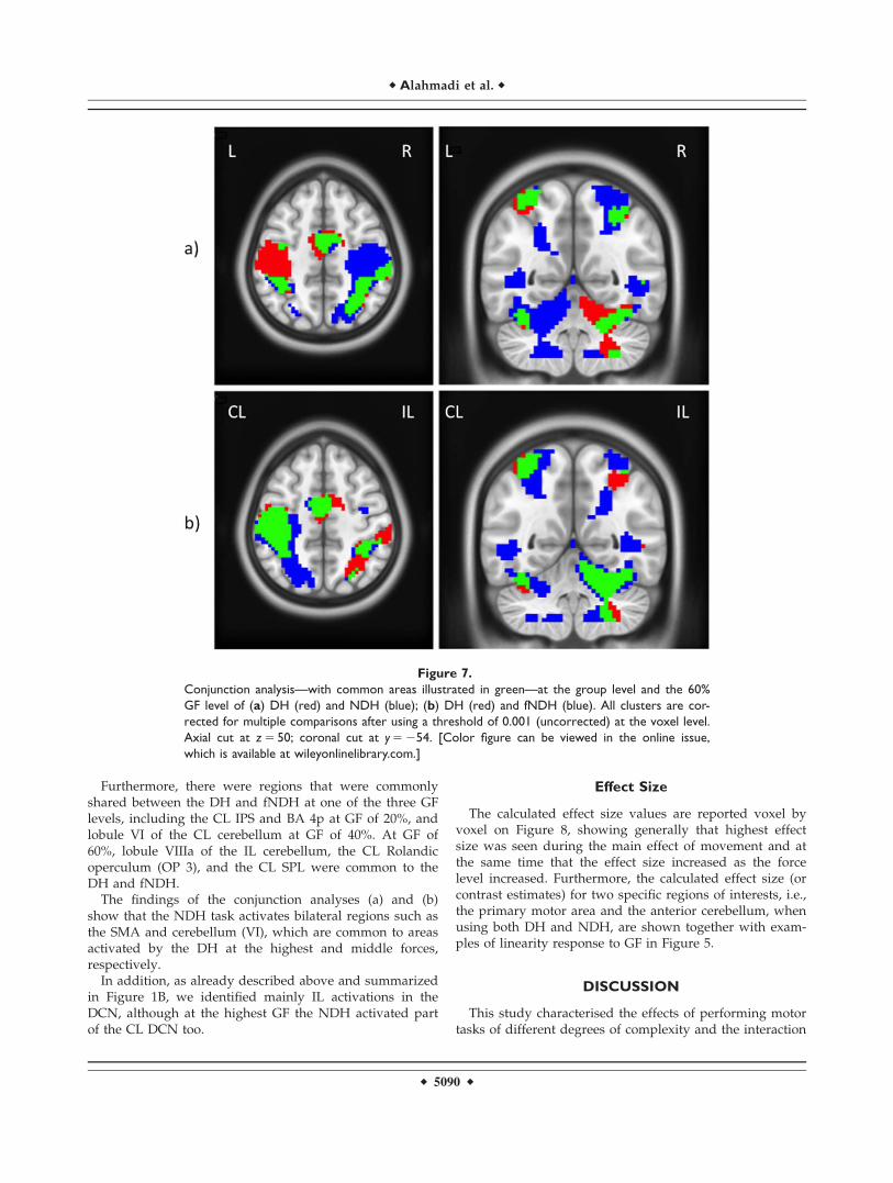
Furthermore, there were regions that were commonlyshared between the DH and fNDH at one of the three GFlevels, including the CL IPS and BA 4p at GF of 20%, andlobule VI of the CL cerebellum at GF of 40%. At GF of60%, lobule VIIIa of the IL cerebellum, the CL Rolandicoperculum (OP 3), and the CL SPL were common to theDH and fNDH.
The findings of the conjunction analyses (a) and (b)show that the NDH task activates bilateral regions such asthe SMA and cerebellum (VI), which are common to areasactivated by the DH at the highest and middle forces,respectively.
In addition, as already described above and summarizedin Figure 1B, we identified mainly IL activations in theDCN, although at the highest GF the NDH activated partof the CL DCN too.
Effect Size
The calculated effect size values are reported voxel byvoxel on Figure 8, showing generally that highest effectsize was seen during the main effect of movement and atthe same time that the effect size increased as the forcelevel increased. Furthermore, the calculated effect size (orcontrast estimates) for two specific regions of interests, i.e.,the primary motor area and the anterior cerebellum, whenusing both DH and NDH, are shown together with exam-ples of linearity response to GF in Figure 5.
DISCUSSION
This study characterised the effects of performing motortasks of different degrees of complexity and the interaction
Figure 7.
Conjunction analysis—with common areas illustrated in green—at the group level and the 60%
GF level of (a) DH (red) and NDH (blue); (b) DH (red) and fNDH (blue). All clusters are cor-
rected for multiple comparisons after using a threshold of 0.001 (uncorrected) at the voxel level.
Axial cut at z 5 50; coronal cut at y 5 254. [Color figure can be viewed in the online issue,
which is available at wileyonlinelibrary.com.]
r Alahmadi et al. r
r 5090 r
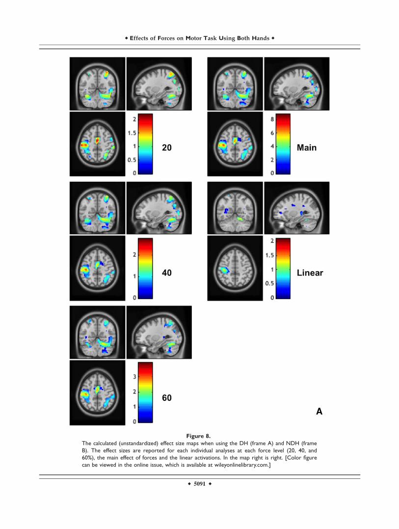
Figure 8.
The calculated (unstandardized) effect size maps when using the DH (frame A) and NDH (frame
B). The effect sizes are reported for each individual analyses at each force level (20, 40, and
60%), the main effect of forces and the linear activations. In the map right is right. [Color figure
can be viewed in the online issue, which is available at wileyonlinelibrary.com.]
r Effects of Forces on Motor Task Using Both Hands r
r 5091 r
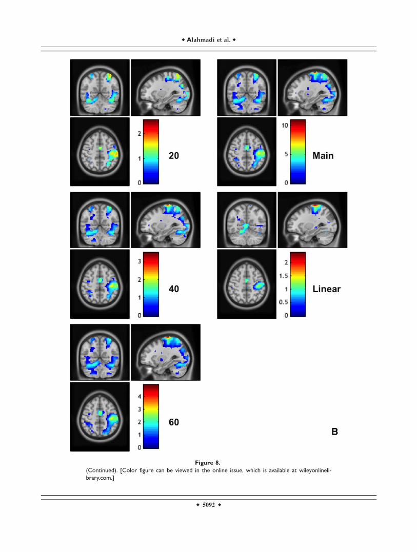
Figure 8.
(Continued). [Color figure can be viewed in the online issue, which is available at wileyonlineli-
brary.com.]
r Alahmadi et al. r
r 5092 r
with its executive complexity in right-handed healthy vol-unteers. The paradigm, performed with either the DH orNDH also required a high degree of visual attention andprocessing. The overall message is that the complexity ofthe paradigm (applying increasing GF levels) interactswith the complexity of the execution of the task (DH ver-sus NDH), with differential and increased enrolment ofextensive motor and nonmotor areas, the right hemisphereand the cerebellum.
Dynamic power grip designs, similar to the one used inthis study, have previously been shown to be a powerfulway to investigate motor functions in healthy controls,aging, and stroke disease [Boudrias et al., 2012; Talelli et al.,2008; Ward et al., 2006]. In the current study, task perform-ance was consistent using either hand for all subjects, bothin terms of reaching the required GF target and maintainingthe grip force for the specified duration. Data was putthrough a statistical analysis plan that was innovative in itsconception: the cascade of the analysis tests was specific tothe study questions, starting from generating fixed effectanalyses (within subjects) with GLM, then designing a seriesof RFXs to elucidate and disentangle specific interactionsbetween task and performance complexity, and finally usingCA for determining common regions of activation.
Main Effect of Task
The main effect of using the DH, irrespective of GF level,was generally in line with previous studies of the powergrip dynamic motor task [Ehrsson et al., 2000; Keiskeret al., 2009; King et al., 2014; Kuhtz-Buschbeck et al., 2008;Ward and Frackowiak, 2003]. This study also demonstratedthat using the NDH recruits larger activations as comparedto the DH, mainly in motor and visual areas in fronto-parieto-occipital regions, as well as in the cerebellum.
Complexity of the Task and its Execution
The complexity of neuronal processing in this study com-bined two factors involved in every-day functions: the taskcomplexity induced by the level of the applied force (from20 to 60% MVC) and the executive complexity induced bythe use of dominant or nondominant executive systems (DHor NDH). Previous fMRI GF studies focused on the effect ofdifferent forces applied using the DH [Keisker et al., 2009;Kuhtz-Buschbeck et al., 2008; Spraker et al., 2012; Talelliet al., 2008; Ward et al., 2006, 2007, 2008]. In these studies,the regions responded in a linear fashion and were mainlylocalised with the CL M1 and IL cerebellum. For the DHhand, we showed a linear increase in activation with GF inthe CL subdivision of M1 or BA 4 (BA 4a and 4p), and S1(BA 2, 3b), while the effect in the cerebellum could beobserved at a lower statistical threshold (P< 0.001) (Fig. 5b).The cytoarchitectural subdivision of BA 4 into BA 4a and 4phas been linked to the fact that these subareas are also func-tionally distinguished [Geyer et al., 1996]. Recently, we also
demonstrated that BA 4p but not 4a responds to increasingGF levels with a BOLD signal changing in a complex nonlin-ear fashion [Alahmadi et al., 2015]. In line with this finding,previous studies have suggested that different neuronalpopulations could behave differently within a cortical area[Alahmadi et al., 2015; Ashe, 1997; Ward and Frackowiak,2003]. Moreover, it has been shown that BA 4p but not BA4a is highly modulated by attention [Binkofski et al., 2002].Although this sort of differentiation was not the focus of thecurrent study, it is possible that attention-modulated sub-ject’s response might explain a differential activation ofBA4a from BA4p. This specific result can be considered asreflecting a task-dependent behaviour rather than executionmodulated behaviour, in line with the hypothesis that atten-tion to performance of the task—i.e., reaching the requestedGF—is similar for both hands. Moreover, previous studieshave shown that the IPS is also involved in modulatingattention [Coull and Frith, 1998; Majerus et al., 2007; Silket al., 2010], indicating that there is a possible functional con-nection between these two regions, especially when finelyvarying GF levels. This could be an interesting topic forfuture investigations.
During the NDH performance and using the linearity anal-ysis, contrasting the DH network, several areas demonstratedgreater or additional activations outside the CL M1 and ILcerebellum; these included the CL premotor cortex (PMv andPMd), hippocampus, and thalamus, which are thought toplay a role in higher-order motor control processes. For exam-ple, the premotor cortex (BA 6) has been shown to beinvolved in executing and planning movements, as well as inmotor control of tasks requiring spatial attention [Begliominiet al., 2008; Simon et al., 2002; Van der Lubbe and Abrahamse,2011]. The findings of the present study suggest that duringNDH tasks, the increased GF is linked with a stronger activa-tion not only of areas involved with movement control, butalso in more cognitive structures such as the hippocampusand prefrontal areas, which are known to respond toincreased attentional demands [Ashe, 1997; Keisker et al.,2009; Ward and Frackowiak, 2003]. In fact, the prefrontal cor-tex has been reported as the area responsible for mediatingpremotor cortex activities [Rowe et al., 2002].
The Role of Each Hemisphere
One question this study is contributing to answer iswhether activations are hemisphere specific, bilateral ordepend on the hand used for the movement. Understand-ing the specificity and lateralization of the activation pat-terns can help understanding the role of different brainregions in the proposed motor task. Looking at both cere-bral hemispheres—and at each GF level—with conjunctionanalyses this study showed that the right hemispherehoused regions activated at all GF levels such as the pre-central gyrus, SMG, fusiform, and visual areas (e.g., V4).Most of these regions are nonprimary motor areas that areinvolved in attentional processing and sensory-spatial
r Effects of Forces on Motor Task Using Both Hands r
r 5093 r
integration. It has been shown that the right hemisphere iscrucial for visual target detection as well as for visual spa-tial attention in which frontal, parietal, and temporal areasplay a key role [Neely et al., 2013; Shulman et al., 2010;Woolley et al., 2010]. This is in line with the engagement(seen in our data) of the posterior parietal cortices, namely,the right SPL and the IPL, which have been shown to playa role in the integration of visual information—thus allow-ing online motor control through modulation of prefrontalmotor areas [Hamzei et al., 2002; Marconi et al., 2001;Neely et al., 2013]. The predominance of right hemisphereregions is also supported by evidence that the right hemi-sphere is specifically involved in grasping networks[Begliomini et al., 2008] and by studies showing that per-forming sequential movements with the NDH producesgreater activations in the CL hemisphere (hence here theright hemisphere) as compared to the DH [J€ancke et al.,1998; Ng et al., 2008; Seong-Gi et al., 1993]. It has beenargued that the recruitment of the motor cortex of right-handers increases when using their NDH and that themore skilled and more widely used cortex requires lesseffort and, hence, less recruitment and signal [Amuntset al., 1996; J€ancke et al., 1998; Ng et al., 2008]. Further-more, in this study we looked at the network that isshared between the DH and NDH. This is distinct fromlooking at an individual pathway. Based on these results,it is arguable that the right hemisphere is more engaged inthe current cohort of right-handed subjects regardless ofthe hand used when a dynamic power grip movement isvisually controlled to produce different GF levels.
On the other hand, the left hemisphere did not contain asingle (significant) region that was commonly activated atevery GF. Compared to previous studies that claimed theprominence of the left hemisphere [Grafton et al., 2002;Marian, 1973; Seong-Gi et al., 1993], this work interrogatedthe task complexity together with its execution using theDH/NDH, therefore the results are reflecting the behav-iour of a shared network between the DH and NDH andnot simply the linearity of the functional response withincreased GF. Compared to finger tapping [Reddy et al.,2000; Cramer et al, 1999; Verstynen et al., 2005], our taskrequired whole hand movements squeezing a ball thatinvokes somatosensory feedback, in addition to attendingto external visual cues, i.e. engaging an additionalecologically-relevant cognitive component [see Kuhtz-Buschbeck et al., 2008; Noble et al., 2013, for more discus-sion]. When analysing the effects of applying increasinglygreater GF the results suggest an increase in grey matterrecruitment, especially in the left hemisphere. It has previ-ously been shown that the left hemisphere is dominant formotor planning [Janssen et al., 2009] both for right andleft-handed subjects [Janssen et al., 2011]. Here, it wasfound that secondary sensorimotor areas, such as the leftpremotor areas, were significantly more active at the high-est GF levels than at the lowest GF. Previous studies sug-gest that one of the key roles of the premotor cortices is in
movement planning [Grafton et al., 2002; Seong-Gi et al.,1993; Verstynen et al., 2005]. This study supports thesefindings and also suggests a key modulatory role for taskcomplexity (as opposed to execution complexity) on lefthemisphere premotor cortex activity.
Regional Involvement
Amongst the various regions highlighted by the analy-sis, it is worth discussing the activation of the right hemi-sphere primary motor (M1) area, parietal and premotorregions, visual areas and the cerebellum, as examples ofthe complexity of the motor network. The readers arereferred to the tables in the Supporting Information forfurther details of the complexity of the network involvedin this task, both with increased GF and with increasedexecution difficulty. Future studies should investigatefunctional and structural connectivity of the grasping net-work, learning from the current results to establishhypothesis-driven models and to examine how motor, vis-ual and associative cortical and cerebellar areas may inter-play especially at high GF, both for DH and NDH tasks.
Primary motor
Amongst the primary motor areas, it is worth discussingthe right M1 because it was activated not only when usingthe NDH (consistent with an expected CL activation of theprimary motor cortex), but also during the DH task. Thisresult confirms the belief that there is a role for the IL M1in performing unimanual DH tasks in right-handed sub-jects, which has previously been reported using varioustechniques, including transcranial magnetic stimulation(TMS) [Bawa et al., 2004], intracortical microstimulation(ICMS) [Brus-Ramer et al., 2009], and near-infrared spec-troscopy (NIRS) [Shibuya et al., 2014], as well as fMRI[Hammond, 2002]; however, its function remains poorlyunderstood. It is possible that the right hemisphere M1 isinvolved in interhemispheric interactions for controllingthe applied force [as also shown by Shibuya et al., 2014],but also in processing the visual feedback cue and translat-ing it into movement, consistent with the dominant role ofthe right hemisphere in visually guided movement. In thestudy presented here, the right M1, IL to the DH, washighlighted by the conjunction analyses as the exact areathat responded CL to the NDH, supporting the hypothesisthat the right M1 is communicating with the CL M1 in DHmotor tasks, performing the transformation of a visualinstruction into a motor action.
Parietal and premotor regions
Among the detected regions, a complex activation pat-tern was observed in the parietal cortex. The primarysomatosensory cortex not only showed a main effect ofmovement but it also showed an extended linear response,especially in BA 3b, between GF and BOLD signal. The
r Alahmadi et al. r
r 5094 r
activation in this somatosensory region is in line with pre-vious fMRI visuomotor grip studies [e.g., Alahmadi et al.,2015; King et al., 2014; Kuhtz-Buschbeck et al., 2008]. Whatis more interesting is that, compared to the other griptypes, the dynamic power grip generates activations thatare uniquely localized in BA 3b, as shown by using anactivation likelihood estimation analyses [King et al.,2014]. In our study, and regardless of the hand used, areaBA 3b responded in a uniquely linear fashion, suggestingthat this area may play a key role in mediating GF level-dependent processing. This behaviour is very similar tothat of M1 and is in line with published evidence of amodulatory effect of the primary sensory cortex on pri-mary motor areas [Petrof et al., 2015], suggesting a directconnection between these two areas, with afferents fromS1 (here 3b) to M1. This seems particularly relevant forperforming the task at higher GF levels [Keisker et al.,2009], but not for the complexity of the task.
One of the important findings is the commonly sharedactivation between the two hands in the right IPS, and IPL(see “the role of each hemisphere” section). These regionswork as a bridge between the visual cortex, especially theextrastriate regions, highly involved in our study, and thepremotor area, which then communicates with the pri-mary sensory and motor areas [Hamzei et al., 2002;Keisker et al., 2009]. Supporting this statement, previousstudies showed that these regions are highly involved incontrolling on-going visually guided movements [Alah-madi et al., 2015; Ehrsson et al., 2001; Frey et al., 2005;Keisker et al., 2009; Martin et al., 2011; Vaillancourt et al.,2003]. Our study suggests that the right PMv and the pos-terior parietal cortex are engaging in the same way regard-less of the used hand. This can be seen using theconjunction analyses and supports the involvement of thepremotor parietal circuitry in the visually-guided graspingnetwork. Moreover, the involvement of PMv as comparedto PMd using either hand is in line with previous studiessuggesting that the PMv is mainly engaged in grippingand grasping movements [Grefkes and Fink, 2005; Rott-schy et al., 2012]. Similarly, the involvement of the anteriorIPS (as a shared region using either hand and at each GFlevel) reflects its role in visually guided grasping move-ment, confirming previous literature [Frey et al., 2005;Grefkes and Fink, 2005; Hamilton and Grafton, 2006; Riceet al., 2006; Tunik et al., 2007]. It is also interesting to seethat most of the parietal areas get activated bilaterally atthe highest GF level. From a task point of view, this sug-gests that the left hemisphere higher order (associative)parietal areas have a modulatory role for task complexity,similarly to the premotor region.
Visual Pathway Areas
The use of visual cues to guide movement has beenshown to activate a number of regions involved in visualprocessing, such as the IPL, SPL, fusiform, middle occipi-
tal, and temporal gyri [Kuhtz-Buschbeck et al., 2008; Nobleet al., 2013], and this pattern of activation has also beenobserved in our study. There were constant activationsdetected in higher order visual areas (the extrastriate vis-ual areas, V4 and V5) as well as in the fusiform gyrus.These activations are known to be related to the transfor-mations and control of guided cue along the dorsal streampathway to the posterior parietal cortex [Driver and Mat-tingley, 1998; Lloyd et al., 2006]. The constant activationsof areas V4 and V5 could be related to modulation ofattention (V4) [Bressler and Silver, 2010] or the guidanceof movement during grasping (V5) [Born and Bradley,2005]. It is also interesting to note the common activationsbetween DH and NDH in the fusiform gyrus, which iscoherent with the knowledge of a link between this gyrusand the visual pathway along the ventral pathway,involved especially during colour processing [Zeki andMarini, 1998]. This is in line with the colorful nature of thevisual stimuls used in the study and suggests that notonly the presence of a visual stimulus but also its naturemodulates the pattern of the activation of the visual sys-tem during visually guided motor tasks. Conversely, vis-ual areas activations were not impacted on by taskperformance or execution performance, but were constantin all conditions of the study. These observations suggestthat during visually guided motor tasks, visual areas arenot modulated by motor task complexity but only by thecharacteristics of the visual cue.
Cerebellum and Deep Cerebellar Nuclei
The other interesting result of this study is the increas-ing involvement of the posterior and right cerebellumwith an increased complexity of the task and its execution.The involvement of the IL motor cerebellum is expected asa result of the main effect of movement. In this study acti-vations in lobules VI, VIIb, and VIIIa were also observed,in agreement with previous sensorimotor studies wherethe posterior cerebellum was shown to play an importantrole in coordinating fine motor movement and performingexecutive functions [Stoodley and Schmahmann, 2009],both needed to correctly perform the motor task used inthis study. The finding that the right cerebellum is acti-vated at each GF level and for both DH and NDH pointsto its key role in motor planning, as well to the presenceof a lateralized functional specialization of the cerebellum.The finding of a bilateral involvement of the cerebellum(especially lobule VI) is interesting and supported by thetwo conjunction analyses and is also in line with a recentreport [Holmstr€om et al., 2011], which interpreted thisresult as an engagement of the cerebellum for error track-ing [Imamizu et al., 2000]. Indeed, the feedback visual sig-nal used in the present study and the need for performingthe squeezing movement at specific GF levels involvedtracking errors and supports the involvement of the bilat-eral cerebellar activity regardless of the hand used. These
r Effects of Forces on Motor Task Using Both Hands r
r 5095 r
results warrant future studies to test specific cerebellarfunctions, such as timing and sensory prediction, involv-ing higher cognitive processing [D’Angelo and Casali,2013; D’Angelo et al., 2013].
In addition, we investigated the activations of the deepnuclei of the cerebellum. Despite their importance, activa-tions of these nuclei are very rarely mentioned in fMRImotor studies probably because of their small size andtherefore the difficulty of visualising them [Diedrichsenet al., 2011; Habas, 2010]. A few groups, though, havefocused their attention on the largest of these nuclei, theright and left deep cerebellar nuclei or DCN [Dimitrovaet al., 2006; Gao et al., 1996; Habas, 2010]. The DCNs are aconvergence point for axons of Purkinje cells coming fromlarge areas of the cerebellar cortex [Diedrichsen et al.,2011] and it is known that they emit fibers that form thebulk of the superior cerebellar peduncles (SCP). It hasbeen shown that the majority of the connections from theSCP are toward associative/non-motor areas [Palesi et al.,2014]. Therefore, it is not surprising to see that the DCNsare increasingly involved by an increased complexity ofthe task as well as of its execution, requiring a greaterassociative and cognitive processing, mimicking the pat-tern observed also for associative parietal areas. Our find-ing is in agreement with a previous report [Gao et al.,1996] that showed a greater DCN activation when increas-ing the cognitive demand of the motor task, requiringprocessing of sensory feedback as well as motor functions.It is also interesting to see that, when using the NDH,there is an increasing activation of the contralateralnucleus, supporting a similar pattern of findings as inother cerebellar and cortical areas and possibly suggestinga functional specialization of the DCN nucleus ipsilateralto the DH for motor control.
Methodological Considerations and Limitations
Our cohort comprised only right-handed subjects.Future studies are needed to assess findings in left-handed subjects, where an extra layer of complexity willbe added to the network. Furthermore, the results of thisstudy apply to a relatively young group of volunteers,hence it would be interesting to study cohorts with largerage range, as brain activation is associated with age[Talelli et al., 2008; Ward and Frackowiak 2003]. Theeffect sizes reported in this study are consistent with anevent related design of a number of subjects typical ofmost visuomotor fMRI studies, ranging from 5 to 14[Ehrsson et al., 2000; Gall�ea et al., 2005, 2008; Goswamiet al., 2011; Keisker et al., 2009, 2010; Kuhtz-Buschbecket al., 2008; Spraker et al., 2007, 2012; Sulzer et al., 2011;Vaillancourt et al., 2003]. We refer the reader to [Friston,2012], for a review of this issue. Also the statistical testsusing random effect classical inference analyses (control-ling for false positives) conducted in this study are inde-pendent (orthogonal).
In terms of the paradigm, an external visually guidedcue has been widely used in previous published work[Alahmadi et al., 2015; Gall�ea et al., 2008; Hilty et al., 2011;Keisker et al., 2009, 2010; Kuhtz-Buschbeck et al., 2008;Neely et al., 2013; Spraker et al., 2007, 2009, 2012; Sulzeret al. 2011; Vaillancourt et al., 2003; Ward, 2004; Ward andFrackowiak, 2003, Ward et al., 2008; Wong et al., 2007]. Tounderline that such motor paradigms are visually guided,they are often referred to as “visuomotor.”
We used the same colour-coded feedback signal cueacross trials and when using either hand; thus, this feedbackis unlikely to confound the complexity of the task as alsoshown by the relative homogeneity of visual areas activa-tions during the different experimental conditions. Our taskrequires visual processing and the use of a squeezeball,which involves a non-negligible degree of tactile sensoryprocessing. Using motor devices similar to that used in thepresent work is well established in GF studies [e.g., Alah-madi et al., 2015; Keisker et al., 2009, 2010; Kuhtz-Buschbecket al., 2008; Sterr et al., 2009; Ward, 2004; Ward and Fracko-wiak, 2003; Ward et al., 2008]. However, as the principalfocus of the current study was to distinguish the effect ofGF level on brain networks and the interaction with thecomplexity of execution with the NDH, it is not possible todirectly assess the contribution of different sensory inputsto our results. Detailed maps of the pattern of brain activa-tions due to unimodal tactile and visual exploration of thesame objects are available [Man et al., 2015]. Comparing ouractivations with these maps, it is possible to hypothesisethat the postcentral gyrus is mainly activated by tactile stim-uli, while the activations observed in the medial and lateraloccipital cortex and in the fusiform gyrus are due to the vis-ual stimuli. Areas thought to play a role in multimodal inte-gration, such as the SPL, were also detected in our study,suggesting the need for the integration of visual and tactileinformation to correctly perform the task.
The range and selection of grip force levels should alsobe considered. In this study, a range of forces from 20 to60% of each subjects’ MVC was used. This range was cho-sen for several reasons. Most of the daily common func-tions require forces of this range [Marshall andArmstrong, 2004]. Moreover, higher force levels could pos-sibly induce fatigability and therefore may not be per-formed adequately. Also, it is important to stress that thisstudy investigated the visuomotor network in the contextof a power grip dynamic task, where all fingers partici-pated in performing the task (as opposed to a precisiontask where two fingers perform the task or to a finger tap-ping task). This is important as it has been shown thatthere are differences and specific activations between thetwo tasks [e.g., Khorrami et al., 2011; King et al., 2014].
Overall, the current study offers a substantial character-isation of visuomotor processing engaged by a dynamicpower grip task executed with the DH and NDH and, atthe same time, illustrates a clear differentiation betweenqualitative and quantitative approaches to fMRI analysis.
r Alahmadi et al. r
r 5096 r
This study shows that event-related designs can be power-ful (e.g., allowing activations to be detected in the deepcerebellar nuclei) when carefully optimized. This sort ofstudies is essential as a prelude to hypothesis-driven con-nectivity analyses that characterise task/execution com-plexity at the network level, both from a functional andstructural point of view [Clayden 2013; Silvestri et al.,2013]. For example, it would be interesting to conduct apsychophysiological interactions (PPI) analyses [Fristonet al., 1997] using M1 (or any other key region affected byGF) as the physiological factor and GF as the (multivari-ate) psychological factor. The keynotes of the distributednetwork could then be used as regions of interests indynamic casual modelling (DCM) [Friston et al., 2003]. Inthis context, the effects of using DH and NDH could bemodelled in terms of increases (or decreases) in connectiv-ity due to the task itself or its execution complexity.
CONCLUSIONS
This study investigated whole brain fMRI activationsusing the DH and NDH to perform a motor task, with theaim of increasing our understanding of shared and specificfunctional networks and their dependency upon task andexecutive complexity. It is evident that a linear increase inthe applied GF is supported by an increased activation ofmotor regions in the left hemisphere, indicative ofincreased neuronal processing with increased task com-plexity; on the other hand, the right hemisphere is exten-sively recruited when adding complexity of task executionby using the NDH. Also, when using the DH, bilateral M1activations were detected, suggesting a role for the rightM1 in performing visuomotor transformation and interhe-mispheric connections. When using the NDH, activationswere seen in different cortical and cerebellar areas, indicat-ing that the NDH can trigger widespread activations thatoverlap with corresponding DH one, hence engaging thedominant hemisphere in controlling its visually guidedmovement. Overall, when task complexity is contextual-ised by execution complexity, several areas are called intoaction ranging from sensory to more cognitive and associa-tive areas, some activated unilaterally and some activatedbilaterally, especially when performing the task with theNDH. These results are not only interesting to understandthe functional and structural basis of motor networks, butcan have important implications for the design and inter-pretation of motor studies that aim to characterise abnor-mal responses in patient groups.
REFERENCES
Alahmadi AA, Samson R, Gasston D, Pardini M, Friston K,
D’Angelo E, Toosy A, Wheeler-Kingshott CA (2015): Complex
motor task associated with non-linear BOLD responses in
cerebro-cortical areas and cerebellum. Brain Struct Funct 1–16.
doi:10.1007/s00429-015-1048-1
Amunts K, Schlaug G, Schleicher A, Steinmetz H, Dabringhaus A,
Roland PE, Zilles K (1996): Asymmetry in the human motor
cortex and handedness. Neuroimage 4:216–222.Ashe J (1997): Force and the motor cortex. Behav Brain Res 87:
255–269.Bawa P, Hamm JD, Dhillon P, Gross PA (2004): Bilateral
responses of upper limb muscles to transcranial magnetic stim-
ulation in human subjects. Exp Brain Res 158:385–390.Begliomini C, Nelini C, Caria A, Grodd W, Castiello U (2008):
Cortical activations in humans grasp-related areas depend on
hand used and handedness. PLoS One 3:e3388–e3388Binkofski F, Fink GR, Geyer S, Buccino G, Gruber O, Shah NJ,
Taylor JG, Seitz RJ, Zilles K, Freund HJ (2002): Neural activity
in human primary motor cortex areas 4a and 4p is modulated
differentially by attention to action. J Neurophysiol 88:514–519.Boudrias MH, Goncalves CS, Penny WD, Park CH, Rossiter HE,
Talelli P, Ward NS (2012): Age-related changes in causal inter-
actions between cortical motor regions during hand grip. Neu-
roimage 59:3398–3405.Brus-Ramer M, Carmel JB, Martin JH (2009): Motor cortex bilateral
motor representation depends on subcortical and interhemi-
spheric interactions. J Neurosci 29:6196–6206.Callaert DV, Vercauteren K, Peeters R, Tam F, Graham S,
Swinnen SP, Sunaert S, Wenderoth N (2011): Hemispheric
asymmetries of motor versus nonmotor processes during
(visuo)motor control. Hum Brain Mapp 32:1311–1329.Caspers S, Eickhoff SB, Geyer S, Scheperjans F, Mohlberg H,
Zilles K, Amunts K (2008): The human inferior parietal lobule
in stereotaxic space. Brain Struct Funct 212:481–495.Choi HJ, Zilles K, Mohlberg H, Schleicher A, Fink GR, Armstrong
E, Amunts K (2006): Cytoarchitectonic identification and prob-
abilistic mapping of two distinct areas within the anterior ven-
tral bank of the human intraparietal sulcus. J Comp Neurol
495:53–69.Clayden JD (2013): Imaging connectivity: MRI and the structural
networks of the brain. Funct Neurol 28:197–203.Coull JT, Frith CD (1998): Differential activation of right superior
parietal cortex and intraparietal sulcus by spatial and nonspa-
tial attention. Neuroimage 8:176–187.Driver J, Mattingley JB (1998): Parietal neglect and visual aware-
ness. Nat Neurosci 1:17–22.D’Angelo E, Casali S (2013): Seeking a unified framework for cere-
bellar function and dysfunction: from circuit operations to cog-
nition. Front Neural Circuits 6:116.D’Angelo E, Solinas S, Garrido J, Casellato C, Pedrocchi A, Mapelli J,
Gandolfi D, Prestori F (2013): Realistic modeling of neurons and
networks: Towards brain simulation. Funct Neurol 28:153–166.Diedrichsen J (2006): A spatially unbiased atlas template of the
human cerebellum. Neuroimage 33:127–138.Diedrichsen J, Maderwald S, K€uper M, Th€urling M, Rabe K,
Gizewski ER, Ladd ME, Timmann D (2011): Imaging the deep
cerebellar nuclei: A probabilistic atlas and normalization pro-
cedure. Neuroimage 54:1786–1794.Diedrichsen J, Zotow E. (submitted): Surface-based display of vol-
ume-averaged cerebellar data.Dimitrova A, de Greiff A, Schoch B, Gerwig M, Frings M,
Gizewski ER, Timmann D (2006): Activation of cerebellar
nuclei comparing finger, foot and tongue movements as
revealed by fMRI. Brain Res Bull 71:233–241.Ehrsson HH, Fagergren A, Jonsson T, Westling G, Johansson RS,
Forssberg H (2000): Cortical activity in precision- versus
power-grip tasks: An fMRI study. J Neurophysiol 83:528–536.
r Effects of Forces on Motor Task Using Both Hands r
r 5097 r
Ehrsson HH, Fagergren A, Forssberg H (2001): Differential fronto-
parietal activation depending on force used in a precision grip
task: An fMRI study. J Neurophysiol 85:2613–2623.Eickhoff SB, Stephan KE, Mohlberg H, Grefkes C, Fink GR,
Amunts K, Zilles K (2005): A new SPM toolbox for combining
probabilistic cytoarchitectonic maps and functional imaging
data. Neuroimage 25:1325–1335.Eickhoff SB, Amunts K, Mohlberg H, Zilles K (2006a): The human
parietal operculum. II. Stereotaxic maps and correlation with
functional imaging results. Cereb Cortex 16:268–279.Eickhoff SB, Schleicher A, Zilles K, Amunts K (2006b): The human
parietal operculum. I. Cytoarchitectonic mapping of subdivi-
sions. Cereb Cortex 16:254–267.Eickhoff SB, Grefkes C, Zilles K, Fink GR (2007a): The somatotopic
organization of cytoarchitectonic areas on the human parietal
operculum. Cereb Cortex 17:1800–1811.Eickhoff SB, Paus T, Caspers S, Grosbras MH, Evans AC, Zilles K,
Amunts K (2007b): Assignment of functional activations to
probabilistic cytoarchitectonic areas revisited. Neuroimage 36:
511–521.Eickhoff SB, Jbabdi S, Caspers S, Laird AR, Fox PT, Zilles K,
Behrens TEJ (2010): Anatomical and functional connectivity of
cytoarchitectonic areas within the human parietal operculum.
J Neurosci Off J Soc Neurosci 30:6409–6421.Fonov V, Evans AC, Botteron K, Almli CR, McKinstry RC, Collins
DL, Brain Development Cooperative G (2011): Unbiased aver-
age age-appropriate atlases for pediatric studies. Neuroimage
54:313–327.Frey SH, Vinton D, Norlund R, Grafton ST (2005): Cortical topog-
raphy of human anterior intraparietal cortex active during vis-
ually guided grasping. Cogn Brain Res 23:397–405.Friston K (2012): Ten ironic rules for non-statistical reviewers.
Neuroimage 61:1300–1310.Friston KJ, Holmes AP, Worsley KJ, Poline J, Frith CD,
Frackowiak RS (1995): Statistical parametric maps in functional
imaging: A general linear approach. Hum Brain Mapp 2:
189–210.Friston KJ, Williams S, Howard R, Frackowiak RS, Turner R
(1996): Movement-related effects in fMRI time-series. Magn
Reson Med 35:346–355.Friston KJ, Buechel C, Fink GR, Morris J, Rolls E, Dolan RJ (1997):
Psychophysiological and modulatory interactions in neuroi-
maging. Neuroimage 6:218–229.Friston KJ, Fletcher P, Josephs O, Holmes A, Rugg MD, Turner R
(1998): Event-related fMRI: Characterizing differential
responses. Neuroimage 7:30–40.Friston KJ, Harrison L, Penny W (2003): Dynamic causal model-
ing. Neuroimage 19:1273–1302.Gall�ea C, de Graaf JB, Bonnard M, Pailhous J (2005): High level of
dexterity: Differential contributions of frontal and parietal
areas. Neuroreport 16:1271–1274.Gall�ea C, Graaf JBD, Pailhous J, Bonnard M (2008): Error process-
ing during online motor control depends on the response accu-
racy. Behav Brain Res 193:117–125.Gao J-H, Parsons LM, Bower JM, Xiong J, Li J, Fox PT (1996): Cer-
ebellum implicated in sensory acquisition and discrimination
rather than motor control. Science 272:545–547.Geyer S. 2003. The Microstructural Border Between the Motor and
the Cognitive Domain in the Human Cerebral Cortex. Wien:
Springer.Geyer S, Ledberg A, Schleicher A, Kinomura S, Schormann T,
Burgel U, Klingberg T, Larsson J, Zilles K, Roland PE (1996):
Two different areas within the primary motor cortex of man.
Nature 382:805–807.Geyer S, Schormann T, Mohlberg H, Zilles K (2000): Areas 3a, 3b,
and 1 of human primary somatosensory cortex. Part 2. Spatial
normalization to standard anatomical space. Neuroimage 11:
684–696.Goswami R, Frances MF, Shoemaker JK (2011): Representation of
somatosensory inputs within the cortical autonomic network.
Neuroimage 54:1211–1220.Grafton ST, Hazeltine E, Ivry RB (2002): Motor sequence learning
with the nondominant left hand. A PET functional imaging
study. Exp Brain Res 146:369–378.Grefkes C, Fink GR (2005): REVIEW: The functional organization
of the intraparietal sulcus in humans and monkeys. J Anat 207:
3–17.Grefkes C, Geyer S, Schormann T, Roland P, Zilles K (2001):
Human somatosensory area 2: Observer-independent cytoarch-
itectonic mapping, interindividual variability, and population
map. Neuroimage 14:617–631.Habas C (2010): Functional imaging of the deep cerebellar nuclei:
A review. Cerebellum 9:22–28.Hamilton AFDC, Grafton ST (2006): Goal representation in human
anterior intraparietal sulcus. J Neurosci Off J Soc Neurosci 26:1133–1137.
Hammond G (2002): Correlates of human handedness in primary
motor cortex: A review and hypothesis. Neurosci Biobehav
Rev 26:285–292.Hamzei F, Dettmers C, Rijntjes M, Glauche V, Kiebel S, Weber B,
Weiller C (2002): Visuomotor control within a distributed
parieto-frontal network. Exp Brain Res 146:273–281.Hilty L, J€ancke L, Luechinger R, Boutellier U, Lutz K (2011): Limi-
tation of physical performance in a muscle fatiguing handgrip
exercise is mediated by thalamo-insular activity. Hum Brain
Mapp 32:2151–2160.Holmstr€om L, de Manzano O, Vollmer B, Forsman L, Valero-
Cuevas FJ, Ull�en F, Forssberg H (2011): Dissociation of brain
areas associated with force production and stabilization duringmanipulation of unstable objects. Exp Brain Res Exp Hirnfor-
schung Exp C�er�eb 215:359–367.Imamizu H, Miyauchi S, Tamada T, Sasaki Y, Takino R, Putz B,
Yoshioka T, Kawato M (2000): Human cerebellar activity reflec-
ting an acquired internal model of a new tool. Nature 403:192–
195.J€ancke L, Peters M, Schlaug G, Posse S, Steinmetz H, M€uller-
G€artner H (1998): Differential magnetic resonance signal
change in human sensorimotor cortex to finger movements of
different rate of the dominant and subdominant hand. Brain
Res Cogn Brain Res 6:279–284.Janssen L, Beuting M, Meulenbroek R, Steenbergen B (2009):
Combined effects of planning and execution constraints on
bimanual task performance. Exp Brain Res 192:61–73.Janssen L, Meulenbroek RG, Steenbergen B (2011): Behavioral evi-
dence for left-hemisphere specialization of motor planning.
Exp Brain Res 209:65–72.Kawashima R, Yamada K, Kinomura S, Yamaguchi T, Matsui H,
Yoshioka S, Fukuda H (1993): Regional cerebral blood flow
changes of cortical motor areas and prefrontal areas in humansrelated to ipsilateral and contralateral hand movement. Brain
research 623:33–40.Keisker B, Hepp-Reymond MC, Blickenstorfer A, Meyer M, Kollias SS
(2009): Differential force scaling of fine-graded power grip force in
the sensorimotor network. Hum Brain Mapp 30:2453–2465.
r Alahmadi et al. r
r 5098 r
Keisker B, Hepp-Reymond MC, Blickenstorfer A, Kollias SS
(2010): Differential representation of dynamic and static powergrip force in the sensorimotor network. Eur J Neurosci 31:
1483–1491.Khorrami MS, Faro SH, Seshadri A, Moonat S, Lidicker J, Hershey
BL, Mohamed FB (2011): Functional MRI of sensory motor cor-
tex: Comparison between finger-to-thumb and hand squeeze
tasks. J Neuroimaging 21:236–240.King M, Rauch HG, Stein DJ, Brooks SJ (2014): The handyman’s
brain: A neuroimaging meta-analysis describing the similarities
and differences between grip type and pattern in humans.
NeuroImage 102:923–937.Kuhtz-Buschbeck JP, Gilster R, Wolff S, Ulmer S, Siebner H,
Jansen O (2008): Brain activity is similar during precision and
power gripping with light force: An fMRI study. Neuroimage
40:1469–1481.Singh, Laxmi NS, Higano S, Takahashi S, Kurihara N, Furuta S,
Tamura H, Shimanuki Y (1998): Comparison of ipsilateral acti-
vation between right and left handers: a functional MR imag-
ing study. Neuroreport 9:1861–1866.Majerus S, Bastin C, Poncelet M, Van der Linden M, Salmon E,
Collette F, Maquet P (2007): Short-term memory and the
left intraparietal sulcus: Focus of attention? Further evidence
from a face short-term memory paradigm. Neuroimage 35:353–367.
Malikovic A, Amunts K, Schleicher A, Mohlberg H, Eickhoff SB,
Wilms M, Palomero-Gallagher N, Armstrong E, Zilles K(2007): Cytoarchitectonic analysis of the human extrastriate
cortex in the region of V5/MT1: A probabilistic, stereotaxic
map of area hOc5. Cereb Cortex 17:562–574.Man K, Damasio A, Meyer K, Kaplan JT (2015): Convergent and
invariant object representations for sight, sound, and touch.
Hum Brain Mapp.Marconi B, Genovesio A, Battaglia-Mayer A, Ferraina S, Squatrito
S, Molinari M, Lacquaniti F, Caminiti R (2001): Eye-hand coor-
dination during reaching. I. Anatomical relationships between
parietal and frontal cortex. Cerebral Cortex (New York, N.Y.)
1991;11:513–527.Marian A (1973): Handedness in families. Ann Hum Genet 3:93–
105.Marshall MM, Armstrong TJ (2004): Observational assessment of
forceful exertion and the perceived force demands of daily
activities. J Occup Rehabil 14:281–294.Martin K, Jacobs S, Frey SH (2011): Handedness-dependent and -
independent cerebral asymmetries in the anterior intraparietal
sulcus and ventral premotor cortex during grasp planning.
Neuroimage 57:502–512.Mayka MA, Corcos DM, Leurgans SE, Vaillancourt DE (2006):
Three-dimensional locations and boundaries of motor and pre-
motor cortices as defined by functional brain imaging: A meta-
analysis. Neuroimage 31:1453–1474.Neely KA, Coombes SA, Planetta PJ, Vaillancourt DE (2013): Seg-
regated and overlapping neural circuits exist for the produc-
tion of static and dynamic precision grip force. Hum Brain
Mapp 34:698–712.Ng MC, Wu EX, Lau HF, Hu Y, Lam EY, Luk KD (2008): Cervical
spinal cord BOLD fMRI study: Modulation of functional acti-
vation by dexterity of dominant and non-dominant hands.
Neuroimage 39:825–831.Noble JW, Eng JJ, Boyd LA (2013): Effect of visual feedback on
brain activation during motor tasks: An FMRI study. Motor
Control 17:298–312.
Oldfield RC (1971): The assessment and analysis of handedness:
the Edinburgh inventory. Neuropsychologia 9:97–113.Palesi F, Tournier J-D, Calamante F, Muhlert N, Castellazzi G,
Chard D, D’Angelo E, Wheeler-Kingshott CA (2014): Contralat-
eral cerebello-thalamo-cortical pathways with prominent involve-
ment of associative areas in humans in vivo. Brain Struct Funct
1–16.Petrof I, Viaene AN, Sherman SM (2015): Properties of the pri-
mary somatosensory cortex projection to the primary motor
cortex in the mouse. J Neurophysiol 113:2400–2407.Reddy H, Matthews PM, Lassonde M, Allison JD, Meador KJ,
Loring DW, Figueroa RE, Wright JC (2000): Functional MRI
cerebral activation and deactivation during finger movement.
Neurology 55:1244–1244.Rice NJ, Tunik E, Grafton ST (2006): The anterior intraparietal sul-
cus mediates grasp execution, independent of requirement to
update: New insights from transcranial magnetic stimulation.
J Neurosci Off J Soc Neurosci 26:8176–8182.Rottschy C, Eickhoff SB, Schleicher A, Mohlberg H, Kujovic M,
Zilles K, Amunts K (2007): Ventral visual cortex in humans:
Cytoarchitectonic mapping of two extrastriate areas. Hum
Brain Mapp 28:1045–1059.Rottschy C, Langner R, Dogan I, Reetz K, Laird AR, Schulz JB,
Fox PT, Eickhoff SB (2012): Modelling neural correlates of
working memory: A coordinate-based meta-analysis. Neuro-
image 60:830–846.Rowe J, Friston K, Frackowiak R, Passingham R (2002): Attention
to action: Specific modulation of corticocortical interactions in
humans. Neuroimage 17:988–998.Salmond CH, Ashburner J, Vargha-Khadem F, Gadian DG,
Friston KJ (2000): Detecting bilateral abnormalities with
voxel-based morphometry. Human brain mapping 11:223–
232.Scheperjans F, Eickhoff SB, H€omke L, Mohlberg H, Hermann K,
Amunts K, Zilles K (2008a): Probabilistic maps, morphometry,
and variability of cytoarchitectonic areas in the human supe-
rior parietal cortex. Cereb Cortex 18:2141–2157.Scheperjans F, Hermann K, Eickhoff SB, Amunts K, Schleicher A,
Zilles K (2008b): Observer-independent cytoarchitectonic map-
ping of the human superior parietal cortex. Cereb Cortex 18:
846–867.Seong-Gi K, James A, Kristy H, Juta ME, Kamil U, Apostolos
PG (1993): Functional magnetic resonance imaging of motor cor-
tex: Hemispheric asymmetry and handedness. Science 261:615–
617.Shibuya K, Kuboyama N, Tanaka J (2014): Changes in ipsilateral
motor cortex activity during a unilateral isometric finger task
are dependent on the muscle contraction force. Physiol Meas
35:417–428.Shulman GL, Pope DLW, Astafiev SV, McAvoy MP, Snyder AZ,
Corbetta M (2010): Right hemisphere dominance during spatialselective attention and target detection occurs outside the dor-
sal frontoparietal network. J Neurosci Off J Soc Neurosci 30:
3640–3651.Silk TJ, Bellgrove MA, Wrafter P, Mattingley JB, Cunnington R
(2010): Spatial working memory and spatial attention rely on
common neural processes in the intraparietal sulcus. Neuro-
image 53:718–724.Silvestri L, Sacconi L, Pavone FS (2013): The connectomics chal-
lenge. Funct Neurol 28:167.Simon SR, Meunier M, Piettre L, Berardi AM, Segebarth CM,
Boussaoud D (2002): Spatial attention and memory versus
r Effects of Forces on Motor Task Using Both Hands r
r 5099 r
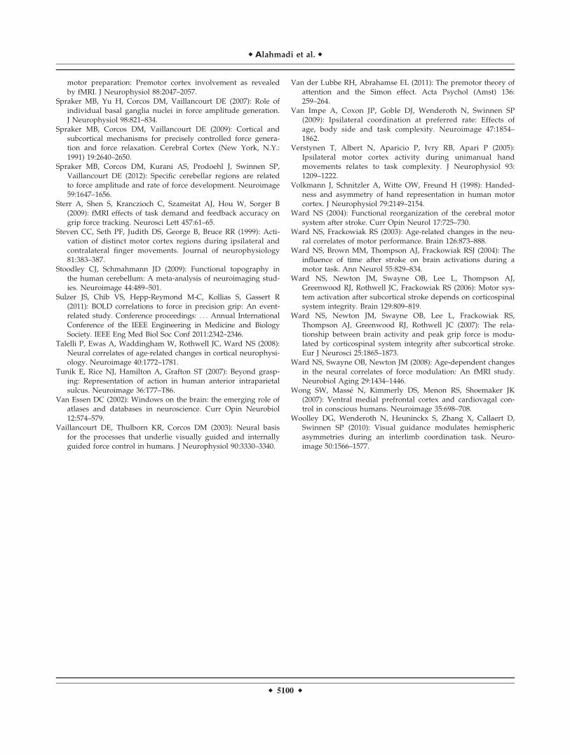
motor preparation: Premotor cortex involvement as revealedby fMRI. J Neurophysiol 88:2047–2057.
Spraker MB, Yu H, Corcos DM, Vaillancourt DE (2007): Role ofindividual basal ganglia nuclei in force amplitude generation.J Neurophysiol 98:821–834.
Spraker MB, Corcos DM, Vaillancourt DE (2009): Cortical andsubcortical mechanisms for precisely controlled force genera-tion and force relaxation. Cerebral Cortex (New York, N.Y.:1991) 19:2640–2650.
Spraker MB, Corcos DM, Kurani AS, Prodoehl J, Swinnen SP,Vaillancourt DE (2012): Specific cerebellar regions are relatedto force amplitude and rate of force development. Neuroimage59:1647–1656.
Sterr A, Shen S, Kranczioch C, Szameitat AJ, Hou W, Sorger B(2009): fMRI effects of task demand and feedback accuracy ongrip force tracking. Neurosci Lett 457:61–65.
Steven CC, Seth PF, Judith DS, George B, Bruce RR (1999): Acti-vation of distinct motor cortex regions during ipsilateral andcontralateral finger movements. Journal of neurophysiology81:383–387.
Stoodley CJ, Schmahmann JD (2009): Functional topography inthe human cerebellum: A meta-analysis of neuroimaging stud-ies. Neuroimage 44:489–501.
Sulzer JS, Chib VS, Hepp-Reymond M-C, Kollias S, Gassert R(2011): BOLD correlations to force in precision grip: An event-related study. Conference proceedings: . . . Annual InternationalConference of the IEEE Engineering in Medicine and BiologySociety. IEEE Eng Med Biol Soc Conf 2011:2342–2346.
Talelli P, Ewas A, Waddingham W, Rothwell JC, Ward NS (2008):Neural correlates of age-related changes in cortical neurophysi-ology. Neuroimage 40:1772–1781.
Tunik E, Rice NJ, Hamilton A, Grafton ST (2007): Beyond grasp-ing: Representation of action in human anterior intraparietalsulcus. Neuroimage 36:T77–T86.
Van Essen DC (2002): Windows on the brain: the emerging role ofatlases and databases in neuroscience. Curr Opin Neurobiol12:574–579.
Vaillancourt DE, Thulborn KR, Corcos DM (2003): Neural basisfor the processes that underlie visually guided and internallyguided force control in humans. J Neurophysiol 90:3330–3340.
Van der Lubbe RH, Abrahamse EL (2011): The premotor theory ofattention and the Simon effect. Acta Psychol (Amst) 136:259–264.
Van Impe A, Coxon JP, Goble DJ, Wenderoth N, Swinnen SP(2009): Ipsilateral coordination at preferred rate: Effects ofage, body side and task complexity. Neuroimage 47:1854–1862.
Verstynen T, Albert N, Aparicio P, Ivry RB, Apari P (2005):Ipsilateral motor cortex activity during unimanual handmovements relates to task complexity. J Neurophysiol 93:1209–1222.
Volkmann J, Schnitzler A, Witte OW, Freund H (1998): Handed-ness and asymmetry of hand representation in human motorcortex. J Neurophysiol 79:2149–2154.
Ward NS (2004): Functional reorganization of the cerebral motorsystem after stroke. Curr Opin Neurol 17:725–730.
Ward NS, Frackowiak RS (2003): Age-related changes in the neu-ral correlates of motor performance. Brain 126:873–888.
Ward NS, Brown MM, Thompson AJ, Frackowiak RSJ (2004): Theinfluence of time after stroke on brain activations during amotor task. Ann Neurol 55:829–834.
Ward NS, Newton JM, Swayne OB, Lee L, Thompson AJ,Greenwood RJ, Rothwell JC, Frackowiak RS (2006): Motor sys-tem activation after subcortical stroke depends on corticospinalsystem integrity. Brain 129:809–819.
Ward NS, Newton JM, Swayne OB, Lee L, Frackowiak RS,Thompson AJ, Greenwood RJ, Rothwell JC (2007): The rela-tionship between brain activity and peak grip force is modu-lated by corticospinal system integrity after subcortical stroke.Eur J Neurosci 25:1865–1873.
Ward NS, Swayne OB, Newton JM (2008): Age-dependent changesin the neural correlates of force modulation: An fMRI study.Neurobiol Aging 29:1434–1446.
Wong SW, Mass�e N, Kimmerly DS, Menon RS, Shoemaker JK(2007): Ventral medial prefrontal cortex and cardiovagal con-trol in conscious humans. Neuroimage 35:698–708.
Woolley DG, Wenderoth N, Heuninckx S, Zhang X, Callaert D,Swinnen SP (2010): Visual guidance modulates hemisphericasymmetries during an interlimb coordination task. Neuro-image 50:1566–1577.
r Alahmadi et al. r
r 5100 r


![Cerebellar Atrophy in Cortical Myoclonic Tremor and Not in ... · presence of head tremor and disease onset represent different ETsubtypes [4, 5], subgroup analyses were performed](https://img.pdfslide.us/doc/110x75/5d66c02588c99356168b4884/cerebellar-atrophy-in-cortical-myoclonic-tremor-and-not-in-presence-of-head.jpg)