Embed Size (px)
Citation preview

INFECTION AND IMMUNITY, Sept. 1991, p. 3254-32600019-9567/91/093254-07$02.00/0Copyright © 1991, American Society for Microbiology
Differential Induction of Tumor Necrosis Factor Alpha in OvinePulmonary Alveolar Macrophages following Infection with
Corynebacterium pseudotuberculosis, Pasteurellahaemolytica, or Lentivirusest
JOHN A. ELLIS,'* MICHAEL D. LAIRMORE,2 DONAL T. O'TOOLE,1 AND MANUEL CAMPOS3
Department of Veterinary Sciences, University of Wyoming, Laramie, Wyoming 820701; Department ofVeterinary Pathobiology, The Ohio State University, Columbus, Ohio 432102; and Veterinary
Infectious Diseases Organization, Saskatoon, Saskatchewan S7N OWO, Canada3
Received 1 April 1991/Accepted 18 June 1991
Soluble mediators such as tumor necrosis factor alpha (TNF-a) may be important in the pathogenesis ofmany chronic pulmonary infections. We examined the ability of Corynebacterium pseudotuberculosis, Pas-teurella haemolytica, and ovine lentiviruses (OvLV) to induce TNF-a secretion by pulmonary alveolarmacrophages (PAM). Bronchoalveolar lavage cells, composed of greater than 90% PAM, were obtained fromnormal sheep. Bronchoalveolar lavage cells were cultured for 2, 24, 48, 72, or 168 h in endotoxin-free RPMImedium (with 10% autologous serum) or in medium containing one of the following additives: lipopolysac-charide, 1-p.m polystyrene beads, C. pseudotuberculosis, P. haemolytica, or one of two plaque-cloned OvLV,85/28 or 85/34. Lipopolysaccharide, C. pseudotuberculosis, and P. haemolytica induced TNF-ft activity in PAMcultures as early as 2 h after inoculation, as assessed by a colorimetric cytotoxicity assay. This activity couldbe blocked by rabbit anti-recombinant bovine TNF-a serum. In contrast, medium alone, polystyrene beads,and productive infection by OvLV did not induce TNF-a activity in PAM cultures. Bacterial pathogens whichinfect pulmonary macrophages may elicit the secretion of TNF-a within the lungs and lead to the cachectic stateassociated with chronic pneumonia.
Tumor necrosis factor alpha (TNF-cx) is a 17-kDa proteinproduced by activated cells of the monocyte-macrophagelineage (5). It is a potent mediator of inflammation andhas been implicated in the promotion of a variety of im-mune-mediated lesions (5). TNF-ao may play a protec-tive role in infectious processes, presumably by activatingmacrophages and thereby enhancing the killing of intracel-lular pathogens (9, 17). However, it is also likely thatthis monokine mediates pathology in microbe-induced shockand in chronic infectious diseases in which macrophageactivation is a characteristic feature (5, 7, 9). Althougha range of pathogens comprising viruses, bacteria, andprotozoal parasites (3, 4, 30) can stimulate TNF-oc secre-
tion by human mononuclear phagocytes, little is knownabout the role of TNF-ao in naturally occurring pulmonarydiseases.A cardinal feature of pulmonary diseases induced by
chronic lentiviral and bacterial infections in sheep iscachexia, and dual infection by these agents often results ina condition known as "thin ewe syndrome" (14). Thepulmonary lesions in sheep with these dual bacterial andlentiviral infections consist of a combination of lymphopro-liferative interstitial pneumonia and bronchopneumonia (13,21). This constellation of lesions resembles that found inpediatric and adult AIDS patients with opportunistic andnonopportunistic pulmonary infections, which are a majorcause of death in humans with lentiviral infections (18, 21,23). Thus, dual infection of ovine pulmonary alveolar mac-
* Corresponding author.t Paper J.A. 1648 of the University of Wyoming Agricultural
Experiment Station.
rophages (PAM) with bacteria and lentiviruses represents a
potential model for examining the role of cytokines in lesiondevelopment in secondary pulmonary infections associatedwith human immunodeficiency virus (HIV).Although there is a plethora of pathophysiological alter-
ations that could lead to the debilitated state in chronicpneumonic infections, one mechanism may involve thehypersecretion of cytokines, such as TNF-cx and inter-leukin-1, with catabolic activities from infected mononuclearphagocytes. Indeed, repeated experimental inoculations ofrecombinant TNF-ot in cattle result in a reversible debilitat-ing condition which is characterized by depression, ano-
rexia, and cachexia and which mimics the chronic diseasestate (6). PAM are the primary target cells of Coryne-bacterium pseudotuberculosis (11) and ovine lentiviruses(OvLV) (20), and there is a marked influx of PAM incases of pneumonic pasteurellosis that often further com-
plicates cases of thin ewe syndrome (10). We have de-monstrated that OvLV-induced lymphoproliferative inter-stitial pneumonia is associated with elevated levels oflocally produced interferon, which may promote lympho-proliferation (21). Previous studies in humans document-ed TNF-cx secretion by PAM in response to microbesthat are associated with pulmonary infections in AIDS(19, 23, 30). To better understand the role of cytokinesin the interaction between bacteria and lentiviruses inovine lungs and in the pathogenesis of chronic pulmonarydiseases of comparative biological interest, we examined theTNF-cx-inducing capabilities of C. pseudotuberculosis, Pas-teurella haemolytica, and lentiviruses in cultures of ovinePAM.
3254
Vol. 59, No. 9
on October 1, 2018 by guest
http://iai.asm.org/
Dow
nloaded from
DIFFERENTIAL INDUCTION OF TNF-a IN OVINE PAM 3255
MATERIALS AND METHODS
Cells. PAM were obtained by postmortem bronchoalveo-lar lavage (12) from clinically normal yearling Rambouilletsheep that tested negative for antibodies to OvLV (31) andC. pseudotuberculosis (22). Two clinically normal sheep,12-3 and 9-25, used in experiments to examine the effects ofP. haemolytica showed low and high levels of seropositivity,respectively, for antibodies to P. haemolytica (15). Lavagecells from the normal lungs were composed of greater thanor equal to 90% PAM, as assessed by morphology, nonspe-
cific esterase staining (32), and reactivity with monoclonalantibody IL-A29 (12). PAM were suspended in endotoxin-free RPMI medium (Sigma Chemical Co., St. Louis, Mo.)supplemented with L-glutamine (2 mM), gentamicin (50jig/ml), and 10% autologous heat-inactivated serum. Thecells were cultured at 37°C with 5% CO2 in 24-well tissueculture plates.
Bacterial preparations. Stock cultures of C. pseudotuber-culosis (ATCC 19410) or P. haemolytica type Al (isolatedfrom a case of bovine pneumonia) were streaked ontocomplete blood agar plates. This serotype of P. haemolyticahas been used to examine the interaction between ovinelung lavage cells and the bacterium (9a). After 48 h at 37°C,single colonies were collected and placed in endotoxin-freeRPMI medium with 10% autologous heat-inactivated serum
but without antibiotics. Following 6 h at 37°C, these suspen-
sions were used as standard cultures to inoculate PAMcultures.OvLV preparations. Two plaque-cloned OvLV, 85/28 and
85/34, with differential pathogenicities for PAM (20) were
grown and their titers were determined in goat synovialmembrane cells (kindly provided by W. P. Cheevers, Wash-ington State University) as previously described (20). Super-natants for which titers were determined were used toinoculate PAM cultures.
Stimulation of PAM. PAM were cultured in 1 ml ofsupplemented medium in 24-well plates (Costar, Cambridge,Mass.) at a concentration of 106/ml and stimulated for 2, 24,48, 72, or 168 h with the following stimuli: (i) medium alone;(ii) latex beads (0.03 to 3.12 ,um; 1:1,000 stock; DukeScientific, Palo Alto, Calif.); (iii) endotoxin (lipopolysaccha-ride [LPS]) (Escherichia coli serotype 055:B5; 0.00125 to12.5 p.g/ml; Sigma); (iv) C. pseudotuberculosis or P. hae-molytica (1/100 or 1/1,000 dilution of a standard suspensionculture); and (v) OvLV 85/28 or 85/34 (multiplicity of infec-tion, 0.1). To test the effect of antibiotics on TNF-a produc-tion induced by bacteria, we did not include gentamicin,which has in vitro activity against both species of bacteria(29), in some cultures. At each time, supernatants were
harvested, centrifuged at 15,000 x g for 10 min, and frozenat -70°C until assayed. Forty-eight-hour cultures were usedto compare the levels of TNF-a induced by pathogens on thebasis of results from preliminary experiments. Bacterial andviral infections of macrophages were confirmed by examina-tion of Wright-Giemsa-stained cytospins, immunocytochem-ical staining of cytospins with monospecific rabbit serum
against C. pseudotuberculosis (prepared in our laboratory)or caprine arthritis-encephalitis virus p-30 (kindly providedby W. P. Cheevers), or electron microscopic examination ofcell pellets or monolayers prepared en face as previouslydescribed (25). Seven independent experiments with seven
different PAM donors were performed.TNF-a assay. TNF-a activity in supernatants was assayed
colorimetrically by its cytoxicity for the WEHI-164, clone13, mouse fibrosarcoma cell line essentially as described
previously (16). In brief, 50,000 WEHI cells in 100 uI werecultured with 100-pl volumes of test supernatants or logdilutions of standard recombinant bovine TNF-a (rBoTNF-cx; kindly provided by Ciba-Geigy, SA) and 10% normalrabbit serum or 10% anti-TNF-ot rabbit serum (prepared inour laboratory by four monthly injections of 50 ,ug of purifiedrBoTNF-(x; neutralizing titer, 1/500). The final concentrationof the supernatant in all assays was 50%. After 20 h ofincubation at 37°C, 25 RI of MTT (5 mg/ml in phosphate-buffered saline; Sigma) was added. Following 2 h of incuba-tion at 37°C, 100 [lI of the supernatant was carefully removedand 100 [lI of extraction buffer containing sodium dodecylsulfate and N,N-dimethylformamide (16) was added. Fol-lowing overnight incubation, color development was quan-titated at 595 nm in a model 3550 microplate reader (Bio-Rad, Richmond, Calif.) with extraction buffer as the blank.Levels of TNF-a activity were quantitated with a standardcurve derived from the rBoTNF-a values. Standard curveswere generated for each assay of PAM supernatants. Alldata are expressed as percent cytotoxicity compared withnegative controls (medium or medium plus additives withoutPAM) and were calculated as follows: [(OD of negativecontrol - OD of test well)/OD of negative control] x 100,where OD is optical density. All assays were performed atleast twice with triplicate wells of each sample supernatantin each experiment.
In preliminary experiments, we used WEHI-164 cells, theparent of the clone 13 cell line, as targets in the bioassay andfound them to be 3 to 4 logs less sensitive than clone 13 cellsto lysis by rBoTNF-a (data not shown). Although the TNF-asensitivity of WEHI-164 cells was increased by pretreatmentwith actinomycin D, WEHI-164 clone 13 cells were usedthroughout the remaining studies.
Immunoblotting. Standard rBoTNF-ot at a concentrationof 0.65 mg/ml was mixed with a buffer containing 62 mMTris-HCl (pH 6.8), 2% sodium dodecyl sulfate, 5% 2-mer-captoethanol, 7.5% glycerol, and 0.01% bromophenol blue,and the mixture was boiled for 5 min. Approximately 10 pgof rBoTNF-a was separated on a 12% mini-polyacrylamidegel electrophoresis gel (Bio-Rad) by standard procedures.Separated proteins were blotted onto Immobilon-P mem-branes (Millipore, Bedford, Mass.) blocked in 3% nonfat drymilk in phosphate-buffered saline for 1 h and probed with a1/200 dilution of normal rabbit serum or anti-rBoTNF-atrabbit serum and then with a 1/200 dilution of peroxidase-conjugated goat anti-rabbit immunoglobulin G (Pel-Frez,Rogers, Ark.). Recognized proteins were visualized withTMB substrate (Kirkegaard and Perry, Gaithersburg, Md.).
Statistical methods. Student's t test was used to determinethe significance of percent cytotoxicity among the varioustreatment groups.
RESULTS
Specificity of the WEHI-164 cell bioassay for ovine TNF-ot.To eliminate the possibility that the additives themselveswere nonspecifically toxic for WEHI-164 clone 13 cells, weincubated samples of additives at the concentrations used toinoculate PAM cultures for 24 h without PAM and testedthem for their direct effects on the fibrosarcoma cells. Nocytotoxic activity was noted when additives alone wereadded to the WEHI-164 cell assay (data not shown). Toconfirm that the lysis of WEHI-164 cells was due to theaction of ruminant TNF-ot, we used a monospecific rabbitantiserum against rBoTNF-at to block cytotoxicity. This
VOL. 59, 1991
on October 1, 2018 by guest
http://iai.asm.org/
Dow
nloaded from
3256 ELLIS ET AL.
0._
0.00
ol
Io'21o-1I 00 101 1 o2 103 1 04 105 106rBoTNF a(ng/ml)
additive
OvLV 85/34
OvLV 85/28P. haemolytica
1/1000P. haemolytica
1/100C. pseudo TB
1/1000C. pseudoTB
1/100beadsLPS
medium
70
0600 50
0 40
O'30
2010
10
0~
I0'3 1 O.2 1 0.1 100 10ol 2
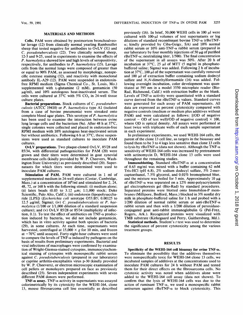
concentration LPS (microgm/ml)FIG. 1. (a) Lysis of WEHI-164 clone 13 cells by rBoTNF-a.
Lysis of target cells was blocked by the inclusion of monospecificanti-rBoTNF-a rabbit serum (final concentration, 10%). Bars repre-sent means of triplicate wells for each dilution tested + standarderrors. (b) Dose-dependent induction of TNF-a- activity by LPS in48-h PAM cultures from one sheep. Lytic activity was blocked bythe inclusion of monospecific anti-rBoTNF-ot rabbit serum, indicat-ing homology between ovine and bovine TNF-ct. NRS, normalrabbit serum.
antiserum reacted with a single protein of approximately 16kDa and a dimer of approximately 32 kDa on immunoblots ofthe rBoTNF-ot standard (data not shown). At a final concen-tration of 10%, this antiserum effectively blocked lysis due tothe presence of rBoTNF-a at concentrations ranging from0.065 to 65,000 pg/ml (Fig. la). Lytic activity in the WEHI-164 cell assay was induced in ovine PAM in a dose-depen-dent fashion by coculturing for 48 h with LPS at dosesranging from 0.00125 to 12.5 ,ug/ml (Fig. lb). Lytic activity insupernatants from ovine PAM cultures stimulated with thisrange of LPS doses was effectively blocked by the anti-rBoTNF-ot serum, indicating the presence of TNF-a and thespecificity of the antibody for both bovine and ovine TNF-a(Fig. lb).
Induction of TNF-a release by C. pseudotuberculosis and P.
additive
OvLV 85/34 anti-TNF b.NRS
OvLV 85/28
P. haemolytica1/100
C. pseudoTB1/100
LPS
medium
0 10 20 30 40 50 60 70 8090100
% cytotoxicityFIG. 2. (a) Microbe-induced TNF-a activity in PAM cultures
derived from sheep and harvested 48 h after the initiation ofculturing with additives. When compared with the negative (medi-um) and positive (LPS) control cultures, PAM cultures infected withC. pseudotuberculosis (C. pseudoTB) and P. haemolytica but notOvLV 85/28 or OvLV 85/34 had significant TNF-a activity. Barsrepresent means of triplicate wells for each animal tested + standarderrors. n, number of different sheep tested; *, significant differences.Each assay was conducted twice, and the final concentration ofsupernatant in each well was 50%. Results of one experiment areshown. (b) Blocking of microbe-induced TNF-a activity by theinclusion of TNF-a-specific antiserum in the assay, showing thatlysis was due to the action of TNF-ot. NRS, normal rabbit serum.
haemolytica. Compared with the low levels of cytotoxicity insupernatants from PAM cultures with medium alone, therewas significant (P < 0.02) WEHI-164 cell lytic activity inPAM cultures inoculated with C. pseudotuberculosis or P.haemolytica (Fig. 2a). The levels of cytotoxicity induced bythese bacterial species were comparable to those induced byLPS in PAM cultures. Lytic activity in these cultures wascompletely blocked by TNF-a-specific antiserum (Fig. 2b).TNF-a activity was induced by both bacterial species in
n=6 8a.
n2n.
n=4nz4~~n7
5n=7
0 10 20 30 40 50 60 70 80 90100
% cytotoxicity
INFECT. IMMUN.
on October 1, 2018 by guest
http://iai.asm.org/
Dow
nloaded from
DIFFERENTIAL INDUCTION OF TNF-a IN OVINE PAM 3257
168
a_
E4._
72
48
24
2
0 10 20 30 40 50 60 70 80 90100% cytotoxicity
N-.._
0)
168
72
48
24
2
0 10 20 30 40 50 60 70 80 90100 a C.psTB% cytotoxicity LPS
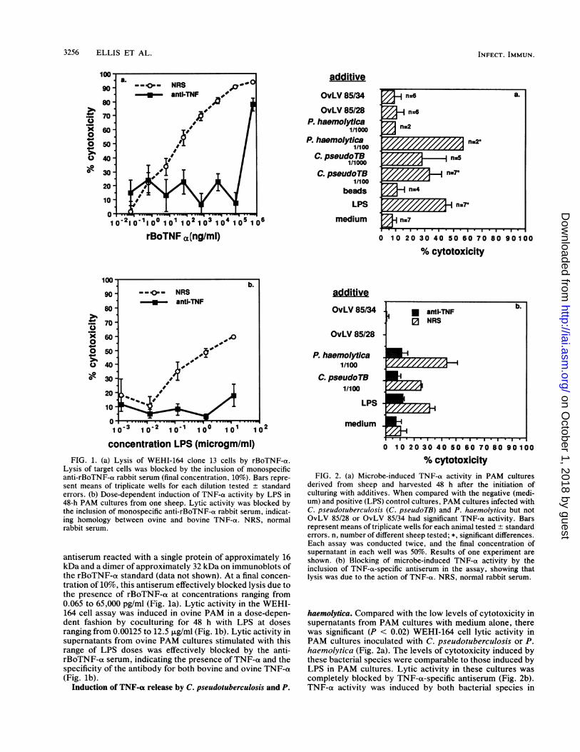
FIG. 3. Kinetics ofTNF-a release in cultures of ovine PAM fromtwo sheep, 650 (a) and 641 (b), indicating variable responses to LPSand C. pseudotuberculosis (C. psTB) determined in WEHI-164 cellassays performed concurrently. No TNF-ca activity was detected inPAM cultures inoculated with latex beads or OvLV clones.
PAM cultured in the presence and absence of gentamicin(data not shown).Lack of TNF-a activity in lentivirus-infected PAM cultures.
In contrast to supernatants from PAM cultures treated withLPS and the two bacterial species assessed, supematantsfrom PAM cultures inoculated with OvLV 85/28 or 85/34 didnot contain significant levels of TNF-a activity comparedwith cultures incubated with medium alone (Fig. 2).
Kinetics of TNF-ox release from PAM. The kinetics ofTNF-a release in PAM cultures inoculated with variousadditives were determined with PAM derived from foursheep (Fig. 3). LPS induced high levels of TNF-a in PAMfrom all four sheep. This induction was detectable as early as2 h after stimulation in two of the four sheep and at 24 h intwo of the four sheep. LPS-induced TNF-a activity peakedat 24 h but remained high throughout the culture period.
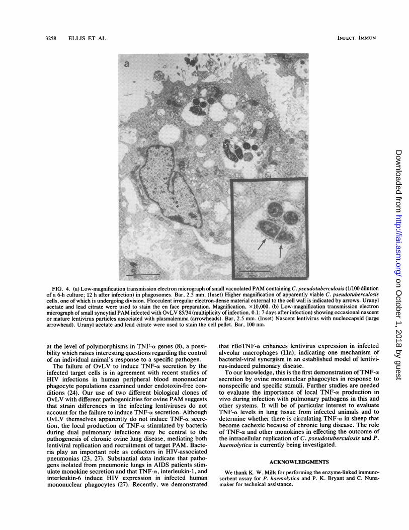
Phagocytosis of C. pseudotuberculosis (Fig. 4a) inducedTNF-a activity in PAM cultures from all four sheep. Thisinduction was detectable as early as 2 h after inoculation(Fig. 3). The levels of C. pseudotuberculosis phagocytosis-induced TNF-a activity peaked from 48 to 72 h after cultur-ing and varied with regard to the degree of response amongindividual lambs. This induction occurred under equivalentculture conditions and doses of bacteria, reflecting individualanimal variations (Fig. 3). In contrast, phagocytosis of latexbeads did not stimulate TNF-ot activity at any time afterinoculation.
Lytic activity that could be blocked by the TNF-ot-specificantiserum was not detected in PAM cultures inoculated withOvLV 85/28 or 85/34 at any of the times tested, despiteimmunocytochemical and ultrastructural evidence of infec-
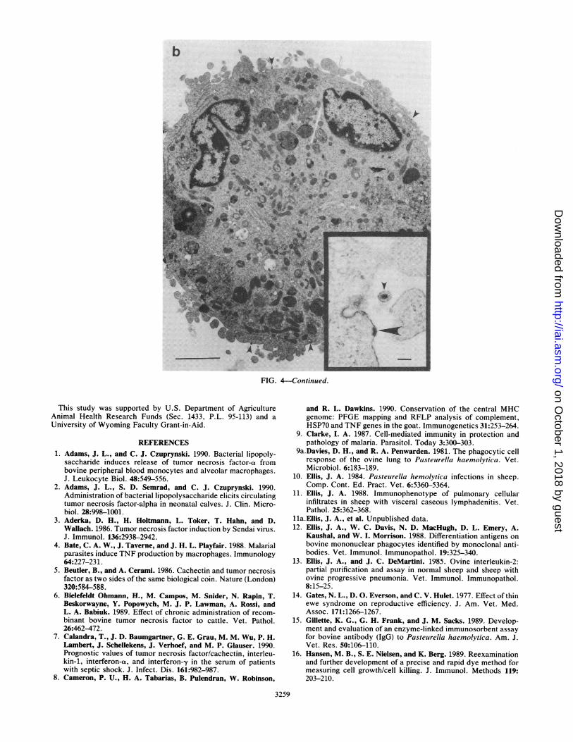
tion of up to 40% ofPAM by OvLV at 168 h after inoculation(Fig. 4b). Syncytium formation typical of the cytopathologyinduced by lentiviruses was also apparent in these cultures(Fig. 4b).
DISCUSSION
The interaction between bacteria and lentiviruses andovine PAM is a model relevant to better defining the role ofcytokines in the pathogenesis of chronic pulmonary diseaseassociated with HIV infections (21). Furthermore, we haveshown that infection ofPAM with the bacterial organisms C.pseudotuberculosis and P. haemolytica, which are com-monly isolated from cases of chronic pneumonia in sheep,results in TNF-a release in vitro. Failure of latex beadingestion to induce TNF-ot indicates that specific compo-nents of the bacteria and not simply the process of phago-cytosis are responsible for this effect. In contrast to infectionwith the bacterial pathogens examined, productive infectionof PAM with OvLV did not result in TNF-a secretion byPAM.
Induction of TNF-a secretion in vitro and in vivo has beenreported for both gram-negative and gram-positive bacteriaor their products in a variety of species (1, 2, 7, 30). Ourresults demonstrate the applicability of WEHI-164 clone 13cells to the bioassay of ovine TNF-a induced by bacterialpathogens. LPS and several mycobacterial antigens havebeen implicated as specific bacterial components that induceTNF-ac secretion (1, 2, 30). The presence of TNF-a in PAMcultures infected with C. pseudotuberculosis and P. hae-molytica and containing antibiotics suggests that these bac-teria need not be alive to induce TNF-ao secretion and thatsome of their soluble products are effective in inducingTNF-a. It is probable that the LPS of P. haemolyticainduces TNF-a in ovine PAM (1, 2); however, other specificconstituents of C. pseudotuberculosis and P. haemolytica,such as the exotoxins of these agents, that stimulate thesecretion of TNF-a remain to be identified. It has beenproposed that vaccination with antigens that stimulateTNF-a secretion to induce an antibody-mediated blockadeof TNF-a secretion or activity may be a rational approach toreducing the effects of disease caused by intracellular para-sites such as plasmodia (26). Thus, the identification ofTNF-a-inducing constituents of the intracellular pathogensC. pseudotuberculosis and P. haemolytica would be usefulin determining how such antigens may be used in prophy-laxis against chronic bacterial infections.We found considerable variability in TNF-ot production in
response to LPS and bacterial pathogens among sheep.Opsonizing antibody that mediates enhanced phagocytosisand processing may be irrelevant to TNF-a production,since all sheep used here were seronegative for C. pseudo-tuberculosis. The induction of TNF-a by LPS suggests thatthe mechanisms involved in TNF-a induction may in fact beindependent of phagocytosis, especially in the case of bac-teria that secrete exotoxins. The variability in TNF-a secre-tion may be attributable to differences in the pulmonarymacrophage subpopulations among the sheep. Several sub-populations of PAM that differ not only morphologically butalso in interleukin-1 production have been described in ratsand other species (28). It would be worthwhile to determinewhether similar functional differences that may account fordifferences in monokine secretion following stimulation withmicroorganisms exist among ruminant PAM. Alternatively,individual differences in susceptibility to infection or inTNF-a secretion in response to the agents tested could occur
l ~~~~~~~~~~~b.mmmI ..P.mmmm0mmP
S0 mZZmi
VOL. 59, 1991
on October 1, 2018 by guest
http://iai.asm.org/
Dow
nloaded from
3258 ELLIS ET AL.
.W. '.`:,.1 .
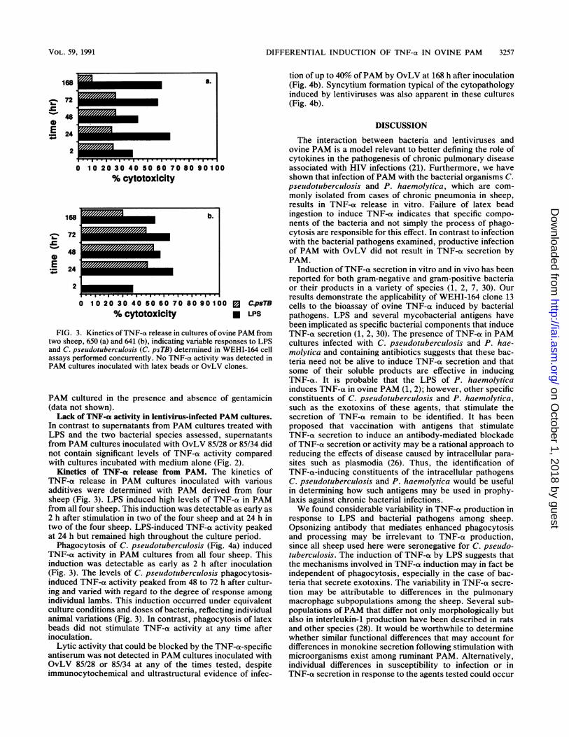
FIG. 4. (a) Low-magnification transmission electron micrograph of small vacuolated PAM containing C. pseudotuberculosis (1/100 dilutionof a 6-h culture; 12 h after infection) in phagosomes. Bar, 2.5 mm. (Inset) Higher magnification of apparently viable C. pseudotuberculosiscells, one of which is undergoing division. Flocculent irregular electron-dense material external to the cell wall is indicated by arrows. Uranylacetate and lead citrate were used to stain the en face preparation. Magnification, x 10,000. (b) Low-magnification transmission electronmicrograph of small syncytial PAM infected with OvLV 85/34 (multiplicity of infection, 0. 1; 7 days after infection) showing occasional nascentor mature lentivirus particles associated with plasmalemma (arrowheads). Bar, 2.5 mm. (Inset) Nascent lentivirus with nucleocapsid (largearrowhead). Uranyl acetate and lead citrate were used to stain the cell pellet. Bar, 100 nm.
at the level of polymorphisms in TNF-ax genes (8), a possi-bility which raises interesting questions regarding the controlof an individual animal's response to a specific pathogen.The failure of OvLV to induce TNF-a secretion by the
infected target cells is in agreement with recent studies ofHIV infections in human peripheral blood mononuclearphagocyte populations examined under endotoxin-free con-ditions (24). Our use of two different biological clones ofOvLV with different pathogenicities for ovine PAM suggeststhat strain differences in the infecting lentiviruses do notaccount for the failure to induce TNF-a secretion. AlthoughOvLV themselves apparently do not induce TNF-a secre-tion, the local production of TNF-ao stimulated by bacteriaduring dual pulmonary infections may be central to thepathogenesis of chronic ovine lung disease, mediating bothlentiviral replication and recruitment of target PAM. Bacte-ria play an important role as cofactors in HIV-associatedpneumonias (23, 27). Substantial data indicate that patho-gens isolated from pneumonic lungs in AIDS patients stim-ulate monokine secretion and that TNF-a, interleukin-1, andinterleukin-6 induce HIV expression in infected humanmononuclear phagocytes (27). Recently, we demonstrated
that rBoTNF-ot enhances lentivirus expression in infectedalveolar macrophages (lla), indicating one mechanism ofbacterial-viral synergism in an established model of lentivi-rus-induced pulmonary disease.To our knowledge, this is the first demonstration ofTNF-o
secretion by ovine mononuclear phagocytes in response tononspecific and specific stimuli. Further studies are neededto evaluate the importance of local TNF-a production invivo during infection with pulmonary pathogens in this andother systems. It will be of particular interest to evaluateTNF-(x levels in lung tissue from infected animals and todetermine whether there is circulating TNF-a in sheep thatbecome cachexic because of chronic lung disease. The roleof TNF-ot and other monokines in effecting the outcome ofthe intracellular replication of C. pseudotuberculosis and P.haemolytica is currently being investigated.
ACKNOWLEDGMENTS
We thank K. W. Mills for performing the enzyme-linked immuno-sorbent assay for P. haemolytica and P. K. Bryant and C. Nuna-maker for technical assistance.
:4zM
i0f
INFECT. IMMUN.
on October 1, 2018 by guest
http://iai.asm.org/
Dow
nloaded from
b
FIG. 4-Continued.
This study was supported by U.S. Department of AgricultureAnimal Health Research Funds (Sec. 1433, P.L. 95-113) and aUniversity of Wyoming Faculty Grant-in-Aid.
REFERENCES1. Adams, J. L., and C. J. Czuprynski. 1990. Bacterial lipopoly-
saccharide induces release of tumor necrosis factor-a frombovine peripheral blood monocytes and alveolar macrophages.J. Leukocyte Biol. 48:549-556.
2. Adams, J. L., S. D. Semrad, and C. J. Czuprynski. 1990.Administration of bacterial lipopolysaccharide elicits circulatingtumor necrosis factor-alpha in neonatal calves. J. Clin. Micro-biol. 28:998-1001.
3. Aderka, D. H., H. Holtmann, L. Toker, T. Hahn, and D.Wallach. 1986. Tumor necrosis factor induction by Sendai virus.J. Immunol. 136:2938-2942.
4. Bate, C. A. W., J. Taverne, and J. H. L. Playfair. 1988. Malarialparasites induce TNF production by macrophages. Immunology64:227-231.
5. Beutler, B., and A. Cerami. 1986. Cachectin and tumor necrosisfactor as two sides of the same biological coin. Nature (London)320:584-588.
6. Bielefeldt Ohmann, H., M. Campos, M. Snider, N. Rapin, T.Beskorwayne, Y. Popowych, M. J. P. Lawman, A. Rossi, andL. A. Babiuk. 1989. Effect of chronic administration of recom-binant bovine tumor necrosis factor to cattle. Vet. Pathol.26:462-472.
7. Calandra, T., J. D. Baumgartner, G. E. Grau, M. M. Wu, P. H.Lambert, J. Schellekens, J. Verhoef, and M. P. Glauser. 1990.Prognostic values of tumor necrosis factor/cachectin, interleu-kin-1, interferon-a, and interferon-y in the serum of patientswith septic shock. J. Infect. Dis. 161:982-987.
8. Cameron, P. U., H. A. Tabarias, B. Pulendran, W. Robinson,
3259
and R. L. Dawkins. 1990. Conservation of the central MHCgenome: PFGE mapping and RFLP analysis of complement,HSP70 and TNF genes in the goat. Immunogenetics 31:253-264.
9. Clarke, I. A. 1987. Cell-mediated immunity in protection andpathology of malaria. Parasitol. Today 3:300-303.
9a.Davies, D. H., and R. A. Penwarden. 1981. The phagocytic cellresponse of the ovine lung to Pasteurella haemolytica. Vet.Microbiol. 6:183-189.
10. Ellis, J. A. 1984. Pasteurella hemolytica infections in sheep.Comp. Cont. Ed. Pract. Vet. 6:5360-5364.
11. Ellis, J. A. 1988. Immunophenotype of pulmonary cellularinfiltrates in sheep with visceral caseous lymphadenitis. Vet.Pathol. 25:362-368.
11a.Ellis, J. A., et al. Unpublished data.12. Ellis, J. A., W. C. Davis, N. D. MacHugh, D. L. Emery, A.
Kaushal, and W. I. Morrison. 1988. Differentiation antigens onbovine mononuclear phagocytes identified by monoclonal anti-bodies. Vet. Immunol. Immunopathol. 19:325-340.
13. Ellis, J. A., and J. C. DeMartini. 1985. Ovine interleukin-2:partial purification and assay in normal sheep and sheep withovine progressive pneumonia. Vet. Immunol. Immunopathol.8:15-25.
14. Gates, N. L., D. 0. Everson, and C. V. Hulet. 1977. Effect of thinewe syndrome on reproductive efficiency. J. Am. Vet. Med.Assoc. 171:1266-1267.
15. Gillette, K. G., G. H. Frank, and J. M. Sacks. 1989. Develop-ment and evaluation of an enzyme-linked immunosorbent assayfor bovine antibody (IgG) to Pasteurella haemolytica. Am. J.Vet. Res. 50:106-110.
16. Hansen, M. B., S. E. Nielsen, and K. Berg. 1989. Reexaminationand further development of a precise and rapid dye method formeasuring cell growth/cell killing. J. Immunol. Methods 119:203-210.
m
on October 1, 2018 by guest
http://iai.asm.org/
Dow
nloaded from
3260 ELLIS ET AL.
17. Havell, E. A. 1989. Evidence that tumor necrosis factor has animportant role in antibacterial resistance. J. Immunol. 143:2894-2899.
18. Joshi, V. V., and J. M. Oleske. 1986. Pulmonary lesions inchildren with the acquired immunodeficiency syndrome: a re-appraisal based on data in additional cases and a followup studyof previously reported cases. Hum. Pathol. 17:641-642.
19. Krishnan, V. L., A. Meager, D. M. Mitchell, and A. J. Pinching.1990. Alveolar macrophages in AIDS patients: increased spon-taneous tumor necrosis factor-alpha production in Pneumocys-tis carinii pneumonia. Clin. Exp. Immunol. 80:156-160.
20. Lairmore, M. D., G. Y. Akita, H. I. Russell, and J. C. DeMar-tini. 1987. Replication and cytopathic effects of ovine lentivirusstrains in alveolar macrophages correlate with in vivo pathoge-nicity. J. Virol. 61:4038-4042.
21. Lairmore, M. D., J. M. Poulson, T. Adducci, and J. C. DeMar-tini. 1988. Lentivirus-induced lymphoproliferative disease.Comparative pathogenicity of phenotypically distinct ovine len-tivirus strains. Am. J. Pathol. 130:80-90.
22. Maki, L. R., S. H. Shen, R. C. Bergstrom, and L. D. Stetzen-bach. 1985. Diagnosis of Corynebacterium pseudotuberculosisinfections in sheep, using an enzyme-linked immunosorbentassay. Am. J. Vet. Res. 46:209-211.
23. Marchevsky, A., M. J. Rosen, G. Chrystal, and J. Kleinerman.1985. Pulmonary complications of the acquired immunodefi-ciency syndrome: a clinicopathologic study of 70 cases. Hum.Pathol. 16:659-670.
24. Molina, J. M., D. T. Scadden, C. Amirault, A. Woon, E.Vannier, C. A. Dinarello, and J. E. Groopman. 1990. Human
immunodeficiency virus does not induce interleukin-1, interleu-kin-6, or tumor necrosis factor in mononuclear cells. J. Virol.64:2901-2906.
25. Moses, R. L., E. G. Underwood, C. C. Vial, and J. B. Delcarpio.1989. In situ electron microscopy of cultured cells. Electron.Microsc. Soc. Am. Bull. 19:60-66.
26. Playfair, J. H. L., J. Taverne, C. A. W. Bate, and J. B. de Souza.1990. The malaria vaccine: anti-parasite or anti-disease? Immu-nol. Today 11:25-27.
27. Rosenberg, Z. F., and A. S. Fauci. 1990. Immunopathogenicmechanisms of HIV infection: cytokine induction of HIVexpression. Immunol. Today 11:176-180.
28. Shellito, J., and H. Benfer Kaltreider. 1984. Heterogeneity ofimmunologic function among subfractions of normal rat alveolarmacrophages. Am. Rev. Respir. Dis. 129:747-754.
29. Timoney, J. F., J. H. Gillespie, F. W. Scott, and J. E. Barlough.1988. The pathogenic bacteria, p. 114, 252. In Hagan andBruner's microbiology and infectious diseases of domestic ani-mals, 8th ed. Comstock Publishing Associates, Ithaca, N.Y.
30. Valone, S. E., E. A. Rich, R. S. Wallis, and J. J. Ellner. 1988.Expression of tumor necrosis factor in vitro by human mono-nuclear phagocytes stimulated with whole Mycobacterium bovisBCG and mycobacterial antigens. Infect. Immun. 56:3313-3315.
31. Windward, L. D., L. Leendertsen, and D. T. Shen. 1979.Microimmunodiffusion test for diagnosis of ovine progressivepneumonia. Am. J. Vet. Res. 40:564-566.
32. Yang, T. J., P. A. Jantzen, and L. J. Williams. 1979. Acidalpha-naphthyl acetate esterase: presence of activity in bovineand human T and B lymphocytes. Immunology 38:85-91.
INFECT. IMMUN.
on October 1, 2018 by guest
http://iai.asm.org/
Dow
nloaded from