Embed Size (px)
Citation preview

INTRODUCTION
Syndecan is a HSPG that consists of core proteinand covalently coupled glycosaminoglycan chains.It is a member of the type I transmembrane protein
family (1-3). There are four types of syndecan genesin mammals (Syndecan-1 to -4). Syndecan-1 is themajor syndecan in epithelial cells. Syndecan-1 knock-out mice show delayed cutaneous and corneal woundhealing (4), as well as increased leukocyte-endothelialinteractions, and angiogenesis (5). Syndecan-2 ispredominates in fibroblasts. It regulates actin skele-tal organization of the Lewis lung carcinoma-derivedcells by interacting with fibronectin (6). Syndecan-3 is abundant in neuronal cells, and syndecan-3 de-ficient mice show enhanced long-term potentiation
ORIGINAL
Differential expression of syndecan isoforms during mouseincisor amelogenesis
Taro Muto1), Keiko Miyoshi1), Seiichi Munesue3), Hiroshi Nakada3), Minoru Okayama3),
Takashi Matsuo2), and Takafumi Noma1)
1)Department of Molecular Biology, and Department of 2)Conservative Dentistry, Institute of Health
Biosciences, The University of Tokushima Graduate School, Tokushima, Japan ; and 3)Department of
Biotechnology, Faculty of Engineering, Kyoto Sangyo University, Kyoto, Japan
Abstract : Syndecans are transmembranous heparan sulfate proteoglycans (HSPGs) withcovalently attached glycosaminoglycan side-chains located on the cell surface. The mam-malian syndecan family is composed of four types of syndecans (syndecan-1 to -4). Syn-decans interact with the intracellular cytoskeleton through the cytoplasmic domains oftheir core proteins and membrane proteins, extracellular enzymes, growth factors, andmatrix components, through their heparan-sulfate chains, to regulate developmental proc-esses.
Here, as a first step to assess the possible roles of syndecan proteins in amelogenesis,we examined the expression patterns of all syndecan isoforms in continuously growingmouse incisors, in which we can overview major differentiation stages of amelogenesisat a glance. Understanding the expression domain of each syndecan isoform during spe-cific developmental stages seems useful for investigating their physiological roles in amelo-genesis.
Immunohistochemical analysis of syndecan core proteins in the lower incisors from post-natal day 1 mice revealed spatially and temporally specific expression patterns, withsyndecan-1 expressed in undifferentiated epithelial and mesenchymal cells, and syndecan-2,-3, and -4 in more differentiated cells. These findings suggest that each syndecan isoformfunctions distinctly during the amelogenesis of the incisors of mice. J. Med. Invest. 54 :331-339, August, 2007
Keywords : amelogenesis, immunohistochemistry, incisor, syndecan
Received for publication May 14, 2007 ; accepted July 17, 2007.
Address correspondence and reprint requests to Takafumi Noma,M.D., Ph.D., Department of Molecular Biology, Institute ofHealth Biosciences, The University of Tokushima GraduateSchool, Kuramoto-cho, Tokushima 770-8504, Japan and Fax : +81-88-633-7326.
The Journal of Medical Investigation Vol. 54 2007
331
and impaired hippocampus-dependent memory (7).Syndecan-4 is widely distributed and is thought tobe an important cell-surface receptor in wound heal-ing and angiogenesis (8).
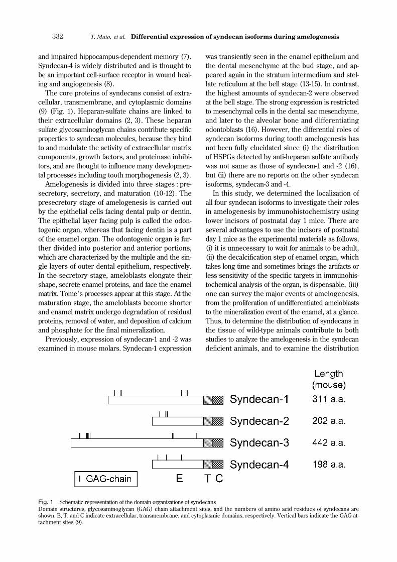
The core proteins of syndecans consist of extra-cellular, transmembrane, and cytoplasmic domains(9) (Fig. 1). Heparan-sulfate chains are linked totheir extracellular domains (2, 3). These heparansulfate glycosaminoglycan chains contribute specificproperties to syndecan molecules, because they bindto and modulate the activity of extracellular matrixcomponents, growth factors, and proteinase inhibi-tors, and are thought to influence many developmen-tal processes including tooth morphogenesis (2, 3).
Amelogenesis is divided into three stages : pre-secretory, secretory, and maturation (10-12). Thepresecretory stage of amelogenesis is carried outby the epithelial cells facing dental pulp or dentin.The epithelial layer facing pulp is called the odon-togenic organ, whereas that facing dentin is a partof the enamel organ. The odontogenic organ is fur-ther divided into posterior and anterior portions,which are characterized by the multiple and the sin-gle layers of outer dental epithelium, respectively.In the secretory stage, ameloblasts elongate theirshape, secrete enamel proteins, and face the enamelmatrix. Tome’s processes appear at this stage. At thematuration stage, the ameloblasts become shorterand enamel matrix undergo degradation of residualproteins, removal of water, and deposition of calciumand phosphate for the final mineralization.
Previously, expression of syndecan-1 and -2 wasexamined in mouse molars. Syndecan-1 expression
was transiently seen in the enamel epithelium andthe dental mesenchyme at the bud stage, and ap-peared again in the stratum intermedium and stel-late reticulum at the bell stage (13-15). In contrast,the highest amounts of syndecan-2 were observedat the bell stage. The strong expression is restrictedto mesenchymal cells in the dental sac mesenchyme,and later to the alveolar bone and differentiatingodontoblasts (16). However, the differential roles ofsyndecan isoforms during tooth amelogenesis hasnot been fully elucidated since (i) the distributionof HSPGs detected by anti-heparan sulfate antibodywas not same as those of syndecan-1 and -2 (16),but (ii) there are no reports on the other syndecanisoforms, syndecan-3 and -4.
In this study, we determined the localization ofall four syndecan isoforms to investigate their rolesin amelogenesis by immunohistochemistry usinglower incisors of postnatal day 1 mice. There areseveral advantages to use the incisors of postnatalday 1 mice as the experimental materials as follows,(i) it is unnecessary to wait for animals to be adult,(ii) the decalcification step of enamel organ, whichtakes long time and sometimes brings the artifacts orless sensitivity of the specific targets in immunohis-tochemical analysis of the organ, is dispensable, (iii)one can survey the major events of amelogenesis,from the proliferation of undifferentiated ameloblaststo the mineralization event of the enamel, at a glance.Thus, to determine the distribution of syndecans inthe tissue of wild-type animals contribute to bothstudies to analyze the amelogenesis in the syndecandeficient animals, and to examine the distribution
Fig. 1 Schematic representation of the domain organizations of syndecansDomain structures, glycosaminoglycan (GAG) chain attachment sites, and the numbers of amino acid residues of syndecans areshown. E, T, and C indicate extracellular, transmembrane, and cytoplasmic domains, respectively. Vertical bars indicate the GAG at-tachment sites (9).
T. Muto, et al. Differential expression of syndecan isoforms during amelogenesis332

of syndecans in animals deficient in amelogenesis.Our present study supplied the method to detectsyndecans, and the normal controls of their expres-sion patterns, as well as the possible roles of syn-decans in amelogenesis are speculated.
MATERIALS AND METHODS
Tissues and Antibodies
Mandibles including incisors were excised fromICR mice at P1. The tissue samples were fixed with4% paraformaldehyde in phosphate buffered saline(PBS) for 4 h at room temperature. For immunohis-tochemistry, 8-12-μm sections of paraffin-embeddedtissues were used.
Polyclonal rabbit IgG antibodies were raised againstthe extracellular domains of mouse syndecan-1, -2,-3, and -4 (SN1Ab, SN2Ab, SN3Ab, and SN4Ab, re-spectively) (17). Pre-immune rabbit IgG was used asa control. Mice were maintained and treated in ac-cordance with the guidelines for Animal Experimentat The University of Tokushima Graduate School,and the experimental protocols were approved bythe Committee of the Ethics on Animal Experimentat The University of Tokushima Graduate School.
Reverse transcription-polymerase chain reaction(RT-PCR)
Lower jaws were excised from three ICR mice atpostnatal day 1 (P1). Total RNA was extracted fromthe jaws using TRI reagent (MRC ; Cincinnati, OH)by following the manufacturer’s instructions. TotalRNA from the jaws was reverse transcribed witholigo dT primer using an RNA PCR kit (AMV) ver.3.0 (Takara Bio ; Ohtsu, Japan). Amplification of syn-decans and GAPDH transcripts was done by aninitial denaturing step of 94��for 6 min followed by40 cycles of polymerase chain reaction (PCR) (94��for 30 s, 57��for 30 s, 72��for 1 min), using TaqDNA polymerase (Promega ; Madison, WI). Theprimers used were as follows : syndecan-1 forward :5’-TCT GAC AAC TTC TCT GGC TCT GG-3’, andreverse : 5’-GCT GTG TTC TCC CCA GAT GTTTC-3’ (545 bp), syndecan-2 forward : 5’-CAC AGACGT GTA CAC CGA GAA AC-3’, and reverse : 5’-CTG CTA TTC ACA GAA CAC TGC AGA TG-3’
(444 bp), syndecan-3 forward : 5’-ACT AGA GCGGAA GGA GGT GCT C-3’, and reverse : 5’-AACCAG GGC TTC CTT CCT CTT G-3’ (474 bp),syndecan-4 forward : 5’-TGC TGG CGG CTC GGATGA CTT TG-3’, and reverse : 5’-CTG CCA AGA
CCT CAG TTC TCT C-3’ (290 bp), and GAPDHforward : 5’-CAT TGA CCT CAA CTA CAT GG-3’,and reverse : 5’-CTC AGT GTA GCC CAG GATGC-3’ (724 bp). The PCR products were sequencedby an ABI PRISM 3100-Avant Genetic Analyzer (Ap-plied Biosystems ; Lincoln Centre Drive, Foster City,CA) using primers used for PCR amplification andtheir identities were confirmed.
Immunohistochemistry
Sections on glass slides were deparaffinized inxylene, and substituted with ethanol followed byPBS. Intrinsic alkaline phosphatase activity was re-moved by using 3% hydrogen peroxide in metha-nol for 20 min at room temperature. After treatmentof the sections by microwave with antigen unmask-ing solution (Vector Laboratories Inc. ; Burlingame,CA), sections were blocked with PBS supplementedwith 4% bovine serum albumin (BSA) and 3% normalhorse serum for 30 min. Incubation of sections wasperformed with primary antibodies diluted with 1%BSA-PBS (30 μg/ml for SN1Ab, SN2Ab, and SN4Ab, 10 μg/ml for SN3Ab, and 1 : 2000 dilution foranti-collagen type IV antiserum) at 4��overnight,followed by incubation with horseradish peroxidase-labeled anti-rabbit Ig polyclonal antibody (HistofineSimple Stain Rat MAX-PO (R)) (Nichirei ; Tokyo,Japan). Finally, peroxidase activity was developedin Histofine Simple Stain DAB solution (Nichirei).The sections were counterstained with hematoxylin,dehydrated, and mounted. Sections from three tofive mice were examined for each staining.
RESULTS
RT-PCR analysis of syndecan expression in the man-dibles of P1 mice
Prior to the immunohistochemical analyses ofsyndecans, the expression of syndecans in the man-dibles of ICR mice at P1 was examined by RT-PCR,the most sensitive system to detect gene expres-sion. The mandibles contain apical loops, immatureand mature enamel organs, dental pulp, and thesurrounding epithelial cells of the continuously grow-ing incisors. Total RNA was extracted from the man-dibles, and RT-PCR analysis was performed to ex-amine which syndecans were expressed. SpecificPCR products were obtained from each reaction,indicating that all syndecan genes are expressed inthe mandibles of P1 mice (Fig. 2).
The Journal of Medical Investigation Vol. 54 August 2007 333

Localization of mouse syndecan core proteins in thelower incisors
The fact that each syndecan gene was expressedin the mandibles led us to study the immunohisto-chemical localization of syndecans in the tissues, es-pecially in the incisors, in which all stages of amelo-genesis can be seen in one section. Tissue slicesalong the longitudinal axis of incisors were stainedwith rabbit polyclonal antibodies raised against ex-tracellular portions of mouse syndecan core pro-teins. The specificity of the antibodies was confirmedin the previous study (17, 18).
Syndecan-1 was detected inside the apical loopand the surrounding region (Fig. 3a). In the ante-rior portion of the odontogenic organ in the prese-cretory stage, syndecan-1 was strongly detected ininner enamel epithelium (ameloblasts), stratum in-termedium, and outer enamel epithelium (Fig. 3aand f). In pulp, most of the cells expressed syndecan-1, but pre-odontoblasts did not (Fig. 3a). In addi-tion, the cells surrounding the outer enamel epithe-lium strongly expressed syndecan-1 (Fig. 3a). In thepresecretory stage of the enamel organ, no syndecan-1 staining was observed in ameloblasts or odon-toblasts (Fig. 3a). Stratum intermedium and outerenamel epithelium were strongly stained with anti-syndecan-1 antibody (Fig. 3a). Moreover, the pulpand the cells surrounding the outer enamel epithe-lium moderately expressed syndecan-1 (Fig. 3a). Inthe secretory stage, syndecan-1 staining was de-tected in the stratum intermedium, the papillarycells, and their surrounding cells, while no syndecan-1 staining was seen in ameloblasts or odontoblasts
(Fig. 3a and k). In the maturation stage, syndecan-1staining was detected in the stratum intermedium,the papillary cells and their surrounding cells, andnot in the ameloblasts or odontoblasts (Fig. 3a).
In contrast to syndecan-1, expression of syndecan-2 was slightly detected in the apical loop and inthe cells surrounding the apical loop (Fig. 3b). Inthe anterior portion of the odontogenic organ inthe presecretory stage, syndecan-2 was slightlystained in the inner enamel epithelium (Fig. 3band g). In the presecretory stage of the enamelorgan, syndecan-2 was detected in ameloblasts,especially in the nuclei and the apical side, thestratum intermedium, and the outer enamel epi-thelium ; however, there was no staining in thestellate reticulum (Fig. 3b). In odontoblasts andthe surrounding cells of the outer enamel epithe-lium, syndecan-2 was moderately stained, and alittle expression was observed in the pulp (Fig. 3b).In the secretory and maturation stages, syndecan-2was strongly detected in ameloblasts, odontoblasts,and the stratum intermedium, and slightly in thepapillary cells, their surrounding cells, and the pulp(Fig. 3b and l).
Expression of syndecan-3, similar to syndecan-2,was slightly detected in the apical loop and the sur-rounding dental pulp (Fig. 3c). Moreover, syndecan-3 was barely observed in the inner enamel epithe-lium in the anterior portion of the odontogenic organin the presecretory stage (Fig. 3c and h). In thepresecretory stage of the enamel organ, syndecan-3was stained in ameloblasts (nuclei and cytosol), thestratum intermedium, and the outer enamel epi-thelium (Fig. 3c). In addition, syndecan-3 was ex-pressed in odontoblasts and the surrounding cells ofthe outer enamel epithelium, and was slightly de-tected in the pulp (Fig. 3c). In the secretory andmaturation stages, syndecan-3 was detected inameloblasts, odontoblasts, the stratum intermedium,the papillary cells and their surrounding cells, andthe pulp (Fig. 3c and m).
Syndecan-4 expression contrasts with syndecan-1and is similar to that of both syndecan-2 and -3,with some distinctions. Little expression was ob-served in the apical loop, the surrounding dental pulp(Fig. 3d), and the inner enamel epithelium in theanterior portion of the odontogenic organ in thepresecretory stage (Fig. 3d and i). In the presecre-tory stage of the enamel organ, syndecan-4 wasslightly stained in this region (Fig. 3d). In the se-cretory stage, syndecan-4 was stained in ameloblasts,odontoblasts, the stratum intermedium, and the pulp.
Fig. 2 RT-PCR analysis of syndecan expression in mandiblesPCR amplification of syndecan cDNAs (syndecan-1 to -4) fromthe mandibles of P1 mice. GAPDH was amplified as a control.RTase : reverse transcriptase treatment for preparation of thecDNA template.
T. Muto, et al. Differential expression of syndecan isoforms during amelogenesis334

However, no staining was seen in the papillary cellsand their surrounding cells (Fig. 3d and n). In thematuration stage, syndecan-4 was stained stronglyin ameloblasts (Fig. 3d). The staining of syndecan-4was detected in ameloblasts, and modestly detectedin odontoblasts, papillary cells, and the pulp region
(Fig. 3d). Further, no syndecan-4 staining was seenin the cells surrounding the papillary cells (Fig. 3d).
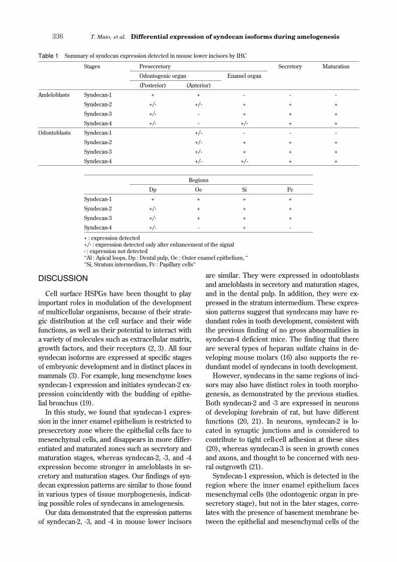
Incisor tissue sections were also stained with nor-mal rabbit IgG as a negative control for the immu-nohistochemistry (Fig. 3e, j, and o). The summaryof syndecan expression is shown in Table 1.
Fig. 3 Immunohistochemical examination of syndecan expression in mouse incisors at P1Anti-syndecan-1 (a, f, k), -2 (b, g, l), -3 (c, h, m), and -4 (d, i, n) antibodies, and normal IgG (e, j, o) staining of lower incisorsfrom P1 mice. (a-e) Overview of the incisors. (f-o) Higher magnification of odontogenic regions of the presecretory stage (f-j),and the secretory stage (k-o) of incisors, as indicated in the red solid and dotted boxes in (a-e), respectively. The surroundingarchitectures are indicated as follows : al : apical loop, p : dental pulp, oe : outer enamel epithelium, a : ameloblasts, o :odontoblasts, si : stratum intermedium, st : stellate reticulum. Scale bars : 500 μm (a-e), or 50 μm (f-o).
The Journal of Medical Investigation Vol. 54 August 2007 335
DISCUSSION
Cell surface HSPGs have been thought to playimportant roles in modulation of the developmentof multicellular organisms, because of their strate-gic distribution at the cell surface and their widefunctions, as well as their potential to interact witha variety of molecules such as extracellular matrix,growth factors, and their receptors (2, 3). All foursyndecan isoforms are expressed at specific stagesof embryonic development and in distinct places inmammals (3). For example, lung mesenchyme losessyndecan-1 expression and initiates syndecan-2 ex-pression coincidently with the budding of epithe-lial bronchus (19).
In this study, we found that syndecan-1 expres-sion in the inner enamel epithelium is restricted topresecretory zone where the epithelial cells face tomesenchymal cells, and disappears in more differ-entiated and maturated zones such as secretory andmaturation stages, whereas syndecan-2, -3, and -4expression become stronger in ameloblasts in se-cretory and maturation stages. Our findings of syn-decan expression patterns are similar to those foundin various types of tissue morphogenesis, indicat-ing possible roles of syndecans in amelogenesis.
Our data demonstrated that the expression patternsof syndecan-2, -3, and -4 in mouse lower incisors
are similar. They were expressed in odontoblastsand ameloblasts in secretory and maturation stages,and in the dental pulp. In addition, they were ex-pressed in the stratum intermedium. These expres-sion patterns suggest that syndecans may have re-dundant roles in tooth development, consistent withthe previous finding of no gross abnormalities insyndecan-4 deficient mice. The finding that thereare several types of heparan sulfate chains in de-veloping mouse molars (16) also supports the re-dundant model of syndecans in tooth development.
However, syndecans in the same regions of inci-sors may also have distinct roles in tooth morpho-genesis, as demonstrated by the previous studies.Both syndecan-2 and -3 are expressed in neuronsof developing forebrain of rat, but have differentfunctions (20, 21). In neurons, syndecan-2 is lo-cated in synaptic junctions and is considered tocontribute to tight cell-cell adhesion at these sites(20), whereas syndecan-3 is seen in growth conesand axons, and thought to be concerned with neu-ral outgrowth (21).
Syndecan-1 expression, which is detected in theregion where the inner enamel epithelium facesmesenchymal cells (the odontogenic organ in pre-secretory stage), but not in the later stages, corre-lates with the presence of basement membrane be-tween the epithelial and mesenchymal cells of the
Table 1 Summary of syndecan expression detected in mouse lower incisors by IHC
Stages Presecretory Secretory Maturation
Odontogenic organ Enamel organ
(Posterior) (Anterior)
Amleloblasts Syndecan-1 + + - - -
Syndecan-2 +/- +/- + + +
Syndecan-3 +/- - + + +
Syndecan-4 +/- - +/- + +
Odontoblasts Syndecan-1 +/- - - -
Syndecan-2 +/- + + +
Syndecan-3 +/- + + +
Syndecan-4 +/- +/- + +
Regions
Dp Oe Si Pc
Syndecan-1 + + + +
Syndecan-2 +/- + + +
Syndecan-3 +/- + + +
Syndecan-4 +/- - + -
+ : expression detected+/- : expression detected only after enhancement of the signal- : expression not detected”Al : Apical loops, Dp : Dental pulp, Oe : Outer enamel epithelium, ””Si, Stratum intermedium, Pc : Papillary cells”
T. Muto, et al. Differential expression of syndecan isoforms during amelogenesis336

enamel organ. Previous findings demonstrated thatboth collagen type IV and HSPG are the componentsof the basement membrane at the incisors (22-24),and collagen type IV in the basement membrane ofthe enamel organ is removed and degraded by dif-ferentiating ameloblasts by means of their engulf-ing system (25), and the 72 kDa type IV collage-nase (MMP-2) secreted by odontoblasts is alsothought to participate in the degradation of thedental basement membrane (26-28). Differentiat-ing ameloblasts may decrease the expression ofsyndecan-1, a possible component of the basementmembrane. Alternatively, restricted localization ofsyndecan-1 in the immature zone may enable thematuration of ameloblasts and odontoblasts, becausedisappearance of the basement membrane locatedbetween them enables the interaction between epi-thelium and mesenchyme (29) that is necessary formaturation of the enamel organ.
HSPGs bind to a wide variety of growth factors us-ing their heparan sulfate chains with different moie-ties. Their binding to growth factors such as fibro-blast growth factor, Wnt, and transforming growthfactor-β (TGF-β) family are known to be critical fortissue development (30-35). For example, duringtooth development, several members of the TGF-βfamily are known to be necessary to complete amelo-genesis. TGF-β1, and bone morphogenic protein(BMP) 2 are shown to induce in vitro ameloblastdifferentiation (36). In addition, BMP4 is neces-sary for ameloblast differentiation and enamel for-mation (37). Moreover, it is known that dental pulpin continuously erupting rat incisors expresses atleast fourteen genes belonging to the TGF-β su-perfamily (38). These findings, together with theexpression patterns of syndecan-2, -3, and -4 in thesecretory and maturation stages of enamel organ,suggest that these syndecans may contribute toamelogenesis via supporting the activities of thesegrowth factors. This scenario supports the expres-sion pattern of syndecan-2, -3, and -4 in ameloblastsin the later portion of presecretory stage, whichprecedes the mineralization of enamel. Alternatively,syndecans themselves may regulate ameloblast dif-ferentiation, using their potential activity as transcrip-tional regulators after releasing the intracellular do-main by protease cleavage of intramembrane do-mains like the notch/delta system (39). Anotherfinding also indicate the possible role of syndecan-4in amelogenesis through its negative regulation ac-tivity for ATF2, a member of activating transcrip-tion factor (ATF) subfamily (40), of which phos-
phorylated form is restricted to secretory to transi-tion zones (41).
In conclusion, we found reciprocal and redundantexpression patterns of syndecan isoforms in thecontinuously erupting mouse incisors, in which wecan survey the whole steps of amelogenesis, fromthe apical to the incisal end. The results suggestthat the syndecan isoforms have both distinct andredundant roles in amelogenesis. The molecularbasis of these roles during tooth development re-mains to be determined as a next step.
ACKNOWLEDGMENTS
This work was partly supported by grants-in-aidfor scientific research (Nos. 17659598, 18791368,and 17689051) from the Ministry of Education, Cul-ture, Sports, Science and Technology of Japan. Wethank Drs. T. Horiguchi, K. Abe, Intan Ruspita, K.Fujisawa and P. Yamaguchi for helpful discussions,and Ms. M. Sawatari for her secretarial help. Wealso thanks to Prof. Y. Hayashi, Mr. A. Kanaya,Mr. H. Niki and Mr. K. Sukegawa for their kindsupport.
REFERENCES
1. Couchman JR : Syndecans : proteoglycan regu-lators of cell-surface microdomains? Nat RevMol Cell Biol 4 : 926-937, 2003
2. Rapraeger AC : Molecular interactions of syn-decans during development. Semin Cell DevBiol 12 : 107-116, 2001
3. Bernfield M, Kokenyesi R, Kato M, Hinkes MT,Spring J, Gallo RL, Lose EJ : Biology of the syn-decans : a family of transmembrane heparan sul-fate proteoglycans. Annu Rev Cell Biol 8 : 365-393, 1992
4. Stepp MA, Gibson HE, Gala PH, Iglesia DD,Pajoohesh-Ganji A, Pal-Ghosh S, Brown M,Aquino C, Schwartz AM, Goldberger O, HinkesMT, Bernfield M : Defects in keratinocyte ac-tivation during wound healing in the syndecan-1-deficient mouse. J Cell Sci 115 : 4517-4531,2002
5. Gotte M, Joussen AM, Klein C, Andre P, WagnerDD, Hinkes MT, Kirchhof B, Adamis AP,Bernfield M : Role of syndecan-1 in leukocyte-endothelial interactions in the ocular vascula-ture. Invest Ophthalmol Vis Sci 43 : 1135-1141,
The Journal of Medical Investigation Vol. 54 August 2007 337

20026. Kusano Y, Oguri K, Nagayasu Y, Munesue S,
Ishihara M, Saiki I, Yonekura H, YamamotoH, Okayama M : Participation of syndecan 2in the induction of stress fiber formation incooperation with integrin alpha5beta1 : struc-tural characteristics of heparan sulfate chainswith avidity to COOH-terminal heparin-bindingdomain of fibronectin. Exp Cell Res 256 : 434-444, 2000
7. Kaksonen M, Pavlov I, Voikar V, Lauri SE,Hienola A, Riekki R, Lakso M, Taira T, RauvalaH : Syndecan-3-deficient mice exhibit enhancedLTP and impaired hippocampus-dependentmemory. Mol Cell Neurosci 21 : 158-172, 2002
8. Echtermeyer F, Baciu PC, Saoncella S, Ge Y,Goetinck PF : Syndecan-4 core protein is suf-ficient for the assembly of focal adhesions andactin stress fibers. J Cell Sci 112 (Pt 20) : 3433-3441, 1999
9. Zimmermann P, David G : The syndecans, tun-ers of transmembrane signaling. Faseb J 13(Suppl) : S91-S100, 1999
10. Ten Cate A, Sharpe P, Roy S, Nanci A : Devel-opment of the tooth and its supporting tissues.In : Nanci A, ed. Ten Cate’s Oral Histology :Development, Structure, and Function (TenCate’s Oral Histology). Mosby, Missouri, 2003,pp. 79-110
11. Nanci A : Enamel : Composition, formation,structure. In : Nanci A, ed. Ten Cate’s OralHistology : Development, Structure, and Func-tion (Ten Cate’s Oral Histology). Mosby, Mis-souri, 2003, pp. 145-191
12. Warshawsky H, Smith CE : Morphological clas-sification of rat incisor ameloblasts. Anat Rec179 : 423-446, 1974
13. Thesleff I, Jalkanen M, Vainio S, Bernfield M :Cell surface proteoglycan expression correlateswith epithelial-mesenchymal interaction dur-ing tooth morphogenesis. Dev Biol 129 : 565-572, 1988
14. Vainio S, Jalkanen M, Thesleff I : Syndecanand tenascin expression is induced by epithelial-mesenchymal interactions in embryonic toothmesenchyme. J Cell Biol 108 : 1945-1953, 1989
15. Vainio S, Jalkanen M, Vaahtokari A, SahlbergC, Mali M, Bernfield M, Thesleff I : Expres-sion of syndecan gene is induced early, is tran-sient, and correlates with changes in mesen-chymal cell proliferation during tooth organo-genesis. Dev Biol 147 : 322-333, 1991
16. Bai XM, Van der Schueren B, Cassiman JJ,Van den Berghe H, David G : Differential ex-pression of multiple cell-surface heparan sul-fate proteoglycans during embryonic tooth de-velopment. J Histochem Cytochem 42 : 1043-1054, 1994
17. Kusano Y, Yoshitomi Y, Munesue S, OkayamaM, Oguri K : Cooperation of syndecan-2 andsyndecan-4 among cell surface heparan sulfateproteoglycans in the actin cytoskeletal organi-zation of Lewis lung carcinoma cells. J Bio-chem (Tokyo) 135 : 129-137, 2004
18. Akita K, Fushiki S, Fujimoto T, Munesue S,Inoue M, Oguri K, Okayama M, Yamashina I,Nakada H : Identification of the core proteincarrying the Tn antigen in mouse brain : spe-cific expression on syndecan-3. Cell Struct Funct26 : 271-278, 2001
19. Brauker JH, Trautman MS, Bernfield M : Syn-decan, a cell surface proteoglycan, exhibits amolecular polymorphism during lung devel-opment. Dev Biol 147 : 285-292, 1991
20. Hsueh YP, Sheng M : Regulated expressionand subcellular localization of syndecan heparansulfate proteoglycans and the syndecan-bindingprotein CASK/LIN-2 during rat brain develop-ment. J Neurosci 19 : 7415-7425, 1999
21. Kinnunen A, Kinnunen T, Kaksonen M, NoloR, Panula P, Rauvala H : N-syndecan and HB-GAM (heparin-binding growth-associated mole-cule) associate with early axonal tracts in therat brain. Eur J Neurosci 10 : 635-648, 1998
22. Murray IC, Leblond CP : Immunoelectron mi-croscopy of endothelial cells in rat incisor sug-gests that most basement membrane compo-nents are produced by young cells, whereasheparan sulfate proteoglycan is produced byboth young and old cells. J Histochem Cyto-chem 36 : 763-773, 1988
23. Laurie GW, Leblond CP, Cournil I, Martin GR :Immunohistochemical evidence for the intra-cellular formation of basement membrane col-lagen (type IV) in developing tissues. J Histo-chem Cytochem 28 : 1267-1274, 1980
24. Laurie GW, Leblond CP, Martin GR : Local-ization of type IV collagen, laminin, heparansulfate proteoglycan, and fibronectin to thebasal lamina of basement membranes. J CellBiol 95 : 340-344, 1982
25. Sawada T, Yamamoto T, Yanagisawa T, TakumaS, Hasegawa H, Watanabe K : Evidence foruptake of basement membrane by differenti-
T. Muto, et al. Differential expression of syndecan isoforms during amelogenesis338

ating ameloblasts in the rat incisor enamel or-gan. J Dent Res 69 : 1508-1511, 1990
26. Sahlberg C, Reponen P, Tryggvason K, ThesleffI : Association between the expression of murine72 kDa type IV collagenase by odontoblasts andbasement membrane degradation during mousetooth development. Arch Oral Biol 37 : 1021-1030, 1992
27. Heikinheimo K, Salo T : Expression of base-ment membrane type IV collagen and type IVcollagenases (MMP-2 and MMP-9) in humanfetal teeth. J Dent Res 74 : 1226-1234, 1995
28. Kjoelby M, Thesleff I, Sahlberg C, FejerskovO, Josephsen K : Degradation of the dentalbasement membrane during mouse tooth de-velopment in vitro . Int J Dev Biol 38 : 455-462,1994
29. Brownell AG, Slavkin HC : Role of basal lam-ina in tissue interactions. Ren Physiol 3 : 193-204, 1980
30. Bernfield M, Gotte M, Park PW, Reizes O,Fitzgerald ML, Lincecum J, Zako M : Func-tions of cell surface heparan sulfate proteogly-cans. Annu Rev Biochem 68 : 729-777, 1999
31. Rapraeger AC : In the clutches of proteogly-cans : how does heparan sulfate regulate FGFbinding? Chem Biol 2 : 645-649, 1995
32. Perrimon N, Bernfield M : Specificities ofheparan sulphate proteoglycans in develop-mental processes. Nature 404 : 725-728, 2000
33. Allen BL, Rapraeger AC : Spatial and tempo-ral expression of heparan sulfate in mouse de-velopment regulates FGF and FGF receptorassembly. J Cell Biol 163 : 637-648, 2003
34. Viviano BL, Paine-Saunders S, Gasiunas N,Gallagher J, Saunders S : Domain-specific modi-fication of heparan sulfate by Qsulf1 modu-
lates the binding of the bone morphogeneticprotein antagonist Noggin. J Biol Chem 279 :5604-5611, 2004
35. Norton WH, Ledin J, Grandel H, Neumann CJ :HSPG synthesis by zebrafish Ext2 and Extl3is required for Fgf10 signalling during limb de-velopment. Development 132 : 4963-4973, 2005
36. Coin R, Haikel Y, Ruch JV : Effects of apatite,transforming growth factor beta-1, bone morpho-genetic protein-2 and interleukin-7 on ameloblastdifferentiation in vitro . Eur J Oral Sci 107 : 487-495, 1999
37. Wang XP, Suomalainen M, Jorgez CJ, MatzukMM, Werner S, Thesleff I : Follistatin regu-lates enamel patterning in mouse incisors byasymmetrically inhibiting BMP signaling andameloblast differentiation. Dev Cell 7 : 719-730,2004
38. Nakashima M, Toyono T, Murakami T, AkamineA : Transforming growth factor-beta superfamilymembers expressed in rat incisor pulp. ArchOral Biol 43 : 745-751, 1998
39. Schulz JG, Annaert W, Vandekerckhove J,Zimmermann P, De Strooper B, David G : Syn-decan 3 intramembrane proteolysis is preseni-lin/gamma-secretase-dependent and modulatescytosolic signaling. J Biol Chem 278 : 48651-48657, 2003
40. Saoncella S, Calautti E, Neveu W, Goetinck PF :Syndecan-4 regulates ATF-2 transcriptional ac-tivity in a Rac1-dependent manner. J Biol Chem279 : 47172-47176, 2004
41. Nishikawa S : Transient increase in anti-p-ATF2 immunoreactivity in the late secretionameloblasts apical to the transition zone of ratincisors. Anat Sci Int 79 : 87-94, 2004
The Journal of Medical Investigation Vol. 54 August 2007 339