Embed Size (px)
Citation preview

Differential Effects of Topical Vitamin E and C E FerulicHTreatments on Ultraviolet Light B-Induced CutaneousTumor Development in Skh-1 MiceErin M. Burns1, Kathleen L. Tober1, Judith A. Riggenbach1, Donna F. Kusewitt2, Gregory S. Young3,
Tatiana M. Oberyszyn1*
1Department of Pathology, The Ohio State University, Columbus, Ohio, United States of America, 2Department of Molecular Carcinogenesis, Science Park, The University
of Texas MD Anderson Cancer Center, Smithville Texas, United States of America, 3Center for Biostatistics, The Ohio State University, Columbus, Ohio, United States of
America
Abstract
Because of the ever-increasing incidence of ultraviolet light B (UVB)-induced skin cancer, considerable attention is beingpaid to prevention through the use of both sunscreens and after sun treatments, many of which contain antioxidants.Vitamin E is included as an antioxidant in many sunscreens and lotions currently on the market. Studies examining theefficacy of vitamin E as a topical preventative agent for UVB-induced skin cancer have yielded conflicting results. A likelycontributor to differences in study outcome is the stability of vitamin E in the particular formulation being tested. In thecurrent study we examined the effects of topical vitamin E alone as well as vitamin E combined with vitamin C and ferulicacid in a more stable topical formula (C E FerulicH). Mice were exposed to UVB for 10 weeks in order to induce skin damage.Then, before the appearance of any cutaneous lesions, mice were treated for 15 weeks with a topical antioxidant, withoutany further UVB exposure. We found that topical C E Ferulic decreased tumor number and tumor burden and prevented thedevelopment of malignant skin tumors in female mice with chronically UVB-damaged skin. In contrast, female micechronically exposed to UVB and treated topically with vitamin E alone showed a trend towards increased tumor growth rateand exhibited increased levels of overall DNA damage, cutaneous proliferation, and angiogenesis compared to vehicle-treated mice. Thus, we have demonstrated that topical 5% alpha tocopherol may actually promote carcinogenesis whenapplied on chronically UVB-damaged skin while treating with a more stable antioxidant compound may offer therapeuticbenefits.
Citation: Burns EM, Tober KL, Riggenbach JA, Kusewitt DF, Young GS, et al. (2013) Differential Effects of Topical Vitamin E and C E FerulicH Treatments onUltraviolet Light B-Induced Cutaneous Tumor Development in Skh-1 Mice. PLoS ONE 8(5): e63809. doi:10.1371/journal.pone.0063809
Editor: Arianna L. Kim, Columbia University Medical Center, United States of America
Received December 7, 2012; Accepted April 5, 2013; Published May 14, 2013
Copyright: � 2013 Burns et al. This is an open-access article distributed under the terms of the Creative Commons Attribution License, which permitsunrestricted use, distribution, and reproduction in any medium, provided the original author and source are credited.
Funding: This study was supported by the National Institutes of Health grant R01 CA133629 (www.nih.gov). The funders had no role in study design, datacollection and analysis, decision to publish, or preparation of the manuscript.
Competing Interests: The authors have declared that no competing interests exist.
* E-mail: [email protected]
Introduction
Over two million people are diagnosed with a form of non-
melanoma skin cancer (NMSC) each year in the United States,
making skin cancer more prevalent than all other cancers
combined [1]. Squamous cell carcinoma (SCC), a malignant form
of NMSC, makes up about 16% of all skin cancers. While the
mortality rate from SCC is relatively low–about 3000 deaths per
year–SCC can be quite disfiguring since most lesions are located
on sun exposed body parts such as the face and arms. Additionally,
topical therapies used to treat precursor actinic keratotic lesions
are not effective on invasive SCC, necessitating invasive surgeries.
With the increasing NMSC incidence, there has been a renewed
focus on sunscreens and other methods of preventing skin cancer,
including antioxidant supplementation in food, sunscreens, and
lotions.
Following UVB exposure, both infiltrating inflammatory cells
and activated epidermal keratinocytes generate reactive oxygen
species (ROS). Endogenous antioxidants play an important role in
detoxifying ROS and maintaining cutaneous homeostasis. If ROS
levels overwhelm the cutaneous antioxidant networks, the cells will
be subjected to oxidative stress [2]. Major mechanisms by which
ROS foster skin tumor development include induction of DNA
damage [3,4], inflammation [5], and angiogenesis [6,7,8].
Endogenous antioxidants in the skin include the enzyme
catalase as well as ascorbic acid and alpha tocopherol (vitamins
C and E, respectively) [9,10,11,12,13]. Catalase, the main
cutaneous antioxidant, detoxifies hydrogen peroxide. Decreased
catalase activity has been linked with both skin carcinogenesis and
progression [10,11]. Glutathione peroxidase (GPx) has been
argued to be even more crucial to maintaining cutaneous
homeostasis as evidenced by a study demonstrating that a small
increase in GPx activity can completely compensate for catalase-
deficient fibroblasts from patients [14]. However, GPx activity has
not been shown to be significantly affected by UV exposure
[15,16,17,18]. A decrease in vitamin C levels following UVB
exposure [19] results in increased DNA damage and apoptosis
[20]. Both human and animal studies have demonstrated
decreased SCC formation with diets containing supplemental
vitamin C [21,22]. Previous studies of the effects of exogenous
PLOS ONE | www.plosone.org 1 May 2013 | Volume 8 | Issue 5 | e63809
vitamin E treatment on skin carcinogenesis have resulted in a
variety of observations, including a 50% decrease in skin cancer
incidence with topical application of vitamin E [23] and an
increase in photocarcinogenesis following treatment with more
stable vitamin E esters [24]. Other studies have reported no
significant association between vitamin E treatment and SCC
development [25,26,27,28,29]. As vitamin E quenches free
radicals, it becomes oxidized. Vitamin C is able to reduce oxidized
vitamin E thus regenerating its activity; therefore, mixing vitamins
E and C stabilizes topical formulations of vitamin E [30]. Ferulic
acid exerts its antioxidant effects by supplying protons or hydrogen
ions to free radicals with phenolic hydroxyl groups [31]. Ferulic
acid also protects against the toxicity of active oxygen, or
superoxide, similarly to superoxide dismutase. Ferulic acid and
several of its derivatives have been shown to decrease tumor
formation in chemically-induced skin carcinogenesis models
[32,33,34,35]. Additionally, ferulic acid further stabilizes vitamins
C and E. Previously, the combination of vitamin C, vitamin E, and
ferulic acid was demonstrated to have photoprotective effects
when applied for four days prior to one UVB exposure [36].
Topical application of this antioxidant combination for four days
prior to UVB exposure also significantly reduced UVB-induced
thymine dimer formation in the epidermis 24 hours post-
irradiation [37]. C E Ferulic is currently being marketed as an
anti-aging treatment and sunscreen additive. However, the
potential of C E Ferulic for preventing skin cancer in chronically
UVB-damaged skin has not been examined.
Many cosmeceuticals are targeted primarily towards women
who often have a history of considerable prior UVB exposure
make use of antioxidant strategies. However, any beneficial effect
of topical antioxidant application to previously sun damaged skin
on skin tumor development has remained controversial. We
examined the efficacy of two topical antioxidant formulations in
preventing UVB-induced cutaneous SCC. Our model mimicked
women who were exposed to UVB regularly in childhood and
early adulthood and then markedly reduced their sun exposure
and began applying topical antioxidants prior to the formation of
any lesions. The current study demonstrated that topical C E
Ferulic treatment effectively reduced tumor number and burden in
female Skh-1 mice. Topical vitamin E treatment alone provided
no preventative benefits, and in fact, resulted in accelerated tumor
growth rate compared to vehicle-treated mice. This difference may
be explained by the resultant increase in catalase activity levels and
DNA damage present in the mice treated with vitamin E
compared to those treated with C E Ferulic. Our study
demonstrates both the potential detrimental effects of treating
chronically UVB-damaged skin with topical vitamin E alone and
the potential benefits of topically treating with a stable combina-
tion antioxidant compound for the prevention of UVB-induced
SCC.
Materials and Methods
Ethics StatementOutbred, female Skh-1 mice (6–8 weeks old, Charles River
Laboratories, Wilmington, MA) were housed in the vivarium at
The Ohio State University according to the requirements
established by the American Association for Accreditation of
Laboratory Animal Care. All procedures were approved by the
Ohio State University Institutional Animal Care and Use
Committee before the initiation of any studies (Protocol Number:
2010A00000083) and all efforts were made to minimize suffering.
MiceThe outbred nature of this strain of mice represents the
variability observed in the human population. Mice (n = 20 treated
with vehicle, n = 10 treated with vitamin E, n = 10 treated with C
E Ferulic) were dorsally exposed to 2240 J/m2 UVB, previously
determined to be 1 minimal erythemal dose (MED), 36weekly on
non-consecutive days for 10 weeks. UVB dose was calculated using
UVX radiometer and UVB sensor (UVP, Upland, CA) and
emitted by Phillips FS40 UV bulbs (American Ultraviolet
Company, Lebanon, IN). After 10 weeks of UVB exposure, the
mice were treated topically with vehicle (SurgilubeH; Savage
Laboratories, Melville, NY), 5 mg vitamin E (d-alpha tocopherol;
Sigma-Aldrich, St. Louis, MO) in vehicle, or 0.1 mL C E Ferulic
(SkinCeuticals) for 15 weeks with no additional UVB exposure.
This dose of vitamin E was chosen based on previously published
results demonstrating efficacy in preventing UVB-induced damage
[38,39] as well as previous preliminary studies in our laboratory.
Tumors larger than 1 mm in diameter were measured weekly with
calipers. After sacrifice, 0.5 cm2 section of dorsal skin and all
tumors were fixed as previously described [40] while remaining
dorsal skin was snap frozen in liquid nitrogen.
Tumor GradingHematoxylin and Eosin (H&E) stained tissue sections of tumors
isolated from mice were graded in a blinded manner by a board-
certified veterinary pathologist (DFK) as previously described [41].
Catalase Activity AssayFrozen dorsal skin was crushed and 15 mg was used for analysis
of catalase activity using the Catalase Assay Kit (Cayman
Chemical, Ann Arbor, MI) according to manufacturer’s instruc-
tions.
Glutathione Peroxidase Activity AssayFrozen dorsal skin was crushed and 20 mg was used for analysis
of glutathione peroxidase activity using the Glutathione Peroxi-
dase Activity Kit (Cayman Chemical) according to manufacturer’s
instructions.
ImmunohistochemistryParaformaldehyde-fixed/OCT-embedded dorsal skin sections
were cut (10 mm) onto Superfrost PlusH microscope slides (Fisher
Scientific) and stored at 280uC for future analysis. Slides were
thawed overnight at room temperature, baked at 60uC for 30 min,
and then rehydrated in Clear Rite 3 and a graded series of
ethanol. Detailed protocols for p53 and Ki67 have been described
previously [42].
Slides were incubated with primary p53 antibody (clone CM5p,
Novocastra (Leica Microsystems Inc.), Buffalo Grove, IL) at a
1:500 dilution in 16Casein at room temperature for 1 hour. p53
foci were counted as 3 or more adjacent p53-positive cells and
examined in 5 fields of view at 2006magnification.
Slides were incubated with primary Ki67 antibody (Dako,
Carpinteria, CA) at a 1:200 dilution in 16Casein overnight at 4uCin a humid chamber. Ki67-positive cells were examined in 5 fields
of view at 6006magnification.
CD31: Endogenous peroxidase activity was blocked with 3%
H2O2 in water for 10 minutes at room temperature. Slides were
incubated in Antigen Unmasking Solution (Vector Laboratories)
for 15 minutes in a microwave. After slides were cooled, they were
blocked with avidin D and biotin (Vector Laboratories), each for
15 minutes, 16 Casein for 30 minutes, and incubated with
primary CD31 antibody (Abcam) at a 1:50 dilution in 16Casein
Alpha Tocopherol and Cutaneous Tumorigenesis
PLOS ONE | www.plosone.org 2 May 2013 | Volume 8 | Issue 5 | e63809

for 1 hour at room temperature. Slides were then incubated with
biotinylated IgG (Vector Laboratories) at a 1:200 dilution in 16Casein, followed by ABC Elite. Slides were incubated in DAB
solution (Vector Laboratories) for 10 minutes at RT. Slides were
washed in deionized water, counterstained, and dehydrated.
CD31-positive vessels were examined in 7 fields of view at 6006magnification.
Statistical AnalysisThe results presented in this paper were part of a larger
experiment involving four treatment groups (of which vitamin E
and C E Ferulic were two) and a single control group. Dunnett’s
adjustment [43,44] for multiplicity was used for comparing the
primary outcome of tumor burden at 24 weeks between the
treatment groups and control in order to restrict the probability of
a type I error to 5%. The number of control mice was inflated
compared to the treatment groups to increase the power of the
comparison [43]. Residual plots verified the model assumptions of
normality and homoscedasticity and a logarithmic transformation
was utilized if necessary. Continuous outcome data were analyzed
using an ANOVA approach with linear contrasts for testing the
comparisons of interest. A mixed-effects regression model with a
random slope and intercept by subject was used to model tumor
growth from the time of tumor origination. For count data,
Poisson regression was used. All analyses were conducted in SAS
version 9.2 (SAS Institute, Cary, NC). p-values #0.05 were
considered statistically significant.
Results
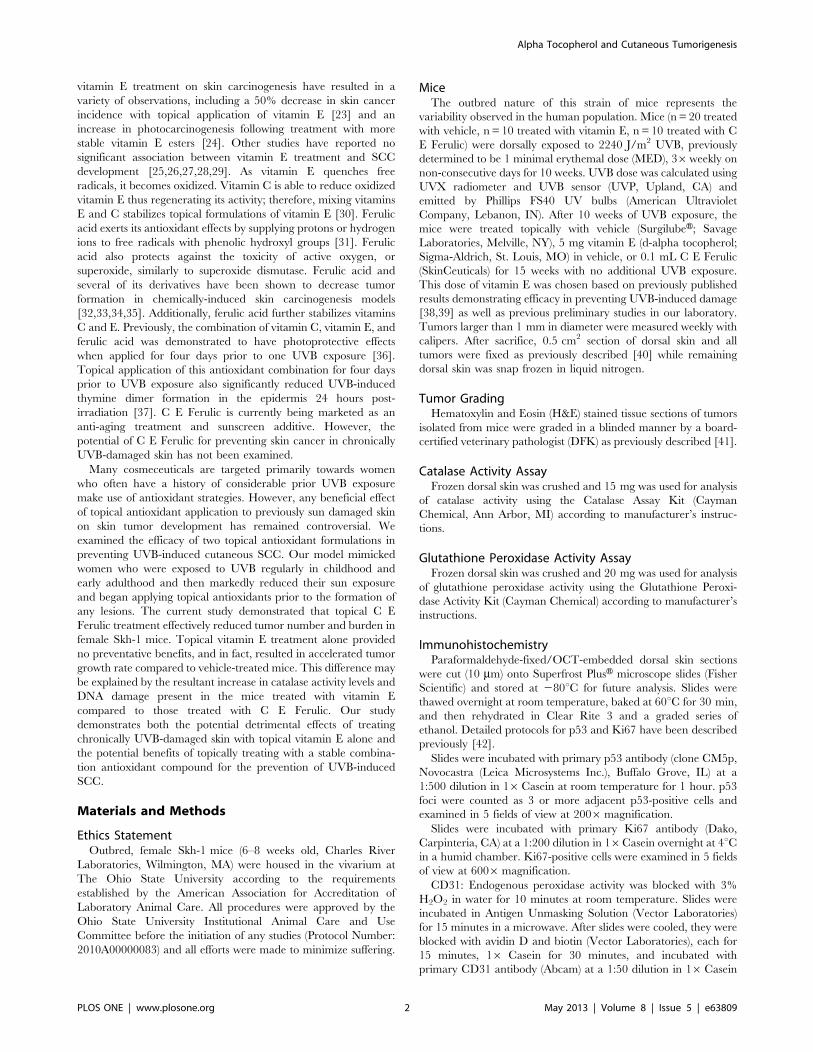
C E Ferulic Topical Treatment Decreased Tumor Numberand BurdenTo examine the effects of topical vitamin E or C E Ferulic
treatment as preventative agents against tumor development, we
exposed female Skh-1 hairless mice to 2240 J/m2 UVB (previously
determined to be 1 MED in our laboratory) three times weekly for
ten weeks to model chronic sun exposure. Mice were then treated
topically with vehicle, vitamin E, or C E Ferulic for 15 weeks
without further UVB exposure to model a lifestyle change. Non-
irradiated female mice treated with either vehicle, vitamin E, or C
E Ferulic did not develop tumors. Additionally, mice that were
exposed to UVB but received no vehicle treatment did not exhibit
a significantly different tumor burden compared to mice treated
with vehicle, indicating that the vehicle had no significant effect on
tumorigenesis in this study (data not shown).
Following 10 weeks of UV exposure alone and 15 weeks of
preventative topical treatment with C E Ferulic without further
UVB exposures, female mice developed 30.7% fewer tumors
compared to the mice treated with vehicle (p=0.0340, Figure 1A).
At the end of the study, female mice treated topically with C E
Ferulic exhibited a 34% decrease in tumor burden compared to
mice treated with vehicle (Figure 1B); however, probably as a
result of variability due to the outbred nature of this strain of mice,
the difference was not statistically significant (p=0.6047). Mice
treated topically with vitamin E demonstrated a trend toward
increased tumor multiplicity with 14.9% more tumors compared
to mice treated with vehicle (p=0.3193, Figure 1A). Mice treated
topically with vitamin E displayed a 20.7% increase in average
tumor burden; the tumor burden in vitamin E-treated mice was
not statistically different from that in mice treated with vehicle
(p=0.9566, Figure 1B).
Examining the change in tumor burden over time, we found
that tumor growth rates did not significantly differ between mice
treated topically with C E Ferulic and those treated with vehicle
(Figure 1C). Mice treated topically with vitamin E, however,
exhibited an increase in tumor growth rate compared to mice
treated with vehicle, 16.6% increase in tumor burden per week
versus 11.1%, respectively, which approached significance
(p=0.0649, Figure 1C).
UVB-irradiated Mice Treated Topically with C E FerulicDeveloped no Malignant TumorsTumors were isolated from mice after 10 weeks of UVB
exposure followed by 15 weeks of topical treatment and scored by
a board-certified veterinary pathologist (DFK). Tumors classified
as papilloma were considered benign and those classified as
microinvasive or fully invasive SCC were considered malignant.
As seen in Table 1, female mice treated with vehicle developed
papillomas, microinvasive SCC, and fully invasive SCC. Female
mice preventatively treated with C E Ferulic developed only
papillomas. Mice preventatively treated with vitamin E developed
both papillomas and microinvasive SCC, with a lower percentage
of malignant tumors compared to vehicle-treated mice.
Topical Vitamin E Treatment Increased the Number ofp53-positive FociAs a measure of total DNA damage, tumor-free, dorsal skin
sections were examined for p53-positive foci via immunohisto-
chemistry. Because the antibody used detected both wild type and
mutant p53, some p53-positive foci represented expanding clones
of keratinocytes with mutated p53. The density of p53-positive foci
in hairless mice has been shown to correlate well with skin tumor
risk [45]. The mean number of p53 foci was not significantly
altered with topical C E Ferulic treatment (Figure 2A) compared
to vehicle-treated mice (Figure 2B). The average number of p53-
positive foci was significantly increased with preventative topical
vitamin E treatment in female skin (p=0.0216, Figure 2C,
quantified in Figure 2D), suggesting a greater risk of skin tumors
in mice treated with vitamin E alone.
Topical Vitamin E Treatment Increased the Number ofCD31-positive Blood VesselsTo examine changes in vasculature, tumor-free dorsal skin
sections were examined for CD31-positive blood vessels via
immunohistochemistry. Several studies have demonstrated a link
between microvessel density, tumor growth, and metastasis
[46,47,48]. The mean number of CD31-positive blood vessels
was not significantly altered with topical C E Ferulic treatment
(Figure 3A) compared to vehicle-treated mice (Figure 3B).
However, the average number of blood vessels was significantly
increased with topical vitamin E treatment (p,0.0001, Figure 3C,
quantified in Figure 3D).
Vitamin E Topical Treatment Increased CutaneousProliferationTo examine changes in proliferation rates, tumor-free, dorsal
skin sections were stained for Ki67. The percentage of Ki67-
positive cells was not significantly altered in C E Ferulic-treated
skin compared to vehicle-treated skin after 10 weeks of UVB
exposure followed by 15 weeks of topical treatment with no
additional UVB exposure. In contrast, the percentage of Ki67-
positive cells exhibited a significant increase with topical vitamin
E treatment compared to vehicle-treated mice (p=0.0004,
Figure 4).
Alpha Tocopherol and Cutaneous Tumorigenesis
PLOS ONE | www.plosone.org 3 May 2013 | Volume 8 | Issue 5 | e63809
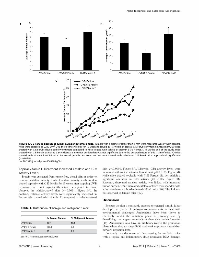
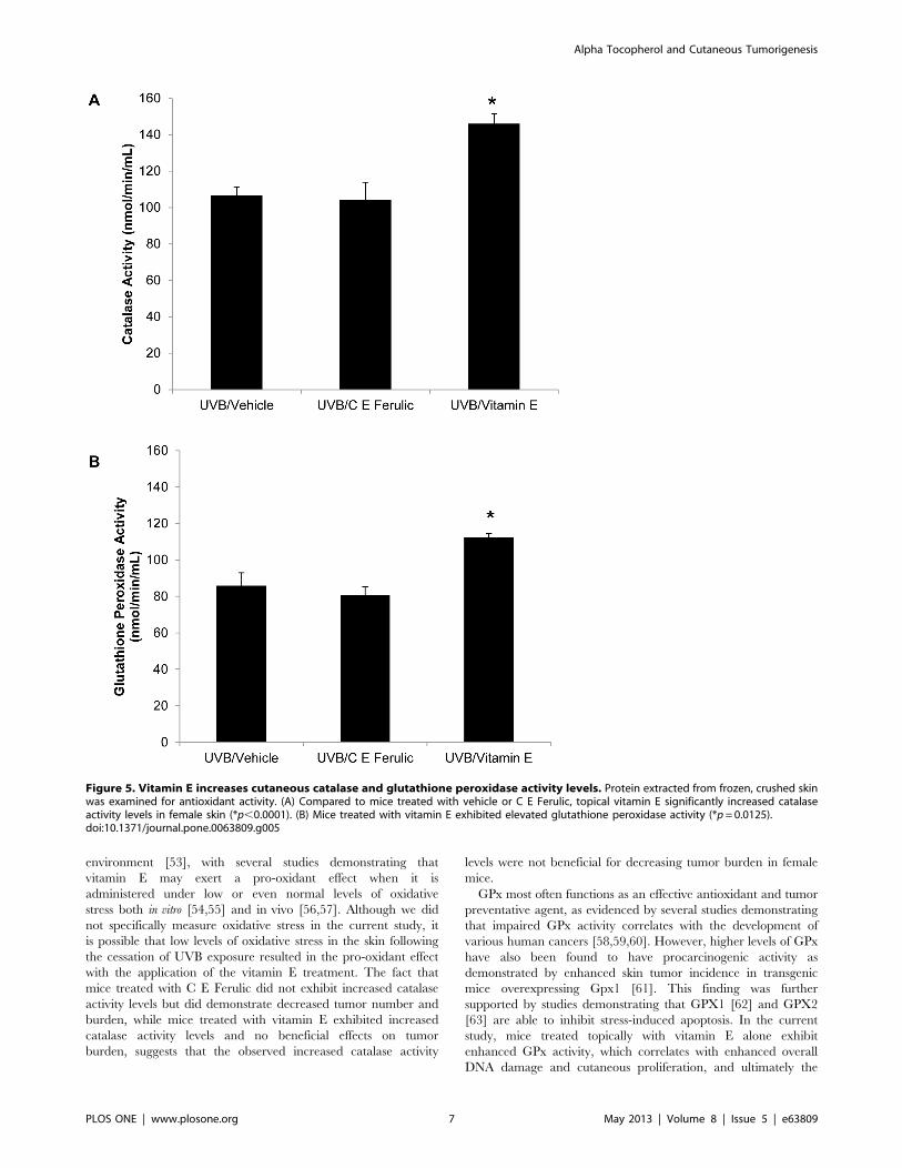
Topical Vitamin E Treatment Increased Catalase and GPxActivity LevelsProtein was extracted from tumor-free, dorsal skin in order to
examine catalase activity levels. Catalase activity levels in skin
treated topically with C E Ferulic for 15 weeks after stopping UVB
exposures were not significantly altered compared to those
observed in vehicle-treated skin (p=0.7631, Figure 5A). In
contrast, catalase activity levels were significantly increased in
female skin treated with vitamin E compared to vehicle-treated
skin (p,0.0001, Figure 5A). Likewise, GPx activity levels were
increased with topical vitamin E treatment (p=0.0125, Figure 5B)
while mice treated topically with C E Ferulic did not exhibit a
significant alteration in GPx activity (p=0.6415, Figure 5B).
Recently, decreased catalase activity was linked with increased
tumor burden, while increased catalase activity corresponded with
a decrease in tumor burden in male Skh-1 mice [40]. This link was
not observed in female mice [42].
Discussion
Because the skin is constantly exposed to external stimuli, it has
developed a system of endogenous antioxidants to deal with
environmental challenges. Antioxidants have been shown to
effectively inhibit the initiation phase of carcinogenesis by
detoxifying carcinogens, especially in chemically induced models
[49]. Antioxidants also have an inhibitory role in the promotion
phase where they scavenge ROS and work to prevent antioxidant
network depletion [49].
Previously, we demonstrated that treating female Skh-1 mice
with a topical anti-inflammatory drug decreased DNA damage,
Figure 1. C E Ferulic decreases tumor number in female mice. Tumors with a diameter larger than 1 mm were measured weekly with calipers.Mice were exposed to 2240 J/m2 UVB three times weekly for 10 weeks followed by 15 weeks of topical C E Ferulic or vitamin E treatment. (A) Micetreated with C E Ferulic developed fewer tumors compared to mice treated with vehicle or vitamin E (*p= 0.0283). (B) At the end of the study, micetreated with C E Ferulic exhibited a 34% decrease in tumor burden that was not significant due to the outbred nature of this strain of mice. (C) Micetreated with vitamin E exhibited an increased growth rate compared to mice treated with vehicle or C E Ferulic that approached significance(p=0.0649).doi:10.1371/journal.pone.0063809.g001
Table 1. Distribution of benign and malignant tumors.
% Benign Tumors % Malignant Tumors
UVB/Vehicle 89.7 10.3
UVB/C E Ferulic 100.0 0.0
UVB/Vitamin E 97.1 2.9
doi:10.1371/journal.pone.0063809.t001
Alpha Tocopherol and Cutaneous Tumorigenesis
PLOS ONE | www.plosone.org 4 May 2013 | Volume 8 | Issue 5 | e63809

tumor number and tumor grade [50]. In the current study we
showed that topically applied C E Ferulic, a stable antioxidant
compound, protects chronically UVB-damaged skin against skin
tumor development. Tumor number and burden were decreased
in C E Ferulic-treated mice compared to vehicle-treated mice.
Confirming the results of several previous studies [25,26,27,28,29],
topical vitamin E provided no beneficial effects with regard to
tumor burden; in fact, topical vitamin E alone resulted in
increased tumor number and burden as well as increased DNA
damage as indicated by p53 stabilization. Additionally, topical
vitamin E treatment contributed to increased proliferation of
epidermal cells, as well as an increase in angiogenesis. Interest-
ingly, catalase and glutathione peroxidase activity were increased
only with vitamin E treatment.
Topical vitamin E increased the number of p53-positive foci
observed in the epidermis, reflecting an increase in DNA damage.
If vitamin E is exerting a pro-oxidant effect, increased alpha
tocopheroxyl radicals may be contributing to increased levels of
DNA damage. However, previous studies indicate that both
vitamins E and C decrease the amount of UVB-induced oxidative
DNA damage, specifically 8-hydroxy-2-deoxyguanosine adducts
[20,51]. Because we saw no change in the level of overall DNA
damage in C E Ferulic-treated mice but elevated levels of DNA
damage in vitamin E-treated mice, it is possible that the
concentrations of the vitamins utilized, as well as the delivery
and treatment schedule, may play important roles in determining
the efficacy of these antioxidants.
ROS, including alpha tocopheroxyl radicals, contribute to
oxidative stress after UVB exposure. Previous studies suggested
that alpha tocopheroxyl radicals generated from alpha tocopherol
play a pivotal role in antioxidant-induced angiogenesis [52]. In the
current study, the increased vessel density observed in vitamin E-
treated mice further supports the mounting evidence that oxidative
stress can act as a trigger for angiogenesis. Interestingly, increased
levels of vitamin C have been demonstrated to prevent the
increased angiogenesis observed with high vitamin E treatment
concentrations; the suggested mechanism is scavenging of alpha
tocopheroxyl radicals [53]. While we only observed a change in
angiogenesis with the vitamin E treatment, it is possible that the C
E Ferulic is scavenging tocopheroxyl radicals at a rate that
prevents an increase in angiogenesis but is not sufficient to cause a
decrease.
It is important to note that mice treated with either C E Ferulic
or vitamin E developed lower percentages of malignant tumors
Figure 2. Mice treated with topical vitamin E have increased numbers of p53-positive foci. Dorsal, tumor-free skin sections wereexamined via immunohistochemistry with an antibody detecting both wild type and mutant p53. Representative images of skin from mice treatedwith (A) vehicle, (B) C E Ferulic, and (C) vitamin E for 15 weeks after 10 weeks of UVB exposure. (D) The average number of p53-positive foci per fieldof view was significantly higher in mice treated with vitamin E compared to vehicle-treated mice (*p= 0.0216).doi:10.1371/journal.pone.0063809.g002
Alpha Tocopherol and Cutaneous Tumorigenesis
PLOS ONE | www.plosone.org 5 May 2013 | Volume 8 | Issue 5 | e63809

compared to vehicle-treated mice. While mice treated topically
with C E Ferulic exhibited decreased tumor multiplicity and
burden, mice treated topically with vitamin E alone developed an
increased tumor number and burden compared to vehicle-treated
mice. It may be possible that vitamin E alone is more effective in
the late phase of tumorigenesis thus affecting tumor progression
but not tumor development as compared to C E Ferulic. This may
explain why, while we did not observe a decrease in tumor number
or burden in mice treated with vitamin E, we did see a smaller
percentage of malignant tumors in mice treated with vitamin E
compared to vehicle-treated mice. It is important to note,
however, that due to the outbred nature of this strain of mice
these differences were not statistically significant.
Of the antioxidants, catalase has been the best studied in
terms of its role in cutaneous homeostasis. Recently, we
demonstrated an important link between restoring catalase
activity after chronic UVB exposures and decreasing tumor
burden in male but not female mice [40,42]. In the current
study, the observed increase in catalase activity in female mice
treated with vitamin E suggests that this antioxidant may
actually be acting as a pro-oxidant. Antioxidant activity and
efficacy depend heavily on the preexisting redox status of the
Figure 3. Mice treated with topical vitamin E have increased numbers of CD31-positive vessels. Dorsal, tumor-free skin sections wereexamined for CD31-positive vessels via immunohistochemistry. Representative images of skin from mice treated with (A) vehicle, (B) C E Ferulic, and(C) vitamin E for 15 weeks after 10 weeks of UVB exposure. (D) The average number of CD31-positive vessels per field of view was significantly higherin mice treated with vitamin E compared to vehicle-treated mice (*p,0.0001).doi:10.1371/journal.pone.0063809.g003
Figure 4. Female skin treated with vitamin E displays increasedlevels of proliferation. Dorsal, tumor-free skin sections were stainedfor Ki67. Mice treated with vitamin E exhibited higher percentages ofKi67-positive cells compared to mice treated with vehicle or micetreated with C E Ferulic (*p= 0.0004).doi:10.1371/journal.pone.0063809.g004
Alpha Tocopherol and Cutaneous Tumorigenesis
PLOS ONE | www.plosone.org 6 May 2013 | Volume 8 | Issue 5 | e63809
environment [53], with several studies demonstrating that
vitamin E may exert a pro-oxidant effect when it is
administered under low or even normal levels of oxidative
stress both in vitro [54,55] and in vivo [56,57]. Although we did
not specifically measure oxidative stress in the current study, it
is possible that low levels of oxidative stress in the skin following
the cessation of UVB exposure resulted in the pro-oxidant effect
with the application of the vitamin E treatment. The fact that
mice treated with C E Ferulic did not exhibit increased catalase
activity levels but did demonstrate decreased tumor number and
burden, while mice treated with vitamin E exhibited increased
catalase activity levels and no beneficial effects on tumor
burden, suggests that the observed increased catalase activity
levels were not beneficial for decreasing tumor burden in female
mice.
GPx most often functions as an effective antioxidant and tumor
preventative agent, as evidenced by several studies demonstrating
that impaired GPx activity correlates with the development of
various human cancers [58,59,60]. However, higher levels of GPx
have also been found to have procarcinogenic activity as
demonstrated by enhanced skin tumor incidence in transgenic
mice overexpressing Gpx1 [61]. This finding was further
supported by studies demonstrating that GPX1 [62] and GPX2
[63] are able to inhibit stress-induced apoptosis. In the current
study, mice treated topically with vitamin E alone exhibit
enhanced GPx activity, which correlates with enhanced overall
DNA damage and cutaneous proliferation, and ultimately the
Figure 5. Vitamin E increases cutaneous catalase and glutathione peroxidase activity levels. Protein extracted from frozen, crushed skinwas examined for antioxidant activity. (A) Compared to mice treated with vehicle or C E Ferulic, topical vitamin E significantly increased catalaseactivity levels in female skin (*p,0.0001). (B) Mice treated with vitamin E exhibited elevated glutathione peroxidase activity (*p=0.0125).doi:10.1371/journal.pone.0063809.g005
Alpha Tocopherol and Cutaneous Tumorigenesis
PLOS ONE | www.plosone.org 7 May 2013 | Volume 8 | Issue 5 | e63809

increased formation of skin tumors. However, because the
cutaneous antioxidant networks are exceedingly complex, further
studies are needed to fully understand the interactions among the
different endogenous antioxidants in the skin, as well as with
exogenously applied antioxidants.
With the ever-increasing skin cancer incidence, antioxidant
supplementation in food, sunscreens, and lotions has become
widespread. While some animal models suggest beneficial effects
from antioxidants, it is important to note that antioxidants are
delivered prior to any UV exposure in many of these studies
[19,23]. Clinical trials examining potential effects of antioxidant
supplementation have yielded contradictory results. The Supple-
mentation in Vitamins and Mineral Antioxidants (SU.VI.MAX)
study revealed that daily supplementation with nutritional doses of
antioxidants decreased the overall incidence of cancer in men, but
had no effect in women [64]. Further, the impact of antioxidant
supplementation on skin cancer incidence, specifically, was
examined within the framework of the SU.VI.MAX study [65],
revealing an increased incidence of skin cancer in women and a
trend towards decreased skin cancer in men receiving antioxidant
supplements. The current study supports these findings in that, as
reported in the SU.VI.MAX study, there is a trend towards
increased tumor burden in female mice treated with vitamin E, a
single antioxidant. In contrast, the group treated with C E Ferulic,
a combination antioxidant exhibited decreased tumor number.
The delivery method of antioxidant supplements also seems to
play a role in the study outcomes. A systematic review of
randomized controlled trials reported that there was no beneficial
effect of oral vitamin or antioxidant supplementation on skin
cancer prevention but that topical antioxidant application did offer
some degree of protection in high risk individuals [66]. Our
current study supported these findings, in that mice with
chronically UVB-damaged skin that were treated topically with
C E Ferulic had decreased numbers of skin tumors compared to
vehicle-treated mice. Further, the antioxidant concentration and
activity in the various products varied greatly; thus, standardized
testing and labeling will be required to allow consumers to more
easily compare these products [67]. Further studies are needed to
understand antioxidant activity in vivo and to measure topical
antioxidant efficacy.
In summary, we have shown in a model of UVB-induced SCC
that topically treating female Skh-1 hairless mice with C E Ferulic
for 15 weeks after 10 weeks of UVB exposure decreased tumor
number and burden and suppressed the formation of malignant
tumors. In contrast, treating with topical vitamin E had no
therapeutic benefits, and, in fact, resulted in increased overall
DNA damage and vessel density, and tended to increase tumor
number, burden and growth rate, possibly due to the pro-oxidant
effects of vitamin E supplementation to chronically UVB-damaged
skin.
Because of the focus on antioxidant supplementation in many
products targeted towards women who have often been exposed to
significant amounts of UVB, these findings are especially relevant.
Overall, our data suggest that topically treating chronically UVB-
damaged skin with 5% vitamin E alone may actually promote
SCC development. Our findings may help explain previous
contradictory evidence regarding antioxidant supplementation
and cancer incidence.
Acknowledgments
We would like to thank Jonathan Schick, Keith Lamping and Katie
Samijlenko for excellent technical support.
Author Contributions
Conceived and designed the experiments: EMB KLT GSY TMO.
Performed the experiments: EMB KLT JAR TMO. Analyzed the data:
EMB DFK GSY. Contributed reagents/materials/analysis tools: DFK
GSY TMO. Wrote the paper: EMB KLT DFK GSY TMO.
References
1. Rogers HW, Weinstock MA, Harris AR, Hinckley MR, Feldman SR, et al.
(2010) Incidence estimate of nonmelanoma skin cancer in the United States,
2006. Arch Dermatol 146: 283–287.
2. Mates JM, Perez-Gomez C, Nunez de Castro I (1999) Antioxidant enzymes and
human diseases. Clin Biochem 32: 595–603.
3. McBride TJ, Preston BD, Loeb LA (1991) Mutagenic spectrum resulting from
DNA damage by oxygen radicals. Biochemistry 30: 207–213.
4. Nishigori C, Hattori Y, Toyokuni S (2004) Role of reactive oxygen species in
skin carcinogenesis. Antioxid Redox Signal 6: 561–570.
5. Halliday GM (2005) Inflammation, gene mutation and photoimmunosuppres-
sion in response to UVR-induced oxidative damage contributes to photo-
carcinogenesis. Mutat Res 571: 107–120.
6. Kuwabara M, Kakinuma Y, Ando M, Katare RG, Yamasaki F, et al. (2006)
Nitric oxide stimulates vascular endothelial growth factor production in
cardiomyocytes involved in angiogenesis. J Physiol Sci 56: 95–101.
7. Rojas A, Figueroa H, Re L, Morales MA (2006) Oxidative stress at the vascular
wall. Mechanistic and pharmacological aspects. Arch Med Res 37: 436–448.
8. Sauer H, Wartenberg M (2005) Reactive oxygen species as signaling molecules
in cardiovascular differentiation of embryonic stem cells and tumor-induced
angiogenesis. Antioxid Redox Signal 7: 1423–1434.
9. Afaq F, Mukhtar H (2001) Effects of solar radiation on cutaneous detoxification
pathways. J Photochem Photobiol B 63: 61–69.
10. Sander CS, Chang H, Salzmann S, Muller CS, Ekanayake-Mudiyanselage S, et
al. (2002) Photoaging is associated with protein oxidation in human skin in vivo.
J Invest Dermatol 118: 618–625.
11. Sander CS, Hamm F, Elsner P, Thiele JJ (2003) Oxidative stress in malignant
melanoma and non-melanoma skin cancer. Br J Dermatol 148: 913–922.
12. Sander CS, Chang H, Hamm F, Elsner P, Thiele JJ (2004) Role of oxidative
stress and the antioxidant network in cutaneous carcinogenesis. Int J Dermatol
43: 326–335.
13. Shindo Y, Witt E, Packer L (1993) Antioxidant defense mechanisms in murine
epidermis and dermis and their responses to ultraviolet light. J Invest Dermatol
100: 260–265.
14. Shindo Y, Hashimoto T (1995) Antioxidant defence mechanism of the skin
against UV irradiation: study of the role of catalase using acatalasaemiafibroblasts. Arch Dermatol Res 287: 747–753.
15. Fuchs J, Huflejt ME, Rothfuss LM, Wilson DS, Carcamo G, et al. (1989)
Impairment of enzymic and nonenzymic antioxidants in skin by UVB
irradiation. J Invest Dermatol 93: 769–773.
16. Shindo Y, Witt E, Han D, Packer L (1994) Dose-response effects of acuteultraviolet irradiation on antioxidants and molecular markers of oxidation in
murine epidermis and dermis. J Invest Dermatol 102: 470–475.
17. Hasegawa T, Kaneko F, Niwa Y (1992) Changes in lipid peroxide levels and
activity of reactive oxygen scavenging enzymes in skin, serum and liver followingUVB irradiation in mice. Life Sci 50: 1893–1903.
18. Fuchs J, Huflejt ME, Rothfuss LM, Wilson DS, Carcamo G, et al. (1989) Acuteeffects of near ultraviolet and visible light on the cutaneous antioxidant defense
system. Photochem Photobiol 50: 739–744.
19. Bissett DL, Chatterjee R, Hannon DP (1990) Photoprotective effect of
superoxide-scavenging antioxidants against ultraviolet radiation-induced chronicskin damage in the hairless mouse. Photodermatol Photoimmunol Photomed 7:
56–62.
20. Darr D, Combs S, Dunston S, Manning T, Pinnell S (1992) Topical vitamin C
protects porcine skin from ultraviolet radiation-induced damage. Br J Dermatol127: 247–253.
21. Pauling L, Willoughby R, Reynolds R, Blaisdell BE, Lawson S (1982) Incidence
of squamous cell carcinoma in hairless mice irradiated with ultraviolet light in
relation to intake of ascorbic acid (vitamin C) and of D, L-alpha-tocopherylacetate (vitamin E). Int J Vitam Nutr Res Suppl 23: 53–82.
22. Kune GA, Bannerman S, Field B, Watson LF, Cleland H, et al. (1992) Diet,alcohol, smoking, serum beta-carotene, and vitamin A in male nonmelanocytic
skin cancer patients and controls. Nutr Cancer 18: 237–244.
23. Gensler HL, Magdaleno M (1991) Topical vitamin E inhibition of immuno-
suppression and tumorigenesis induced by ultraviolet irradiation. Nutr Cancer15: 97–106.
24. Gensler HL, Aickin M, Peng YM, Xu M (1996) Importance of the form of
topical vitamin E for prevention of photocarcinogenesis. Nutr Cancer 26: 183–
191.
Alpha Tocopherol and Cutaneous Tumorigenesis
PLOS ONE | www.plosone.org 8 May 2013 | Volume 8 | Issue 5 | e63809

25. Fung TT, Spiegelman D, Egan KM, Giovannucci E, Hunter DJ, et al. (2003)
Vitamin and carotenoid intake and risk of squamous cell carcinoma of the skin.Int J Cancer 103: 110–115.
26. Breslow RA, Alberg AJ, Helzlsouer KJ, Bush TL, Norkus EP, et al. (1995)
Serological precursors of cancer: malignant melanoma, basal and squamous cellskin cancer, and prediagnostic levels of retinol, beta- carotene, lycopene, alpha-
tocopherol, and selenium. Cancer Epidemiol Biomarkers Prev 4: 837–842.27. Karagas MR, Greenberg ER, Nierenberg D, Stukel TA, Morris JS, et al. (1997)
Risk of squamous cell carcinoma of the skin in relation to plasma selenium,
alpha-tocopherol, beta-carotene, and retinol: a nested case-control study. CancerEpidemiol Biomarkers Prev 6: 25–29.
28. Dorgan JF, Boakye NA, Fears TR, Schleicher RL, Helsel W, et al. (2004) Serumcarotenoids and alpha-tocopherol and risk of nonmelanoma skin cancer. Cancer
Epidemiol Biomarkers Prev 13: 1276–1282.29. van der Pols JC, Heinen MM, Hughes MC, Ibiebele TI, Marks GC, et al. (2009)
Serum antioxidants and skin cancer risk: an 8-year community-based follow-up
study. Cancer Epidemiol Biomarkers Prev 18: 1167–1173.30. Chan AC (1993) Partners in defense, vitamin E and vitamin C. Can J Physiol
Pharmacol 71: 725–731.31. Fazary AE, Ju YH (2007) Feruloyl esterases as biotechnological tools: current
and future perspectives. Acta Biochim Biophys Sin (Shanghai) 39: 811–828.
32. Huang MT, Smart RC, Wong CQ, Conney AH (1988) Inhibitory effect ofcurcumin, chlorogenic acid, caffeic acid, and ferulic acid on tumor promotion in
mouse skin by 12-O-tetradecanoylphorbol-13-acetate. Cancer Res 48: 5941–5946.
33. Asanoma M, Takahashi K, Miyabe M, Yamamoto K, Yoshimi N, et al. (1994)Inhibitory effect of topical application of polymerized ferulic acid, a synthetic
lignin, on tumor promotion in mouse skin two-stage tumorigenesis. Carcino-
genesis 15: 2069–2071.34. Murakami A, Kadota M, Takahashi D, Taniguchi H, Nomura E, et al. (2000)
Suppressive effects of novel ferulic acid derivatives on cellular responses inducedby phorbol ester, and by combined lipopolysaccharide and interferon-gamma.
Cancer Lett 157: 77–85.
35. Murakami A, Nakamura Y, Koshimizu K, Takahashi D, Matsumoto K, et al.(2002) FA15, a hydrophobic derivative of ferulic acid, suppresses inflammatory
responses and skin tumor promotion: comparison with ferulic acid. Cancer Lett180: 121–129.
36. Lin FH, Lin JY, Gupta RD, Tournas JA, Burch JA, et al. (2005) Ferulic acidstabilizes a solution of vitamins C and E and doubles its photoprotection of skin.
J Invest Dermatol 125: 826–832.
37. Lin JY, Selim MA, Shea CR, Grichnik JM, Omar MM, et al. (2003) UVphotoprotection by combination topical antioxidants vitamin C and vitamin E. J
Am Acad Dermatol 48: 866–874.38. Lopez-Torres M, Thiele JJ, Shindo Y, Han D, Packer L (1998) Topical
application of alpha-tocopherol modulates the antioxidant network and
diminishes ultraviolet-induced oxidative damage in murine skin. Br J Dermatol138: 207–215.
39. Berton TR, Conti CJ, Mitchell DL, Aldaz CM, Lubet RA, et al. (1998) Theeffect of vitamin E acetate on ultraviolet-induced mouse skin carcinogenesis. Mol
Carcinog 23: 175–184.40. Sullivan NJ, Tober KL, Burns EM, Schick JS, Riggenbach JA, et al. (2012) UV
light B-mediated inhibition of skin catalase activity promotes Gr-1+ CD11b+myeloid cell expansion. J Invest Dermatol 132: 695–702.
41. Thomas-Ahner JM, Wulff BC, Tober KL, Kusewitt DF, Riggenbach JA, et al.
(2007) Gender differences in UVB-induced skin carcinogenesis, inflammation,and DNA damage. Cancer Res 67: 3468–3474.
42. Burns EM, Tober KL, Riggenbach JA, Schick JS, Lamping KN, et al. (2013)
Preventative topical diclofenac treatment differentially decreases tumor burdenin male and female Skh-1 mice in a model of UVB-induced cutaneous squamous
cell carcinoma. Carcinogenesis 34: 370–377.43. Dunnett C (1955) A multiple comparison procedure for comparing several
treatments with a control. Journal of the American Statistical Association 50:
1096–1121.44. Hsu J (1992) The factor analytic approach to simultaneous inference in the
general linear model. Journal of Computational and Graphical Statistics 1: 151–168.
45. Rebel H, Mosnier LO, Berg RJ, Westerman-de Vries A, van Steeg H, et al.(2001) Early p53-positive foci as indicators of tumor risk in ultraviolet-exposed
hairless mice: kinetics of induction, effects of DNA repair deficiency, and p53
heterozygosity. Cancer Res 61: 977–983.
46. Vermeulen PB, Verhoeven D, Hubens G, Van Marck E, Goovaerts G, et al.(1995) Microvessel density, endothelial cell proliferation and tumour cell
proliferation in human colorectal adenocarcinomas. Ann Oncol 6: 59–64.
47. Fox SB, Gatter KC, Bicknell R, Going JJ, Stanton P, et al. (1993) Relationship ofendothelial cell proliferation to tumor vascularity in human breast cancer.
Cancer Res 53: 4161–4163.
48. Mattern J, Koomagi R, Volm M (1996) Association of vascular endothelialgrowth factor expression with intratumoral microvessel density and tumour cell
proliferation in human epidermoid lung carcinoma. Br J Cancer 73: 931–934.
49. Walaszek Z, Hanausek M, Slaga TJ (2004) Mechanisms of chemoprevention.Chest 125: 128S–133S.
50. Wilgus TA, Koki AT, Zweifel BS, Kusewitt DF, Rubal PA, et al. (2003)
Inhibition of cutaneous ultraviolet light B-mediated inflammation and tumorformation with topical celecoxib treatment. Mol Carcinog 38: 49–58.
51. Ichihashi M, Funasaka Y, Ohashi A, Chacraborty A, Ahmed NU, et al. (1999)
The inhibitory effect of DL-alpha-tocopheryl ferulate in lecithin on melanogen-esis. Anticancer Res 19: 3769–3774.
52. Daghini E, Zhu XY, Versari D, Bentley MD, Napoli C, et al. (2007) Antioxidant
vitamins induce angiogenesis in the normal pig kidney. Am J Physiol RenalPhysiol 293: F371–381.
53. Stocker R, Keaney JF Jr (2004) Role of oxidative modifications in
atherosclerosis. Physiol Rev 84: 1381–1478.
54. Kontush A, Finckh B, Karten B, Kohlschutter A, Beisiegel U (1996) Antioxidantand prooxidant activity of alpha-tocopherol in human plasma and low density
lipoprotein. J Lipid Res 37: 1436–1448.
55. Neuzil J, Thomas SR, Stocker R (1997) Requirement for, promotion, orinhibition by alpha-tocopherol of radical-induced initiation of plasma lipoprotein
lipid peroxidation. Free Radic Biol Med 22: 57–71.
56. Keaney JF, Jr., Gaziano JM, Xu A, Frei B, Curran-Celentano J, et al. (1994)Low-dose alpha-tocopherol improves and high-dose alpha-tocopherol worsens
endothelial vasodilator function in cholesterol-fed rabbits. J Clin Invest 93: 844–851.
57. Versari D, Daghini E, Rodriguez-Porcel M, Sattler K, Galili O, et al. (2006)
Chronic antioxidant supplementation impairs coronary endothelial function andmyocardial perfusion in normal pigs. Hypertension 47: 475–481.
58. Chu FF, Esworthy RS, Chu PG, Longmate JA, Huycke MM, et al. (2004)
Bacteria-induced intestinal cancer in mice with disrupted Gpx1 and Gpx2 genes.Cancer Res 64: 962–968.
59. Ratnasinghe D, Tangrea JA, Andersen MR, Barrett MJ, Virtamo J, et al. (2000)
Glutathione peroxidase codon 198 polymorphism variant increases lung cancerrisk. Cancer Res 60: 6381–6383.
60. Ravn-Haren G, Olsen A, Tjonneland A, Dragsted LO, Nexo BA, et al. (2006)
Associations between GPX1 Pro198Leu polymorphism, erythrocyte GPXactivity, alcohol consumption and breast cancer risk in a prospective cohort
study. Carcinogenesis 27: 820–825.
61. Lu YP, Lou YR, Yen P, Newmark HL, Mirochnitchenko OI, et al. (1997)Enhanced skin carcinogenesis in transgenic mice with high expression of
glutathione peroxidase or both glutathione peroxidase and superoxidedismutase. Cancer Res 57: 1468–1474.
62. Gouaze V, Andrieu-Abadie N, Cuvillier O, Malagarie-Cazenave S, Frisach MF,
et al. (2002) Glutathione peroxidase-1 protects from CD95-induced apoptosis.J Biol Chem 277: 42867–42874.
63. Yan W, Chen X (2006) GPX2, a direct target of p63, inhibits oxidative stress-
induced apoptosis in a p53-dependent manner. J Biol Chem 281: 7856–7862.
64. Hercberg S, Galan P, Preziosi P, Bertrais S, Mennen L, et al. (2004) TheSU.VI.MAX Study: a randomized, placebo-controlled trial of the health effects
of antioxidant vitamins and minerals. Arch Intern Med 164: 2335–2342.
65. Hercberg S, Ezzedine K, Guinot C, Preziosi P, Galan P, et al. (2007)Antioxidant supplementation increases the risk of skin cancers in women but not
in men. J Nutr 137: 2098–2105.
66. Bath-Hextall F, Leonardi-Bee J, Somchand N, Webster A, Delitt J, et al. (2007)Interventions for preventing non-melanoma skin cancers in high-risk groups.
Cochrane Database Syst Rev: CD005414.
67. Chen LL, Wang SQ (2012) From the bottle to the skin: challenges in evaluatingantioxidants. Photodermatol Photoimmunol Photomed 28: 228–234.
Alpha Tocopherol and Cutaneous Tumorigenesis
PLOS ONE | www.plosone.org 9 May 2013 | Volume 8 | Issue 5 | e63809


![CHEMISTRY & BIOLOGY INTERFACE...cinnamoyl ester hydrolases, include ferulic / para-coumaric acid esterases [6].Moreover, ferulic acid esterases (FAEs; EC 3.1.1.73) and microorganisms](https://img.pdfslide.us/doc/110x75/5e62fc0fd110d451973c6f71/chemistry-biology-cinnamoyl-ester-hydrolases-include-ferulic-para-coumaric.jpg)
![FERULIC ACID - · PDF fileclarified butter originated from India] ... Food Chem. 55, 9800-4, ... ferulic acid on the regulation of blood pressure and the hepatic lipid metabolic profile](https://img.pdfslide.us/doc/110x75/5ab992967f8b9ab62f8e0b6d/ferulic-acid-butter-originated-from-india-food-chem-55-9800-4-ferulic.jpg)