Embed Size (px)
Citation preview
Differential Diagnosis of Temporal Bone and Skull Base
Lesions
Russell D. Briggs, M.D.
Faculty Advisor: Arun K. Gadre, M.D.
The University of Texas Medical Branch
Department of Otolaryngology
Grand Rounds Presentation
December 2001
Introduction
Wide spectrum of diseases
Primary tumors, inflammatory processes, metastases
Diagnosis improved with HRCT/MRI
Location
Imaging characteristics

Lesions of the Middle Ear and Mastoid
Cholesteatoma
Not a true neoplasm (accumulation of keratin debris)
May be congenital or acquired
Diagnosis is usually clinical

Lesions of the Middle Ear and Mastoid
Cholesteatoma
HRCT is of value in preoperative assessment
Erosion of scutum, antrum expansion, ossicular destruction, erosion of otic capsule or tegmen
MRI of limited use
Lesions of the Middle Ear and Mastoid
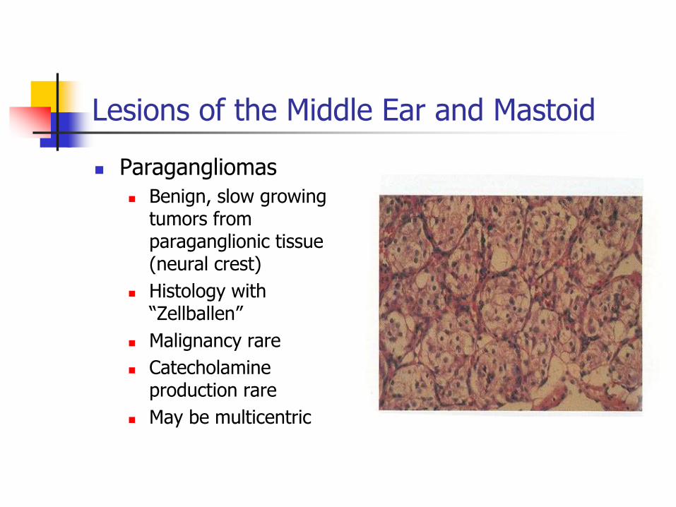
Paragangliomas
Benign, slow growing tumors from paraganglionic tissue (neural crest)
Histology with “Zellballen”
Malignancy rare
Catecholamine production rare
May be multicentric
Lesions of the Middle Ear and Mastoid
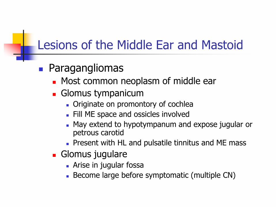
Paragangliomas Most common neoplasm of middle ear
Glomus tympanicum Originate on promontory of cochlea
Fill ME space and ossicles involved
May extend to hypotympanum and expose jugular or petrous carotid
Present with HL and pulsatile tinnitus and ME mass
Glomus jugulare Arise in jugular fossa
Become large before symptomatic (multiple CN)
Lesions of the Middle Ear and Mastoid
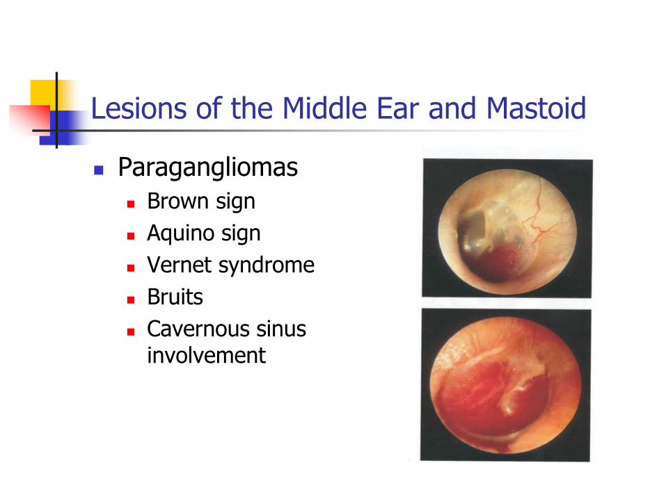
Paragangliomas
Brown sign
Aquino sign
Vernet syndrome
Bruits
Cavernous sinus involvement
Lesions of the Middle Ear and Mastoid
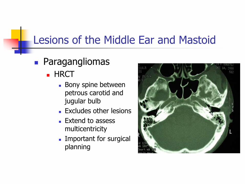
Paragangliomas
HRCT
Bony spine between petrous carotid and jugular bulb
Excludes other lesions
Extend to assess multicentricity
Important for surgical planning
Lesions of the Middle Ear and Mastoid
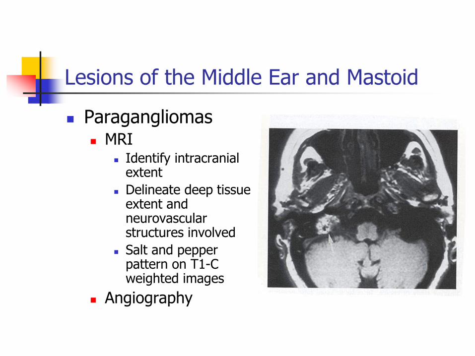
Paragangliomas MRI
Identify intracranial extent
Delineate deep tissue extent and neurovascular structures involved
Salt and pepper pattern on T1-C weighted images
Angiography
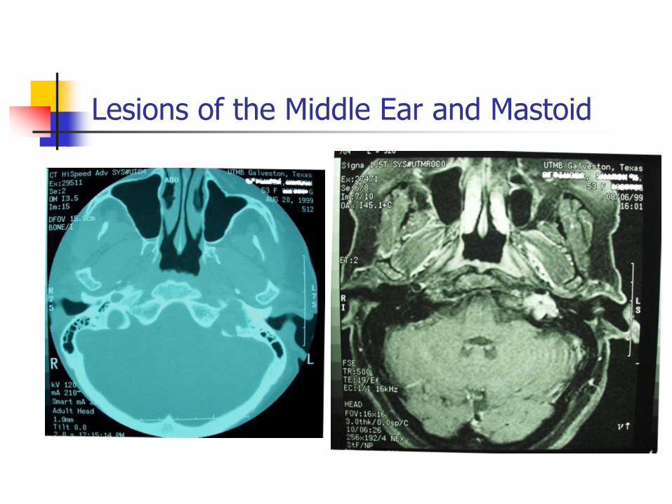
Lesions of the Middle Ear and Mastoid
Lesions of the Middle Ear and Mastoid
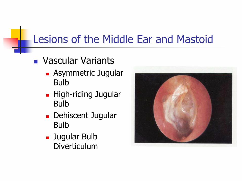
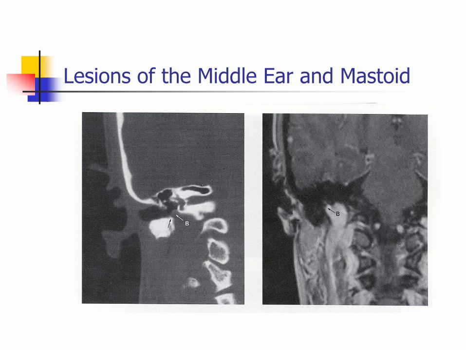
Vascular Variants
Asymmetric Jugular Bulb
High-riding Jugular Bulb
Dehiscent Jugular Bulb
Jugular Bulb Diverticulum
Lesions of the Middle Ear and Mastoid
Lesions of the Middle Ear and Mastoid
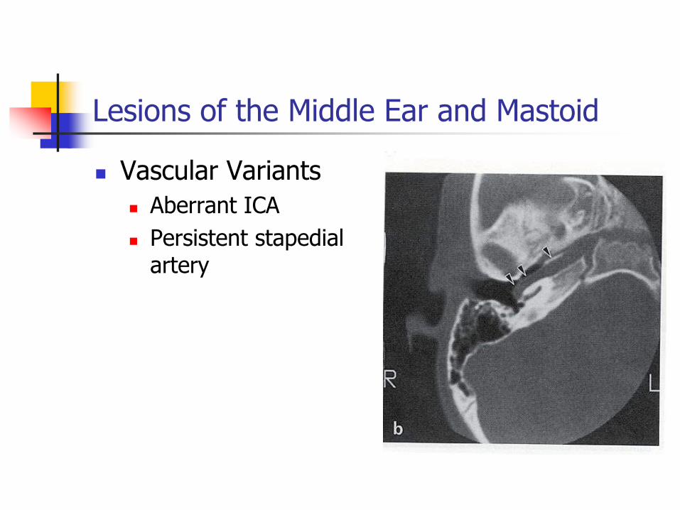
Vascular Variants
Aberrant ICA
Persistent stapedial artery
Lesions of the Middle Ear and Mastoid
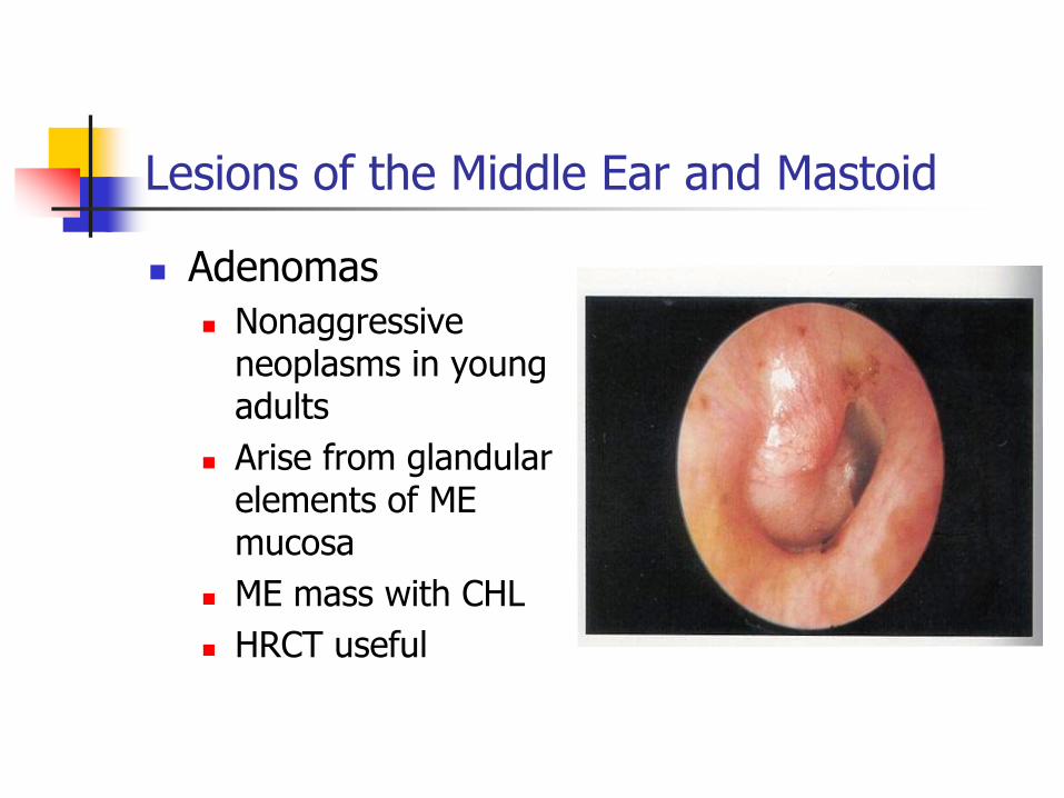
Adenomas
Nonaggressive neoplasms in young adults
Arise from glandular elements of ME mucosa
ME mass with CHL
HRCT useful
Lesions of the Middle Ear and Mastoid
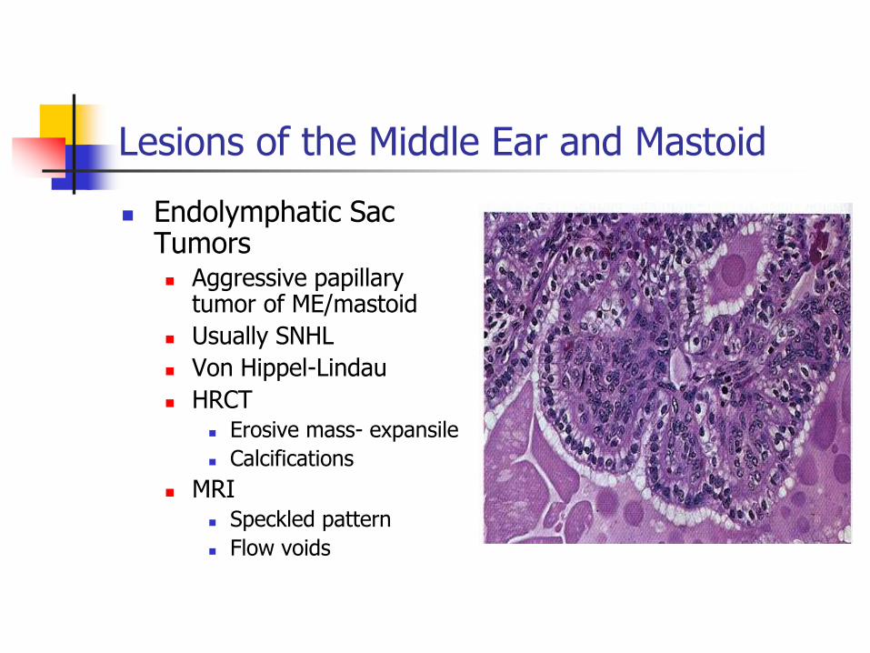
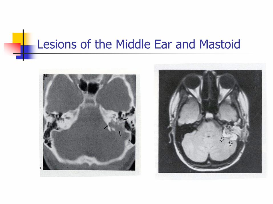
Endolymphatic Sac Tumors Aggressive papillary
tumor of ME/mastoid
Usually SNHL
Von Hippel-Lindau
HRCT Erosive mass- expansile
Calcifications
MRI Speckled pattern
Flow voids
Lesions of the Middle Ear and Mastoid
Lesions of the Middle Ear and Mastoid
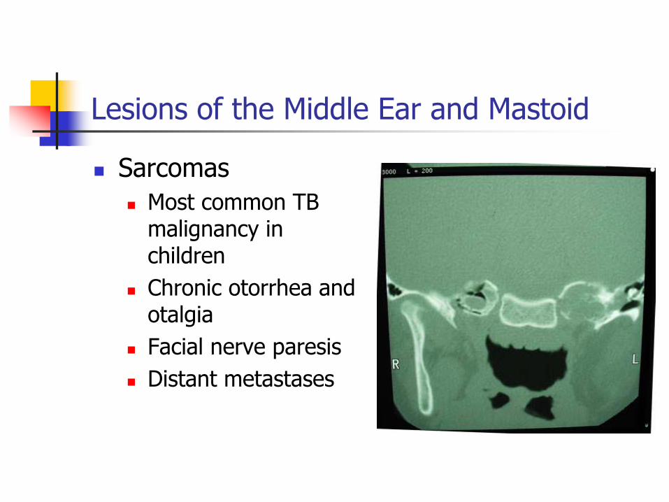
Sarcomas
Most common TB malignancy in children
Chronic otorrhea and otalgia
Facial nerve paresis
Distant metastases
Lesions of the Middle Ear and Mastoid
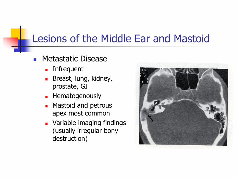
Metastatic Disease
Infrequent
Breast, lung, kidney, prostate, GI
Hematogenously
Mastoid and petrous apex most common
Variable imaging findings (usually irregular bony destruction)
Lesions of the Middle Ear and Mastoid
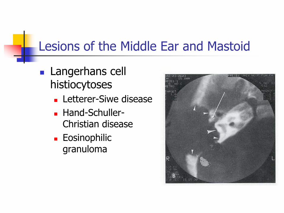
Langerhans cell histiocytoses
Letterer-Siwe disease
Hand-Schuller-Christian disease
Eosinophilic granuloma

Lesions of the Petrous Apex and Clivus
Anatomy
Petrous apex divided by IAC
AM- clivus
AS- floor of middle cranial fossa
Lateral- cochlea/labyrinth
IAC- posterior
Lesions of the Petrous Apex and Clivus
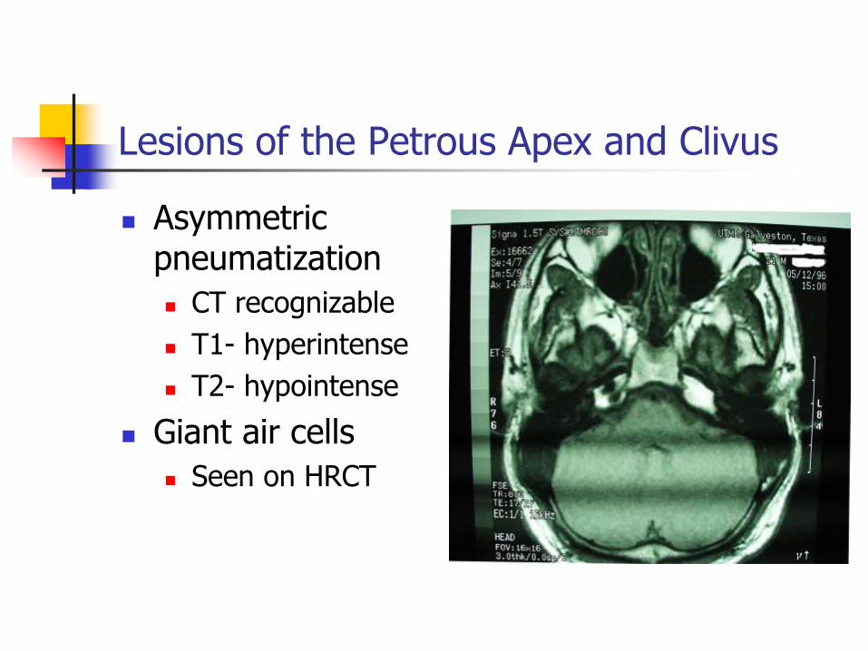
Asymmetric pneumatization
CT recognizable
T1- hyperintense
T2- hypointense
Giant air cells
Seen on HRCT
Lesions of the Middle Ear and Mastoid
Lesions of the Petrous Apex and Clivus
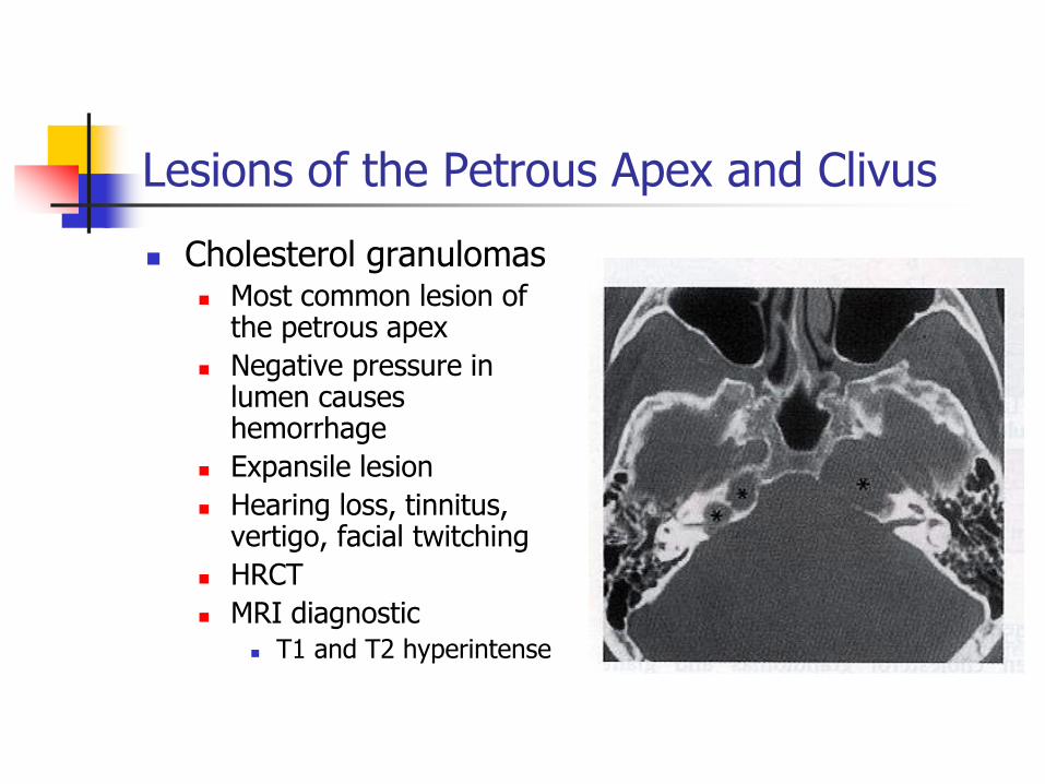
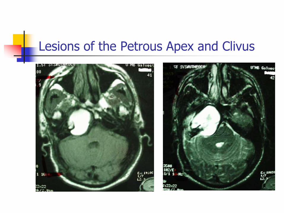
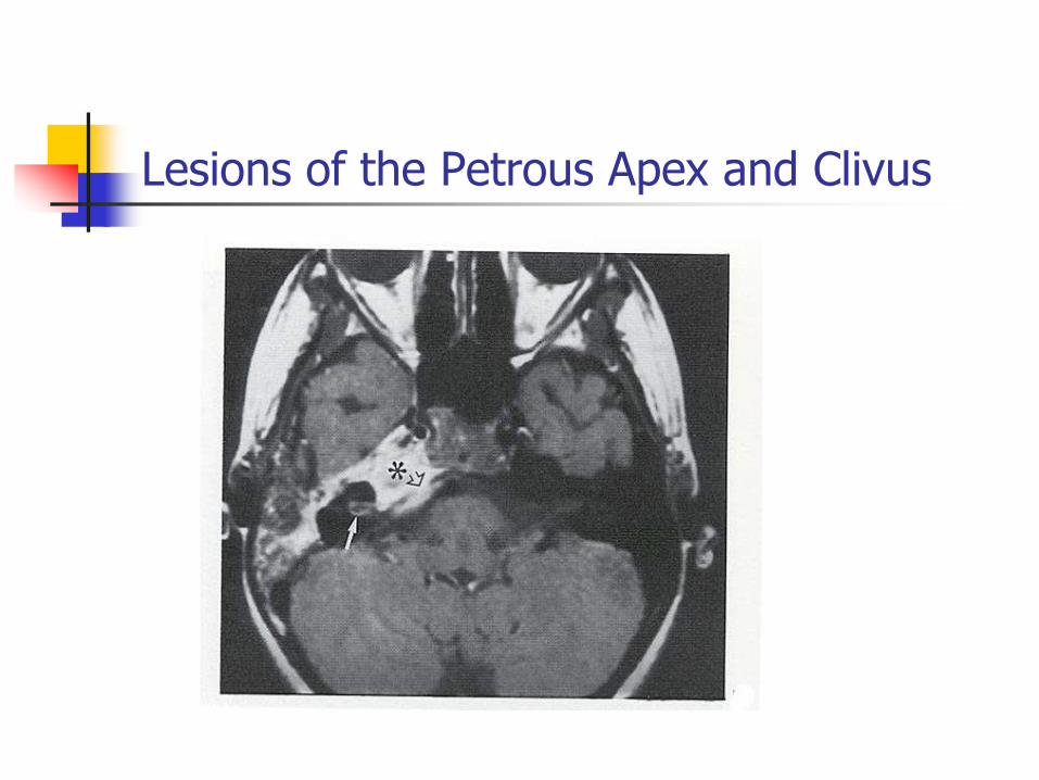
Cholesterol granulomas Most common lesion of
the petrous apex
Negative pressure in lumen causes hemorrhage
Expansile lesion
Hearing loss, tinnitus, vertigo, facial twitching
HRCT
MRI diagnostic T1 and T2 hyperintense
Lesions of the Petrous Apex and Clivus
Lesions of the Petrous Apex and Clivus
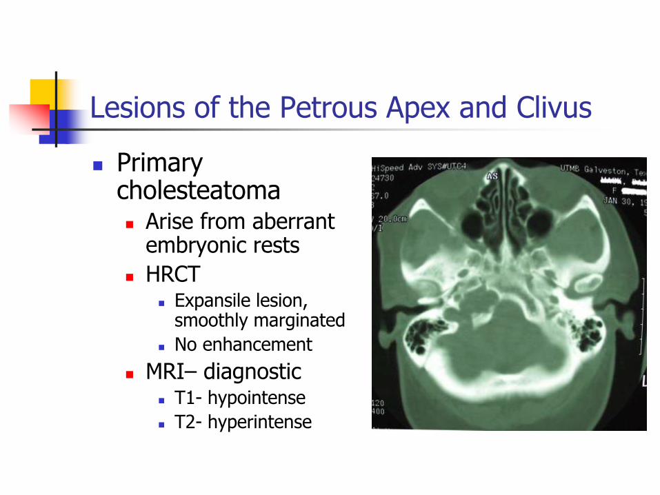
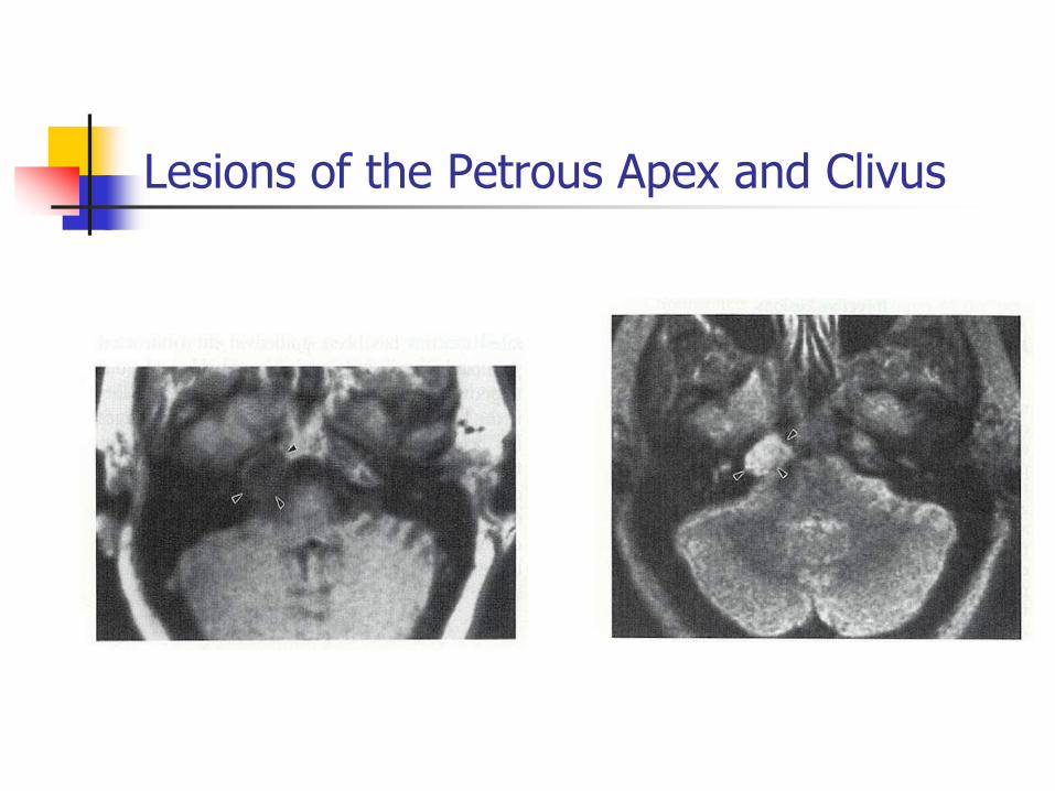
Primary cholesteatoma Arise from aberrant
embryonic rests
HRCT Expansile lesion,
smoothly marginated
No enhancement
MRI– diagnostic T1- hypointense
T2- hyperintense

Lesions of the Petrous Apex and Clivus
Lesions of the Petrous Apex and Clivus
Lesions of the Petrous Apex and Clivus
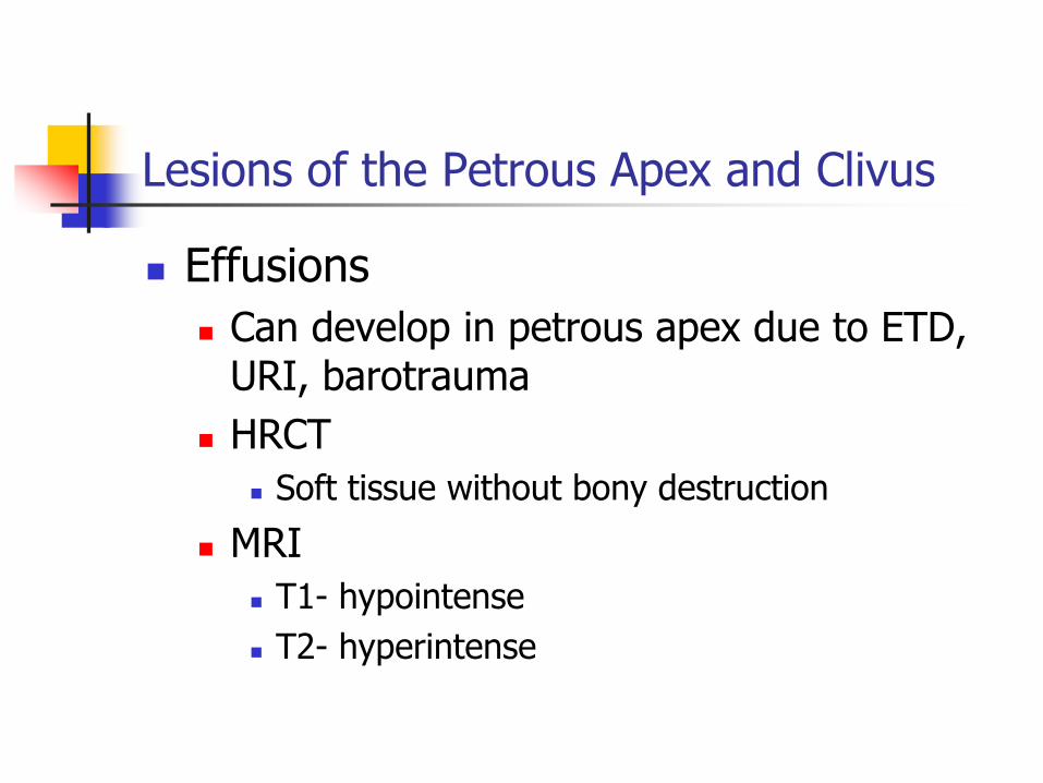
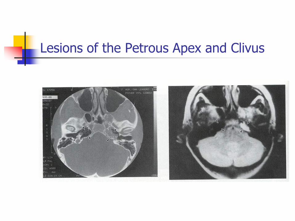
Effusions
Can develop in petrous apex due to ETD, URI, barotrauma
HRCT
Soft tissue without bony destruction
MRI
T1- hypointense
T2- hyperintense
Lesions of the Petrous Apex and Clivus
Lesions of the Petrous Apex and Clivus
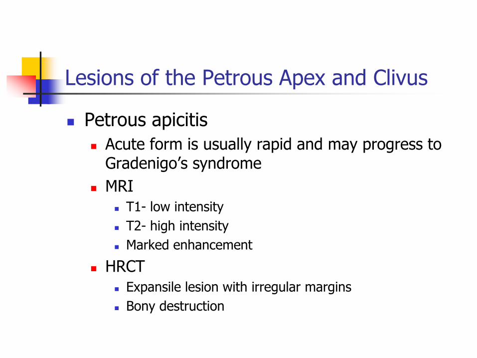
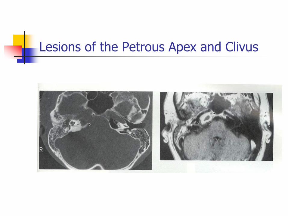
Petrous apicitis
Acute form is usually rapid and may progress to Gradenigo’s syndrome
MRI
T1- low intensity
T2- high intensity
Marked enhancement
HRCT
Expansile lesion with irregular margins
Bony destruction
Lesions of the Petrous Apex and Clivus
Lesions of the Petrous Apex and Clivus

Lesions of the Petrous Apex and Clivus
Skull Base Osteomyelitis Usually after chronic OE
in diabetics or immunocompromised
HRCT Soft tissue density
Demineralization
Irregular lytic lesions
MRI Increased signal on T2
Enhancement
Technetium/Gallium
Lesions of the Petrous Apex and Clivus
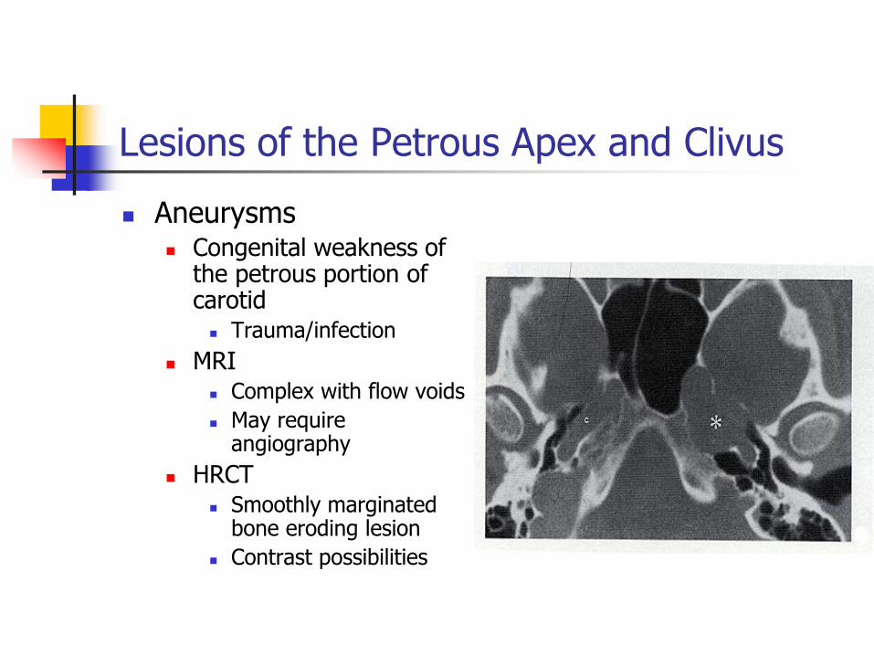
Aneurysms Congenital weakness of
the petrous portion of carotid
Trauma/infection
MRI Complex with flow voids
May require angiography
HRCT Smoothly marginated
bone eroding lesion
Contrast possibilities
Lesions of the Petrous Apex and Clivus
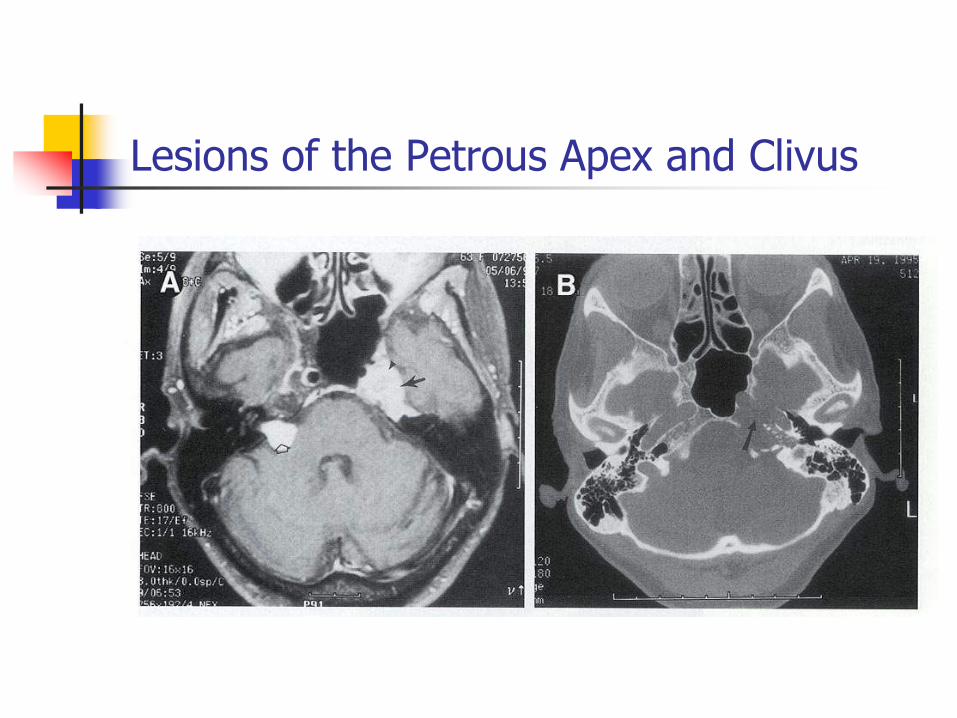
Chondrosarcoma
Arises from embryonic rests of cartilage at foramen lacerum and petrous apex
Headaches and multiple cranial neuropathies
HRCT
Irregular bone destruction
Enhances
Calcifications (popcorn)
MRI
Enhances markedly with gadolinium (chordomas)
Lesions of the Petrous Apex and Clivus
Lesions of the Petrous Apex and Clivus
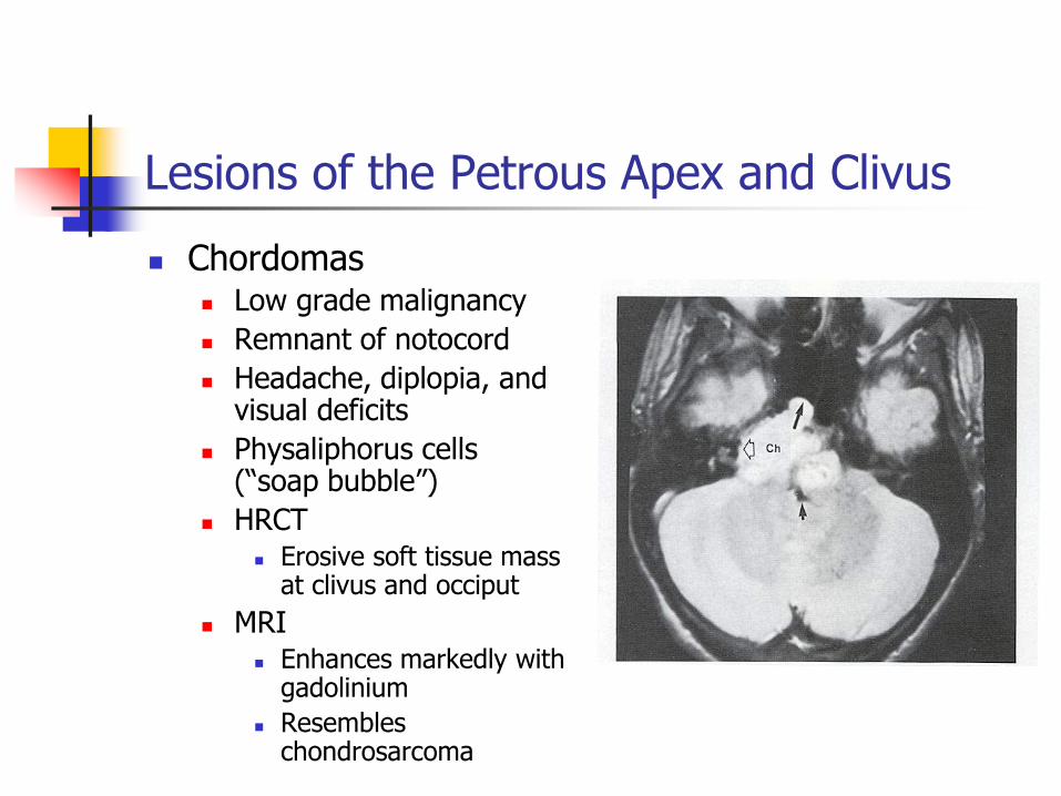
Chordomas Low grade malignancy
Remnant of notocord
Headache, diplopia, and visual deficits
Physaliphorus cells (“soap bubble”)
HRCT Erosive soft tissue mass
at clivus and occiput
MRI Enhances markedly with
gadolinium
Resembles chondrosarcoma
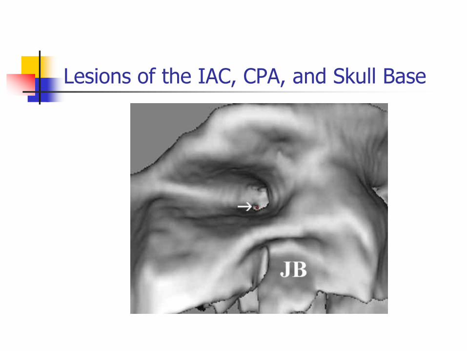
Lesions of the IAC, CPA, and Skull Base
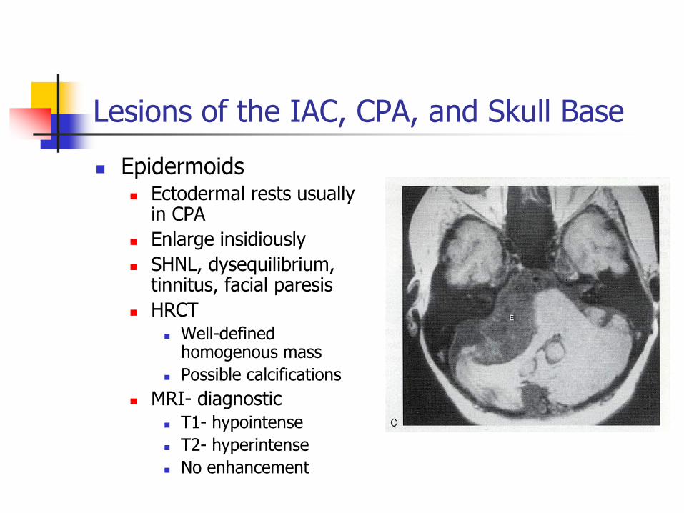
Epidermoids Ectodermal rests usually
in CPA
Enlarge insidiously
SHNL, dysequilibrium, tinnitus, facial paresis
HRCT Well-defined
homogenous mass
Possible calcifications
MRI- diagnostic T1- hypointense
T2- hyperintense
No enhancement
Lesions of the IAC, CPA, and Skull Base
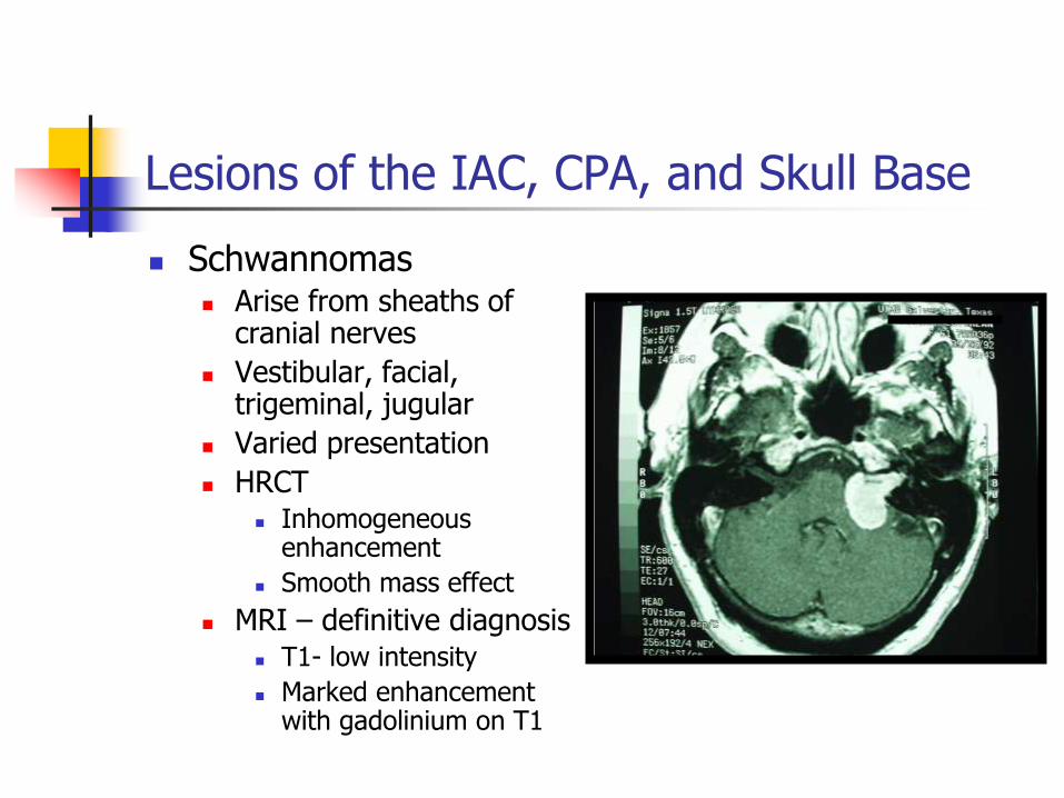
Schwannomas Arise from sheaths of
cranial nerves
Vestibular, facial, trigeminal, jugular
Varied presentation
HRCT Inhomogeneous
enhancement
Smooth mass effect
MRI – definitive diagnosis T1- low intensity
Marked enhancement with gadolinium on T1
Lesions of the IAC, CPA, and Skull Base
Lesions of the IAC, CPA, and Skull Base
Lesions of the IAC, CPA, and Skull Base
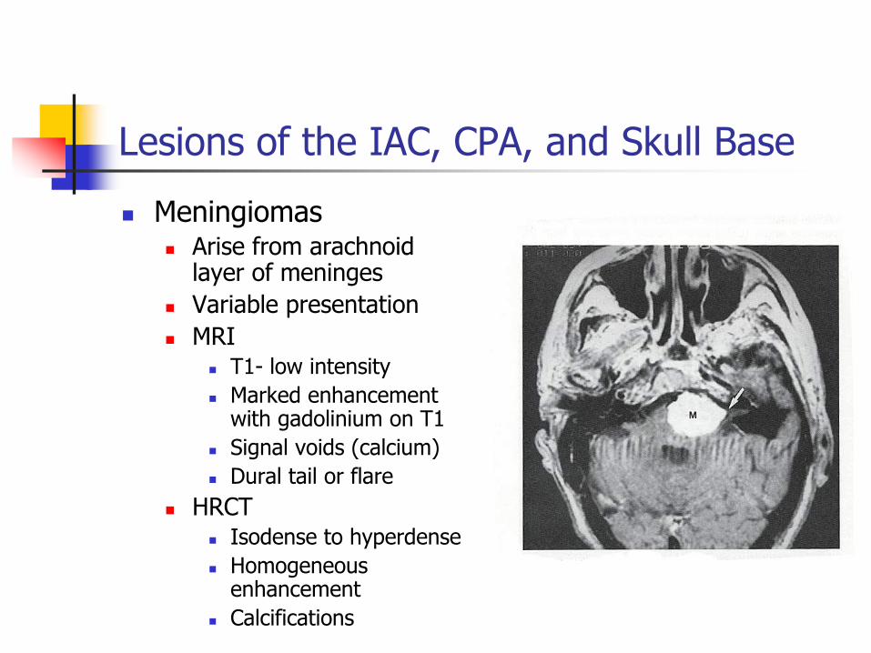
Meningiomas Arise from arachnoid
layer of meninges
Variable presentation
MRI T1- low intensity
Marked enhancement with gadolinium on T1
Signal voids (calcium)
Dural tail or flare
HRCT Isodense to hyperdense
Homogeneous enhancement
Calcifications
Lesions of the IAC, CPA, and Skull Base
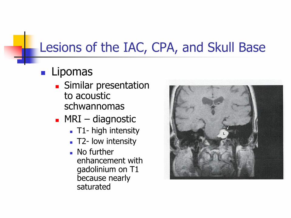
Lipomas Similar presentation
to acoustic schwannomas
MRI – diagnostic T1- high intensity
T2- low intensity
No further enhancement with gadolinium on T1 because nearly saturated
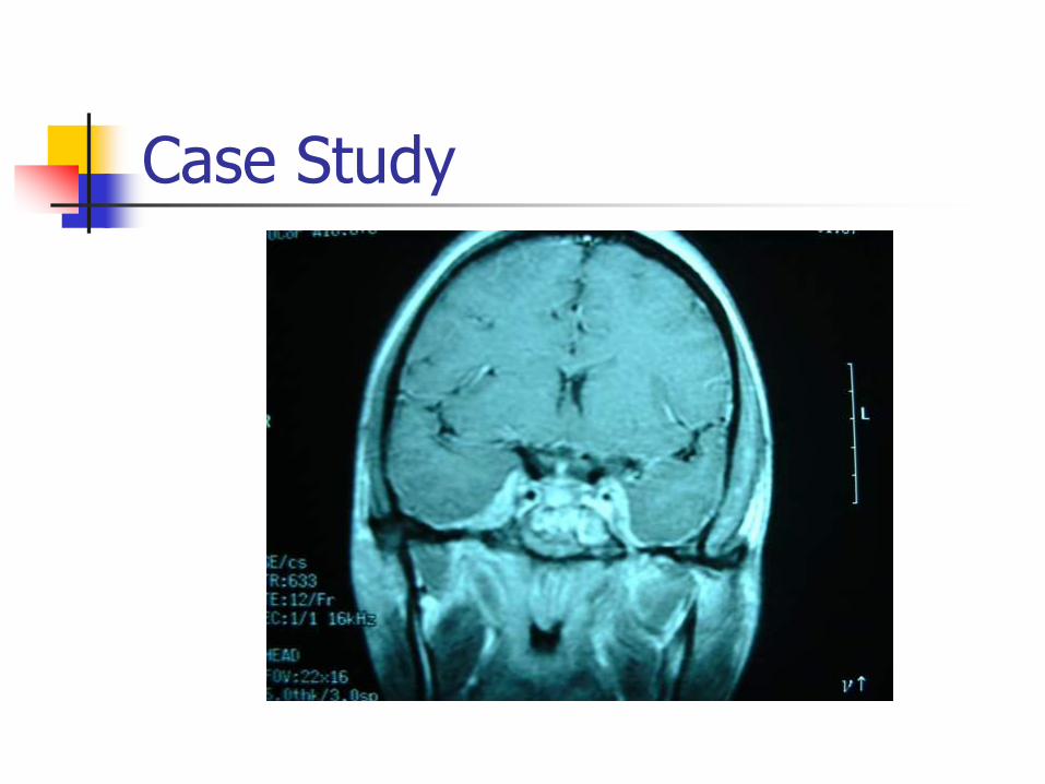
Case Study
21 yo bf present s to clinic with complaint of “drainage from left ear”

Case Study
21 yo bf present s to clinic with complaint of “drainage from left ear”
Pain in left ear, behind left eye and forehead, developed double vision
Experienced fevers, chills, N/V
Swelling in left face
Similar episode one month prior- no money for Abx

Case Study
PMH: ear infections “all life”, no hospitalizations
PSH: none
Meds: castor oil left ear, Tylenol
SH/FH: N/C
ROS: N/C
Case Study
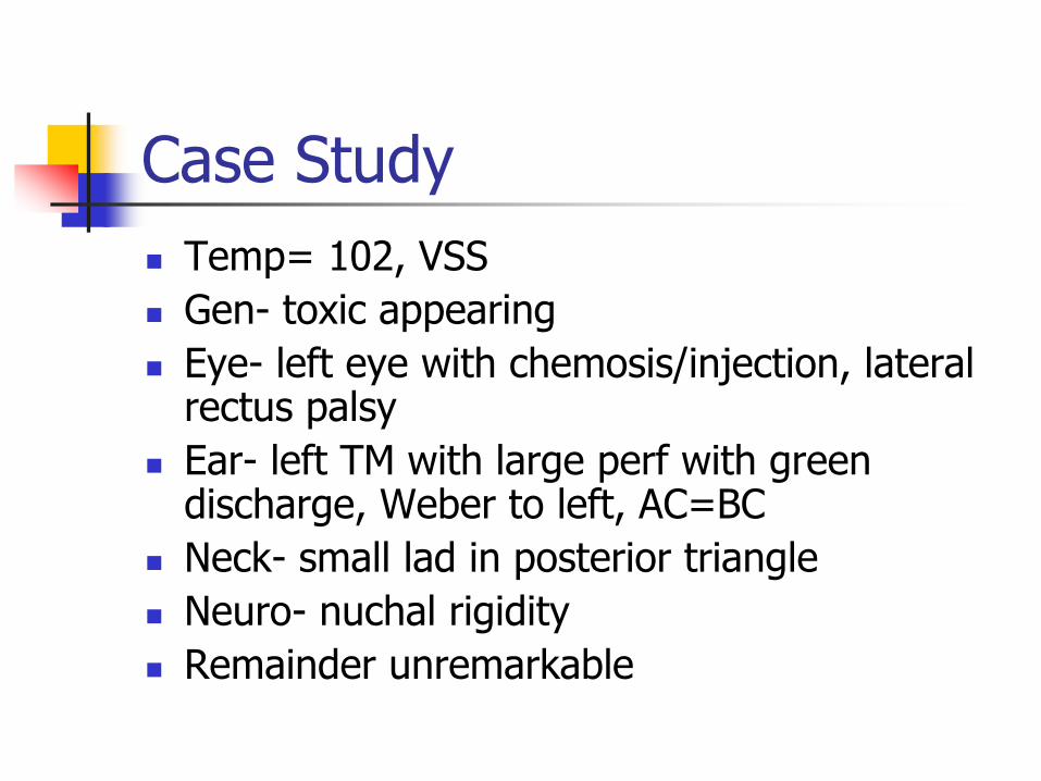
Temp= 102, VSS
Gen- toxic appearing
Eye- left eye with chemosis/injection, lateral rectus palsy
Ear- left TM with large perf with green discharge, Weber to left, AC=BC
Neck- small lad in posterior triangle
Neuro- nuchal rigidity
Remainder unremarkable

Case Study
Labs- WBC= 19.3 with left shift
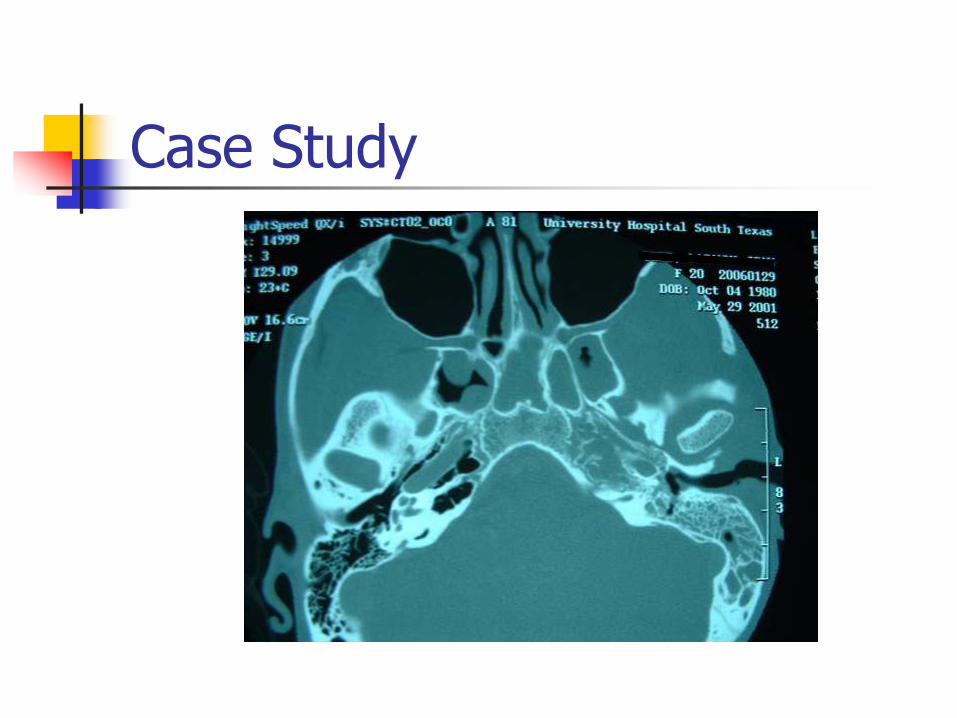
Case Study
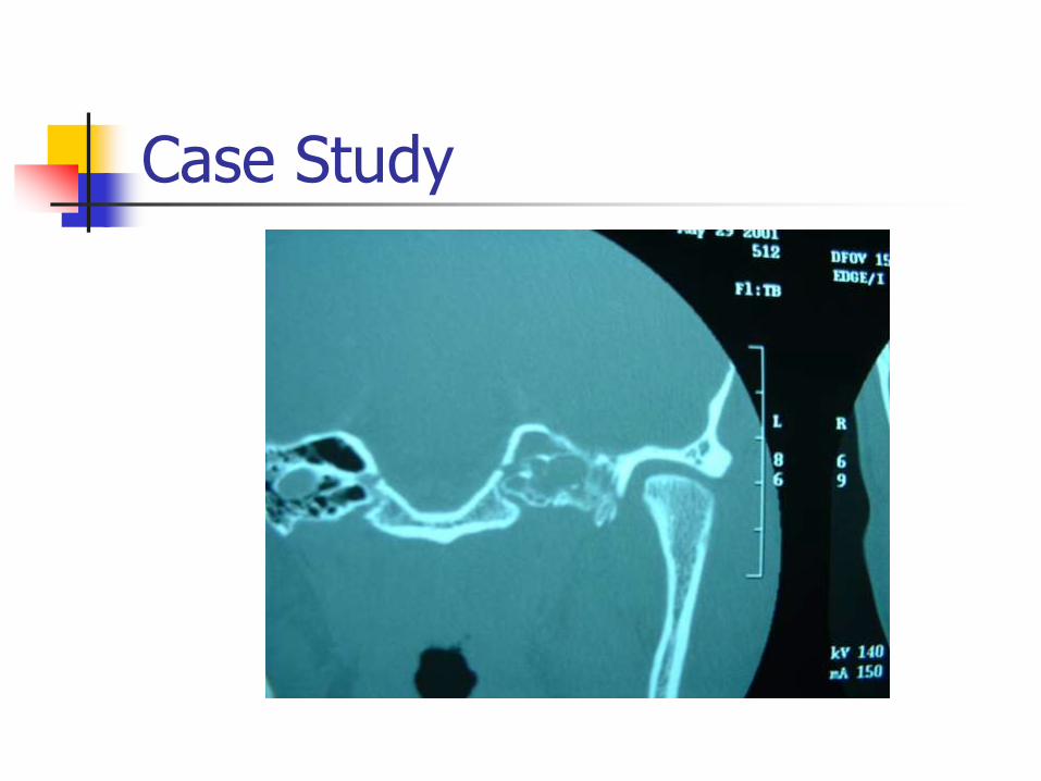
Case Study
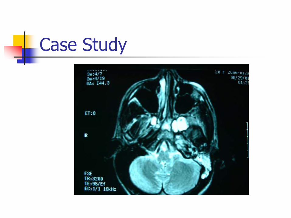
Case Study
Case Study