Embed Size (px)
Citation preview

I. I. LENKOVA, S. N. KACHALOV
DIFFERENTIAL DIAGNOSIS
OF JAW CYSTS
Minsk BSMU 2020

1
МИНИСТЕРСТВО ЗДРАВООХРАНЕНИЯ РЕСПУБЛИКИ БЕЛАРУСЬ
БЕЛОРУССКИЙ ГОСУДАРСТВЕННЫЙ МЕДИЦИНСКИЙ УНИВЕРСИТЕТ
КАФЕДРА ЧЕЛЮСТНО-ЛИЦЕВОЙ ХИРУРГИИ
И. И. ЛЕНЬКОВА, С. Н. КАЧАЛОВ
ДИФФЕРЕНЦИАЛЬНАЯ ДИАГНОСТИКА
КИСТ ЧЕЛЮСТЕЙ
DIFFERENTIAL DIAGNOSIS
OF JAW CYSTS
Учебно-методическое пособие
Минск БГМУ 2020

2
УДК 616.716-003.4-079.4(075.8)-054.6
ББК 56.6я73
Л46
Рекомендовано Научно-методическим советом университета в качестве
учебно-методического пособия 15.01.2020 г., протокол № 5
Р е ц е н з е н т ы: д-р мед. наук, проф., зав. каф. челюстно-лицевой хирургии
Белорусской медицинской академии последипломного образования А. С. Артюшкевич;
канд. мед. наук, доц. каф. стоматологии детского возраста Белорусского государст-
венного медицинского университета А. К. Корсак
Ленькова, И. И.
Л46 Дифференциальная диагностика кист челюстей = Differential diagnosis of jaw
cysts : учебно-методическое пособие / И. И. Ленькова, С. Н. Качалов. – Минск :
БГМУ, 2020. – 20 с.
ISBN 978-985-21-0526-2.
Рассмотрены вопросы клиники, диагностики кист челюстей, дифференциальная диагностика.
Изложены основные лечебные принципы.
Предназначено студентов 5-го курса стоматологического факультета, будет полезно препода-
вателям.
УДК 616.716-003.4-079.4(075.8)-054.6
ББК 56.6я73
___________________________________________
Учебное издание
Ленькова Ирина Иосифовна
Качалов Сергей Николаевич
ДИФФЕРЕНЦИАЛЬНАЯ ДИАГНОСТИКА КИСТ ЧЕЛЮСТЕЙ
DIFFERENTIAL DIAGNOSIS OF JAW CYSTS
Учебно-методическое пособие
На английском языке
Ответственный за выпуск А. С. Ластовка
Переводчик А. А. Царикова
Компьютерная вёрстка Н. М. Федорцовой
Подписано в печать 25.03.20. Формат 6084/16. Бумага писчая «Xerox office».
Ризография. Гарнитура «Times».
Усл. печ. л. 1,16. Уч.-изд. л. 1,11. Тираж 60 экз. Заказ 175.
Издатель и полиграфическое исполнение: учреждение образования
«Белорусский государственный медицинский университет».
Свидетельство о государственной регистрации издателя, изготовителя,
распространителя печатных изданий № 1/187 от 18.02.2014.
Ул. Ленинградская, 6, 220006, Минск.
ISBN 978-985-21-0526-2 © Ленькова И. И., Качалов С. Н., 2020
© УО «Белорусский государственный
медицинский университет», 2020

3
The issue of diagnosis, differential diagnosis, treatment and prevention of
cysts of the jaws is one of the most important for surgeons, dentists and
maxillofacial surgeons.
Odontogenic jaw cysts are a very common pathology. Among operations
performed by dental surgeons on an outpatient basis, operations for odontogenic
jaw cysts occupy one of the first places after a tooth extraction operation.
Among patients admitted to hospitals of maxillofacial surgery, patients with
perforated cysts account for about 8 %. Half of them (46 %) are patients with
suppurative cysts of the jaws [1].
This handbook provides basic information regarding the most common jaw
cysts, the issues of diagnosis, differential diagnosis and their treatment.
Purpose: based on the results of clinical examination, clinical and
radiological data and the results of additional examination methods, to be able to
diagnose jaw cysts, to conduct differential diagnostics with benign growths of
the maxillofacial area.
Tasks of the lesson:
1) to learn how to make an examination plan for patients with cysts of the
jaws, to collect history, to identify clinical symptoms; to be able to carry out
differential diagnostics with benign bone growths of the maxillofacial region;
2) to learn how to read the radiological signs of cysts of the jaws and carry
out a comparative description with the x-ray picture of the jaw tumors, how to
make a plan for the integrated treatment of patients with tumor-like formations
of the jaws.
Requirements for the initial level of knowledge. In order to fully master
the topic, it is necessary to revise the material from the following sections:
• Human morphology:
– topography-anatomical features of the bones of the face;
– blood supply to the head and neck;
– innervation of the maxillofacial region;
– morphology of benign tumors of the bones of the facial skeleton.
Test questions and tasks from related disciplines:
1. List the bones involved in the structure of the maxillofacial region.
2. Anatomical features of the upper jaw.
3. Anatomical features of the localization of the teeth of the upper jaw in
relation to the maxillary sinus.
4. Anatomical structure of the lower jaw.
5. Blood supply and innervation of the upper and lower jaw.
4
LEARNING MATERIAL
Odontogenic cysts are referred to as cavity formations localized inside
the bone, having a connective tissue capsule, lined from the inside with stratified
squamous epithelium, and filled with liquid or semi-liquid contents with
cholesterol crystals. The cavity of an odontogenic cyst is usually filled with
yellow liquid, opalescent due to the presence of cholesterol crystals in it, which
are clearly visible in the form of sparkles when they are absorbed on the surface
of a gauze pad. Sometimes the cyst cavity is filled with a curd gray-white mass.
The epithelium, lining the cyst cavity, originates from the remnants of the tooth-
forming epithelial plate, i.e. Malasse epithelial islets or from the epithelium of
the dental follicle.
Cysts of the jaws are divided into odontogenic and non-odontogenic.
The jaw bones, with some exceptions, are the only bones in which cysts with
epithelial lining are found.
The most detailed classification of jaw cysts is the clinico-morphological
classification of tumors and tumor-like jaw formations, designed by
I. I. Ermolaev, V. V. Panikarovsky, A. I. Paces, B. D. Kabakov took part.,
V. M. Benzianova and S. Ya. Balsevich (1975). Cysts of the jaws are presented
in the section “B. Tumor-like formations” and are divided into:
I. Epithelial cysts.
1. Odontogenic cysts:
a) primary cyst;
b) eruption cyst;
c) paradental cyst;
d) gingival cyst;
e) a tooth-containing cyst;
e) follicular cyst;
g) root (radicular) cyst.
2. Non odontogenic cysts:
a) cyst of the incisal canal (naso-palatine);
b) a globulomaxillary (fissural) cyst;
c) cholesteatoma.
II. Non-epithelial bone cysts:
a) aneurysmatic cysts;
b) traumatic cysts;
c) hemorrhagic cysts.
The morphological classification of odontogenic tumors according to
Yermolaev (1964), which distinguishes two groups of cysts, those of
inflammatory origin and those due to a developmental defect of the tooth-
forming epithelium, is considered to be successful and still used by many
specialists.

5
1. Odontogenic cysts of inflammatory origin:
– radicular;
– tooth-containing;
– paradental.
2. Odontogenic cysts due to a developmental defect of the tooth-forming
epithelium:
– primary;
– follicular;
– teething cysts;
– gingival.
Methods for diagnosing jaw cysts. For the diagnosis of cysts of the jaws,
the following methods are used:
– clinical data;
– X-ray diagnostics;
– morphological diagnosis.
ETIOLOGY AND PATHOGENESIS
OF ODONTOGENIC JAW CYSTS
A cyst is referred to as an abnormal space inside the epithelium lining.
Odontogenic cysts are connected with the teeth, are localized in the bone, and
form a connective tissue capsule lined with epithelial tissue, the cavity of which
is filled with yellowish fluid. Odontogenic cysts are divided into two groups:
inflammatory and dysontogenetic.
The etiology and pathogenesis of the odontogenic jaw cysts are based on
various factors. In the development of cysts of inflammatory genesis,
the primary role is played by chronic inflammatory processes in the periapical
tissues, which lead to the development of radicular cysts of the jaws, since such
formations are in the root zone. The pathogenesis of a radicular cyst has three
stages: initiation, cyst formation, and growth. At the initiation stage,
the stimulation and proliferation of epithelial cells that make up the complex
granulomas, as well as the growth of granulation tissue, occur. Under
the influence of frequent exacerbations of apical periodontitis and malnutrition
of the central divisions of the granuloma, necrosis of some of its sections,
located between epithelial cords (islets), occurs. As a result, cavities are formed
in the thickness of the granuloma, surrounded by the epithelium, which is
the inner lining of the future cyst. The growth of the cyst occurs due to
the intracystic pressure resulting from the accumulation of transudate, which is
constantly produced by the membrane of the formation (Fig. 1). The increase in
the volume of the cyst further occurs due to an increase in the internal

6
hydrostatic pressure with an increase in oncotic pressure in the cyst cavity due to
the presence of such protein macromolecules as albumin, globulin, fibrinogen.
Figure 1. Diagram of the formation of an odontogenic cyst of inflammatory genesis
In the pathogenesis of the formation of dysontogenetic odontogenic cysts
lies a spontaneous proliferation of odontogenic epithelial islets that remain
inside the jaws. Associated with the influence of exogenous and endogenous
factors on epithelial tissue remaining in the bone as a result of a violation of
the laying of the tooth-forming organ (islets Malyasse). But the development of
cysts can also be the result of metaplasia of endothelial cells and the growing
season of the epithelium from the surface of the oral mucosa through
the marginal periodontium (Schuster, I. G. Lukomsky) (Fig. 2). The increase of
the cyst size is associated with accumulation of cystic fluid.
Figure 2. On the left: diagram of the formation of a dysontogenetic cyst, on the right: cysts of
inflammatory genesis

7
INFLAMMATORY ODONTOGENIC CYSTS
Clinic of a radicular (root) cyst. As a rule, a cyst is found in the region of
a previously treated or “periodontitis” tooth (Fig. 3), as well as the tooth that
was injured, less frequently in the region of the extracted tooth (residual cyst).
The absence of any clinical symptoms is characteristic of the initial period of
cyst development, except symptoms of periodontitis in case of its aggravation.
The cyst grows slowly over many months and even years. In the lower jaw,
the first signs of bone destruction are found on the vestibular surface of
the alveolar process; they are characterized by prolapse (protrusion) of the cyst
under the mucous membrane. If the cyst comes from the roots of the second or
third molar of the lower jaw, then it may be located closer to the lingual surface,
since from the vestibular side there is a thick layer of compact and spongy bone.
The neurovascular bundle on the lower jaw is pushed aside by the cyst as it
grows and is not involved in the pathological process (Fig. 4).
Figure 3. Radicular cyst of the upper jaw Figure 4. Radicular cyst of the mandible
In 1 % of cases, we encounter a calcified odontogenic cyst. It is a rare type
of cyst and can occur at any age, mostly between second and third decade of life.
The diameter ranges from 2 to 4 cm and swelling pain may be present. Intrabony
expansions may produce hard bony expansion and may perforate cortical bones.
It can also extend to soft tissue. It can present asymptomatically (Fig. 5).
In the case of a cyst from a tooth, whose root is facing the palate, thinning
and even resorption of the palatal plate is observed. The cyst, which develops
within the boundaries of the maxillary and nasal cavities, extends in their
direction (Fig. 6).
On external examination, the deformation of the face, as a rule, is not
marked. Regional lymph nodes are not enlarged. Smoothness or protrusion of
the transitional fold of the arch of the vestibule of the oral cavity of a rounded
shape with fairly clear boundaries is revealed in the oral cavity. The mucous

8
membrane covering the cyst does not change in color. On palpation,
the overlying bone tissue bends; with a sharp thinning, the so-called parchment
crunch (Dupuytren’s symptom) is determined, in the case of the absence of
a bone there is fluctuation. If there is a significant defect in the jaw bone under
the mucous membrane, a bone window is palpated. A convergence of crowns of
adjacent teeth may be observed. Percussion of the causal tooth is painless.
Electroodontal diagnostics of intact teeth located in the cyst zone reveals
a decrease in electrical excitability (the pulp reacts to a current of more than
6–8 mA) due to compression of the nerve endings by the cyst.
Figure 5. Calcified odontogenic cyst of the lower jaw (Turtle Cyst)
Figure 6. Root cyst, germinated in the maxillary sinus on the left
The inflammation of a radicular cyst is accompanied by the following
symptoms: fever, pain, swelling, and hyperemia of the mucous membrane in
the area of the cyst. Clinically, the process proceeds according to the type of
odontogenic inflammatory disease (periostitis, less often osteomyelitis),
accompanied by regional lymphadenitis, purulent-inflammatory processes in

9
soft tissues. Developing in the upper jaw, a cyst can cause chronic inflammation
of the maxillary sinus, or emit a clinic of odontogenic sinusitis. The transition of
the root cyst to the malignant form is not observed. Radiologically, a cyst is
characterized by the presence of a single homogeneous section of bone tissue
destruction, round or oval in shape with clear boundaries. The root of
the causative tooth is turned into the cystic cavity, periodontal gap is absent.
The root of the causative tooth in the cyst cavity is not resorbed.
Pathological anatomy. The cyst sheath is formed by connective tissue tight
to the bone, and inside there is a lining of stratified squamous epithelium, which
is well supplied by the middle vascular layer. When germinating into the nasal
cavity or maxillary sinus, cysts can be lined with a cylindrical, cubic or ciliated
epithelium. In the shell of a cyst, almost always there are areas of hyperplasia,
erosion or necrosis of part or all of the shell, which is explained by the presence
of an inflammatory process. Characteristic of radicular cysts is the presence of
free cholesterol in the cystic contents and walls.
Differential diagnosis is carried out with other types of cysts of the jaws
and with cystic forms of tumors of the jaw bones (ameloblastoma,
osteoblastoma). A radicular cyst differs from an adamantinoma (an odontogenic
tumor of epithelial origin) in that a clearly defined (rather than a bay-shaped)
defect in the bone is associated with a causal “periodontitis” tooth. In addition,
the history of the disease will be significantly different. A radicular cyst is
differentiated with an osteoblastoclastoma (cystic and cellular form) —
an osteogenic non odontogenic tumor. Clinically, a tumor is characterized by
a spindle-shaped thickening of the jaw. The tumor has 2 types: central (localized
in the bone) and peripheral, which is localized on the mucous membrane of
the alveolar process. However, the leading in the diagnosis is the result of
morphological studies of formations.
Treatment of radicular cysts is surgical or conservative surgical.
The treatment plan includes the removal of a cyst and a causal tooth
(if indicated). In the case of the preservation of the causal tooth, it is necessary
to fill the root canal to the apex with a non-absorbable filling material in
the preoperative period. If the intact teeth, standing nearby, are turned into
the cyst cavity, endodontic treatment is also carried out. During the resection of
the root apex, only 1/3 of its length can be removed in order to maintain a stable
tooth fixation. If more than 1/3 of the length of the root stands in the cavity of
the cyst, the tooth must be removed.
Treatment. According to the indications, 2 methods of treating cysts
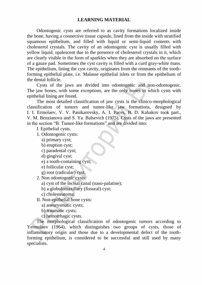
are used: cystotomy (Parch-1), and cystectomy (Parch-2).
Cystotomy (Parch-1) is a method of surgical treatment (partial removal), in
which the external wall of the cyst and the adjacent cortical jaw plate are
resected. The remaining intraosseous cavity, lined with cystic membrane, is
connected with the vestibule of the oral cavity (Fig. 7).
10
Figure 7. Cystotomy scheme
Indications for cystotomy:
1) old age, debilitated and depleted patients (due to the low potential of
bone tissue regeneration);
2) patients with concomitant diseases, when prolonged traumatic (radical)
surgery is undesirable or impossible;
3) extensive cysts of the lower jaw with a sharp thinning of the base of
the jaw (thick bone less than 1–0.5 cm);
4) early age, in view of the impossibility of complete removal of the cyst
membrane without injuring the rudiments of the teeth.
Cystectomy (Parch-2) is a radical operation consisting in the complete
removal of the cyst membrane with subsequent suturing of the surgical wound.
Indications for cystectomy are: 1) a cyst of small size, located within 1–2 intact teeth;
2) an extensive cyst of the lower jaw, in which there are no teeth in its zone
and a sufficient thickness (up to 1 cm) of the jaw base is preserved;
3) a large cyst on the upper jaw, which has no teeth in this area, with
a preserved bony wall of the bottom of the nasal cavity;
4) a cyst adjacent to the maxillary sinus or pushing it without symptoms of
sinus inflammation.
Residual cyst occurs as a consequence of incomplete surgical extirpation of
the apical cytogranule after tooth extraction. Its clinical and histological
characteristics are identical to the root cyst. Radiographically, it manifests itself
as radiolucency of various sizes in the area of the previous extraction of
the tooth (Fig. 8). Treatment is cystectomy.

11
Figure 8. Residual cyst in the region of the extracted tooth 46, adjacent to the intact 45
Paradental cyst (eruption cyst, retromolar cyst). In the case of difficulty in
the eruption of the third molar of the lower jaw, a bone pocket filled with fibrous
tissue is formed between the jaw bone and the tooth. Paradental cysts develop
from epithelial cells (squamous epithelium of the oral cavity) ectopied into
the fibrous tissue from the mucosal surface. In the presence of inflammation,
epithelial cells, differentiating, form small cavities. Over time, the message of
the bone pocket with the oral cavity is stopped and the epithelial cavities are
isolated. As they grow, they merge into a single cavity with the formation of
a cyst. A feature of this cyst is the presence of fibrous strands connecting the
shell with the periodontal of impacted tooth. Macroscopically, the cyst envelope
and its contents are no different from radicular and follicular cysts.
Clinic. The cyst grows asymptomatically, so it is extremely difficult to
identify it in the initial stage of development. As it grows, it appears as a small,
limited and slightly painful swelling in the area of an impacted wisdom tooth
(Fig. 9). The inflammation is accompanied by the symptoms characteristic of
retromolar periostitis.
Figure 9. Paradental cyst in the area of a dystopic laxed lower wisdom tooth (covering an impacted
tooth)
X-ray determines the destruction of bone tissue behind the impacted lower
wisdom tooth in the form of a sickle-shaped half moon (Wassmund semi-moon).

12
A small paradental cyst encloses the crown of an impacted tooth in part, for
large ones, the tooth is completely covered. Differential diagnosis is performed
with cysts of the jaws and cystic forms of tumors of the jaw bones. Treatment:
Both cystotomy and cystectomy are used with mandatory removal of
the causative tooth.
Tooth-containing cysts are formed in children, in the period of mixed bite,
localized in the area of temporary, caries-affected molars, repeatedly subjected
to unsuccessful treatment or untreated (root cyst of a temporary tooth). In this
case, the crown of the forming permanent tooth is immersed in the cavity of
the cyst, and the root with the growth zone is located outside the shell.
The treatment consists of cystotomy, in order to preserve the germ of
a permanent tooth and remove the causal temporary one.
ODONTOGENIC CYSTS DUE TO A DEVELOPMENTAL DEFECT
OF THE TOOTH-FORMING EPITHELIUM
A follicular cyst is more often found at a younger age but can be observed
at any age, developing around a crown of the impacted tooth. Localization site
in either the upper or lower jaw. These cysts are associated with impaired
development of the tooth germ, may occur at any stage of tooth formation. Most
often, a follicular cyst occurs after the end of tooth development (contains
a fully formed tooth).
Clinic of follicular cyst is in many respects similar to that of radicular cyst.
Cyst growth is asymptomatic. On examination of the patient, it is possible to
identify a preserved milk tooth and the absence of a permanent tooth in
the dentition (except for the development of a cyst from a supernumerary tooth).
Follicular cysts rarely suppurate.
Pathological anatomy. A follicular cyst is characterized by the presence
of a single-chamber cavity filled with clear liquid with cholesterol crystals.
The crown of an impacted tooth is facing the cyst cavity.
Radiographically determined are the focus of destruction of bone tissue,
homogeneous structure, round or oval in shape with clear even boundaries like
a monocystic lesion and the presence of a retained tooth with a crown facing
the cyst cavity. The root of the tooth is usually located outside the cyst (Fig. 10).
Differential diagnosis should be performed with cysts of the jaws and
cystic forms of tumors of the jaw bones (ameloblastoma, osteoblastoclastoma).
The treatment consists in the complete removal of the cystic formation
together with the causative tooth; in children it is cystotomy, in view of
impossible complete removal of the cyst membrane without trauma to
the rudiments of the teeth.

13
Figure 10. Follicular cyst in the lower jaw on the right (impacted tooth 43)
The primary cyst (keratocyst) develops, as a rule, on the lower jaw and
makes up 1 % of all jaw cysts. It is found in middle-aged and older people.
It has the ability to epithelium of the cyst envelope to keratinization, it can recur
and turn into a malignant form. With a considerable size, the cyst spreads into
the body, the angle and the branch of the jaw, causing great destruction of
the bone.
Clinic. The disease begins imperceptibly and does not manifest itself for
a long time. In some patients, a cyst is detected due to the addition of
the inflammatory process, sometimes it is found by chance during X-ray
examination for other diseases, and it is also determined if it is large when
the body, angle, and jaw branch are affected. There is no connection between
the appearance of a cyst and the pathology of the teeth. In general, the clinical
symptoms of a primary odontogenic cyst are not different from other jaw cysts.
Radiographically it appears as a focus of destruction of bone tissue with
clear contours. With a significant cyst size, uneven destruction of the bone
creates the impression of a multi-chamber formation. The coronary and condylar
processes are involved in the process, the cortical plate becomes thinner and
absent in some areas, the periodontal crevices of the roots of the teeth projecting
to the cyst area are usually determined (Fig. 11).
Figure 11. Primary cysts in the body and the angle of the mandible

14
Keratocyst must be differentiated from ameloblastoma, osteoblastoma and
other jaw cysts. Treatment is usually cystectomy. Teething cysts are formed
during the permanent teeth eruption in children, less often during milk teeth
eruption. If we consider the official statistics, in 40 % of all reported cases this is
the eruption of milk teeth; in the remaining 60 % in the permanent bite.
NON-ODONTOGENIC JAW CYSTS
Pathogenetic non-odontogenic cysts of the jaws are not associated either
with the teeth or with impaired development of the tooth-forming epithelium.
They are formed due to the violation of facial embryogenesis (embryonic
dysplasia). These are so-called fissural (fissure) cysts and develop in the
embryonic period on the border of the embryonic facial processes. Localization
in the upper jaw is rare. Depending on the location site, the following fissural
cysts are distinguished: nasopalatine, globulomaxillary, and nasoalveolar cysts.
Nasopalatine duct cysts (NPDC), also known as Incisive canal cysts,
develop from the embryonic remnants of the epithelium. The latter connects
the bottom of the nasal cavity and the oral cavity. A cyst can form in any part of
the canal, but much more often in its lower parts. Pathological structure of
the cyst depends on its localization. In the upper section of the canal (closer to
the nasal cavity), the cysts are lined with a cylindrical or shimmering epithelium,
in the lower sections — with multi-layered flat.
Clinic. The nasal cyst is located between the central incisors. Cyst growth
is slow, painless. With an increase in its size, destruction of the palatine bone
occurs in the anterior section, behind the intact central incisors, a hemispherical
protrusion with clear boundaries appears. On puncture, clear liquid with
cholesterol crystals can be found. The cysts can be inflamed.
Radiographically, in the area of the site
where the incisal foramen should be located,
the center of bone tissue destruction with clear
boundaries, rounded in shape, located strictly
along the midline is determined. The intact
roots of the incisors are projected onto it with
the periodontal gap preserved (Fig. 12).
When conducting a differential diagnosis
with a radicular cyst, it is necessary to
determine on the radiograph the presence or
destruction of the periodontal gap of the tooth,
which is projected onto the cyst. The latter
indicates an existing radicular cyst. The final
diagnosis is established after histopathological
examination. Figure 12. Cyst of the incisal canal

15
Treatment: cystectomy with preservation of the central incisors, but in
the preoperative period endodontic treatment of the latter is carried out.
Globulomaxillary cyst (intramaxillary, spherical — maxillary) is localized
between the intact lateral incisor and the canine of the upper jaw. It is formed
from epithelial cells at the junction of the frontal (processus globularis) and the
maxillary (processus maxillaris) facial embryonic processes. The sheath is thin,
containing flat cubic and cylindrical epithelium (Fig. 13, a).
The clinic is similar to other cysts, but it can grow into the nasal cavity or
the maxillary sinus. The inflammation is rare. The contents of the cyst are clear
liquid with cholesterol crystals. Diagnosis is complicated if the lateral incisor or
canine is destroyed (periodontal), in this case the differential diagnosis with
a radicular cyst is carried out.
Treatment: cystectomy with tooth preservation; endodontic treatment in
the preoperative period (Fig. 13, b).
Figure 13. Globulomaxillary cyst:
a — condition before cystectomy; b — condition after cystectomy
Nasoalveolar cyst (nasolabial cyst of the nasal vestibule). This congenital
soft tissue cyst is localized in the region of the upper lip between the canine and
the lateral incisor. It develops from the remnants of the embryonic epithelium on
the border of three embryonic processes, i.e. frontal, external nasal and
maxillary. The cyst sheath is lined with flat, cubic, transitional or ciliated
epithelium.
Clinic. The cyst size increase causes deformation (depression) of the outer
cortical plate. It is palpable as a sedentary, elastic protrusion of a rounded shape
with clear boundaries and disconnected with the surrounding tissues in the area
of the nasolabial sulcus under the base of the wing of the nose (Fig. 14).
There may be a narrowing of the entrance to the nose. Its content is clear,
yellowish, somewhat viscous, liquid with cholesterol crystals. On X-ray,
the cyst is extraossal and cannot be detected on the roentgenogram, in some
a b
16
cases small rounded enlightenment (due to the deepening) of the bone tissue
at the site of the cyst is determined. The teeth in the cyst zone are intact.
The diagnosis is specified during surgery.
Figure 14. Nasoalveolar cyst
NON-EPITHELIAL BONE CYSTS
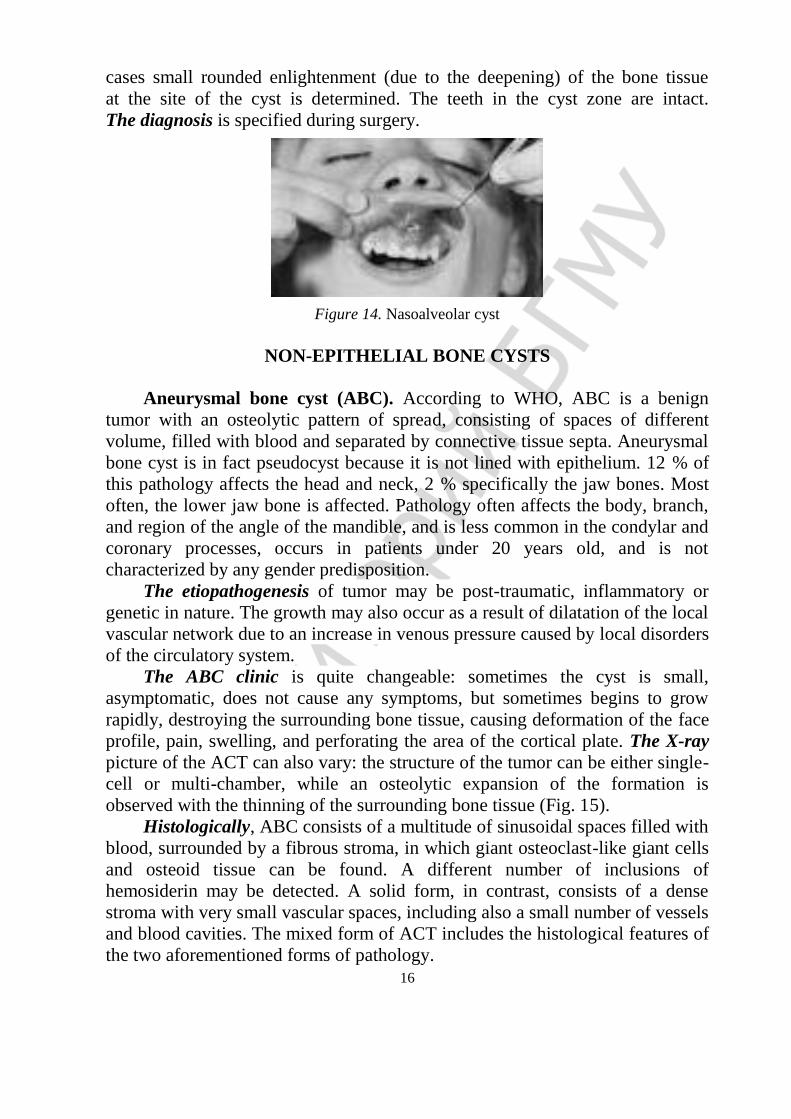
Aneurysmal bone cyst (ABC). According to WHO, ABC is a benign
tumor with an osteolytic pattern of spread, consisting of spaces of different
volume, filled with blood and separated by connective tissue septa. Aneurysmal
bone cyst is in fact pseudocyst because it is not lined with epithelium. 12 % of
this pathology affects the head and neck, 2 % specifically the jaw bones. Most
often, the lower jaw bone is affected. Pathology often affects the body, branch,
and region of the angle of the mandible, and is less common in the condylar and
coronary processes, occurs in patients under 20 years old, and is not
characterized by any gender predisposition.
The etiopathogenesis of tumor may be post-traumatic, inflammatory or
genetic in nature. The growth may also occur as a result of dilatation of the local
vascular network due to an increase in venous pressure caused by local disorders
of the circulatory system.
The ABC clinic is quite changeable: sometimes the cyst is small,
asymptomatic, does not cause any symptoms, but sometimes begins to grow
rapidly, destroying the surrounding bone tissue, causing deformation of the face
profile, pain, swelling, and perforating the area of the cortical plate. The X-ray
picture of the ACT can also vary: the structure of the tumor can be either single-
cell or multi-chamber, while an osteolytic expansion of the formation is
observed with the thinning of the surrounding bone tissue (Fig. 15).
Histologically, ABC consists of a multitude of sinusoidal spaces filled with
blood, surrounded by a fibrous stroma, in which giant osteoclast-like giant cells
and osteoid tissue can be found. A different number of inclusions of
hemosiderin may be detected. A solid form, in contrast, consists of a dense
stroma with very small vascular spaces, including also a small number of vessels
and blood cavities. The mixed form of ACT includes the histological features of
the two aforementioned forms of pathology.
17
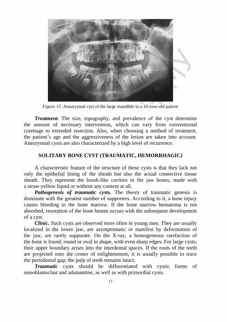
Figure 15. Aneurysmal cyst of the large mandible in a 10-year-old patient
Treatment. The size, topography, and prevalence of the cyst determine
the amount of necessary intervention, which can vary from conventional
curettage to extended resection. Also, when choosing a method of treatment,
the patient’s age and the aggressiveness of the lesion are taken into account.
Aneurysmal cysts are also characterized by a high level of recurrence.
SOLITARY BONE CYST (TRAUMATIC, HEMORRHAGIC)
A characteristic feature of the structure of these cysts is that they lack not
only the epithelial lining of the sheath but also the actual connective tissue
sheath. They represent the brush-like cavities in the jaw bones, made with
a straw-yellow liquid or without any content at all.
Pathogenesis of traumatic cysts. The theory of traumatic genesis is
dominant with the greatest number of supporters. According to it, a bone injury
causes bleeding in the bone marrow. If the bone marrow hematoma is not
absorbed, resorption of the bone beams occurs with the subsequent development
of a cyst.
Clinic. Such cysts are observed more often in young men. They are usually
localized in the lower jaw, are asymptomatic or manifest by deformation of
the jaw, are rarely suppurate. On the X-ray, a homogeneous rarefaction of
the bone is found, round or oval in shape, with even sharp edges. For large cysts,
their upper boundary arises into the interdental spaces. If the roots of the teeth
are projected onto the center of enlightenment, it is usually possible to trace
the periodontal gap; the pulp of teeth remains intact.
Traumatic cysts should be differentiated with cystic forms of
osteoblastoclast and adamantine, as well as with primordial cysts.

18
The diagnosis of a traumatic cyst can usually be established only during
the operation on the basis of the absence of a cystic membrane. Surgical
treatment involves the simple opening of the cystic cavity, followed by suturing
the wound tightly. The blood clot formed in the cavity leads to further bone
regeneration in the cyst area.
TEST QUESTIONS
1. What is the classification of jaw cysts used in practical work of
maxillofacial surgeons and dentists.
2. List the methods for diagnosing jaw cysts.
3. Give the definition of a cyst.
4. Name the structure of the sheath of the cyst.
5. Name the types of odontogenic cysts of inflammatory genesis.
6. Name the types of odontogenic cysts developing in the process of
dysontogenesis.
7. What are the possible options for the surgical treatment of cysts of
the jaws, the indications for them.
8. Identify the characteristic clinical signs of a root cyst.
9. Specify the growths with which the differential diagnosis of cysts of
the jaws is required.
10. Name non-epithelial bone cysts.
Answers to test questions:
1. Classification of odontogenic tumors according to Yermolaev (1964),
clinical and morphological classification of tumors and tumor-like formations.
2. Clinical data, radiological and morphological diagnostics.
3. A cyst is understood as an “abnormal” space inside the lining of
the epithelium.
4. The outer layer is the connective tissue, the inner layer is the epithelium,
between them there is a lesion.
5. Radicular, tooth-containing, paradental.
6. Follicular, primary, cyst of the incisal canal (naso-palatine),
globulomaxillary (fissural) cyst, cholesteatoma.
7. Cystotomy — Parch 1, cystectomy — Parch 2.
8. When the cyst is small in size, the process is asymptomatic, with a large
size, deformity of the alveolar process, Dupuytren symptom, convergence of
dental crowns associated with the cyst. In inflammation there appear
corresponding signs.
9. The differential diagnosis of cysts of the jaws is required with other cyst
types as well as with cystic forms of tumors of the jaw bones (ameloblastoma,
osteoblastoma).
10. Aneurysmal, traumatic, hemorrhagic.
19
CASE STUDIES
Case 1. A 54-year-old man turned to a dental surgeon to resolve the issue
of implant placement, wanted to replace the existing orthopedic structures.
An orthopantomogram (OPG) available. During external examination without
pathological changes. Regional lymph nodes not palpable. The mouth opens in
full. Palpation of the lower jaw did not reveal its deformation, oral mucosa was
pink, moist, without pathological elements. Orthognathic bite. Dental rows are
made of non-removable metal structures.
The OPTG shows a focus of bone tissue destruction, with clear even
boundaries, associated with the tips of the roots of the tooth 47. The tooth 47 has
been previously treated endodontically. The buccal canals are sealed to 1/2
the length of the root. Buccal roots protrude 1/2 to the cyst cavity. The size of
the cyst is 1.5 × 2.0 cm.
Questions:
1. Suggest a clinical diagnosis.
2. Specify the complications that are possible with the further course of
the disease.
3. Specify other additional and appropriate examination methods.
4. Offer a method for the treatment of identified pathology.
Answers:
1. Radicular cyst of the lower jaw on the right in the area of the tooth 47.
2. Traumatic neuropathy of the third branch of the trigeminal nerve on
the right, pathological fracture of the lower jaw body on the right, suppuration of
the radicular cyst.
3. Cone-beam computed tomography (CBCT).
4. Tooth extraction 47, cystectomy on the lower jaw on the right.
Case 2. A man, 30 y.o. turned to a dental surgeon about the displacement
of the medial incisors of the upper jaw. OPTG was available. External
examination without pathological changes. Regional lymph nodes are not
palpable. The mouth opens in full, the oral mucosa is pink, moist, without
pathological elements. Palpation of the upper jaw did not reveal its deformation.
Orthognathic bite. The OPTG is a center of bone tissue destruction between
the roots of the teeth 11 and 12, with clear even borders, 1.5 cm in diameter.
The teeth 11 and 21 are intact.
Questions:
1. Suggest a clinical diagnosis.
2. Specify the complications that are possible with the further course of
the disease.
3. Suggest additional survey methods.
4. Offer a method for the treatment of identified pathology.

20
Answers:
1. Cyst of the nasolabial (incisal) canal.
2. Suppuration of the cyst.
3. Cone-beam computed tomography (CBCT), conducting electrical donor
diagnostics (EDI) of teeth 11 and 12.
4. Cystectomy with endodontic dental treatment in the preoperative period.
REFERENCE
1. Аветиков, Д. С. Современные подходы к классификации кист челюстей /
Д. С. Аветиков, И. В. Яценко // Стоматологiчнi аспекти : сб. науч. тр. Украинской меди-
цинской стоматологической академии. г. Полтава, «Проблеми екології та медицини».
2012. Т. 16, № 1–2. С. 1–6.
2. Бернадский, Ю. И. Основы хирургической стоматологии / Ю. И. Бернадский.
Киев : Здоров’я, 1970. С. 354–361.
3. Ефимов, Ю. В. Хирургическое лечение околокорневых кист челюстей /
Ю. В. Ефимов // Стоматология. 1993. № 3. С. 26–27.
4. Кисты челюстей и их лечение / И. В. Иванов [и др.] // Новое в стоматологии :
сб. науч. тр. Юга России. 2000. С. 152–157.
5. Карапетян, И. С. Опухоли и опухолеподобные поражения органов полости
рта, челюстей, лица и шеи / И. С. Карапетян, Е. Л. Губайлуллина, Л. H. Цегельник.
Москва : МИА, 2004. 232 с.
6. Солнцев, А. М. Кисты челюстно-лицевой области и шеи / А. М. Солнцев,
В. С. Колесов. Киев : Здоров’я, 1982. 144 с.
7. Соловьев, М. М. Оперативное лечение одонтогенных кист / М. М. Соловьев,
Г. М. Семенов, Д. В. Галецкий. Санкт-Петербург : Наука, 2004. 127 с.
8. Соловьев, Ю. П. Новые нозологические формы классификации опухолей
костей / Ю. П. Соловьев. Москва : Медицина, 1998. С. 57–61.
9. Тимофеев, А. А. Челюстно-лицевая хирургия / А. А. Тимофеев. Киев : Медицина,
2010. С. 57–61.
10. Калужская, С. М. Зубосодержащие кисты у детей и подростков / С. М. Калуж-
ская, Л. В. Макаренкова // Вестник Смоленской медицинской академии. 2007. № 2.
С. 109–110.
11. Giant aneurismal bone cy of the mandible. A case report and review of literature /
Gaurau Bharadwaj [et al.] // National Journal of maxillofacial surgery. 2013. № 4. С. 107–10.

21


![A Cystic Mass in the Popliteal Fossa and Its Differential ......[2]. Therefore, surgeons may mistake ganglionic cysts in the popliteal fossa for Baker’s cysts or meniscal cysts](https://img.pdfslide.us/doc/110x75/5f8ba0d5beaa983e540e6dd7/a-cystic-mass-in-the-popliteal-fossa-and-its-differential-2-therefore.jpg)