Embed Size (px)
Citation preview
NeuroImage 75 (2013) 236–245
Contents lists available at SciVerse ScienceDirect
NeuroImage
j ourna l homepage: www.e lsev ie r .com/ locate /yn img
Differential connectivity within the Parahippocampal Place Area
Christopher Baldassano a,⁎, Diane M. Beck b, Li Fei-Fei a
a Department of Computer Science, Stanford University, Stanford, CA, USAb Beckman Institute and Department of Psychology, University of Illinois at Urbana-Champaign, Urbana, IL, USA
⁎ Corresponding author at: Department of Computer ScSerra Mall, Stanford, CA 94305, USA. Fax: +1 650 725 74
E-mail address: [email protected] (C. Baldas
1053-8119/$ – see front matter © 2013 Elsevier Inc. Allhttp://dx.doi.org/10.1016/j.neuroimage.2013.02.073
a b s t r a c t
a r t i c l e i n f oArticle history:Accepted 25 February 2013Available online 16 March 2013
Keywords:Parahippocampal Place AreaPPAFunctional connectivityfMRI
The Parahippocampal Place Area (PPA) has traditionally been considered a homogeneous region of interest, butrecent evidence from both human studies and animal models has suggested that PPA may be composed of func-tionally distinct subunits. To investigate this hypothesis, we utilize a functional connectivity measure for fMRIthat can estimate connectivity differences at the voxel level. Applying this method to whole-brain data fromtwo experiments, we provide the first direct evidence that anterior and posterior PPA exhibit distinct connectivitypatterns, with anterior PPA more strongly connected to regions in the default mode network (including theparieto-medial temporal pathway) and posterior PPA more strongly connected to occipital visual regions. Weshow that object sensitivity in PPA also has an anterior–posterior gradient, with stronger responses to abstractobjects in posterior PPA. These findings cast doubt on the traditional view of PPA as a single coherent region,and suggest that PPA is composed of one subregion specialized for the processing of low-level visual featuresand object shape, and a separate subregion more involved in memory and scene context.
© 2013 Elsevier Inc. All rights reserved.
Introduction
Over the past two decades, functional magnetic resonance imaging(fMRI) has identified a number of category-selective regions involvedin visual processing. Most of these regions have been defined based ondifferential activation to one category of stimuli over another, but thishypothesis-driven approach to mapping brain regions has significantdrawbacks. Adjacent areas that have similar response profiles to thepresented stimuli, but different functions, may be mistakenly conflated;for example, functionally distinct subregions have been identified in bothobject-sensitive lateral occipital complex (LOC) (Grill-Spector et al.,1999) and the extrastriate body area (Weiner and Grill-Spector, 2011).
Another visual region that has been proposed as a candidate forsubdivision is the Parahippocampal Place Area (PPA) (Epstein andKanwisher, 1998). This scene-sensitive area has been heavily implicatedin visual scene perception, though the precise nature of the representa-tion in this area has been controversial. Leading models have arguedthat PPA represents local scene geometry (Epstein et al., 2003), spatialexpanse (Kravitz et al., 2011a; Park et al., 2011), space-defining objects(Mullally and Maguire, 2011), or contextual relationships (Bar, 2004).All of these models have implicitly assumed that PPA is a homogeneousunit performing a single functional role, but this view has recently beencalled into question. In the last several years, a number of researchershave suggested that PPA could have multiple functional components.Differences in spatial frequency response (Rajimehr et al., 2011),
ience, Stanford University, 35311.sano).
rights reserved.
varying deficits resulting from PPA lesions (Epstein, 2008), PPA's over-lapwithmultiple visual fieldmaps (Arcaro et al., 2009), and a clusteringmeta-analysis (Sewards, 2011) all hint at the possibility that PPA maybe comprised of at least two functionally distinct subunits along itsposterior–anterior axis. However, studies explicitly searching for a dis-tinction between posterior and anterior PPA have failed to identifymajor differences (Cant and Xu, 2012; Epstein and Morgan, 2012).
Anatomical data from a proposed macaque homologue of PPApresents an interesting possibility for identifying subregions of humanPPA. Although the definition of macaque PPA is still a matter of ongoingresearch (Nasr et al., 2011; Rajimehr et al., 2011; Sewards, 2011), a pos-sible candidate spans cytoarchitectonically defined parahippocampalareas TH, TF, and TFO (Kravitz et al., 2011b). The most anterior area,TH, is primarily connected to retrosplenial cortex (RSC) (Kravitz et al.,2011b; Suzuki, 2009) and is also connected to the caudal inferior parietallobule (cIPL) through a parieto-medial temporal pathway (Cavada andGoldman-Rakic, 1989; Kravitz et al., 2011b). The more posterior TF isconnected to a similar set of regions, but receives stronger input fromventral visual areas V4 and TEO (Suzuki, 2009). The specific connectivityproperties of themost posterior area (TFO) are not yet known, but it hasbeen shown that TFO has a neuronal architecture highly similar to that ofventral visual regions (Saleem et al., 2007). In short, these macaqueparahippocampal regions exhibit an anterior–posterior gradient, withthe anterior side most related to RSC and cIPL and the posterior sidemost related to ventral visual areas.
Connectivity results in humans, using both diffusion tensor imaging(DTI) and fMRI, have shown that the parahippocampal region isconnected to occipital visual cortex (Kim and Kim, 2005; Libby et al.,2012; Rushworth et al., 2006) as well as RSC and posterior parietal
237C. Baldassano et al. / NeuroImage 75 (2013) 236–245
cortex (Caspers et al., 2011; Kahn et al., 2008; Libby et al., 2012;Rushworth et al., 2006; Uddin et al., 2010), and PPA is known to com-bine both spatial and object identity information (Harel et al., 2012).However, it is not known whether the posterior and anterior parts ofthe PPA connect differentially to these two networks. If human PPA cor-responds to some or all of the macaque areas TH/TF/TFO, it should bepossible to identify an anterior–posterior gradient in the functional con-nectivity properties of PPA. Such a finding would not only reinforce theproposed link between PPA and these macaque parahippocampalregions, but also demonstrate that PPA is actually composed of at leasttwo regions operating on different types of visual information, sheddingnew light on the controversy over its functional properties.
To test whether voxels within PPA have differing connectivity prop-erties, we apply our recentmethod for learning voxel-level connectivitymaps (Baldassano et al., 2012). Unlike standard functional connectivitymeasures that examine each voxel independently, our method con-siders all PPA voxels simultaneously to identify subtle differences inconnectivity between voxels. After examining how several predefinedROIs connect to PPA, we perform a whole-brain searchlight analysis toidentify the distinct cortical networks that connect preferentially toanterior or posterior PPA. We then demonstrate that these connectivitygradients are paired with gradients in functional selectivity, by evaluat-ing the response to scenes and objects across PPA. Finally, we show thatthe connectivity gradients within PPA extend beyond PPA's borders,placing PPA in the context of ventral occipital and parahippocampalregions.
Materials and methods
Regularized connectivity method
Investigating our hypothesis requires a method that characterizesfunctional connectivity patterns within a region of interest (ROI), atthe voxel level. A number of studies have used fMRI functional connec-tivity measures to investigate structure within ROIs (Chai et al., 2009;Cohen et al., 2008; Kim et al., 2010; Margulies et al., 2007, 2009; Royet al., 2009; Zhang et al., 2008), but most previous approaches eitherdo not measure connectivity at the voxel level (requiring spatialdownsampling to a small number of subregions) and/or learn connec-tivity weights separately for each voxel (decreasing sensitivity andmaking comparisons between voxels more difficult). In our datasets,the PPA connectivity effects are too subtle to be detected by learningweights separately for each voxel (see Supplementary Fig. 1), and re-quire the use of a method which can learn voxel-level connectivitymaps that consider all voxels simultaneously. Support vector regressioncan learn these type of voxel-level connectivity maps (Heinzle et al.,2011), but does not utilize information about the spatial arrangementof the voxels and therefore requires a relatively large amount of data.To address this issue, we developed amethod for examining connectiv-ity differences within ROIs that is specifically tailored to small trainingsets typical in the fMRI setting. This method has been shown to recovervoxel-level connectivity propertiesmore accurately and efficiently thanprevious approaches (Baldassano et al., 2012).
The most common type of analysis for computing functionalconnectivity between two regions A1 and A2 measures how well themean of all voxel timecourses in A1 predicts the mean timecourse inA2. We generalize this approach to identify voxel-level connectivity dif-ferences, by learning aweightedmean over the voxel timecourses in A1
that best predicts themean timecourse in A2. The learnedweights of thevoxels in A1 will then indicate the strength of the functional connectionbetween each voxel and region A2. Simply allowing each voxel weightto be learned independently leads to severe overfitting on typicalfMRI datasets, but fMRI data naturally satisfies some regularity assump-tions that can constrain our model. In particular, voxel connectivityproperties are likely to be spatially correlated, with nearby voxels typi-cally having more similar connectivity properties than spatially distant
voxels. This reflects a common view of cortical organization, and isespecially applicable to blood-oxygen-level dependent (BOLD) signalssuch as fMRI, since the hemodynamic response is spatially smooth. Toincorporate this assumption, we add a spatial regularization term toour model, which encourages each voxel in A1 to have a connectivityweight similar to its spatially adjacent neighbors.
The learned connectivity maps are therefore a compromisebetween two objectives. Our first goal is tomatch theweighted averageof the A1 timecourses to the mean A2 timecourse, by adjusting theweights. Our second goal is to make the weights spatially smooth, toprevent overfitting and allow our weights to generalize to independentdata runs. The relative importance of this second goal is controlled by ahyperparameter λ, allowing us to trade off between having all weightsbe learned independently (λ = 0) and having all weights be identical(λ = ∞).
Mathematically, the connectivity weights are learned by minimiz-ing the convex optimization objective
Minimizea;b
‖ aT ⋅A1 þ bÞ−meanυ A2Þ‖22 þ λ‖D⋅a‖22��
where a is the connectivityweightmap, b is a constant offset, A1 and A2
are the (# voxels × # timepoints) data matrices from two ROIs, andmeanυ denotes an average across voxels.D is the voxel connectivityma-trix, whichwe design to penalize themean squared difference betweenthe weight ai of voxel i, and the weights of voxel i's neighbors:
jjD⋅aj 22 ¼ ∑N
i¼11nij j ∑j∈ni
ai−aj� �2��� where N is the number of voxels in
A1 and ni is the set of i's neighbors. The optimal a (for a given choiceof λ) can be found efficiently by using a convex optimization packagesuch as CVX (Grant and Boyd, 2011). For further details and validationexperiments, see Baldassano et al. (2012).
The following sections describe the collection of the datasets used tolearn the connectivity weights a. As will be shown in the Results, PPA'sfunctional connectivity properties are not sensitive to the choice ofexperimental dataset; the specific details of the stimuli and tasks inthese experiments are provided only for reference purposes.
Localizer and object-in-scene experiments
Participants10 subjects (3 female) with normal or corrected-to-normal vision
participated in the object-in-scene and localizer fMRI experiment. Thestudy protocol was approved by the Stanford University InstitutionalReview Board, and all subjects gave their written informed consent.
Scanning parametersImaging data were acquired with a 3 T G.E. Healthcare scanner. A
gradient echo, echo-planar sequence was used to obtain functionalimages [volume repetition time (TR), 2 s; echo time (TE), 30 ms; flipangle, 80°; matrix, 128 × 128 voxels; FOV, 20 cm; 29 oblique 3 mmslices with 1 mm gap; in-plane resolution, 1.56 × 1.56 mm]. The func-tional data was motion-corrected and each voxel's mean value wasscaled to equal 100 (no spatial smoothing was applied). We collecteda high-resolution (1 × 1 × 1 mm voxels) structural scan (SPGR; TR,5.9 ms; TE, 2.0 ms, flip angle, 11°) in each scanning session. The struc-tural scan was used to calculate a transformation between eachsubject's brain and the Talairach atlas.
Localizer stimuli and procedureFor the localizer experiment, subjects performed 2 runs, eachwith 12
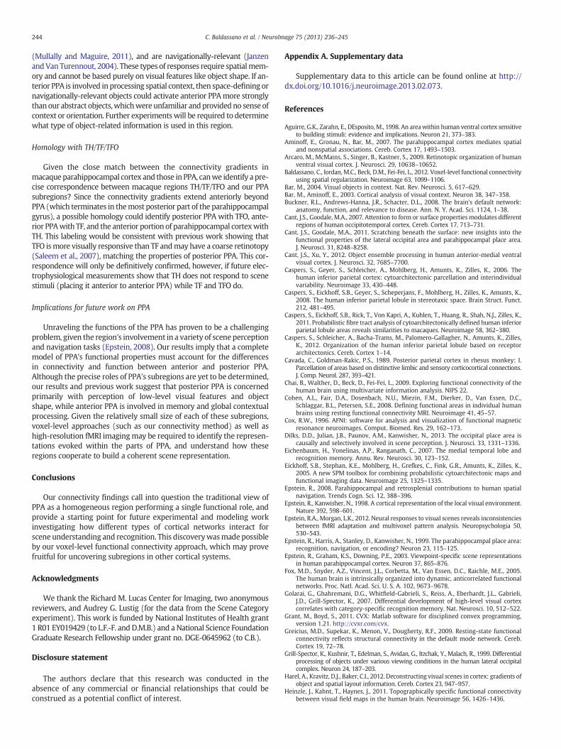
blocks drawn equally from six categories: child faces, adult faces, indoorscenes, outdoor scenes, objects (abstract sculptures with no semanticmeaning), and scrambled objects (these stimuli have been used in previ-ous studies such as Golarai et al., 2007). Images (240 × 240 pixels;subtending 12.8 × 12.8° of visual angle) were presented at fixation.
238 C. Baldassano et al. / NeuroImage 75 (2013) 236–245
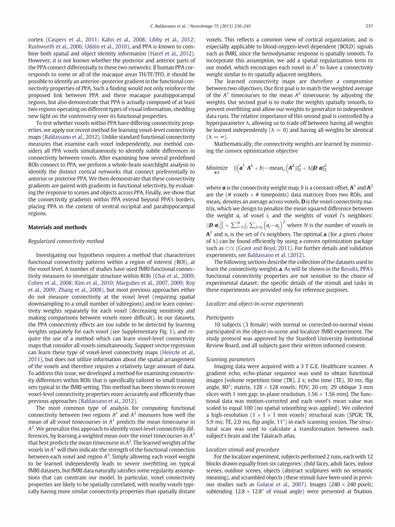
Examples of scene and object stimuli are shown in Fig. 1a. Blocks wereseparated by 12 s fixation cross periods, and consisted of 12 imagepresentations, each of which consisted of a 900 ms image followed bya 100 ms fixation cross. Each image was presented exactly once, withthe exception of two images during each block that were repeatedtwice in a row. Subjects were asked to maintain fixation at the centerof the screen, and respond via button-press whenever an image wasrepeated. The total number of timepoints was 300 (150 per run).
Object-in-scene stimuli and procedureFor the object-in-scene experiment,wepresented two types of stim-
uli, as shown in Fig. 1b: (1) boats and cars on a blankwhite background(isolated objects); and (2) boats and cars with a street or water scenebackground (objects in context). Images (450 × 450 pixels; subtending24 × 24° of visual angle) were presented 100 pixels (5°) away from fix-ation in randomly determined directions. Subjects were informed thateach image contained either a boat or a car, and were asked to indicateas quickly as possible whether the object was on the left half of theimage or the right half of the image (using a button box). Subjectsperformed 4 runs, with 16 blocks per run (with a 14 s gap betweenblocks) and 9 images per block. The first 8 blocks of each run showeda boat or car placed in a photographic scene; for each block, the objectcould violate a semantic relationship (appearing in the wrong type ofscene, e.g. a boat on a city street) and/or a geometric relationship(appearing in the wrong position in the scene, e.g. a car above a treerather than on the street). Each presentation consisted of a 500 ms fix-ation cross, an image flashed for 100 ms, a 300 ms mask, and then a1300 ms response period (blank gray screen). The last 8 blocks ofeach run showed a boat or car on a white background; these imageswere identical to those presented in thefirst eight blocks, with the back-grounds removed (and presented in a different random order). Eachpresentation consisted of a 500 ms fixation cross, an image flashed for350 ms, and then a 1300 ms response period (blank gray screen). Thetotal number of timepoints was 1224 (306 per run). Timepoints wereclassified as “resting” if they occurred more than 4 s after the end ofone stimulus block and less than 4 s after the start of the next stimulusblock.
Functional region of interest definitionRegressors for faces, scenes, objects, and scrambled objects in the
localizer experiment were constructed by using the standard blockhemodynamic model in AFNI (Cox, 1996). LOC, PPA, RSC, and TOSwere defined using the following contrasts: LOC, top 500 voxels for
a b
Fig. 1. Sample stimuli used in our experiments. (a) Scene and object stimuli from the localizobject-in-scene stimuli from the object-in-scene experiment. (c) Beach and mountain stim
Objects > Scrambled near lateral occipital surface; PPA, top 300 voxelsfor Scenes > Objects near parahippocampal gyrus; RSC, top 200 voxelsfor Scenes > Objects near retrosplenial cortex; TOS, top 200 voxels forScenes > Objects near the transverse occipital sulcus. The volume ofeach ROI in mm3 was chosen conservatively, based on previous results(Golarai et al., 2007). Consistent with the meta-analysis by Nasr et al.(2011), PPA in our subjects was found to be centered on the collateralsulcus adjacent to the parahippocampal gyrus.
Scene category experiment
Participants8 subjects (4 female) with normal or corrected-to-normal vision
participated in the scene category fMRI experiment (these subjectsdid not overlap with those in the object-in-scene experiment). Thestudy protocol was approved by the University of Illinois InstitutionalReview Board, and all subjects gave their written informed consent.
Scanning parametersFunctional imaging data were acquiredwith a 3 T Siemens Trio scan-
ner. A gradient echo, echo-planar sequencewas used to obtain functionalimages [volume repetition time (TR), 1.75 s; echo time (TE), 30 ms; flipangle, 90°; matrix, 64 × 64 voxels; FOV, 19 cm; 29 oblique 3 mm sliceswith 0 mm gap; in-plane resolution, 3.0 × 3.0 mm]. The functionaldata was motion-corrected and each voxel's mean value was scaled toequal 100 (no spatial smoothing was applied). We collected a high-resolution structural scan for each subject; 4 subjects were scanned ina 3 T Siemens Trio scanner (MPRAGE; 1 × 1 × 1.2 mm, TR, 1900 ms;TE, 2.25 ms, flip angle, 9°) and 4 subjects were scanned in a 3 T SiemensAllegra (MPRAGE; 1.25 × 1.25 × 1.25 mm, TR, 2000 ms; TE, 2.22 ms,flip angle, 8°). The structural scanwas used to calculate a transformationbetween each subject's brain and the Talairach atlas.
Stimuli and procedureImages (800 × 600 pixels; subtending 24 × 18° of visual angle) were
presented in the center of the display using a back-projection system(Resonance Technologies) operating at a resolution of 800 × 600 pixelsat 60 Hz. For each run, subjects were instructed to count the number ofimages belonging to a target category (beaches, cities, highways ormoun-tains; see example stimuli in Fig. 1c). On average, there were 16 targetimages per run, ranging from 15 to 17 targets. Stimuli were presentedin blocks of 8 images with a display time of 1.75 s for each image. Imageswithin a block were primarily from the same natural scene category;
c
er experiment, which also included faces and scrambled objects. (b) Isolated object anduli from the scene category experiment, which also included cities and highways.
239C. Baldassano et al. / NeuroImage 75 (2013) 236–245
however, in order to increase the difficulty of the counting task, one ortwo outgroup images from different scene categories (intrusions) occa-sionally appearedwithin a block. A fixation crosswas presented through-out each block, and subjects were instructed to maintain fixation. Therewere 8 blocks in each run (2blocks for eachnatural scene category), inter-leavedwith 12 s fixation periods to allow for the hemodynamic responseto return to baseline levels. A session contained 16 such runs, and theorder of categories and intrusion images were counterbalanced andrandomized across blocks. The total number of timepoints was 2064(129 per run). Timepoints were classified as “resting” if they occurredmore than 4 s after the end of one stimulus block and less than 4 s afterthe start of the next stimulus block.
Functional region of interest definitionROIs were defined using an independent localizer scan, consisting of
blocks of face, object, scrambled object, landscape, and cityscape images.Each block consisted of 20 images presented for 450 ms each with a330 ms interstimulus interval. Each of the five types of stimuli waspresented four times during a run, with 12 s fixation periods after twoor three blocks. Subjects completed two runs, performing a one-backtask during the localizer by pressing a button every time an image wasrepeated. Regressors for faces, scenes, objects, and scrambled objectswere constructed by using the standard block hemodynamic model inAFNI (Cox, 1996), and the following contrasts were used to defineROIs: LOC, Objects > Scrambled near lateral occipital surface; PPA,Scenes > Objects near parahippocampal gyrus; RSC, Scenes > Objectsnear retrosplenial cortex; TOS, Scenes > Objects near the transverseoccipital sulcus. A threshold of p b 2 · 10−3 (uncorrected) was applied,and was tightened to break clusters if necessary.
Caudal IPL definition
Caudal IPL is a region strongly connected to macaqueparahippocampal cortex (Kravitz et al., 2011b) for which we do nothave a functional localizer. In order to evaluate the match between themacaque and human connectivity patterns, we sought to anatomicallydefine a human region equivalent to cIPL. The two caudal-most areasof human IPL (defined using probabilistic cytoarchitectonic maps) arePGa and PGp, which are thought to correspond to the caudal-most sec-tions of macaque IPL, PG and Opt (Caspers et al., 2011). Of these, PGpexhibits significantly stronger functional and structural connectivitywith the parahippocampal gyrus (Uddin et al., 2010), giving the bestmatch with the proposed parieto-medial temporal pathway targetingparahippocampal areas from cIPL. We therefore define cIPL in allsubjects using the Eickhoff–Zilles PGp probabilistic cytoarchitectonicmap (Eickhoff et al., 2005, based on Caspers et al., 2006, 2008). Wethresholded the map at p > 0.5, and transformed the map into eachsubject's native space. Since cIPL slightly overlapped TOS in some sub-jects, any voxels shared between cIPL and TOS were excluded fromboth regions (no other ROIs included overlapping voxels).
PPA connectivity analysis: ROIs
We first learned PPA connectivity maps for four pre-defined seedregions: lateral occipital complex (LOC), transverse occipital sulcus(TOS, also referred to as the “occipital place area” in Dilks et al., 2013),retrosplenial cortex (RSC), and caudal inferior parietal lobule (cIPL) bysetting A1 to be PPA and A2 to be one of the four seed regions. Toavoid functional connectivity idiosyncratic to a specific experiment ortask, we used data from both the object-in-scene experiment and thescene category experiment (see above).
We first validated that our method could learn meaningfulvoxel-level connectivity maps which provide better generalizationperformance, compared to a connectivity map which is constantover left PPA and constant over right PPA. For each seed region andsubject, we learned a connectivity map using one training run, and
tuned the smoothness parameter λ tomaximize the fraction of varianceexplained on a validation set consisting of all but one of the remainingruns. The classifier was then retrained on both the training run and val-idation set (using the selected λ value) and tested on the final held-outtesting run. Results were averaged across all choices of training run,with a random testing run being chosen for each training run. Theseresults were compared to those from ROI-level connectivity maps, inwhich all PPA voxels in each hemisphere were constrained to take onthe same value (equivalent to λ → ∞).
We then learned a weight map over PPA for each subject and foreach seed region using all experimental runs, with λ chosen such thatthe average fraction of variance explained, when training on one runand testing on the other runs, was maximized. Wemeasured the corre-lation between the connectivity weights and the anterior–posteriorvoxel coordinates, to obtain a simple measure of how the learnedweights in PPA varied along the anterior–posterior axis. The correlationwas computed separately for left and right PPA (exceptwhere specified,results below are collapsed across left and right PPA).
PPA connectivity analysis: whole-brain
To explore the connectivity patterns between PPA and the rest of thebrain, we performed a whole-brain searchlight connectivity analysis inwhich our seed region was densely sampled throughout the entire cor-tex.WefixedA1 to be PPA, and then placed a 3 × 3 × 3 voxel searchlightA2 at each point on a lattice with 2 voxel spacing. For each searchlight,we used all experimental runs to learn a map of connectivity weightsin PPA, and then measured the correlation between the learned weightsand the anterior–posterior axis. We obtained an anterior PPA vs. posteri-or PPA preference for each brain voxel by averaging the correlation valueof all searchlights which included that voxel. In order to speed up com-putation, we used a single value of λ = 5.5 for all subjects, equal to theaverage of the optimal λ values in the ROI experiment (in log space).Group-level statistics were computed by transforming each subject'sresults into Talairach space.
Scene- and object-sensitivity analysis
After identifying connectivity differences among PPA voxels, weinvestigated whether these connectivity gradients corresponded to func-tional differences in stimulus selectivity. Tomeasure the response proper-ties of individual PPA voxels, we examined the statistics from theregressors in the localizer experiment. For each voxel, the t-statisticsfrom the scene and object regressors were recorded, and each voxelwas also given a binary label of “significantly activated” or “not signifi-cantly activated” based on whether its false discovery rate (FDR) foreach category was less than or greater than 0.05. To detect a sensitivitygradient across PPA, the correlation between the anterior–posterior axisand the t-statistics was computed. For visualization purposes, eachsubject's PPAvoxelswere binned into 10 bins running anterior–posterior,and themean t-statistic andpercentage of activated voxelswas calculatedfor each bin, to give a sensitivity profile.
LOC/TOS vs. RSC/cIPL connectivity
After discovering that LOC/TOS and RSC/cIPL connect preferentiallyto different voxels in PPA (see Results), we sought to place these con-nectivity gradients in the context of the entire parahippocampal region.For each cortical voxel, we averaged the coefficients for the voxel's cor-relations with LOC and TOS, and compared it to the average of the coef-ficients for the voxel's correlations with RSC and cIPL. We transformedeach subject's correlation maps into Talairach space, and identifiedvoxels at the group level that showed a consistent different across sub-jects for LOC/TOS functional connectivity vs. RSC/cIPL functional con-nectivity. In addition to the parahippocampal region, we searched allof cortex for voxels with this connectivity pattern.

240 C. Baldassano et al. / NeuroImage 75 (2013) 236–245
Results
Sincewe are interested in the intrinsic connectivity properties of PPA(rather than functional correlations idiosyncratic to a specific stimulusset), we localized PPA in two separate groups of subjects, each ofwhich then performed a different experimental task with different stim-uli. Although both experiments included scenes, in one case (identifyingscene category) the sceneswere directly relevant to the task,while in theother (locating a target object in scenes) scenes were not the primaryfocus. Given these datasets, dowe see connectivity differences in anteriorversus posterior PPA analogous to those in macaque parahippocampalcortex? Note that, since the connectivity patterns were similar in bothdatasets (see Supplementary Fig. 4), all connectivity results below arecollapsed across both experiments.
PPA connectivity analysis: ROIs
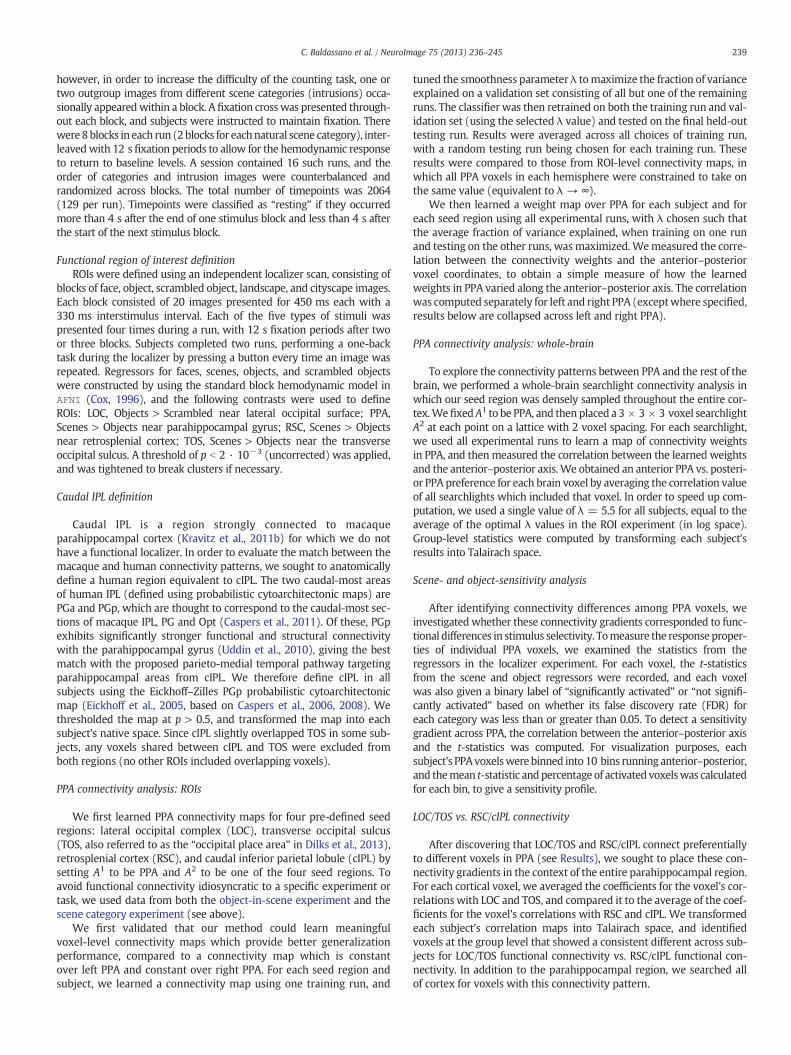
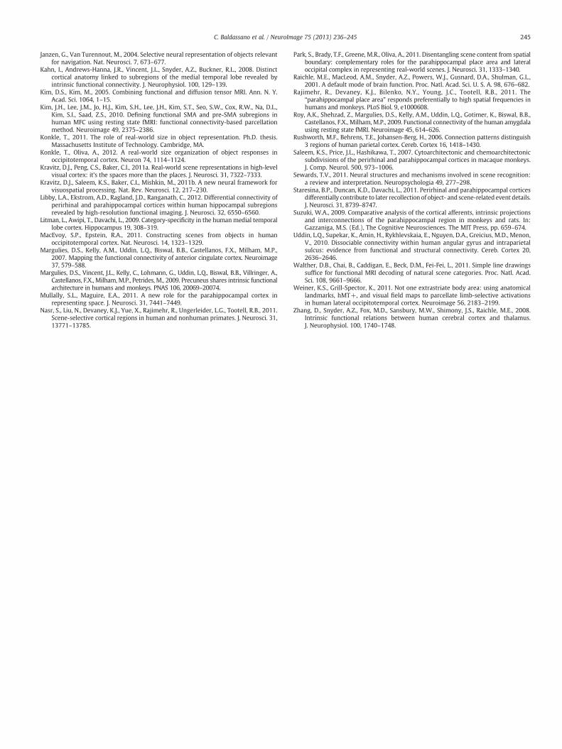
We began our investigation of PPA's connectivity structure by learn-ing PPA connectivity maps for four seed regions: two other scene-sensitive regions (TOS and RSC), an object-sensitive area in ventraloccipital cortex (LOC), and a posterior parietal region known to exhibitparahippocampal connectivity (cIPL). We first confirmed that, for eachindividual subject, we could learn weight maps over PPA (describingits connectivity with each of these regions) that generalize well acrossruns. As shown in Fig. 2a, spatially smooth voxel-level connectivitymaps in PPA predict activity in LOC, TOS, RSC, or cIPL better than amap which has only a single weight for left PPA and a single weightfor right PPA (LOC: t17 = 4.42; TOS: t17 = 4.63; RSC:t17 = 7.80; cIPL:t17 = 3.28; all p b 0.01, two-tailed paired t-test). These results werecomputed by choosing λ tomaximize the fraction of variance explained(on an independent validation set) but improvement over the traditionalconstant-weight connectivity held for a wide range of regularizationstrengths λ (see Supplementary Fig. 2). Although all regions showed atleast some activity related to PPA's timecourse, a significantly smalleramount of the cIPL timecourse can be predicted by PPA (ROI-level:LOC > cIPL: t17 = 7.31; TOS > cIPL: t17 = 10.58; RSC > cIPL: t17 =10.23; Voxel-level: LOC > cIPL: t17 = 6.58; TOS > cIPL: t17 = 12.12;RSC > cIPL: t17 = 11.81; all p b 0.01 two-tailed paired t-test), consistentwith its proposed role as a general processing hub in parietal cortexwithconnections to many regions besides PPA (Caspers et al., 2011; 2012).
Since meaningful voxel-level weight maps can be learned for indi-vidual subjects, we can ask whether these weight maps show anyanterior–posterior differences which are consistent across subjects. IfPPA shows the same gradient of connectivity as TH/TF/TFO, we expectthe posterior portion of PPA to be more strongly connected to occipitalvisual regions LOC and TOS, with the anterior portion of PPA morestrongly connected to RSC and cIPL. As shown in Fig. 2b, this is preciselywhat we observed; LOC and TOS connectivity weights tend to increasemoving anterior to posterior, while RSC and cIPL weights increase inthe opposite direction (LOC: t17 = 3.10,p b 0.01; TOS: t17 = 2.72,p = 0.01; RSC: t17 = −3.76,p b 0.01; cIPL: t17 = −3.24,p b 0.01;two-tailed t-test after z-transform). These results are collapsed acrossleft and right PPA; both hemispheres showed similar connectivity pat-terns, though effects were somewhat stronger in left PPA, by an averageof 0.13 (t17 = 2.20,p = 0.042; two-tailed t-test after z-transform). Wedid not observe significant differences along the inferior–superior axisor medial–lateral axis, except for preferential connectivity of cIPL tomedial PPA (see Supplementary Fig. 3).
PPA connectivity analysis: whole-brain
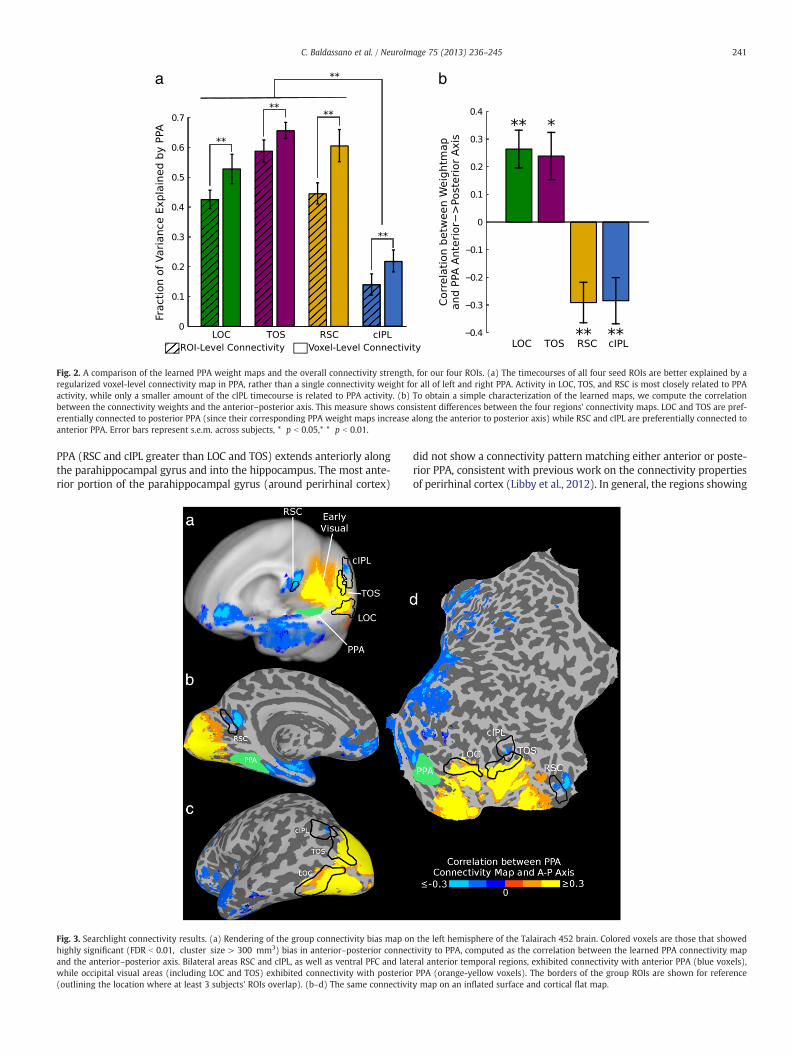
Having established a consistent posterior–anterior gradient of con-nectivity between our regions of interest and PPA, we then performeda searchlight analysis to search for other brain regions with posterior–anterior PPA connectivity gradients; rather than using our fixed ROIsas seed regions, we swept a 3 × 3 × 3 voxel searchlight throughout
the entire cortex. As in Fig. 2b, we learn a PPA connectivity map foreach seed region and compute the correlation of this map with theanterior–posterior axis; those seed regions which induce a PPA weightmap that is positively correlated with the anterior–posterior axis arepreferentially connected to posterior PPA, while those inducing a nega-tively correlated weight map are preferentially connected to anteriorPPA. The traditional (homogeneous) model of PPA predicts that consis-tent preferential connectivity should only occur for seed regions directlyadjacent to posterior or anterior PPA (whichwill be correlated with thenearer part of PPA due to local noise). If PPA contains subregions similarto those in macaque, however, we would expect a number of regionsthroughout cortex to show preferential connectivity patterns whichare both consistently non-zero and in opposite directions.
Our results are shown in Fig. 3. As predicted by our subregionhypothesis, seed regions in occipital visual areas (including LOC andTOS) showed preferential connectivity to posterior PPA, while RSCand cIPL showed preferential connectivity to anterior PPA. Note thatthese results cannot be explained by local noise correlations, since RSCand cIPL are physically closer to the posterior edge of PPA. We alsoobserved connectivity to anterior PPA in ventral prefrontal cortex(PFC) (primarily on the medial surface) and on the lateral surface ofthe anterior temporal lobe. Regions immediately anterior to PPA,including the hippocampus and anterior parahippocampal gyrus, showpreferential correlation with anterior PPA, but it is unclear if this effectis driven by intrinsic connectivity or local noise correlations. Coronaland axial slices are shown in Fig. 4, demonstrating that these connectiv-ity patterns are bilaterally symmetric. This result can also be obtained byusing only “resting” timepoints from between stimulus blocks or using adifferent value for λ, and is apparent for both the scene and object tasks(see Supplementary Fig. 4), suggesting that this connectivity pattern isintrinsic rather than task-specific. The fraction of variance explained forthe searchlights is consistent with our ROI analysis, showing the stron-gest coupling between PPA and visual regions including LOC, TOS, andRSC (see Supplementary Fig. 5).
Scene- and object-sensitivity analysis
Do these connectivity differences give rise to differences in functionalresponse to stimulus categories? Although the functional roles of anteriorand posterior PPA are likely complex, a simple functional anterior–posterior distinction can be seen in the scene and object responsesduring our localizer experiment. The selectivities of the PPA voxels toscenes and objects are shown in Fig. 5, binned based on position alongthe anterior–posterior axis. At the posterior side of PPA, the sensitivityto both scenes and objects is high, with nearly all voxels respondingto scene stimuli and a majority of voxels responding to object stimuli.Moving posterior to anterior, scene selectivity decreases somewhat(average correlation between t-statistic and posterior–anterior axis of0.25, t10 = 3.00,p = 0.01 two-tailed t-test after z-transform), althoughmost voxels respond significantly to scene stimuli across all of PPA.Object sensitivity, however, substantially decreases (average correla-tion between t-statistic and posterior–anterior axis of 0.32, t10 = 3.39,p b 0.01 two-tailed t-test after z-transform), with a majority of voxelsat the anterior edge showing no significant response to object stimuli.
LOC/TOS vs. RSC/cIPL connectivity
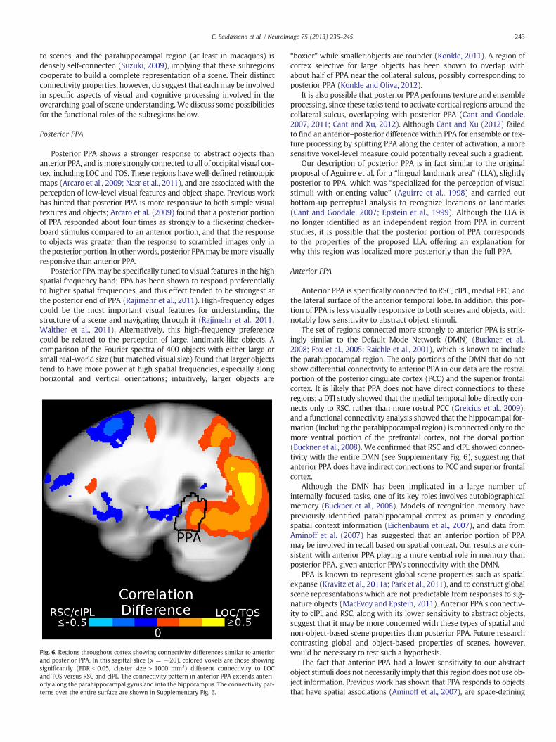
A number of studies have examined functional and connectivitygradients along the entire parahippocampal gyrus, which includes (inaddition to PPA) a portion of parahippocampal cortex anterior to PPA,and the perirhinal cortex (Aminoff et al., 2007; Bar and Aminoff, 2003;Libby et al., 2012; Litman et al., 2009; Staresina et al., 2011). In orderto examine how our gradients within PPA fit into the connectivitypatterns of the broader medial temporal lobe, we searched for voxelswhich showed the same connectivity differences we observed withinPPA. As shown in Fig. 6, the pattern of connectivity observed in anterior
a b
Fig. 2. A comparison of the learned PPA weight maps and the overall connectivity strength, for our four ROIs. (a) The timecourses of all four seed ROIs are better explained by aregularized voxel-level connectivity map in PPA, rather than a single connectivity weight for all of left and right PPA. Activity in LOC, TOS, and RSC is most closely related to PPAactivity, while only a smaller amount of the cIPL timecourse is related to PPA activity. (b) To obtain a simple characterization of the learned maps, we compute the correlationbetween the connectivity weights and the anterior–posterior axis. This measure shows consistent differences between the four regions' connectivity maps. LOC and TOS are pref-erentially connected to posterior PPA (since their corresponding PPA weight maps increase along the anterior to posterior axis) while RSC and cIPL are preferentially connected toanterior PPA. Error bars represent s.e.m. across subjects, * p b 0.05,* * p b 0.01.
241C. Baldassano et al. / NeuroImage 75 (2013) 236–245
PPA (RSC and cIPL greater than LOC and TOS) extends anteriorly alongthe parahippocampal gyrus and into the hippocampus. The most ante-rior portion of the parahippocampal gyrus (around perirhinal cortex)
Fig. 3. Searchlight connectivity results. (a) Rendering of the group connectivity bias map onhighly significant (FDR b 0.01, cluster size > 300 mm3) bias in anterior–posterior connecand the anterior–posterior axis. Bilateral areas RSC and cIPL, as well as ventral PFC and latewhile occipital visual areas (including LOC and TOS) exhibited connectivity with posterior(outlining the location where at least 3 subjects' ROIs overlap). (b–d) The same connectivit
did not show a connectivity pattern matching either anterior or poste-rior PPA, consistent with previous work on the connectivity propertiesof perirhinal cortex (Libby et al., 2012). In general, the regions showing
the left hemisphere of the Talairach 452 brain. Colored voxels are those that showedtivity to PPA, computed as the correlation between the learned PPA connectivity mapral anterior temporal regions, exhibited connectivity with anterior PPA (blue voxels),PPA (orange-yellow voxels). The borders of the group ROIs are shown for referencey map on an inflated surface and cortical flat map.
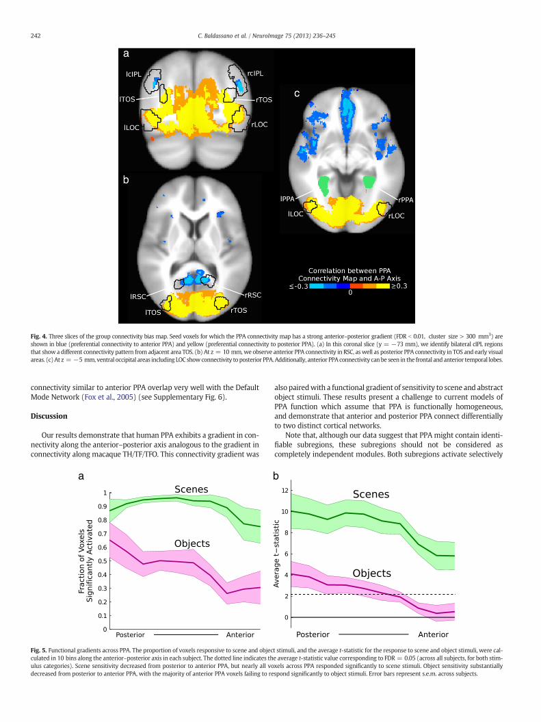
Fig. 4. Three slices of the group connectivity bias map. Seed voxels for which the PPA connectivity map has a strong anterior–posterior gradient (FDR b 0.01, cluster size > 300 mm3) areshown in blue (preferential connectivity to anterior PPA) and yellow (preferential connectivity to posterior PPA). (a) In this coronal slice (y = −73 mm), we identify bilateral cIPL regionsthat show a different connectivity pattern from adjacent area TOS. (b) At z = 10 mm,we observe anterior PPA connectivity in RSC, aswell as posterior PPA connectivity in TOS and early visualareas. (c) At z = −5 mm, ventral occipital areas including LOC showconnectivity to posterior PPA. Additionally, anterior PPA connectivity canbe seen in the frontal and anterior temporal lobes.
242 C. Baldassano et al. / NeuroImage 75 (2013) 236–245
connectivity similar to anterior PPA overlap very well with the DefaultMode Network (Fox et al., 2005) (see Supplementary Fig. 6).
Discussion
Our results demonstrate that human PPA exhibits a gradient in con-nectivity along the anterior–posterior axis analogous to the gradient inconnectivity along macaque TH/TF/TFO. This connectivity gradient was
a
Fig. 5. Functional gradients across PPA. The proportion of voxels responsive to scene and objecculated in 10 bins along the anterior–posterior axis in each subject. The dotted line indicates thulus categories). Scene sensitivity decreased from posterior to anterior PPA, but nearly all vodecreased from posterior to anterior PPA, with the majority of anterior PPA voxels failing to re
also pairedwith a functional gradient of sensitivity to scene and abstractobject stimuli. These results present a challenge to current models ofPPA function which assume that PPA is functionally homogeneous,and demonstrate that anterior and posterior PPA connect differentiallyto two distinct cortical networks.
Note that, although our data suggest that PPA might contain identi-fiable subregions, these subregions should not be considered ascompletely independent modules. Both subregions activate selectively
b
t stimuli, and the average t-statistic for the response to scene and object stimuli, were cal-e average t-statistic value corresponding to FDR = 0.05 (across all subjects, for both stim-xels across PPA responded significantly to scene stimuli. Object sensitivity substantiallyspond significantly to object stimuli. Error bars represent s.e.m. across subjects.
243C. Baldassano et al. / NeuroImage 75 (2013) 236–245
to scenes, and the parahippocampal region (at least in macaques) isdensely self-connected (Suzuki, 2009), implying that these subregionscooperate to build a complete representation of a scene. Their distinctconnectivity properties, however, do suggest that eachmay be involvedin specific aspects of visual and cognitive processing involved in theoverarching goal of scene understanding. We discuss some possibilitiesfor the functional roles of the subregions below.
Posterior PPA
Posterior PPA shows a stronger response to abstract objects thananterior PPA, and ismore strongly connected to all of occipital visual cor-tex, including LOC and TOS. These regions have well-defined retinotopicmaps (Arcaro et al., 2009; Nasr et al., 2011), and are associated with theperception of low-level visual features and object shape. Previous workhas hinted that posterior PPA is more responsive to both simple visualtextures and objects; Arcaro et al. (2009) found that a posterior portionof PPA responded about four times as strongly to a flickering checker-board stimulus compared to an anterior portion, and that the responseto objects was greater than the response to scrambled images only intheposterior portion. In otherwords, posterior PPAmay bemore visuallyresponsive than anterior PPA.
Posterior PPAmay be specifically tuned to visual features in the highspatial frequency band; PPA has been shown to respond preferentiallyto higher spatial frequencies, and this effect tended to be strongest atthe posterior end of PPA (Rajimehr et al., 2011). High-frequency edgescould be the most important visual features for understanding thestructure of a scene and navigating through it (Rajimehr et al., 2011;Walther et al., 2011). Alternatively, this high-frequency preferencecould be related to the perception of large, landmark-like objects. Acomparison of the Fourier spectra of 400 objects with either large orsmall real-world size (butmatched visual size) found that larger objectstend to have more power at high spatial frequencies, especially alonghorizontal and vertical orientations; intuitively, larger objects are
Fig. 6. Regions throughout cortex showing connectivity differences similar to anteriorand posterior PPA. In this sagittal slice (x = −26), colored voxels are those showingsignificantly (FDR b 0.05, cluster size > 1000 mm3) different connectivity to LOCand TOS versus RSC and cIPL. The connectivity pattern in anterior PPA extends anteri-orly along the parahippocampal gyrus and into the hippocampus. The connectivity pat-terns over the entire surface are shown in Supplementary Fig. 6.
“boxier” while smaller objects are rounder (Konkle, 2011). A region ofcortex selective for large objects has been shown to overlap withabout half of PPA near the collateral sulcus, possibly corresponding toposterior PPA (Konkle and Oliva, 2012).
It is also possible that posterior PPA performs texture and ensembleprocessing, since these tasks tend to activate cortical regions around thecollateral sulcus, overlapping with posterior PPA (Cant and Goodale,2007, 2011; Cant and Xu, 2012). Although Cant and Xu (2012) failedto find an anterior–posterior difference within PPA for ensemble or tex-ture processing by splitting PPA along the center of activation, a moresensitive voxel-level measure could potentially reveal such a gradient.
Our description of posterior PPA is in fact similar to the originalproposal of Aguirre et al. for a “lingual landmark area” (LLA), slightlyposterior to PPA, which was “specialized for the perception of visualstimuli with orienting value” (Aguirre et al., 1998) and carried outbottom-up perceptual analysis to recognize locations or landmarks(Cant and Goodale, 2007; Epstein et al., 1999). Although the LLA isno longer identified as an independent region from PPA in currentstudies, it is possible that the posterior portion of PPA correspondsto the properties of the proposed LLA, offering an explanation forwhy this region was localized more posteriorly than the full PPA.
Anterior PPA
Anterior PPA is specifically connected to RSC, cIPL, medial PFC, andthe lateral surface of the anterior temporal lobe. In addition, this por-tion of PPA is less visually responsive to both scenes and objects, withnotably low sensitivity to abstract object stimuli.
The set of regions connected more strongly to anterior PPA is strik-ingly similar to the Default Mode Network (DMN) (Buckner et al.,2008; Fox et al., 2005; Raichle et al., 2001), which is known to includethe parahippocampal region. The only portions of the DMN that do notshow differential connectivity to anterior PPA in our data are the rostralportion of the posterior cingulate cortex (PCC) and the superior frontalcortex. It is likely that PPA does not have direct connections to theseregions; a DTI study showed that the medial temporal lobe directly con-nects only to RSC, rather than more rostral PCC (Greicius et al., 2009),and a functional connectivity analysis showed that the hippocampal for-mation (including the parahippocampal region) is connected only to themore ventral portion of the prefrontal cortex, not the dorsal portion(Buckner et al., 2008). We confirmed that RSC and cIPL showed connec-tivity with the entire DMN (see Supplementary Fig. 6), suggesting thatanterior PPA does have indirect connections to PCC and superior frontalcortex.
Although the DMN has been implicated in a large number ofinternally-focused tasks, one of its key roles involves autobiographicalmemory (Buckner et al., 2008). Models of recognition memory havepreviously identified parahippocampal cortex as primarily encodingspatial context information (Eichenbaum et al., 2007), and data fromAminoff et al. (2007) has suggested that an anterior portion of PPAmay be involved in recall based on spatial context. Our results are con-sistent with anterior PPA playing a more central role in memory thanposterior PPA, given anterior PPA's connectivity with the DMN.
PPA is known to represent global scene properties such as spatialexpanse (Kravitz et al., 2011a; Park et al., 2011), and to construct globalscene representations which are not predictable from responses to sig-nature objects (MacEvoy and Epstein, 2011). Anterior PPA's connectiv-ity to cIPL and RSC, along with its lower sensitivity to abstract objects,suggest that it may be more concerned with these types of spatial andnon-object-based scene properties than posterior PPA. Future researchcontrasting global and object-based properties of scenes, however,would be necessary to test such a hypothesis.
The fact that anterior PPA had a lower sensitivity to our abstractobject stimuli does not necessarily imply that this region does not use ob-ject information. Previous work has shown that PPA responds to objectsthat have spatial associations (Aminoff et al., 2007), are space-defining
244 C. Baldassano et al. / NeuroImage 75 (2013) 236–245
(Mullally and Maguire, 2011), and are navigationally-relevant (JanzenandVan Turennout, 2004). These types of responses require spatialmem-ory and cannot be based purely on visual features like object shape. If an-terior PPA is involved in processing spatial context, then space-defining ornavigationally-relevant objects could activate anterior PPAmore stronglythanour abstract objects,whichwereunfamiliar andprovidedno sense ofcontext or orientation. Further experimentswill be required to determinewhat type of object-related information is used in this region.
Homology with TH/TF/TFO
Given the close match between the connectivity gradients inmacaqueparahippocampal cortex and those in PPA, canwe identify a pre-cise correspondence between macaque regions TH/TF/TFO and our PPAsubregions? Since the connectivity gradients extend anteriorly beyondPPA (which terminates in themost posterior part of the parahippocampalgyrus), a possible homology could identify posterior PPA with TFO, ante-rior PPAwith TF, and the anterior portion of parahippocampal cortexwithTH. This labeling would be consistent with previous work showing thatTFO ismore visually responsive than TF andmay have a coarse retinotopy(Saleem et al., 2007), matching the properties of posterior PPA. This cor-respondence will only be definitively confirmed, however, if future elec-trophysiological measurements show that TH does not respond to scenestimuli (placing it anterior to anterior PPA) while TF and TFO do.
Implications for future work on PPA
Unraveling the functions of the PPA has proven to be a challengingproblem, given the region's involvement in a variety of scene perceptionand navigation tasks (Epstein, 2008). Our results imply that a completemodel of PPA's functional properties must account for the differencesin connectivity and function between anterior and posterior PPA.Although the precise roles of PPA's subregions are yet to be determined,our results and previous work suggest that posterior PPA is concernedprimarily with perception of low-level visual features and objectshape, while anterior PPA is involved in memory and global contextualprocessing. Given the relatively small size of each of these subregions,voxel-level approaches (such as our connectivity method) as well ashigh-resolution fMRI imagingmay be required to identify the represen-tations evoked within the parts of PPA, and understand how theseregions cooperate to build a coherent scene representation.
Conclusions
Our connectivity findings call into question the traditional view ofPPA as a homogeneous region performing a single functional role, andprovide a starting point for future experimental and modeling workinvestigating how different types of cortical networks interact forscene understanding and recognition. This discoverywasmade possibleby our voxel-level functional connectivity approach, which may provefruitful for uncovering subregions in other cortical systems.
Acknowledgments
We thank the Richard M. Lucas Center for Imaging, two anonymousreviewers, and Audrey G. Lustig (for the data from the Scene Categoryexperiment). This work is funded by National Institutes of Health grant1 R01 EY019429 (to L.F.-F. and D.M.B.) and a National Science FoundationGraduate Research Fellowship under grant no. DGE-0645962 (to C.B.).
Disclosure statement
The authors declare that this research was conducted in theabsence of any commercial or financial relationships that could beconstrued as a potential conflict of interest.
Appendix A. Supplementary data
Supplementary data to this article can be found online at http://dx.doi.org/10.1016/j.neuroimage.2013.02.073.
References
Aguirre, G.K., Zarahn, E., DEsposito, M., 1998. An areawithin human ventral cortex sensitiveto building stimuli: evidence and implications. Neuron 21, 373–383.
Aminoff, E., Gronau, N., Bar, M., 2007. The parahippocampal cortex mediates spatialand nonspatial associations. Cereb. Cortex 17, 1493–1503.
Arcaro, M., McMains, S., Singer, B., Kastner, S., 2009. Retinotopic organization of humanventral visual cortex. J. Neurosci. 29, 10638–10652.
Baldassano, C., Iordan, M.C., Beck, D.M., Fei-Fei, L., 2012. Voxel-level functional connectivityusing spatial regularization. Neuroimage 63, 1099–1106.
Bar, M., 2004. Visual objects in context. Nat. Rev. Neurosci. 5, 617–629.Bar, M., Aminoff, E., 2003. Cortical analysis of visual context. Neuron 38, 347–358.Buckner, R.L., Andrews-Hanna, J.R., Schacter, D.L., 2008. The brain's default network:
anatomy, function, and relevance to disease. Ann. N. Y. Acad. Sci. 1124, 1–38.Cant, J.S., Goodale, M.A., 2007. Attention to form or surface properties modulates different
regions of human occipitotemporal cortex. Cereb. Cortex 17, 713–731.Cant, J.S., Goodale, M.A., 2011. Scratching beneath the surface: new insights into the
functional properties of the lateral occipital area and parahippocampal place area.J. Neurosci. 31, 8248–8258.
Cant, J.S., Xu, Y., 2012. Object ensemble processing in human anterior-medial ventralvisual cortex. J. Neurosci. 32, 7685–7700.
Caspers, S., Geyer, S., Schleicher, A., Mohlberg, H., Amunts, K., Zilles, K., 2006. Thehuman inferior parietal cortex: cytoarchitectonic parcellation and interindividualvariability. Neuroimage 33, 430–448.
Caspers, S., Eickhoff, S.B., Geyer, S., Scheperjans, F., Mohlberg, H., Zilles, K., Amunts, K.,2008. The human inferior parietal lobule in stereotaxic space. Brain Struct. Funct.212, 481–495.
Caspers, S., Eickhoff, S.B., Rick, T., Von Kapri, A., Kuhlen, T., Huang, R., Shah, N.J., Zilles, K.,2011. Probabilistic fibre tract analysis of cytoarchitectonically defined human inferiorparietal lobule areas reveals similarities to macaques. Neuroimage 58, 362–380.
Caspers, S., Schleicher, A., Bacha-Trams, M., Palomero-Gallagher, N., Amunts, K., Zilles,K., 2012. Organization of the human inferior parietal lobule based on receptorarchitectonics. Cereb. Cortex 1–14.
Cavada, C., Goldman-Rakic, P.S., 1989. Posterior parietal cortex in rhesus monkey: I.Parcellation of areas based on distinctive limbic and sensory corticocortical connections.J. Comp. Neurol. 287, 393–421.
Chai, B., Walther, D., Beck, D., Fei-Fei, L., 2009. Exploring functional connectivity of thehuman brain using multivariate information analysis. NIPS 22.
Cohen, A.L., Fair, D.A., Dosenbach, N.U., Miezin, F.M., Dierker, D., Van Essen, D.C.,Schlaggar, B.L., Petersen, S.E., 2008. Defining functional areas in individual humanbrains using resting functional connectivity MRI. Neuroimage 41, 45–57.
Cox, R.W., 1996. AFNI: software for analysis and visualization of functional magneticresonance neuroimages. Comput. Biomed. Res. 29, 162–173.
Dilks, D.D., Julian, J.B., Paunov, A.M., Kanwisher, N., 2013. The occipital place area iscausally and selectively involved in scene perception. J. Neurosci. 33, 1331–1336.
Eichenbaum, H., Yonelinas, A.P., Ranganath, C., 2007. The medial temporal lobe andrecognition memory. Annu. Rev. Neurosci. 30, 123–152.
Eickhoff, S.B., Stephan, K.E., Mohlberg, H., Grefkes, C., Fink, G.R., Amunts, K., Zilles, K.,2005. A new SPM toolbox for combining probabilistic cytoarchitectonic maps andfunctional imaging data. Neuroimage 25, 1325–1335.
Epstein, R., 2008. Parahippocampal and retrosplenial contributions to human spatialnavigation. Trends Cogn. Sci. 12, 388–396.
Epstein, R., Kanwisher, N., 1998. A cortical representation of the local visual environment.Nature 392, 598–601.
Epstein, R.A., Morgan, L.K., 2012. Neural responses to visual scenes reveals inconsistenciesbetween fMRI adaptation and multivoxel pattern analysis. Neuropsychologia 50,530–543.
Epstein, R., Harris, A., Stanley, D., Kanwisher, N., 1999. The parahippocampal place area:recognition, navigation, or encoding? Neuron 23, 115–125.
Epstein, R., Graham, K.S., Downing, P.E., 2003. Viewpoint-specific scene representationsin human parahippocampal cortex. Neuron 37, 865–876.
Fox, M.D., Snyder, A.Z., Vincent, J.L., Corbetta, M., Van Essen, D.C., Raichle, M.E., 2005.The human brain is intrinsically organized into dynamic, anticorrelated functionalnetworks. Proc. Natl. Acad. Sci. U. S. A. 102, 9673–9678.
Golarai, G., Ghahremani, D.G., Whitfield-Gabrieli, S., Reiss, A., Eberhardt, J.L., Gabrieli,J.D., Grill-Spector, K., 2007. Differential development of high-level visual cortexcorrelates with category-specific recognition memory. Nat. Neurosci. 10, 512–522.
Grant, M., Boyd, S., 2011. CVX: Matlab software for disciplined convex programming,version 1.21. http://cvxr.com/cvx.
Greicius, M.D., Supekar, K., Menon, V., Dougherty, R.F., 2009. Resting-state functionalconnectivity reflects structural connectivity in the default mode network. Cereb.Cortex 19, 72–78.
Grill-Spector, K., Kushnir, T., Edelman, S., Avidan, G., Itzchak, Y., Malach, R., 1999. Differentialprocessing of objects under various viewing conditions in the human lateral occipitalcomplex. Neuron 24, 187–203.
Harel, A., Kravitz, D.J., Baker, C.I., 2012. Deconstructing visual scenes in cortex: gradients ofobject and spatial layout information. Cereb. Cortex 23, 947–957.
Heinzle, J., Kahnt, T., Haynes, J., 2011. Topographically specific functional connectivitybetween visual field maps in the human brain. Neuroimage 56, 1426–1436.
245C. Baldassano et al. / NeuroImage 75 (2013) 236–245
Janzen, G., Van Turennout, M., 2004. Selective neural representation of objects relevantfor navigation. Nat. Neurosci. 7, 673–677.
Kahn, I., Andrews-Hanna, J.R., Vincent, J.L., Snyder, A.Z., Buckner, R.L., 2008. Distinctcortical anatomy linked to subregions of the medial temporal lobe revealed byintrinsic functional connectivity. J. Neurophysiol. 100, 129–139.
Kim, D.S., Kim, M., 2005. Combining functional and diffusion tensor MRI. Ann. N. Y.Acad. Sci. 1064, 1–15.
Kim, J.H., Lee, J.M., Jo, H.J., Kim, S.H., Lee, J.H., Kim, S.T., Seo, S.W., Cox, R.W., Na, D.L.,Kim, S.I., Saad, Z.S., 2010. Defining functional SMA and pre-SMA subregions inhuman MFC using resting state fMRI: functional connectivity-based parcellationmethod. Neuroimage 49, 2375–2386.
Konkle, T., 2011. The role of real-world size in object representation. Ph.D. thesis.Massachusetts Institute of Technology. Cambridge, MA.
Konkle, T., Oliva, A., 2012. A real-world size organization of object responses inoccipitotemporal cortex. Neuron 74, 1114–1124.
Kravitz, D.J., Peng, C.S., Baker, C.I., 2011a. Real-world scene representations in high-levelvisual cortex: it's the spaces more than the places. J. Neurosci. 31, 7322–7333.
Kravitz, D.J., Saleem, K.S., Baker, C.I., Mishkin, M., 2011b. A new neural framework forvisuospatial processing. Nat. Rev. Neurosci. 12, 217–230.
Libby, L.A., Ekstrom, A.D., Ragland, J.D., Ranganath, C., 2012. Differential connectivity ofperirhinal and parahippocampal cortices within human hippocampal subregionsrevealed by high-resolution functional imaging. J. Neurosci. 32, 6550–6560.
Litman, L., Awipi, T., Davachi, L., 2009. Category-specificity in the humanmedial temporallobe cortex. Hippocampus 19, 308–319.
MacEvoy, S.P., Epstein, R.A., 2011. Constructing scenes from objects in humanoccipitotemporal cortex. Nat. Neurosci. 14, 1323–1329.
Margulies, D.S., Kelly, A.M., Uddin, L.Q., Biswal, B.B., Castellanos, F.X., Milham, M.P.,2007. Mapping the functional connectivity of anterior cingulate cortex. Neuroimage37, 579–588.
Margulies, D.S., Vincent, J.L., Kelly, C., Lohmann, G., Uddin, L.Q., Biswal, B.B., Villringer, A.,Castellanos, F.X., Milham,M.P., Petrides, M., 2009. Precuneus shares intrinsic functionalarchitecture in humans and monkeys. PNAS 106, 20069–20074.
Mullally, S.L., Maguire, E.A., 2011. A new role for the parahippocampal cortex inrepresenting space. J. Neurosci. 31, 7441–7449.
Nasr, S., Liu, N., Devaney, K.J., Yue, X., Rajimehr, R., Ungerleider, L.G., Tootell, R.B., 2011.Scene-selective cortical regions in human and nonhuman primates. J. Neurosci. 31,13771–13785.
Park, S., Brady, T.F., Greene, M.R., Oliva, A., 2011. Disentangling scene content from spatialboundary: complementary roles for the parahippocampal place area and lateraloccipital complex in representing real-world scenes. J. Neurosci. 31, 1333–1340.
Raichle, M.E., MacLeod, A.M., Snyder, A.Z., Powers, W.J., Gusnard, D.A., Shulman, G.L.,2001. A default mode of brain function. Proc. Natl. Acad. Sci. U. S. A. 98, 676–682.
Rajimehr, R., Devaney, K.J., Bilenko, N.Y., Young, J.C., Tootell, R.B., 2011. The“parahippocampal place area” responds preferentially to high spatial frequencies inhumans and monkeys. PLoS Biol. 9, e1000608.
Roy, A.K., Shehzad, Z., Margulies, D.S., Kelly, A.M., Uddin, L.Q., Gotimer, K., Biswal, B.B.,Castellanos, F.X., Milham, M.P., 2009. Functional connectivity of the human amygdalausing resting state fMRI. Neuroimage 45, 614–626.
Rushworth, M.F., Behrens, T.E., Johansen-Berg, H., 2006. Connection patterns distinguish3 regions of human parietal cortex. Cereb. Cortex 16, 1418–1430.
Saleem, K.S., Price, J.L., Hashikawa, T., 2007. Cytoarchitectonic and chemoarchitectonicsubdivisions of the perirhinal and parahippocampal cortices in macaque monkeys.J. Comp. Neurol. 500, 973–1006.
Sewards, T.V., 2011. Neural structures and mechanisms involved in scene recognition:a review and interpretation. Neuropsychologia 49, 277–298.
Staresina, B.P., Duncan, K.D., Davachi, L., 2011. Perirhinal and parahippocampal corticesdifferentially contribute to later recollection of object- and scene-related event details.J. Neurosci. 31, 8739–8747.
Suzuki, W.A., 2009. Comparative analysis of the cortical afferents, intrinsic projectionsand interconnections of the parahippocampal region in monkeys and rats. In:Gazzaniga, M.S. (Ed.), The Cognitive Neurosciences. The MIT Press, pp. 659–674.
Uddin, L.Q., Supekar, K., Amin, H., Rykhlevskaia, E., Nguyen, D.A., Greicius, M.D., Menon,V., 2010. Dissociable connectivity within human angular gyrus and intraparietalsulcus: evidence from functional and structural connectivity. Cereb. Cortex 20,2636–2646.
Walther, D.B., Chai, B., Caddigan, E., Beck, D.M., Fei-Fei, L., 2011. Simple line drawingssuffice for functional MRI decoding of natural scene categories. Proc. Natl. Acad.Sci. 108, 9661–9666.
Weiner, K.S., Grill-Spector, K., 2011. Not one extrastriate body area: using anatomicallandmarks, hMT+, and visual field maps to parcellate limb-selective activationsin human lateral occipitotemporal cortex. Neuroimage 56, 2183–2199.
Zhang, D., Snyder, A.Z., Fox, M.D., Sansbury, M.W., Shimony, J.S., Raichle, M.E., 2008.Intrinsic functional relations between human cerebral cortex and thalamus.J. Neurophysiol. 100, 1740–1748.


![Coexisting Cholinergic and Parahippocampal …nzbri.org/resources/publications/220/Cassel_Neurodegenerative... · 1660–2854/08/0055–0304$24.50/0 Accessible online at: ... 16 ]P](https://img.pdfslide.us/doc/110x75/5aaf38a97f8b9a190d8d0f4e/coexisting-cholinergic-and-parahippocampal-nzbriorgresourcespublications220casselneurodegenerative16602854080055030424500.jpg)