Embed Size (px)
Citation preview
1
3
4
5
6
7 Q1
8
910
1 2
131415
16171819202122
2 3
39
40
41
42
43
44
45
46
47
48
49
50
51
52
53
54
55
56
57
58
59
Biochemical and Biophysical Research Communications xxx (2014) xxx–xxx
YBBRC 32373 No. of Pages 6, Model 5G
9 July 2014
Contents lists available at ScienceDirect
Biochemical and Biophysical Research Communications
journal homepage: www.elsevier .com/locate /ybbrc
Differential concentration-specific effects of caffeine on cell viability,oxidative stress, and cell cycle in pulmonary oxygen toxicity in vitro
http://dx.doi.org/10.1016/j.bbrc.2014.06.1320006-291X/� 2014 Published by Elsevier Inc.
Abbreviations: BPD, bronchopulmonary dysplasia; RDS, respiratory distresssyndrome; ROS, reactive oxygen species.⇑ Corresponding author. Fax: +1 832 825 3204.
E-mail address: [email protected] (K. Lingappan).
Please cite this article in press as: K.K. Tiwari et al., Differential concentration-specific effects of caffeine on cell viability, oxidative stress, and cell cpulmonary oxygen toxicity in vitro, Biochem. Biophys. Res. Commun. (2014), http://dx.doi.org/10.1016/j.bbrc.2014.06.132
Kirti Kumar Tiwari, Chun Chu, Xanthi Couroucli, Bhagavatula Moorthy, Krithika Lingappan ⇑Department of Pediatrics, Section of Neonatology, Texas Children’s Hospital, Baylor College of Medicine, 1102 Bates Avenue, MC: FC530.01, Houston, TX 77030, USA
a r t i c l e i n f o
24252627282930313233
Article history:Received 24 June 2014Available online xxxx
Keywords:HyperoxiaA549MLE 12Bronchopulmonary dysplasiaPulmonary epithelial cells
343536
a b s t r a c t
Caffeine is used to prevent bronchopulmonary dysplasia (BPD) in premature neonates. Hyperoxia con-tributes to the development of BPD, inhibits cell proliferation and decreases cell survival. The mecha-nisms responsible for the protective effect of caffeine in pulmonary oxygen toxicity remain largelyunknown. A549 and MLE 12 pulmonary epithelial cells were exposed to hyperoxia or maintained in roomair, in the presence of different concentrations (0, 0.05, 0.1 and 1 mM) of caffeine. Caffeine had a differ-ential concentration-specific effect on cell cycle progression, oxidative stress and viability, with 1 mMconcentration being deleterious and 0.05 mM being protective. Reactive oxygen species (ROS) generationduring hyperoxia was modulated by caffeine in a similar concentration-specific manner. Caffeine at1 mM, but not at the 0.05 mM concentration decreased the G2 arrest in these cells. Taken together thisstudy shows the novel funding that caffeine has a concentration-specific effect on cell cycle regulation,ROS generation, and cell survival in hyperoxic conditions.
� 2014 Published by Elsevier Inc.
37
38
60
61
62
63
64
65
66
67
68
69
70
71
72
73
74
75
76
1. Introduction
Supplemental oxygen is often used in the treatment of lung dis-eases such as respiratory distress syndrome (RDS) in prematureneonates. Exposure to high concentrations of inhaled oxygen(hyperoxia) combined with other factors such as mechanical ven-tilation, sepsis, etc. leads to lung injury and development of bron-chopulmonary dysplasia (BPD) in these fragile patients [1].
Exposure to hyperoxia leads to increased production of reactiveoxygen species (ROS), inhibition of cell proliferation, cell cyclearrest and eventually cell death [2]. Hyperoxia leads to activationof different cell cycle checkpoints depending on factors such asthe cell type and the p53 status of the cell. Cells with wild typep53 arrest in the G1 phase and cells with deficient p53 arrest inthe S or G2 phase [3–6]. Repair of genotoxic effects of hyperoxiais essential for subsequent tissue recovery.
Caffeine has been observed to decrease the incidence of BPD inpremature neonates [7]. The mechanisms responsible for the pro-tective effect of caffeine in pulmonary oxygen toxicity remainunknown. Variable concentrations ranging from micromolar tohigh millimolar have been used in studies evaluating effects of caf-
feine on cell cycle progression in vitro. The goal of this study was todetermine the effects of caffeine, at concentrations that are clini-cally relevant in BPD patients on pulmonary epithelial cellsexposed to hyperoxia, in vitro. We tested the hypothesis that caf-feine will elicit concentration-specific effects on cell cycle progres-sion, oxidative stress, and viability in human and murinepulmonary epithelial (A549: intact p53 and MLE 12: disruptedp53) cell lines exposed to hyperoxia. We used 0.05 mM (equivalentto 10 mg/kg, molecular weight of caffeine: 194.19 g/mol) and0.1 mM (equivalent to 20 mg/kg) concentrations to model the doseranges used clinically in premature neonates. In the current study,we demonstrate that caffeine has differential effects on cell cycleprogression, cell viability, and oxidative stress in pulmonary epi-thelial cell lines exposed to hyperoxia depending on theconcentration.
77
78
79
80
81
2. Materials and methods
2.1. Cell culture and caffeine preparation
A549 human lung epithelial cells and MLE 12 SV40 transformedmouse epithelial cells were obtained from the American Type Cul-ture Collection (Rockville, MD). Both of these cell lines have type IIalveolar epithelial cell characteristics. A549 cells have an intactp53-dependent G1 checkpoint. MLE 12 cells express the SV40 large
ycle in
82
83
84
85
86
87
88
89
90
91
92
93
94
95
96
97
98
99
100
101
102
103
104
105
106
107
108
109
110
111
112
113
114
115
116
117
118
119
120
121
122
123
124
125
126
127
128
129
130
131
132
133
134
135
136
137
138
139
140
141
142
143
144
145
146
147
148
149
150
151
152
153
154
155
156
157
158
159
160
161
162
163
164
165
166
167
168
169
170
171
172
173
174
175
176
177
178
179
180
181
182
183
184
185
186
187
188
189
190
191
192
193
2 K.K. Tiwari et al. / Biochemical and Biophysical Research Communications xxx (2014) xxx–xxx
YBBRC 32373 No. of Pages 6, Model 5G
9 July 2014
T antigen, which binds to p53 leading to uncontrolled cellular pro-liferation and disrupts the p53 mediated G1 checkpoint [30]. Cellswere cultured in DMEM/F-12, 50/50, (Cell Gro, Manassas, VA) sup-plemented with 10% fetal bovine serum, 50 U penicillin/ml, and50 lg/ml streptomycin in a 5% CO2/95% air atmosphere at 37 �C.Caffeine was purchased from Sigma Aldrich (St. Louis, MO, USA)and varying concentrations of caffeine (0.05, 0.1 and 1 mM) wereprepared in 1� Dulbecco’s Phosphate-Buffered Saline (Cell Gro,Manassas, VA, USA). We used 0.05 mM (ffi10 mg/kg) and 0.1 mM(ffi20 mg/kg) concentration to model the dose ranges used clini-cally in premature neonates. All cells were routinely passagedevery 3 days.
2.2. Exposure of cells to hyperoxia
Hyperoxia experiments were conducted in a Plexiglas sealedchamber into which a mixture of 95% O2 and 5% CO2 was circulatedcontinuously. The chamber was placed in a Forma Scientific water-jacketed incubator at 37 �C. Once the O2 level inside the chamberreached 95%, the cells were placed inside the chamber for thedesired length of time (up to 72 h). For the study of caffeine effects,exponentially growing cells were cultured for 24 h in medium.Cells were exposed to caffeine for 4 h at 5% CO2/95% air atmo-sphere before subjecting them to hyperoxia or control conditions.For each protocol described below, three or four independentexperiments were performed.
2.3. Trypan blue exclusion for cell viability
Cells were treated with caffeine as described before and wereexposed to room air or hyperoxia for up to 72 h. After harvesting,they were diluted 1:1 in 0.4% Trypan Blue dye (Cat # 145–0013)from Bio-Rad laboratories Inc. 10 ll was loaded on counting slides(Cat # 145–0011). TC20™ Automated Cell Counter (Bio-Rad labora-tories Inc.) was used to obtain the number of total cells and livecells.
2.4. Measurement of ROS generation
The ROS-Glo™ Assay (Promega Inc. Madison, WI) was used tomeasure the level of hydrogen peroxide (H2O2), directly in cell cul-ture according to the manufacturer’s recommendations. Cells wereplated at a density of 100,000 cells/well in a 96 well plate and incu-bated overnight for attachment. Cells were treated with varyingcaffeine concentrations at 0.05 mM, 0.1 mM and 1 mM. Plates wereplaced in hyperoxia or normoxia for 6 h, 12 h and 24 h. H2O2 sub-strate solution was added to the plates 6 h before read and theplates were replaced in hyperoxia chamber. The detection solutionwas added to the plates 20 min before each read and incubated atroom temperature and relative luminescence was recorded usingSpectraMax M3 microplate reader (Molecular Devices LLC, Sunny-vale, CA).
2.5. Cell cycle analysis
Asynchronously proliferating cultures at room air or afterhyperoxia exposure with or without caffeine were subjected toflow cytometry analysis for assessment of the cell cycle. Quantita-tive DNA content analysis in cells was performed using the nucleicacid stain propidium iodide followed by flow cytometry (Abcam,Cat. # ab139418). Briefly, cells were grown on six-well plates to60–70% confluence, after which they were treated with caffeineand exposed to room air or hyperoxia for up to 72 h. Cells wereharvested in single cell suspension and fixed in 66% ethanol at4 �C. Cells were stained with propidium iodide and RNase A andincubated at 37 �C for 30 min. Cell-cycle distribution was deter-
Please cite this article in press as: K.K. Tiwari et al., Differential concentration-spulmonary oxygen toxicity in vitro, Biochem. Biophys. Res. Commun. (2014), h
mined by using flow cytometry (FACSort, Becton Dickinson) andModFit LT software (Verity Software House, Topsham, ME) givingus the percentage of cells in different cell cycle stages.
2.6. In-Cell ELISA measuring Cdk2 (pTyr15) and Histone H3 (pSer10)
We used quantitative immunocytochemistry (In-Cell ELISAAssay Kit purchased from Abcam (Cat # ab140363) to measure lev-els of Cdk2 protein phosphorylated Tyr15 (elevated in the G1/Sphase) and Histone H3 protein phosphorylated Ser10 (elevated inG2/M phase) levels in A549 and MLE 12 cells exposed to roomair or hyperoxia for 24, 48 or 72 h. Phosphorylation of Cdk2 atTyr15 indicates that a cell is at the G1/S transition [31]. Phosphor-ylation of Histone H3 at Ser10 is tightly correlated with chromo-some condensation during mitosis [32]. Hence, Histone H3pSer10 signal indicates a mitotic cell with condensed DNA.
2.7. Statistical analysis
Results are reported as means ± standard error of the mean(SEM). Data were analyzed using 2-way analysis of variance (themain effects were: caffeine concentration and hyperoxia), followedby Bonferroni posttests for comparisons against control conditionsusing GraphPad version 5. Significance was assigned for P < 0.05.
3. Results
3.1. Effects of caffeine on cell viability following hyperoxia exposure inA549 or MLE 12 cells
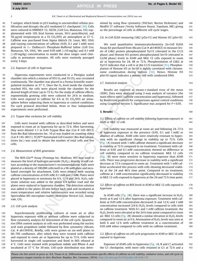
Cell viability was measured at room air and following 24–72 hof hyperoxia exposure in the presence (0.05, 0.1 and 1 mM) orabsence of caffeine. A549 cells were relatively resistant to hyper-oxia with no significant change in viability up to 72 h. Cells(Fig. 1A) treated with 1 mM caffeine showed a significant decreasein viability at 72 h compared to no treatment. Treatment with caf-feine at 0.05 and 0.1 mM concentration showed no difference incell viability compared to no caffeine treatment. MLE 12 cells(Fig. 1B) were more sensitive to hyperoxia exposure than A549cells. There was progressive decrease in viability with a significantdecrease at 72 h compared to room air. Treatment with 1 mM caf-feine accelerated this process with cells showing decreased viabil-ity at the 24 and 48 h time point. Compared to no treatment,caffeine at 1 mM concentration significantly decreased in viabilityin MLE 12 cells exposed to hyperoxia at the 24 and 72 h time point.
3.2. Effects of caffeine on ROS levels in A549 or MLE 12 cells exposed tohyperoxia
In A549 cells (Fig. 2A), there was a significant increase in H2O2
levels at 6 and 12 h after hyperoxia exposure. Treatment with caf-feine at 0.05 mM concentration decreased (6 and 12 h) and 1 mMconcentration increased (24 h) H2O2 levels compared to cells withno caffeine treatment. With 0.1 and 1 mM caffeine treatment, theH2O2 levels were elevated at the 24 h time point compared to roomair. MLE 12 cells (Fig. 2B) showed a similar elevation in H2O2 levelscompared to room air at 6 h. Attenuation of H2O2 levels was seen atboth 6 and 12 h with caffeine treatment at a concentration of0.05 mM when compared to cells with no caffeine treatment.
3.3. Effects of caffeine on cell cycle progression in A549 or MLE 12 cellsexposed to hyperoxia
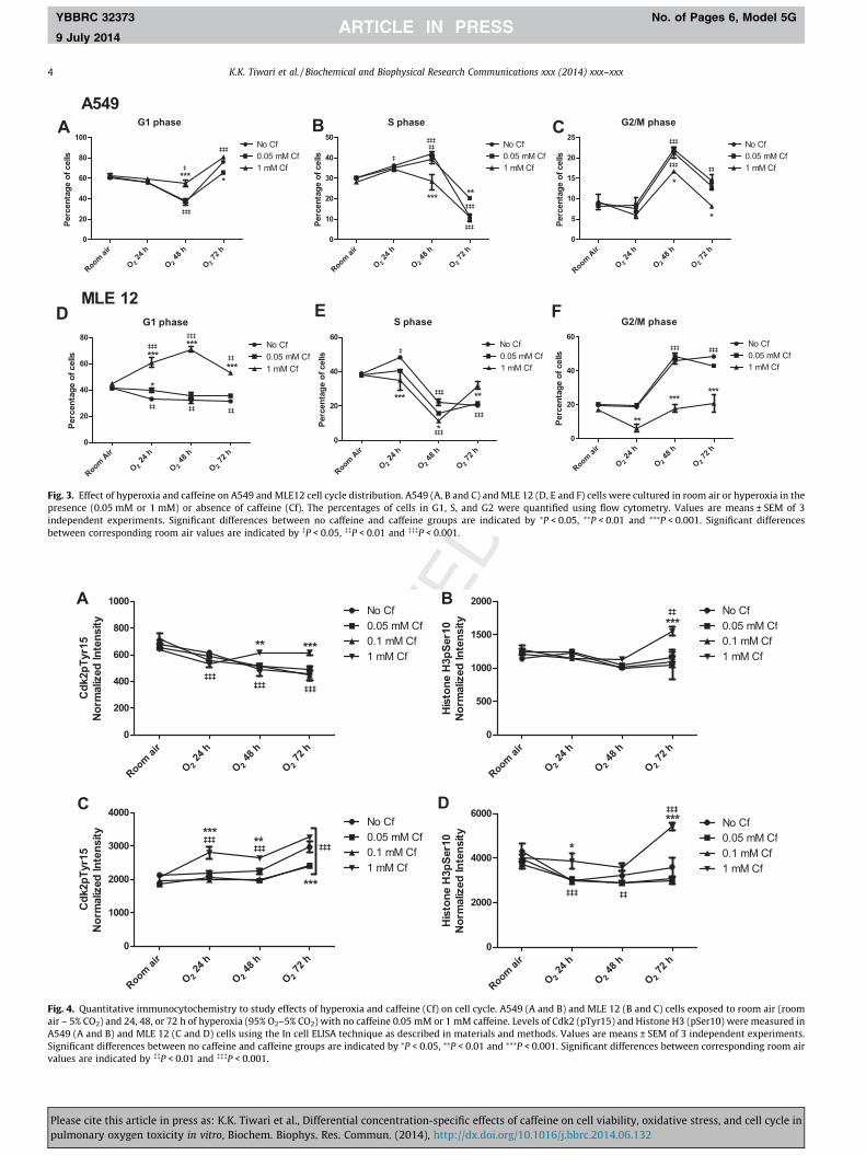
Exposure of A549 cells to hyperoxia (Fig. 3A, B and C) activatedthe G1 checkpoint, with more cells retained in G1 at 72 h and a
pecific effects of caffeine on cell viability, oxidative stress, and cell cycle inttp://dx.doi.org/10.1016/j.bbrc.2014.06.132

194
195
196
197
198
199
200
201
202
203
204
205
206
207
208
209
210
211
212
213
214
215
216
217
218
219
220
221
222
223
224
225
226
227
228
229
230
231
232
233
A B
Fig. 2. Effects of hyperoxia and caffeine (Cf) on reactive oxygen species (H2O2) production. A549 (A) and MLE 12 (B) cells exposed to room air (room air – 5% CO2) and 24, 48,or 72 h of hyperoxia (95% O2–5% CO2) with No Cf, 0.05 mM Cf or 1 mM Cf were subjected to the ROS-Glo™ luminescent H2O2 assay. Values are means ± SEM of 3 independentexperiments. Significant differences between No Cf and Cf groups are indicated by ⁄P < 0.05, ⁄⁄P < 0.01 and ⁄⁄⁄P < 0.001. Significant differences between corresponding roomair values are indicated by �P < 0.05 and ���P < 0.001.
A B
Fig. 1. Effects of hyperoxia and caffeine (Cf) on cell viability. A549 (A) and MLE 12 (B) cells exposed to room air (room air – 5% CO2) and 24, 48, or 72 h of hyperoxia (95% O2–5% CO2) in the presence (0.05 mM or 1 mM) or absence of caffeine were subjected to trypan blue exclusion as described in materials and methods. Values are means ± SEM of3 independent experiments. Significant differences between No Cf and Cf groups are indicated by ⁄P < 0.05 and ⁄⁄⁄P < 0.001. Significant differences between correspondingroom air values are indicated by ���P < 0.001.
K.K. Tiwari et al. / Biochemical and Biophysical Research Communications xxx (2014) xxx–xxx 3
YBBRC 32373 No. of Pages 6, Model 5G
9 July 2014
significant decrease in the number of cells in S and G2 phase. At48 h, there was decrease in percentage of cells in G1 and a corre-sponding increase in cells in S and G2 phase of the cell cycle. At0.05 mM, caffeine decreased the G1 retention at 72 h and had morecells in the S phase compared to no caffeine. On the other hand, at1 mM concentration, caffeine increased the fraction of cells in G1(48 h), and decreased the accumulation of cells in G2 (48 and72 h) compared to other groups (0.05 mM and no caffeine).
In MLE12 cells, hyperoxia significantly decreased the percent-age of cells in G1 and increased the percentage of cells in S(24 h) and G2 phase (48 and 72 h) (Fig. 3D, E and F). At 0.05 mMconcentration, the effect on cell cycle progression was similar tocells with no caffeine. Caffeine at 1 mM concentration markedlyreduced the number of cells in G2 phase at 24, 48 and 72 h timepoint. This was accompanied with an increase in the number ofcells in G1 phase (24, 48 and 72 h) and S phase at 72 h.
234
235
236
237
238
239
240
241
242
243
244
245
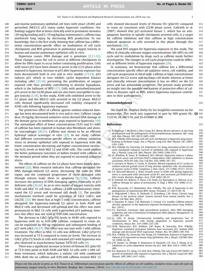
3.4. Effects of caffeine on Cdk2 (pTyr15) and Histone H3 (pSer10) inA549 and MLE 12 cells exposed to hyperoxia
In A549 cells (Fig. 4A), there was a decrease in Cdk2 (pTyr15)levels at 24, 48 and 72 h time point in cells with no caffeine, 0.05and 0.1 mM caffeine compared to room air levels. With 1 mM caf-feine this decrease was not seen. Histone H3 (pSer10) levels(Fig. 4B) showed no change in cells with no caffeine, 0.05 and0.1 mM caffeine, but with 1 mM concentration, there was a signif-icant increase at 72 h time point compared both to room air levelsand other cell populations.
Please cite this article in press as: K.K. Tiwari et al., Differential concentration-spulmonary oxygen toxicity in vitro, Biochem. Biophys. Res. Commun. (2014), h
In MLE 12 cells, there was a significant increase in Cdk2(pTyr15) expression (Fig. 4C) at 72 h time point in all cell subpop-ulations, with the 1 mM concentration showing an earlier increaseat the 24 and 48 h time points compared to room air levels. Cellstreated with 1 mM caffeine had higher (24 and 48 h) and thosetreated with 0.05 and 0.1 mM caffeine had lower expression(72 h) of Cdk2 (pTyr15) compared to cells with no caffeine. Therewas a decrease in Histone H3 (pSer10) levels (Fig. 4D) with hyper-oxia exposure except in cells treated with 1 mM caffeine, whichshowed a significant increase in levels at 72 h compared to roomair and no caffeine group. Caffeine did not alter viability, ROS lev-els, or cell cycle profiles of cultures exposed to room air.
4. Discussion
Hyperoxia leads to lung injury in animal models and causesgrowth arrest and eventually cell death in cultured cells [2,8,9]. Itcontributes to the development of diseases such as BPD in prema-ture neonates. Caffeine has been used in neonates to decrease theincidence BPD [7]. Despite being one of the most commonly useddrugs in neonates, the mechanisms responsible for the protectiveeffect of caffeine in premature infants especially in the setting ofhyperoxic lung injury are not known. A recent study in neonatalmice has shown deleterious effects of caffeine on alveolar develop-ment in a murine model of hyperoxia-induced alveolar hypoplasia[10]. In this study, we provide evidence that caffeine has a differen-tial concentration-specific effect on cell cycle progression,checkpoint activation, cell viability and oxidative stress in human
pecific effects of caffeine on cell viability, oxidative stress, and cell cycle inttp://dx.doi.org/10.1016/j.bbrc.2014.06.132
A
C D
B
Fig. 4. Quantitative immunocytochemistry to study effects of hyperoxia and caffeine (Cf) on cell cycle. A549 (A and B) and MLE 12 (B and C) cells exposed to room air (roomair – 5% CO2) and 24, 48, or 72 h of hyperoxia (95% O2–5% CO2) with no caffeine 0.05 mM or 1 mM caffeine. Levels of Cdk2 (pTyr15) and Histone H3 (pSer10) were measured inA549 (A and B) and MLE 12 (C and D) cells using the In cell ELISA technique as described in materials and methods. Values are means ± SEM of 3 independent experiments.Significant differences between no caffeine and caffeine groups are indicated by ⁄P < 0.05, ⁄⁄P < 0.01 and ⁄⁄⁄P < 0.001. Significant differences between corresponding room airvalues are indicated by ��P < 0.01 and ���P < 0.001.
A B C
D E F
Fig. 3. Effect of hyperoxia and caffeine on A549 and MLE12 cell cycle distribution. A549 (A, B and C) and MLE 12 (D, E and F) cells were cultured in room air or hyperoxia in thepresence (0.05 mM or 1 mM) or absence of caffeine (Cf). The percentages of cells in G1, S, and G2 were quantified using flow cytometry. Values are means ± SEM of 3independent experiments. Significant differences between no caffeine and caffeine groups are indicated by ⁄P < 0.05, ⁄⁄P < 0.01 and ⁄⁄⁄P < 0.001. Significant differencesbetween corresponding room air values are indicated by �P < 0.05, ��P < 0.01 and ���P < 0.001.
4 K.K. Tiwari et al. / Biochemical and Biophysical Research Communications xxx (2014) xxx–xxx
YBBRC 32373 No. of Pages 6, Model 5G
9 July 2014
Please cite this article in press as: K.K. Tiwari et al., Differential concentration-specific effects of caffeine on cell viability, oxidative stress, and cell cycle inpulmonary oxygen toxicity in vitro, Biochem. Biophys. Res. Commun. (2014), http://dx.doi.org/10.1016/j.bbrc.2014.06.132
246
247
248
249
250
251
252
253
254
255
256
257
258
259
260
261
262
263
264
265
266
267
268
269
270
271
272
273
274
275
276
277
278
279
280
281
282
283
284
285
286
287
288
289
290
291
292
293
294
295
296
297
298
299
300
301
302
303
304
305
306
307
308
309
310
311
312
313
314
315
316
317
318
319
320
321
322
323
324
325
326
327
328
329
330
331
332
333
334
335Q2Q3
336
337
338339340341342343344345346347348349350351352353354355356357358359360361362363364365366367368369370371372373374375376377378379380381382383384
K.K. Tiwari et al. / Biochemical and Biophysical Research Communications xxx (2014) xxx–xxx 5
YBBRC 32373 No. of Pages 6, Model 5G
9 July 2014
and murine pulmonary epithelial cell lines with intact (A549) andperturbed (MLE12) p53 status under hyperoxic conditions. Ourfindings suggest that at doses clinically used in premature neonates(20 mg/kg loading and 5–10 mg/kg/day maintenance), caffeine mayameliorate lung injury by decreasing ROS production. To ourknowledge, this is the first study showing that caffeine has a differ-ential concentration-specific effect on modulation of cell cyclecheckpoints and ROS generation in pulmonary oxygen toxicity inhuman and murine pulmonary epithelial cells in vitro.
Hyperoxia causes DNA damage and is genotoxic [2,11,12].These changes cause the cell to arrest at different checkpoints toallow for DNA repair to occur before continuing proliferation. Cellswith intact p53 have been shown to arrest in the G1/S phase whenexposed to hyperoxia. Increase in p53 after hyperoxia exposure hasbeen documented both in vivo and in vitro models [13,14]. p53induces p21 which in turn inhibits cyclin dependent kinasesincluding cdk2 [15,16] preventing the proliferation of oxygenexposed cells thus possibly contributing to alveolar hypoplasia,which is the hallmark of BPD [17]. Cells with perturbed/mutatedp53 arrest in the G2/M phase and are also more susceptible to oxy-gen toxicity [3,18]. In this study, A549 cells exhibited arrest in theG1 phase and the MLE12 cells arrested in the G2 phase. MLE12cells showed significantly decreased cell viability compared toA549 cells following hyperoxia exposure.
The protective effect of caffeine against oxidant-induced dam-age has been documented both in vivo and in vitro. Caffeine (singledose 10 mg/kg) decreased oxidative-stress derived DNA damage inthe dentate gyrus in newborn rat pups exposed to hyperoxia [19].The antioxidant effect of lower concentrations (0.01 and 0.1 mM)of caffeine has been reported in human skin fibroblasts and alveo-lar macrophages [20,21]. Caffeine was shown to be an efficienthydroxyl radical scavenger in vitro [22]. In our study, caffeinehad different concentration-specific effects on cellular oxidativestress in alveolar epithelial cells exposed to hyperoxia with thelower concentration decreasing and higher concentration increas-ing H2O2 levels in both MLE 12 and A549 cells. This could explainthe better pulmonary outcomes in babies treated with caffeine inthe neonatal period when they are exposed to increased oxidativestress.
The effects of caffeine on the G2 phase have been widely docu-mented [23]. Most research indicates that caffeine abrogates theDNA damage-induced G2 arrest, decreasing the time for DNArepair, and the continued progression of these damaged cellsthrough mitosis leads them to apoptosis [2,23,24]. Caffeineincreases the potency of DNA damaging agents, especially in p53deficient cells [25,26]. In an in vitro model of oxygen toxicity withA549 and MLE 15 cell lines, caffeine (2 mM concentration) elimi-nated the G2 arrest and increased cell death [3]. The possiblemechanisms involved could be activation of Cdc2 (cdk1) andCdc25C [23]. We show that at high (1 mM) concentration, caffeineabrogated the hyperoxia-induced G2 arrest in both A549 andMLE12 cells and decreased cell survival and this effect was morepronounced in MLE 12 cells with a perturbed p53 pathway, how-ever this effect was not seen at 0.05 mM concentration.
The decrease in Cdk2 (pTyr15) levels in A549 cells exposed tohyperoxia with no or 0.05 mM caffeine, compared to room air,can be explained by the up regulation and subsequent binding ofp21 with cdk2 [16,27]. This effect was not seen with 1 mM caffeinetreatment. The effect in MLE 12 cells was different. Cdk2 (pTyr15)was increased at 72 h compared to room air levels. The increase inCdk2 (pTyr15) levels in cells with mutated p53 with hyperoxia wasalso observed in asynchronous human T47D-H3 cells [4].
There was a significant increase in levels of Histone H3 (pSer10)at 72 h time point in both A549 and MLE 12 cells with 1 mM caf-feine treatment, which indicates a mitotic cell with condensedDNA. Both the no caffeine and 0.05 mM caffeine treated MLE 12
Please cite this article in press as: K.K. Tiwari et al., Differential concentration-spulmonary oxygen toxicity in vitro, Biochem. Biophys. Res. Commun. (2014), h
cells showed decreased levels of Histone H3 (pSer10) comparedto room air consistent with G2/M phase arrest. Gabrielli et al.(2007) showed that p21-activated kinase 1, which has an anti-apoptotic function in spindle checkpoint-arrested cells, is a targetfor caffeine inhibition and that caffeine at high concentrationinduced apoptosis in cells arrested in this phase through thismechanism [28].
We used 95% oxygen for hyperoxia exposure in this study. Theeffect of clinically relevant oxygen concentrations (40–60%) on cellcycle and its modulation by drugs such as caffeine needs furtherinvestigation. The changes in cell cycle progression could be differ-ent at different levels of hyperoxia exposure [29].
In summary, we demonstrate that caffeine has a differentialconcentration-specific effect on oxidative stress, cell viability, andcell cycle progression in these cells. Caffeine at high concentrationsabrogates the G2 arrest and increases cell death, whereas at lowerand clinically relevant concentration it has no effect on the G2arrest and has antioxidant properties. The present study providesan insight into the possible mechanism of protective effect of caf-feine in diseases such as BPD, where hyperoxia exposure contrib-utes to their pathogenesis.
Acknowledgments
We thank Dr. Stephen Welty for his insightful comments on themanuscript. This work was supported in part by NIH grants HL-112516, HL-087174, and ES-019689 to BM.
References
[1] A. Madurga, I. Mizíková, J. Ruiz-Camp, R.E. Morty, Recent advances in late lungdevelopment and the pathogenesis of bronchopulmonary dysplasia, AJP: LungCell. Mol. Physiol. 305 (2013) L893–L905.
[2] M.A. O’Reilly, DNA damage and cell cycle checkpoints in hyperoxic lung injury:braking to facilitate repair, Am. J. Physiol. Lung Cell. Mol. Physiol. 281 (2001)L291–L305.
[3] M.A. O’Reilly, R.J. Staversky, J.N. Finkelstein, P.C. Keng, Activation of the G2 cellcycle checkpoint enhances survival of epithelial cells exposed to hyperoxia,Am. J. Physiol. Lung Cell. Mol. Physiol. 284 (2003) L368–L375.
[4] J.F. Bilodeau, R. Faure, B. Piedboeuf, M.E. Mirault, Hyperoxia induces S-phasecell-cycle arrest and p21(Cip1/Waf1)-independent Cdk2 inhibition in humancarcinoma T47D-H3 cells, Exp. Cell Res. 256 (2000) 347–357.
[5] S. Corroyer, B. Maitre, V. Cazals, A. Clement, Altered regulation of G1 cyclins inoxidant-induced growth arrest of lung alveolar epithelial cells. Accumulationof inactive cyclin E-DCK2 complexes, J. Biol. Chem. 271 (1996) 25117–25125.
[6] S.A. McGrath-Morrow, J. Stahl, Growth arrest in A549 cells during hyperoxicstress is associated with decreased cyclin B1 and increased p21(Waf1/Cip1/Sdi1) levels, Biochim. Biophys. Acta 1538 (2001) 90–97.
[7] B. Schmidt, R.S. Roberts, P. Davis, L.W. Doyle, K.J. Barrington, A. Ohlsson, et al.,Caffeine therapy for apnea of prematurity, N. Engl. J. Med. 354 (2006) 2112–2121.
[8] B.W. Buczynski, E.T. Maduekwe, M.A. O’Reilly, The role of hyperoxia in thepathogenesis of experimental BPD, Semin. Perinatol. 37 (2013) 69–78.
[9] A. Hilgendorff, I. Reiss, H. Ehrhardt, O. Eickelberg, C.M. Alvira, Chronic lungdisease in the preterm infant: lessons learned from animal models, Am. J.Respir. Cell Mol. Biol. (2013).
[10] S. Dayanim, B. Lopez, T.M. Maisonet, S. Grewal, V.A. Londhe, Caffeine inducesalveolar apoptosis in the hyperoxia-exposed developing mouse lung, Pediatr.Res. (2013).
[11] P.C. Burcham, Genotoxic lipid peroxidation products: their DNA damagingproperties and role in formation of endogenous DNA adducts, Mutagenesis 13(1998) 287–305.
[12] J.J. Gille, H. Joenje, Chromosomal instability and progressive loss ofchromosomes in HeLa cells during adaptation to hyperoxic growthconditions, Mutat. Res. 219 (1989) 225–230.
[13] W.M. Maniscalco, R.H. Watkins, J.M. Roper, R. Staversky, M.A. O’Reilly,Hyperoxic ventilated premature baboons have increased p53, oxidant DNAdamage and decreased VEGF expression, Pediatr. Res. 58 (2005) 549–556.
[14] M.A. O’Reilly, R.J. Staversky, B.R. Stripp, J.N. Finkelstein, Exposure to hyperoxiainduces p53 expression in mouse lung epithelium, Am. J. Respir. Cell Mol. Biol.18 (1998) 43–50.
[15] J.W. Harper, S.J. Elledge, K. Keyomarsi, B. Dynlacht, L.H. Tsai, P. Zhang, et al.,Inhibition of cyclin-dependent kinases by p21, Mol. Biol. Cell 6 (1995) 387–400.
[16] R.C. Rancourt, P.C. Keng, C.E. Helt, M.A. O’Reilly, The role of p21(CIP1/WAF1) ingrowth of epithelial cells exposed to hyperoxia, Am. J. Physiol. Lung Cell. Mol.Physiol. 280 (2001) L617–L626.
pecific effects of caffeine on cell viability, oxidative stress, and cell cycle inttp://dx.doi.org/10.1016/j.bbrc.2014.06.132
385386387388389390391392393394 Q4395396397398399400401402403404405406407
408409410411412413414415416417418419420421422423424425426427428429430
6 K.K. Tiwari et al. / Biochemical and Biophysical Research Communications xxx (2014) xxx–xxx
YBBRC 32373 No. of Pages 6, Model 5G
9 July 2014
[17] M.A. O’Reilly, R.H. Watkins, R.J. Staversky, W.M. Maniscalco, Induced p21Cip1in premature baboons with CLD: implications for alveolar hypoplasia, Am. J.Physiol. Lung Cell. Mol. Physiol. 285 (2003) L964–L971.
[18] J.-F. Bilodeau, A. Patenaude, B. Piedboeuf, C. Carrier, P. Petrov, R. Faure, et al.,Glutathione peroxidase-1 expression enhances recovery of human breastcarcinoma cells from hyperoxic cell cycle arrest, Free Radic. Biol. Med. 33(2002) 1279–1289.
[19] S. Endesfelder, I. Zaak, U. Weichelt, C. Bührer, T. Schmitz, Caffeine protectsneuronal cells against injury caused by hyperoxia in the immature brain, FreeRadic. Biol. Med. (2013).
[20] J.I. Silverberg, M. Patel, N. Brody, J. Jagdeo, Caffeine protects human skinfibroblasts from acute reactive oxygen species-induced necrosis, J. DrugsDermatol. 11 (2012) 1342–1346.
[21] M. Jafari, A. Rabbani, Studies on the mechanism of caffeine action in alveolarmacrophages: caffeine elevates cyclic adenosine monophosphate level andprostaglandin synthesis, Metab. Clin. Exp. 53 (2004) 687–692.
[22] X. Shi, N.S. Dalal, A.C. Jain, Antioxidant behaviour of caffeine: efficientscavenging of hydroxyl radicals, Food Chem. Toxicol. 29 (1991) 1–6.
[23] A.M. Bode, Z. Dong, The enigmatic effects of caffeine in cell cycle and cancer,Cancer Lett. 247 (2007) 26–39.
[24] M. Takagi, T. Shigeta, M. Asada, S. Iwata, S. Nakazawa, Y. Kanke, et al., DNAdamage-associated cell cycle and cell death control is differentially modulatedby caffeine in clones with p53 mutations, Leukemia 13 (1999) 70–77.
431
Please cite this article in press as: K.K. Tiwari et al., Differential concentration-spulmonary oxygen toxicity in vitro, Biochem. Biophys. Res. Commun. (2014), h
[25] C.C. Lau, A.B. Pardee, Mechanism by which caffeine potentiates lethality ofnitrogen mustard, Proc. Natl. Acad. Sci. U.S.A. 79 (1982) 2942–2946.
[26] S.N. Powell, J.S. DeFrank, P. Connell, M. Eogan, F. Preffer, D. Dombkowski, et al.,Differential sensitivity of p53(�) and p53(+) cells to caffeine-inducedradiosensitization and override of G2 delay, Cancer Res. 55 (1995) 1643–1648.
[27] S.C. Gehen, P.F. Vitiello, R.A. Bambara, P.C. Keng, M.A. O’Reilly, Downregulationof PCNA potentiates p21-mediated growth inhibition in response to hyperoxia,Am. J. Physiol. Lung Cell. Mol. Physiol. 292 (2007) L716–L724.
[28] B. Gabrielli, Y.Q. Chau, N. Giles, A. Harding, F. Stevens, H. Beamish, Caffeinepromotes apoptosis in mitotic spindle checkpoint-arrested cells, J. Biol. Chem.282 (2007) 6954–6964.
[29] J.S. Shenberger, P.S. Dixon, Oxygen induces S-phase growth arrest andincreases p53 and p21(WAF1/CIP1) expression in human bronchial smooth-muscle cells, Am. J. Respir. Cell Mol. Biol. 21 (1999) 395–402.
[30] K.A. Wikenheiser, J.C. Clark, R.I. Linnoila, M.T. Stahlman, J.A. Whitsett, Simianvirus 40 large T antigen directed by transcriptional elements of the humansurfactant protein C gene produces pulmonary adenocarcinomas in transgenicmice, Cancer Res. 52 (1992) 5342–5352.
[31] P. Zarzov, A. Decottignies, G. Baldacci, P. Nurse, G(1)/S CDK is inhibited torestrain mitotic onset when DNA replication is blocked in fission yeast, Embo J.21 (2002) 3370–3376.
[32] C. Prigent, Phosphorylation of serine 10 in histone H3, what for?, J Cell Sci. 116(2003) 3677–3685.
pecific effects of caffeine on cell viability, oxidative stress, and cell cycle inttp://dx.doi.org/10.1016/j.bbrc.2014.06.132