Embed Size (px)
Citation preview
Different Susceptibility to the Parkinson’s Toxin MPTP inMice Lacking the Redox Master Regulator Nrf2 or ItsTarget Gene Heme Oxygenase-1Nadia G. Innamorato1,2, Agnieszka Jazwa1,3, Ana I. Rojo1,2, Concepcion Garcıa1,4, Javier Fernandez-
Ruiz1,4, Anna Grochot–Przeczek3, Anna Stachurska3, Alicja Jozkowicz3, Jozef Dulak3*, Antonio
Cuadrado1,2*
1 Centro de Investigacion Biomedica en Red sobre Enfermedades Neurodegenerativas (CIBERNED), Instituto de Investigacion Sanitaria La Paz (IdiPaz), Madrid, Spain,
2 Departamento de Bioquımica e Instituto de Investigaciones Biomedicas ‘‘Alberto Sols’’ Consejo Superior de Investigaciones Cientıficas (CSIC), Universidad Autonoma de
Madrid (UAM), Madrid, Spain, 3 Department of Medical Biotechnology, Faculty of Biochemistry, Biophysics and Biotechnology, Jagiellonian University, Krakow, Poland,
4 Departamento de Bioquımica y Biologıa Molecular, Instituto Universitario de Investigacion en Neuroquımica, Facultad de Medicina, Universidad Complutense de Madrid
(UCM), Madrid, Spain
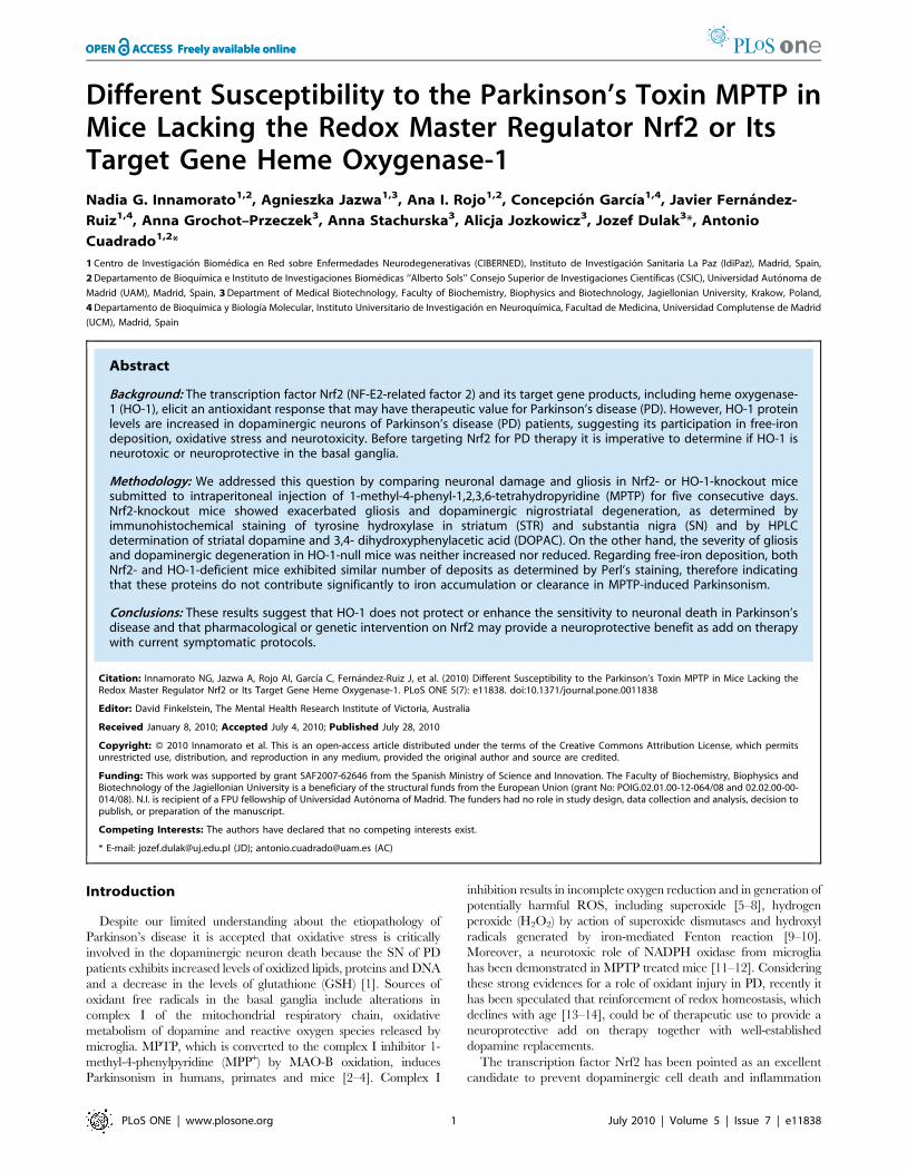
Abstract
Background: The transcription factor Nrf2 (NF-E2-related factor 2) and its target gene products, including heme oxygenase-1 (HO-1), elicit an antioxidant response that may have therapeutic value for Parkinson’s disease (PD). However, HO-1 proteinlevels are increased in dopaminergic neurons of Parkinson’s disease (PD) patients, suggesting its participation in free-irondeposition, oxidative stress and neurotoxicity. Before targeting Nrf2 for PD therapy it is imperative to determine if HO-1 isneurotoxic or neuroprotective in the basal ganglia.
Methodology: We addressed this question by comparing neuronal damage and gliosis in Nrf2- or HO-1-knockout micesubmitted to intraperitoneal injection of 1-methyl-4-phenyl-1,2,3,6-tetrahydropyridine (MPTP) for five consecutive days.Nrf2-knockout mice showed exacerbated gliosis and dopaminergic nigrostriatal degeneration, as determined byimmunohistochemical staining of tyrosine hydroxylase in striatum (STR) and substantia nigra (SN) and by HPLCdetermination of striatal dopamine and 3,4- dihydroxyphenylacetic acid (DOPAC). On the other hand, the severity of gliosisand dopaminergic degeneration in HO-1-null mice was neither increased nor reduced. Regarding free-iron deposition, bothNrf2- and HO-1-deficient mice exhibited similar number of deposits as determined by Perl’s staining, therefore indicatingthat these proteins do not contribute significantly to iron accumulation or clearance in MPTP-induced Parkinsonism.
Conclusions: These results suggest that HO-1 does not protect or enhance the sensitivity to neuronal death in Parkinson’sdisease and that pharmacological or genetic intervention on Nrf2 may provide a neuroprotective benefit as add on therapywith current symptomatic protocols.
Citation: Innamorato NG, Jazwa A, Rojo AI, Garcıa C, Fernandez-Ruiz J, et al. (2010) Different Susceptibility to the Parkinson’s Toxin MPTP in Mice Lacking theRedox Master Regulator Nrf2 or Its Target Gene Heme Oxygenase-1. PLoS ONE 5(7): e11838. doi:10.1371/journal.pone.0011838
Editor: David Finkelstein, The Mental Health Research Institute of Victoria, Australia
Received January 8, 2010; Accepted July 4, 2010; Published July 28, 2010
Copyright: � 2010 Innamorato et al. This is an open-access article distributed under the terms of the Creative Commons Attribution License, which permitsunrestricted use, distribution, and reproduction in any medium, provided the original author and source are credited.
Funding: This work was supported by grant SAF2007-62646 from the Spanish Ministry of Science and Innovation. The Faculty of Biochemistry, Biophysics andBiotechnology of the Jagiellonian University is a beneficiary of the structural funds from the European Union (grant No: POIG.02.01.00-12-064/08 and 02.02.00-00-014/08). N.I. is recipient of a FPU fellowship of Universidad Autonoma of Madrid. The funders had no role in study design, data collection and analysis, decision topublish, or preparation of the manuscript.
Competing Interests: The authors have declared that no competing interests exist.
* E-mail: [email protected] (JD); [email protected] (AC)
Introduction
Despite our limited understanding about the etiopathology of
Parkinson’s disease it is accepted that oxidative stress is critically
involved in the dopaminergic neuron death because the SN of PD
patients exhibits increased levels of oxidized lipids, proteins and DNA
and a decrease in the levels of glutathione (GSH) [1]. Sources of
oxidant free radicals in the basal ganglia include alterations in
complex I of the mitochondrial respiratory chain, oxidative
metabolism of dopamine and reactive oxygen species released by
microglia. MPTP, which is converted to the complex I inhibitor 1-
methyl-4-phenylpyridine (MPP+) by MAO-B oxidation, induces
Parkinsonism in humans, primates and mice [2–4]. Complex I
inhibition results in incomplete oxygen reduction and in generation of
potentially harmful ROS, including superoxide [5–8], hydrogen
peroxide (H2O2) by action of superoxide dismutases and hydroxyl
radicals generated by iron-mediated Fenton reaction [9–10].
Moreover, a neurotoxic role of NADPH oxidase from microglia
has been demonstrated in MPTP treated mice [11–12]. Considering
these strong evidences for a role of oxidant injury in PD, recently it
has been speculated that reinforcement of redox homeostasis, which
declines with age [13–14], could be of therapeutic use to provide a
neuroprotective add on therapy together with well-established
dopamine replacements.
The transcription factor Nrf2 has been pointed as an excellent
candidate to prevent dopaminergic cell death and inflammation
PLoS ONE | www.plosone.org 1 July 2010 | Volume 5 | Issue 7 | e11838
[15]. Nrf2 is the master regulator of genes containing Antioxidant
Response Elements [16] in their promoters, which constitute the
antioxidant and detoxifying phase II response. These genes
include HO-1, NAD(P)H:quinone oxidoreductase-1 (NOQ1) and
enzymes involved in GSH metabolism such as c-glutamylcysteine
synthetase, and glutathione S-transferase [17–19]. Nrf2 has a very
short half life due to its interaction with the BTB-Kelch protein
Keap1 that promotes Nrf2 degradation by the proteasome [20].
Physiological or pharmacological stimuli promote the dissociation
of the Nrf2–Keap1 complex resulting in stabilization of Nrf2,
translocation to the nucleus and induction of phase II cytoprotec-
tive genes [21–22]. Based on the large number of experimental
evidences that support this model, dissociation of Nrf2 from
Keap1 by electrophilic drugs provides a major strategy to increase
Nrf2 transcriptional activity.
A crucial role of Nrf2 has been demonstrated in protection of
dopaminergic neurons against oxidative stress, detoxification of
mitochondrial complex I inhibitors and down-regulation of genes
involved in the brain innate immune response. We and others
have demonstrated that Nrf2-null (Nrf22/2) mice are more
sensitive to MPTP treatment [23–25]. Moreover, Nrf22/2 mice
exhibit exacerbated inflammation compared to wild type litter-
mates as determined by release of pro-inflammatory cytokines in
basal ganglia [25]. However, while Nrf2 holds promise for PD
therapy, a very important concern is to determine the role of one
of its target genes, HO-1 in PD pathology.
The inducible phase II enzyme HO-1, together with the
constitutively expressed isoenzyme HO-2, degrades free heme
into three products: carbon monoxide (CO), biliverdin (which is
rapidly converted to bilirubin), and free iron [26–28]. At low
concentrations at least CO and bilirubin exert cytoprotective
and anti-inflammatory activities [29]. In fact, up-regulation of
HO-1 expression confers adaptive survival response to oxidative
and inflammatory insults both in vitro and in vivo [24] [30–33].
By contrast, the relevance of iron in PD etiopathology is well
documented in patients and in animal models. For instance,
iron-deficient rats are resistant to 6-OHDA neurotoxicity [34].
Moreover, iron chelators attenuate 6-OHDA- [35] and MPTP-
induced [36] toxicity. It has been suggested that dopaminergic
degeneration is a result of excessive redox-active metals within
the SN, which initiates a cascade of events, such as a-synuclein
oligomerization, mitochondrial dysfunction, cytotoxicity, and a
rise of cytosolic free calcium [16]. In postmortem biopsies,
immunohistochemical analysis indicates a moderate increase in
HO-1 protein levels in the cytoplasm of dopaminergic neurons
of SN of PD patients, compared to control individuals.
Moreover, there is a potent HO-1 immunoreactivity in Lewy
bodies [9] [37]. These evidences have led to the speculation that
increased HO-1 levels might be the main responsible for the
accumulation of free iron and subsequent damage to the
nigrostriatal system [38].
Therefore, in order to determine the therapeutic value of Nrf2-
activating drugs for PD it is necessary to determine if HO-1, which
will be up-regulated by Nrf2 activators through Nrf2-depedent
transcription, has a neuroprotective or neurotoxic role. We have
addressed this question in HO-1-knockout (HO-12/2) mice and
Nrf22/2 mice submitted to the sub-acute model of MPTP. Our
results, present two new findings: 1) HO-12/2 mice are similarly
sensitive to MPTP as wild type littermates, suggesting that the
increase in free iron, at least in MPTP-induced parkinsonism is not
related to HO-1. 2) Nrf2 is more efficient than HO-1 in eliciting a
neuroprotective and anti-inflammatory response to MPTP indi-
cating that, in addition to HO-1, other phase II enzymes are also
required for an effective neuroprotection.
Materials and Methods
Animals and treatmentsAll animal protocols were approved by the Ethical Committee
for Research of the Universidad Autonoma de Madrid and by the
Institutional Animal Care and Use Committee at the Jagiellonian
University following institutional, Spanish, Polish and European
guidelines (Boletın Oficial del Estado (BOE) of 18 March 1988;
and 86/609/EEC, 2003/65/EC European Council Directives).
Animals were housed at room temperature under a 12 h light-dark
cycle. Food and water was provided ad libitum. Mouse genotyping
was done according to [39] and [40]. Six months-old male wild
type C57BL/6 mice and Nrf2-knockout littermates (courtesy of Dr
Masayuki Yamamoto, Sendai, Japan) and C57BL/66FVB mice
and HO-1-knockout littermates (courtesy of Dr. Anupam Agarwal,
Birmingham, USA) were used. MPTP (Sigma-Aldrich; 30 mg/kg)
was prepared in saline solution just before use. The compound was
administered by intraperitoneal (i.p.) injection once a day for 5
consecutive days. Once the experimental schedule was completed,
animals were anesthetized with 8 mg/kg ketamine and 1.2 mg/kg
xylazine and perfused.
Analysis of mRNA levels by semiquantitative PCRTotal RNA was extracted using TRIzol reagent according to
the manufacturer’s instructions (Invitrogen). One mg of RNA from
the different treatments was reverse-transcribed in 20 ml using
High Capacity RNA-to-cDNA Kit (Applied Biosystems) according
to manufacturer’s instructions. Semiquantitative amplification of
cDNA was performed in 40 ml of Go Taq Flexi Colorless PCR
buffer (Promega) containing 0.5 U of Go Taq Flexi DNA
polymerase (Promega) and 30 pmol of synthetic gene-specific
primers. Primer sequences were: HO-1 forward 59-CACA-
GATGGCGTCACTTCCGTC-39; HO-1 reverse, 59-GTGAG-
GACCCACTGGGAGGAG-39; b-actin forward, 59-TCCT-
TCCTGGGCATGGAG-39; b-actin reverse, 59-AGGAGGAG-
CAATGATCTTGATCTT-39. To ensure that equal amounts of
cDNA were added to the PCR, the b-actin housekeeping gene was
amplified. PCR cycles (optimized for every gene) proceeded as
follows: initial denaturation for 4 min at 94uC, 1 min at 94uC(denaturation), 1 min at 58uC (annealing), and 1 min at 72uC(elongation). The amplified PCR products were resolved in 5%
PAGE and visualized with SYBR Safe DNA Stain (Invitrogen).
Striatal DA and DOPACStriata were homogenized in 50 volumes of ice-cold 0.2 N
perchloric acid containing 0.2 mM sodium disulphide and
0.45 mM EDTA. Dihydroxybenzylamine was added as an
internal standard. The homogenates were then centrifuged and
the supernatants injected into the HPLC system. This consisted of
a Spectra-Physics 8810 isocratic pump. The column was an RP-
18 (Spherisorb ODS-2; 125 mm, 4.6 mm, 5 mm particle size;
Waters, Massachusetts, USA). The mobile phase consisted of
100 mm citric acid, 100 mm sodium acetate, 1.2 mm heptane
sulphonate, 1 mm EDTA and 7% methanol (pH 3.9) and the
flow rate was 0.8 ml/min. The effluent was monitored with a
coulochemical detector (Coulochem II, ESA) using a procedure
of oxidation and reduction (conditioning cell, +360 mV; analyt-
ical cell no. 1, +50 mV; analytical cell no. 2, 340 mV). The signal
was recorded from the analytical cell no. 2, with a sensitivity of
50 nA (10 pg per sample), on a Spectra-Physics 4290 integrator,
and DA and DOPAC levels were obtained as area under the
peaks, corrected for the amount of total protein in the tissue
sample.
MPTP on Nrf2 or HO-1 Knockouts
PLoS ONE | www.plosone.org 2 July 2010 | Volume 5 | Issue 7 | e11838
ImmunohistochemistryAnimals were perfused through the left ventricle with saline
solution, followed by 4% paraformaldehyde in 0.1 M phosphate
buffer, pH 7.4, for 15 min. Brains were removed and cryopro-
tected by soaking in 30% sucrose solution in phosphate buffer until
they sank. Parallel series of 40-mm-thick coronal sections were
obtained in a freezing microtome. Sections from control and
experimental animals were processed with the same solutions and
processing times. Sections were rinsed in 100 mM Tris–HCl,
pH 7.6, and 225 mM NaCl (TBS). Antigen retrieval was
performed by microwave heating of the sections for 15–20 min
in 10 mM sodium citrate buffer, pH 6.0. Tissue peroxidase was
inactivated by incubating in 10% methanol and 3% hydrogen
peroxide in TBS for 30 min. After three washes in TBS sections
were incubated for 3 h in blocking solution (10% goat serum,
0.3% Triton X-100 in TBS), and then for 48 h at 4uC with mouse
anti-tyrosine hydroxylase (TH), rabbit anti-glial fibrillar acidic
protein (GFAP) (1:250; Millipore, Billerica, MA) or anti-Iba1
(1:1000; Abcam). Sections were rinsed in TBS, then incubated
with rabbit anti-mouse or anti-rabbit secondary antibodies at 1/
5000 dilution for 1 h at room temperature (Vector Labs).
Immunoreagents were diluted in 1% goat serum and 0.2% Triton
X-100 in TBS. Sections were subsequently developed by avidin-
biotin peroxidase complex system following manufacturer’s
instructions (ABC Kit, Vector Labs). Sections were mounted on
gelatin-coated slides, air-dried and finally dehydrated in graded
alcohols, cleared in xylene and coverslipped. Control sections were
treated with the same protocol but omitting the primary antibody.
Stereological quantificationCell counts were performed every four sections (40 mm thick)
through the SN using Stereo Investigator Software (MicroBrightfield,
Colchester, VT, USA) attached to an E800 Nikon microscope
(Nikon, Montreal, QC, Canada). SN, excluding ventral tegmental
area (VTA), was delineated in low magnification (5X objective) and a
point grid was overlaid onto each section. For Nissl staining, TH-
stained sections were rehydrated in xylene and graded alcohols and
incubated in Nissl’s solution (0.1% Cresyl Violet (Sigma), 2.5 ml
acetic acid 10%) at room temperature for 15 min. Sections were
rinsed in deionised water for 5 min and dehydrated in graded
alcohols, cleared in xylene and coverslipped. Stained cells were
counted by the optical fractionator method at high magnification
(63X objective, N.A. 1.25) distinguishing between TH-positive (TH
and Nissl positive) and TH-negative (Nissl positive). TH- and Nissl-
stained neurons were counted only when their nuclei were optimally
visualized within one focal plane. Nissl-stained neurons were
differentiated from non-neuronal cells by clearly defined nucleus,
cytoplasm, and a prominent nucleolus. Total numbers of neurons in
the SN were calculated as described in [41].
DAB-enhanced Perls iron stainingSections were rinsed in deionized water for 30 min and incubated
in Perls solution (5% potassium ferrocyanide : 5% HCl, 1:1, V:V) at
room temperature for 30 min. The reaction was stopped by rinsing in
deionized water for 30 min. Staining was enhanced by incubation in
0.5% 39-39-diaminobenzidine tetrahydrochloride (Sigma-Aldrich) in
Tris-ClH buffer, pH 8.0, for 20 min, and then developed in the same
buffer containing 0.003% hydrogen peroxide (Sigma-Aldrich) for
20 min. Finally, sections were incubated in Nissl’s solution (0.1%
Cresyl Violet (Sigma), 2.5 ml acetic acid 10%) at room temperature
for 15 min, rinsed in deionized water for 5 min, dehydrated in
graded alcohols, cleared in xylene and coverslipped.
ImmunofluorescenceBrains were fixed as described for immunohistochemistry.
Parallel series of 40-mm-thick coronal sections were obtained in
a freezing microtome. Sections were rinsed in TBS. After three
washes, the sections were incubated for 3 h in blocking solution
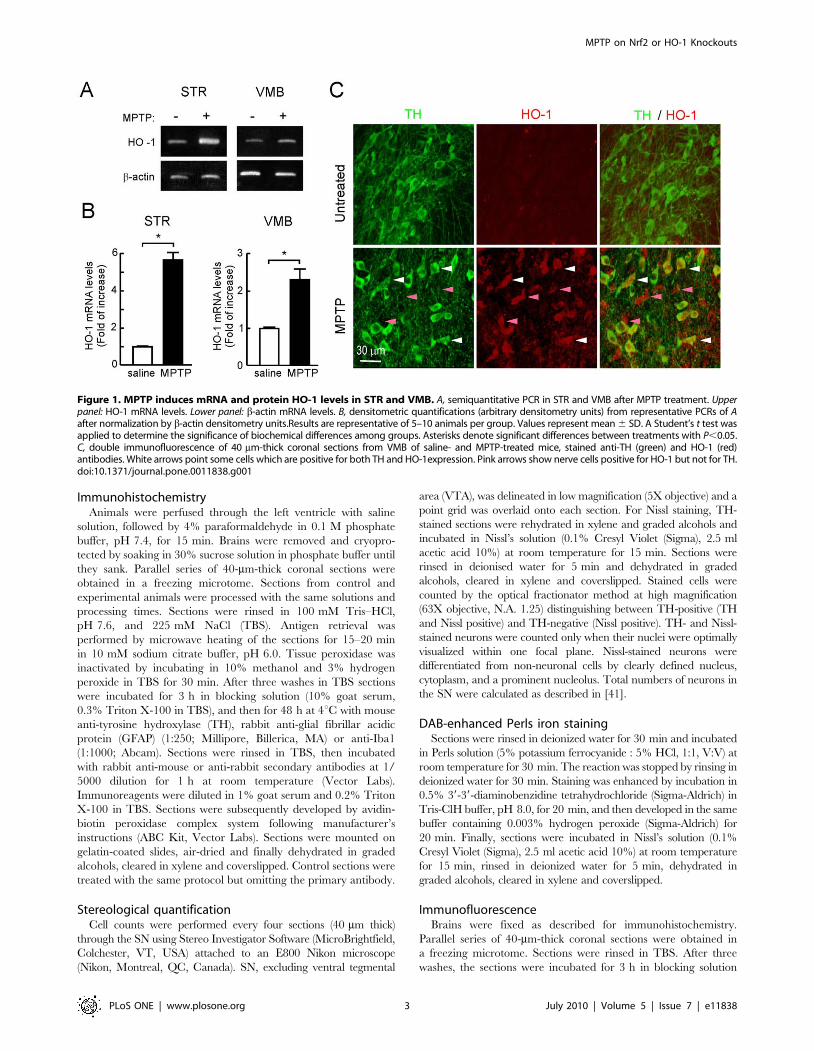
Figure 1. MPTP induces mRNA and protein HO-1 levels in STR and VMB. A, semiquantitative PCR in STR and VMB after MPTP treatment. Upperpanel: HO-1 mRNA levels. Lower panel: b-actin mRNA levels. B, densitometric quantifications (arbitrary densitometry units) from representative PCRs of Aafter normalization by b-actin densitometry units.Results are representative of 5–10 animals per group. Values represent mean 6 SD. A Student’s t test wasapplied to determine the significance of biochemical differences among groups. Asterisks denote significant differences between treatments with P,0.05.C, double immunofluorescence of 40 mm-thick coronal sections from VMB of saline- and MPTP-treated mice, stained anti-TH (green) and HO-1 (red)antibodies. White arrows point some cells which are positive for both TH and HO-1expression. Pink arrows show nerve cells positive for HO-1 but not for TH.doi:10.1371/journal.pone.0011838.g001
MPTP on Nrf2 or HO-1 Knockouts
PLoS ONE | www.plosone.org 3 July 2010 | Volume 5 | Issue 7 | e11838
(10% goat serum, 0.3% Triton X-100 in TBS), and then for 48 h
at 4uC in the following primary antibodies: rabbit anti-TH (1:500;
Millipore) and mouse anti-HO-1 (1:100, Stressgen). Sections were
rinsed in TBS and washed three times and then incubated with
secondary antibodies for 45 min: Alexa Fluor 488 anti-rabbit or
Alexa Fluor 546 anti-mouse at a 1:100 dilution (Invitrogen).
Immunoreagents were diluted in 1% goat or rabbit serum and
0.2% Triton X-100 in TBS. Sections were mounted on gelatin-
coated slides, air-dried and finally dehydrated in graded alcohols,
cleared in xylene and coverslipped. Control sections were treated
with the same protocol but omitting the primary antibody. The
fluorescence images were captured using appropriate filters in a
Leica DMIRE2TCS SP2 confocal microscope (Nussloch, Ger-
many). The lasers used were Ar 488 nm for green fluorescence
and Ar/HeNe 543 nm for red fluorescence.
ImmunoblottingSTR and VMB were removed rapidly and homogenized on ice
with lysis buffer (Tris-HCl, pH 7.5, 20 mM; NaCl, 137 mM; NaF,
20 mM; sodium pyrophosphate, 1 mM; Na3VO4, 1 mM; Nonidet
P-40,1%; glycerol, 10%; phenyl methyl sulphonyl fluoride, 1 mM;
and leupeptin, 1 mg/ml). Protein extracts were cleared by
centrifugation and 30 mg protein were resolved by SDS-PAGE
and transferred to Immobilon-P membranes (Millipore). Blots
were analyzed with the appropriate antibodies: anti-TH (1:2000;
Millipore), anti-GFAP (1:2000; Dako, Denmark), anti-Iba1
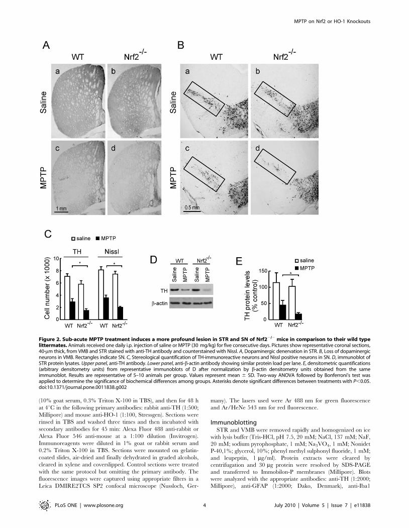
Figure 2. Sub-acute MPTP treatment induces a more profound lesion in STR and SN of Nrf22/2 mice in comparison to their wild typelittermates. Animals received one daily i.p. injection of saline or MPTP (30 mg/kg) for five consecutive days. Pictures show representative coronal sections,40-mm thick, from VMB and STR stained with anti-TH antibody and counterstained with Nissl. A, Dopaminergic denervation in STR. B, Loss of dopaminergicneurons in VMB. Rectangles indicate SN. C, Stereological quantification of TH-immunoreactive neurons and Nissl positive neurons in SN. D, immunoblot ofSTR protein lysates. Upper panel, anti-TH antibody. Lower panel, anti-b-actin antibody showing similar protein load per lane. E, densitometric quantifications(arbitrary densitometry units) from representative immunoblots of D after normalization by b-actin densitometry units obtained from the sameimmunoblot. Results are representative of 5–10 animals per group. Values represent mean 6 SD. Two-way ANOVA followed by Bonferroni’s test wasapplied to determine the significance of biochemical differences among groups. Asterisks denote significant differences between treatments with P,0.05.doi:10.1371/journal.pone.0011838.g002
MPTP on Nrf2 or HO-1 Knockouts
PLoS ONE | www.plosone.org 4 July 2010 | Volume 5 | Issue 7 | e11838
(1:1000; Abcam, Cambridge, UK), anti-HO-1 (1:2000; Millipore),
anti-NQO1 (1:500, Abcam), anti-HO-2 (1:500, Stressgen), anti-
GCL-C, anti-GCL-M (both were a kind gift of Dr Terrance
Kavanagh, University of Washington, USA) and anti-b-actin
(1:1000; Santa Cruz Biotechnology). Appropriate peroxidase-
conjugated secondary antibodies (1:10,000) were used to detect the
proteins of interest by enhanced chemiluminescence (Advanced
ECL, GE Healthcare).
Quantification and statisticsDifferent band intensities corresponding to immunoblot detection
of protein samples were quantified using the MCID software (MCID,
Cambridge, UK). Two-way ANOVA with Bonferroni’s post hoc test or
Student’s t test was used to assess differences between groups.
Results
MPTP induces HO-1 expression in mouse substantia nigraMice were submitted to the sub-acute model of MPTP
administration consisting on one daily intraperitoneal injection
of MPTP (30 mg/kg) for five consecutive days. HO-1 transcript
levels in stratum (STR) and ventral midbrain (VMB) were
analyzed 16 h after the last MPTP injection by semiquantitative
PCR. As shown in Fig. 1A and 1B, messenger HO-1 levels were
increased at both locations after the MPTP treatment. We also
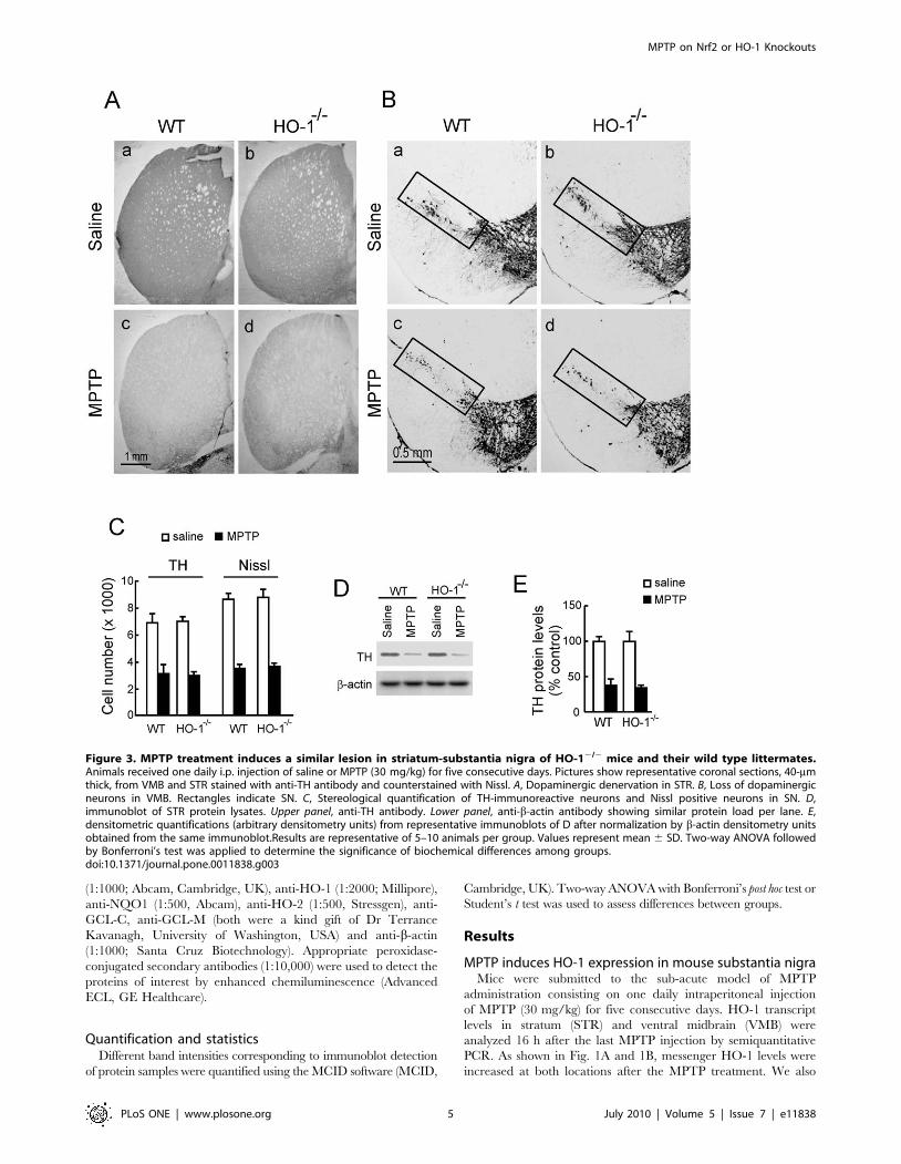
Figure 3. MPTP treatment induces a similar lesion in striatum-substantia nigra of HO-12/2 mice and their wild type littermates.Animals received one daily i.p. injection of saline or MPTP (30 mg/kg) for five consecutive days. Pictures show representative coronal sections, 40-mmthick, from VMB and STR stained with anti-TH antibody and counterstained with Nissl. A, Dopaminergic denervation in STR. B, Loss of dopaminergicneurons in VMB. Rectangles indicate SN. C, Stereological quantification of TH-immunoreactive neurons and Nissl positive neurons in SN. D,immunoblot of STR protein lysates. Upper panel, anti-TH antibody. Lower panel, anti-b-actin antibody showing similar protein load per lane. E,densitometric quantifications (arbitrary densitometry units) from representative immunoblots of D after normalization by b-actin densitometry unitsobtained from the same immunoblot.Results are representative of 5–10 animals per group. Values represent mean 6 SD. Two-way ANOVA followedby Bonferroni’s test was applied to determine the significance of biochemical differences among groups.doi:10.1371/journal.pone.0011838.g003
MPTP on Nrf2 or HO-1 Knockouts
PLoS ONE | www.plosone.org 5 July 2010 | Volume 5 | Issue 7 | e11838
analyzed HO-1 expression by immunofluorescence in SN by
double staining with anti-TH and anti-HO-1 antibodies (Fig. 1C).
In control saline-treated mice, HO-1 immunoreactivity was barely
detectable, in agreement with the concept that this inducible
enzyme exhibits very low basal level of expression. By contrast, in
MPTP-treated mice, HO-1 was increased in several nerve cells
including some TH-positive dopaminergic neurons. Therefore, the
MPTP mouse model reproduces the increase in HO-1 expression
that has been reported in the human PD pathology.
MPTP induction of nigrostriatal dopaminergicdegeneration on HO-12/2 and Nrf22/2 mice and theirwild type littermates
We analyzed TH immunoreactivity as a phenotypic marker for
dopaminergic neuron bodies and fibers. Mice treated with 30 mg/
kg/day of MPTP for 5 consecutive days exhibited a drastic
reduction in TH-immunoreactivity and Nissl staining in both STR
(Fig. 2A) and VMB (Fig. 2B), indicating damage onto the
nigrostriatal tract. However, Nrf22/2 mice showed a more
pronounced reduction in TH-immunoreactivity in comparison
with their wild type littermates. Stereological counts of nigral
dopaminergic neurons, scored from either TH-immunoreactive
and Nissl-stained sections, indicated that the Nrf22/2 mice used in
this study (6-months old) have about 15% fewer nigral dopami-
nergic neurons than their Nrf2+/+ littermates. Interestingly, in 2
weeks-old animals there was not any significant difference in cell
counts or TH-staining (data not shown), therefore suggesting that
Nrf2-defficiency leads to spontaneous dopaminergic degeneration.
After the MPTP treatment the absolute loss in dopaminergic cells
was greater in Nrf22/2 mice compared to MPTP-treated wild
type mice (Fig. 2C). However, changes in cell numbers between
Nrf2+/+ and Nrf2 2/2 mice were more similar when cell numbers
were analyzed relative to their baseline controls. Thus the
percentage of reduction between saline and MPTP-treated groups
was 41% 6 5% in Nrf2+/+ mice v.s. 28% 6 3% in Nrf22/2 mice
(p,0.0046) for TH-stained cells and 42% 6 3% in Nrf2+/+ v.s.
30% + 2% in Nrf22/2 mice (p,0.0009). Because the changes in
nigrostriatal dopaminergic cell numbers were statistically signifi-
cant but still modest when considered relative to their saline
controls, these results were further quantified by densitometry of
TH-stained STR sections (data not shown) and by immunoblot of
STR protein lysates analyzed with anti-TH antibody (Fig. 2D and
2E) further supporting the concept that Nrf22/2 are more
sensitive to MPTP. By contrast, HO-12/2 mice exhibited similar
damage to the nigrostriatal pathway as their wild type littermates,
as determined by TH-immunohistochemistry of STR (Fig. 3A)
and VMB (Fig. 3B) sections, stereological counts of TH-
immuoreactivity and Nissl staining (Fig. 3C), and by immunoblot
quantification with anti-TH antibody (Fig. 3D and 3E). Therefore,
while Nrf22/2 mice are more sensitive to MPTP-induced
dopaminergic damage, HO-12/2 mice are similarly sensitive to
this toxin as wild type mice.
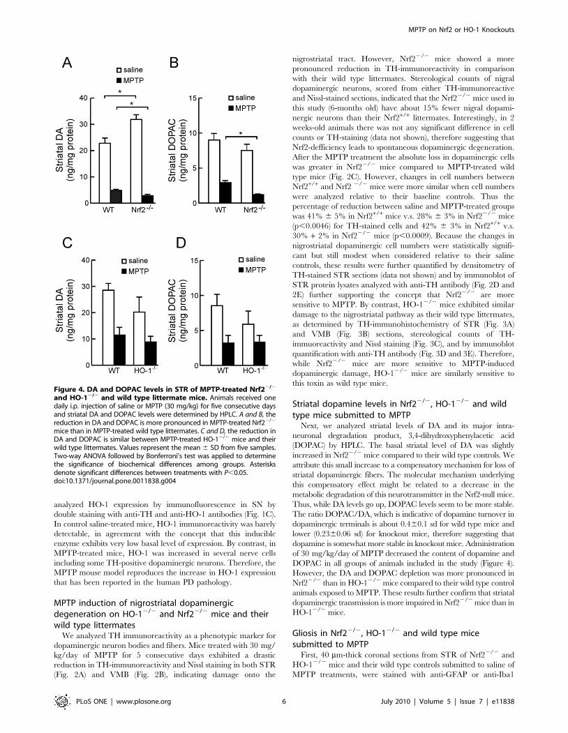
Striatal dopamine levels in Nrf22/2, HO-12/2 and wildtype mice submitted to MPTP
Next, we analyzed striatal levels of DA and its major intra-
neuronal degradation product, 3,4-dihydroxyphenylacetic acid
(DOPAC) by HPLC. The basal striatal level of DA was slightly
increased in Nrf22/2 mice compared to their wild type controls. We
attribute this small increase to a compensatory mechanism for loss of
striatal dopaminergic fibers. The molecular mechanism underlying
this compensatory effect might be related to a decrease in the
metabolic degradation of this neurotransmitter in the Nrf2-null mice.
Thus, while DA levels go up, DOPAC levels seem to be more stable.
The ratio DOPAC/DA, which is indicative of dopamine turnover in
dopaminergic terminals is about 0.460.1 sd for wild type mice and
lower (0.2360.06 sd) for knockout mice, therefore suggesting that
dopamine is somewhat more stable in knockout mice. Administration
of 30 mg/kg/day of MPTP decreased the content of dopamine and
DOPAC in all groups of animals included in the study (Figure 4).
However, the DA and DOPAC depletion was more pronounced in
Nrf22/2 than in HO-12/2 mice compared to their wild type control
animals exposed to MPTP. These results further confirm that striatal
dopaminergic transmission is more impaired in Nrf22/2 mice than in
HO-12/2 mice.
Gliosis in Nrf22/2, HO-12/2 and wild type micesubmitted to MPTP
First, 40 mm-thick coronal sections from STR of Nrf22/2 and
HO-12/2 mice and their wild type controls submitted to saline of
MPTP treatments, were stained with anti-GFAP or anti-Iba1
Figure 4. DA and DOPAC levels in STR of MPTP-treated Nrf22/2
and HO-12/2 and wild type littermate mice. Animals received onedaily i.p. injection of saline or MPTP (30 mg/kg) for five consecutive daysand striatal DA and DOPAC levels were determined by HPLC. A and B, thereduction in DA and DOPAC is more pronounced in MPTP-treated Nrf22/2
mice than in MPTP-treated wild type littermates. C and D, the reduction inDA and DOPAC is similar between MPTP-treated HO-12/2 mice and theirwild type littermates. Values represent the mean 6 SD from five samples.Two-way ANOVA followed by Bonferroni’s test was applied to determinethe significance of biochemical differences among groups. Asterisksdenote significant differences between treatments with P,0.05.doi:10.1371/journal.pone.0011838.g004
MPTP on Nrf2 or HO-1 Knockouts
PLoS ONE | www.plosone.org 6 July 2010 | Volume 5 | Issue 7 | e11838
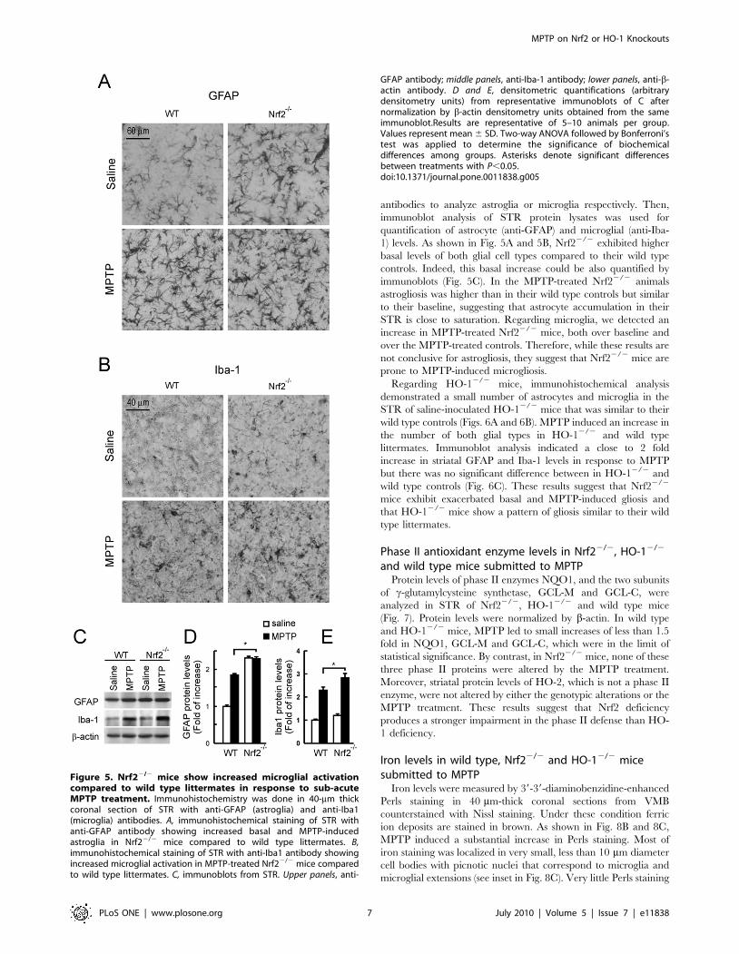
antibodies to analyze astroglia or microglia respectively. Then,
immunoblot analysis of STR protein lysates was used for
quantification of astrocyte (anti-GFAP) and microglial (anti-Iba-
1) levels. As shown in Fig. 5A and 5B, Nrf22/2 exhibited higher
basal levels of both glial cell types compared to their wild type
controls. Indeed, this basal increase could be also quantified by
immunoblots (Fig. 5C). In the MPTP-treated Nrf22/2 animals
astrogliosis was higher than in their wild type controls but similar
to their baseline, suggesting that astrocyte accumulation in their
STR is close to saturation. Regarding microglia, we detected an
increase in MPTP-treated Nrf22/2 mice, both over baseline and
over the MPTP-treated controls. Therefore, while these results are
not conclusive for astrogliosis, they suggest that Nrf22/2 mice are
prone to MPTP-induced microgliosis.
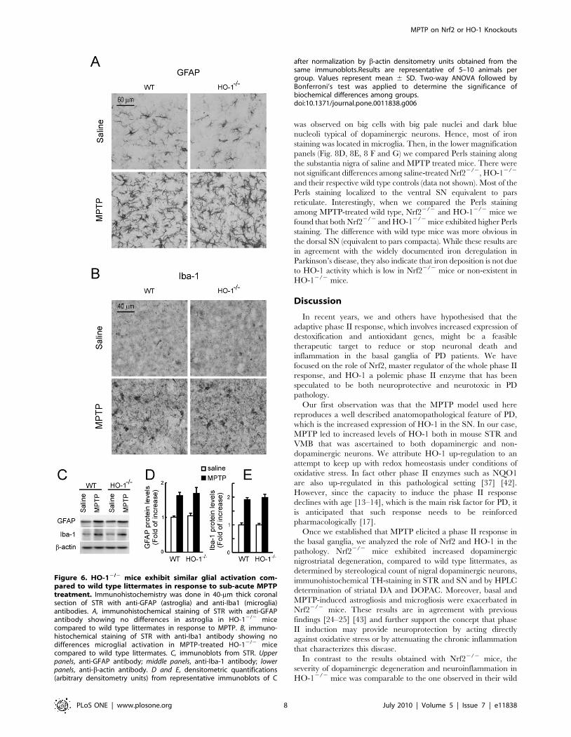
Regarding HO-12/2 mice, immunohistochemical analysis
demonstrated a small number of astrocytes and microglia in the
STR of saline-inoculated HO-12/2 mice that was similar to their
wild type controls (Figs. 6A and 6B). MPTP induced an increase in
the number of both glial types in HO-12/2 and wild type
littermates. Immunoblot analysis indicated a close to 2 fold
increase in striatal GFAP and Iba-1 levels in response to MPTP
but there was no significant difference between in HO-12/2 and
wild type controls (Fig. 6C). These results suggest that Nrf22/2
mice exhibit exacerbated basal and MPTP-induced gliosis and
that HO-12/2 mice show a pattern of gliosis similar to their wild
type littermates.
Phase II antioxidant enzyme levels in Nrf22/2, HO-12/2
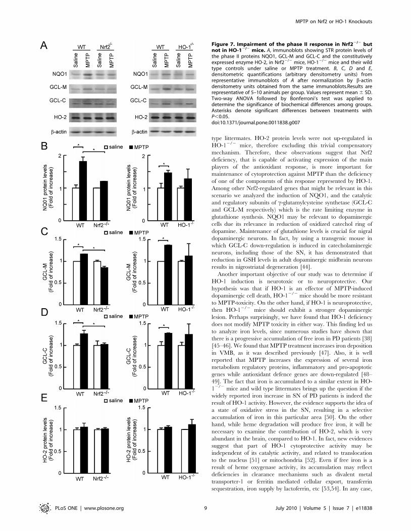
and wild type mice submitted to MPTPProtein levels of phase II enzymes NQO1, and the two subunits
of c-glutamylcysteine synthetase, GCL-M and GCL-C, were
analyzed in STR of Nrf22/2, HO-12/2 and wild type mice
(Fig. 7). Protein levels were normalized by b-actin. In wild type
and HO-12/2 mice, MPTP led to small increases of less than 1.5
fold in NQO1, GCL-M and GCL-C, which were in the limit of
statistical significance. By contrast, in Nrf22/2 mice, none of these
three phase II proteins were altered by the MPTP treatment.
Moreover, striatal protein levels of HO-2, which is not a phase II
enzyme, were not altered by either the genotypic alterations or the
MPTP treatment. These results suggest that Nrf2 deficiency
produces a stronger impairment in the phase II defense than HO-
1 deficiency.
Iron levels in wild type, Nrf22/2 and HO-12/2 micesubmitted to MPTP
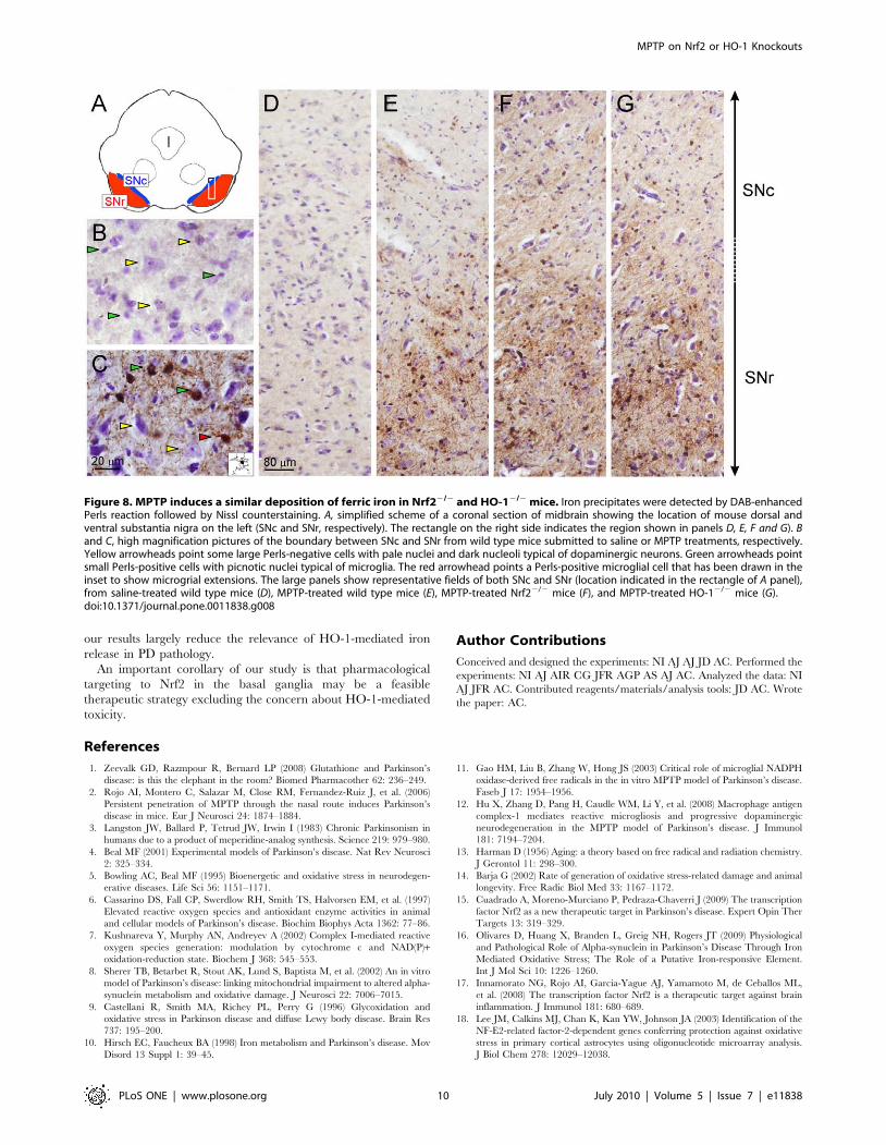
Iron levels were measured by 39-39-diaminobenzidine-enhanced
Perls staining in 40 mm-thick coronal sections from VMB
counterstained with Nissl staining. Under these condition ferric
ion deposits are stained in brown. As shown in Fig. 8B and 8C,
MPTP induced a substantial increase in Perls staining. Most of
iron staining was localized in very small, less than 10 mm diameter
cell bodies with picnotic nuclei that correspond to microglia and
microglial extensions (see inset in Fig. 8C). Very little Perls staining
Figure 5. Nrf22/2 mice show increased microglial activationcompared to wild type littermates in response to sub-acuteMPTP treatment. Immunohistochemistry was done in 40-mm thickcoronal section of STR with anti-GFAP (astroglia) and anti-Iba1(microglia) antibodies. A, immunohistochemical staining of STR withanti-GFAP antibody showing increased basal and MPTP-inducedastroglia in Nrf22/2 mice compared to wild type littermates. B,immunohistochemical staining of STR with anti-Iba1 antibody showingincreased microglial activation in MPTP-treated Nrf22/2 mice comparedto wild type littermates. C, immunoblots from STR. Upper panels, anti-
GFAP antibody; middle panels, anti-Iba-1 antibody; lower panels, anti-b-actin antibody. D and E, densitometric quantifications (arbitrarydensitometry units) from representative immunoblots of C afternormalization by b-actin densitometry units obtained from the sameimmunoblot.Results are representative of 5–10 animals per group.Values represent mean 6 SD. Two-way ANOVA followed by Bonferroni’stest was applied to determine the significance of biochemicaldifferences among groups. Asterisks denote significant differencesbetween treatments with P,0.05.doi:10.1371/journal.pone.0011838.g005
MPTP on Nrf2 or HO-1 Knockouts
PLoS ONE | www.plosone.org 7 July 2010 | Volume 5 | Issue 7 | e11838
was observed on big cells with big pale nuclei and dark blue
nucleoli typical of dopaminergic neurons. Hence, most of iron
staining was located in microglia. Then, in the lower magnification
panels (Fig. 8D, 8E, 8 F and G) we compared Perls staining along
the substantia nigra of saline and MPTP treated mice. There were
not significant differences among saline-treated Nrf22/2, HO-12/2
and their respective wild type controls (data not shown). Most of the
Perls staining localized to the ventral SN equivalent to pars
reticulate. Interestingly, when we compared the Perls staining
among MPTP-treated wild type, Nrf22/2 and HO-12/2 mice we
found that both Nrf22/2 and HO-12/2 mice exhibited higher Perls
staining. The difference with wild type mice was more obvious in
the dorsal SN (equivalent to pars compacta). While these results are
in agreement with the widely documented iron deregulation in
Parkinson’s disease, they also indicate that iron deposition is not due
to HO-1 activity which is low in Nrf22/2 mice or non-existent in
HO-12/2 mice.
Discussion
In recent years, we and others have hypothesised that the
adaptive phase II response, which involves increased expression of
destoxification and antioxidant genes, might be a feasible
therapeutic target to reduce or stop neuronal death and
inflammation in the basal ganglia of PD patients. We have
focused on the role of Nrf2, master regulator of the whole phase II
response, and HO-1 a polemic phase II enzyme that has been
speculated to be both neuroprotective and neurotoxic in PD
pathology.
Our first observation was that the MPTP model used here
reproduces a well described anatomopathological feature of PD,
which is the increased expression of HO-1 in the SN. In our case,
MPTP led to increased levels of HO-1 both in mouse STR and
VMB that was ascertained to both dopaminergic and non-
dopaminergic neurons. We attribute HO-1 up-regulation to an
attempt to keep up with redox homeostasis under conditions of
oxidative stress. In fact other phase II enzymes such as NQO1
are also up-regulated in this pathological setting [37] [42].
However, since the capacity to induce the phase II response
declines with age [13–14], which is the main risk factor for PD, it
is anticipated that such response needs to be reinforced
pharmacologically [17].
Once we established that MPTP elicited a phase II response in
the basal ganglia, we analyzed the role of Nrf2 and HO-1 in the
pathology. Nrf22/2 mice exhibited increased dopaminergic
nigrostriatal degeneration, compared to wild type littermates, as
determined by stereological count of nigral dopaminergic neurons,
immunohistochemical TH-staining in STR and SN and by HPLC
determination of striatal DA and DOPAC. Moreover, basal and
MPTP-induced astrogliosis and microgliosis were exacerbated in
Nrf22/2 mice. These results are in agreement with previous
findings [24–25] [43] and further support the concept that phase
II induction may provide neuroprotection by acting directly
against oxidative stress or by attenuating the chronic inflammation
that characterizes this disease.
In contrast to the results obtained with Nrf22/2 mice, the
severity of dopaminergic degeneration and neuroinflammation in
HO-12/2 mice was comparable to the one observed in their wild
Figure 6. HO-12/2 mice exhibit similar glial activation com-pared to wild type littermates in response to sub-acute MPTPtreatment. Immunohistochemistry was done in 40-mm thick coronalsection of STR with anti-GFAP (astroglia) and anti-Iba1 (microglia)antibodies. A, immunohistochemical staining of STR with anti-GFAPantibody showing no differences in astroglia in HO-12/2 micecompared to wild type littermates in response to MPTP. B, immuno-histochemical staining of STR with anti-Iba1 antibody showing nodifferences microglial activation in MPTP-treated HO-12/2 micecompared to wild type littermates. C, immunoblots from STR. Upperpanels, anti-GFAP antibody; middle panels, anti-Iba-1 antibody; lowerpanels, anti-b-actin antibody. D and E, densitometric quantifications(arbitrary densitometry units) from representative immunoblots of C
after normalization by b-actin densitometry units obtained from thesame immunoblots.Results are representative of 5–10 animals pergroup. Values represent mean 6 SD. Two-way ANOVA followed byBonferroni’s test was applied to determine the significance ofbiochemical differences among groups.doi:10.1371/journal.pone.0011838.g006
MPTP on Nrf2 or HO-1 Knockouts
PLoS ONE | www.plosone.org 8 July 2010 | Volume 5 | Issue 7 | e11838
type littermates. HO-2 protein levels were not up-regulated in
HO-12/2 mice, therefore excluding this trivial compensatory
mechanism. Therefore, these observations suggest that Nrf2
deficiency, that is capable of activating expression of the main
players of the antioxidant response, is more important for
maintenance of cytoprotection against MPTP than the deficiency
of one of the components of this response represented by HO-1.
Among other Nrf2-regulated genes that might be relevant in this
scenario we analyzed the induction of NQO1, and the catalytic
and regulatory subunits of c-glutamylcysteine synthetase (GCL-C
and GCL-M respectively) which is the rate limiting enzyme in
glutathione synthesis. NQO1 may be relevant to dopaminergic
cells due its relevance in reduction of oxidized catechol ring of
dopamine. Maintenance of glutathione levels is crucial for nigral
dopaminergic neurons. In fact, by using a transgenic mouse in
which GCL-C down-regulation is induced in catecholaminergic
neurons, including those of the SN, it has demonstrated that
reduction in GSH levels in adult dopaminergic midbrain neurons
results in nigrostriatal degeneration [44].
Another important objective of our study was to determine if
HO-1 induction is neurotoxic or to neuroprotective. Our
hypothesis was that if HO-1 is an effector of MPTP-induced
dopaminergic cell death, HO-12/2 mice should be more resistant
to MPTP-toxicity. On the other hand, if HO-1 is neuroprotective,
then HO-12/2 mice should exhibit a stronger dopaminergic
lesion. Perhaps surprisingly, we have found that HO-1 deficiency
does not modify MPTP toxicity in either way. This finding led us
to analyze iron levels, since numerous studies have shown that
there is a progressive accumulation of free iron in PD patients [38]
[45–46]. We found that MPTP treatment increases iron deposition
in VMB, as it was described previously [47]. Also, it is well
reported that MPTP increases the expression of several iron
metabolism regulatory proteins, inflammatory and pro-apoptotic
genes while antioxidant defence genes are down-regulated [48–
49]. The fact that iron is accumulated to a similar extent in HO-
12/2 mice and wild type littermates brings up the question if the
widely reported iron increase in SN of PD patients is indeed the
result of HO-1 activity. However, the evidence supports the idea of
a state of oxidative stress in the SN, resulting in a selective
accumulation of iron in this particular area [50]. On the other
hand, while heme degradation will produce free iron, it will be
necessary to examine the contribution of HO-2, which is very
abundant in the brain, compared to HO-1. In fact, new evidences
suggest that part of HO-1 cytoprotective activity may be
independent of its catalytic activity, and related to translocation
to the nucleus [51] or mitochondria [52]. Even if free iron is a
result of heme oxygenase activity, its accumulation may reflect
deficiencies in clearance mechanisms such as divalent metal
transporter-1 or ferritin mediated cellular export, transferrin
sequestration, iron supply by lactoferrin, etc [53,54]. In any case,
Figure 7. Impairment of the phase II response in Nrf22/2 butnot in HO-12/2 mice. A, immunoblots showing STR protein levels ofthe phase II proteins NQO1, GCL-M and GCL-C and the constitutivelyexpressed enzyme HO-2, in Nrf22/2 mice, HO-12/2 mice and their wildtype controls under saline or MPTP treatment. B, C, D and E,densitometric quantifications (arbitrary densitometry units) fromrepresentative immunoblots of A after normalization by b-actindensitometry units obtained from the same immunoblots.Results arerepresentative of 5–10 animals per group. Values represent mean 6 SD.Two-way ANOVA followed by Bonferroni’s test was applied todetermine the significance of biochemical differences among groups.Asterisks denote significant differences between treatments withP,0.05.doi:10.1371/journal.pone.0011838.g007
MPTP on Nrf2 or HO-1 Knockouts
PLoS ONE | www.plosone.org 9 July 2010 | Volume 5 | Issue 7 | e11838
our results largely reduce the relevance of HO-1-mediated iron
release in PD pathology.
An important corollary of our study is that pharmacological
targeting to Nrf2 in the basal ganglia may be a feasible
therapeutic strategy excluding the concern about HO-1-mediated
toxicity.
Author Contributions
Conceived and designed the experiments: NI AJ AJ JD AC. Performed the
experiments: NI AJ AIR CG JFR AGP AS AJ AC. Analyzed the data: NI
AJ JFR AC. Contributed reagents/materials/analysis tools: JD AC. Wrote
the paper: AC.
References
1. Zeevalk GD, Razmpour R, Bernard LP (2008) Glutathione and Parkinson’s
disease: is this the elephant in the room? Biomed Pharmacother 62: 236–249.
2. Rojo AI, Montero C, Salazar M, Close RM, Fernandez-Ruiz J, et al. (2006)
Persistent penetration of MPTP through the nasal route induces Parkinson’s
disease in mice. Eur J Neurosci 24: 1874–1884.
3. Langston JW, Ballard P, Tetrud JW, Irwin I (1983) Chronic Parkinsonism in
humans due to a product of meperidine-analog synthesis. Science 219: 979–980.
4. Beal MF (2001) Experimental models of Parkinson’s disease. Nat Rev Neurosci
2: 325–334.
5. Bowling AC, Beal MF (1995) Bioenergetic and oxidative stress in neurodegen-
erative diseases. Life Sci 56: 1151–1171.
6. Cassarino DS, Fall CP, Swerdlow RH, Smith TS, Halvorsen EM, et al. (1997)
Elevated reactive oxygen species and antioxidant enzyme activities in animal
and cellular models of Parkinson’s disease. Biochim Biophys Acta 1362: 77–86.
7. Kushnareva Y, Murphy AN, Andreyev A (2002) Complex I-mediated reactive
oxygen species generation: modulation by cytochrome c and NAD(P)+oxidation-reduction state. Biochem J 368: 545–553.
8. Sherer TB, Betarbet R, Stout AK, Lund S, Baptista M, et al. (2002) An in vitro
model of Parkinson’s disease: linking mitochondrial impairment to altered alpha-
synuclein metabolism and oxidative damage. J Neurosci 22: 7006–7015.
9. Castellani R, Smith MA, Richey PL, Perry G (1996) Glycoxidation and
oxidative stress in Parkinson disease and diffuse Lewy body disease. Brain Res
737: 195–200.
10. Hirsch EC, Faucheux BA (1998) Iron metabolism and Parkinson’s disease. Mov
Disord 13 Suppl 1: 39–45.
11. Gao HM, Liu B, Zhang W, Hong JS (2003) Critical role of microglial NADPH
oxidase-derived free radicals in the in vitro MPTP model of Parkinson’s disease.
Faseb J 17: 1954–1956.
12. Hu X, Zhang D, Pang H, Caudle WM, Li Y, et al. (2008) Macrophage antigen
complex-1 mediates reactive microgliosis and progressive dopaminergic
neurodegeneration in the MPTP model of Parkinson’s disease. J Immunol
181: 7194–7204.
13. Harman D (1956) Aging: a theory based on free radical and radiation chemistry.
J Gerontol 11: 298–300.
14. Barja G (2002) Rate of generation of oxidative stress-related damage and animal
longevity. Free Radic Biol Med 33: 1167–1172.
15. Cuadrado A, Moreno-Murciano P, Pedraza-Chaverri J (2009) The transcription
factor Nrf2 as a new therapeutic target in Parkinson’s disease. Expert Opin Ther
Targets 13: 319–329.
16. Olivares D, Huang X, Branden L, Greig NH, Rogers JT (2009) Physiological
and Pathological Role of Alpha-synuclein in Parkinson’s Disease Through Iron
Mediated Oxidative Stress; The Role of a Putative Iron-responsive Element.
Int J Mol Sci 10: 1226–1260.
17. Innamorato NG, Rojo AI, Garcia-Yague AJ, Yamamoto M, de Ceballos ML,
et al. (2008) The transcription factor Nrf2 is a therapeutic target against brain
inflammation. J Immunol 181: 680–689.
18. Lee JM, Calkins MJ, Chan K, Kan YW, Johnson JA (2003) Identification of the
NF-E2-related factor-2-dependent genes conferring protection against oxidative
stress in primary cortical astrocytes using oligonucleotide microarray analysis.
J Biol Chem 278: 12029–12038.
Figure 8. MPTP induces a similar deposition of ferric iron in Nrf22/2 and HO-12/2 mice. Iron precipitates were detected by DAB-enhancedPerls reaction followed by Nissl counterstaining. A, simplified scheme of a coronal section of midbrain showing the location of mouse dorsal andventral substantia nigra on the left (SNc and SNr, respectively). The rectangle on the right side indicates the region shown in panels D, E, F and G). Band C, high magnification pictures of the boundary between SNc and SNr from wild type mice submitted to saline or MPTP treatments, respectively.Yellow arrowheads point some large Perls-negative cells with pale nuclei and dark nucleoli typical of dopaminergic neurons. Green arrowheads pointsmall Perls-positive cells with picnotic nuclei typical of microglia. The red arrowhead points a Perls-positive microglial cell that has been drawn in theinset to show microgrial extensions. The large panels show representative fields of both SNc and SNr (location indicated in the rectangle of A panel),from saline-treated wild type mice (D), MPTP-treated wild type mice (E), MPTP-treated Nrf22/2 mice (F), and MPTP-treated HO-12/2 mice (G).doi:10.1371/journal.pone.0011838.g008
MPTP on Nrf2 or HO-1 Knockouts
PLoS ONE | www.plosone.org 10 July 2010 | Volume 5 | Issue 7 | e11838
19. Shih AY, Johnson DA, Wong G, Kraft AD, Jiang L, et al. (2003) Coordinate
regulation of glutathione biosynthesis and release by Nrf2-expressing gliapotently protects neurons from oxidative stress. J Neurosci 23: 3394–3406.
20. Lo SC, Li X, Henzl MT, Beamer LJ, Hannink M (2006) Structure of the
Keap1:Nrf2 interface provides mechanistic insight into Nrf2 signaling. Embo J25: 3605–3617.
21. Katsuoka F, Motohashi H, Ishii T, Aburatani H, Engel JD, et al. (2005) Geneticevidence that small maf proteins are essential for the activation of antioxidant
response element-dependent genes. Mol Cell Biol 25: 8044–8051.
22. Dinkova-Kostova AT, Holtzclaw WD, Cole RN, Itoh K, Wakabayashi N, et al.(2002) Direct evidence that sulfhydryl groups of Keap1 are the sensors regulating
induction of phase 2 enzymes that protect against carcinogens and oxidants.Proc Natl Acad Sci U S A 99: 11908–11913.
23. Burton NC, Kensler TW, Guilarte TR (2006) In vivo modulation of theParkinsonian phenotype by Nrf2. Neurotoxicology 27: 1094–1100.
24. Chen PC, Vargas MR, Pani AK, Smeyne RJ, Johnson DA, et al. (2009) Nrf2-
mediated neuroprotection in the MPTP mouse model of Parkinson’s disease:Critical role for the astrocyte. Proc Natl Acad Sci U S A 106: 2933–2938.
25. Rojo AI, Innamorato NG, Martin-Moreno AM, De Ceballos ML,Yamamoto M, et al. (2009) Nrf2 regulates microglial dynamics and neuroin-
flammation in experimental Parkinson’s disease. Glia 58: 588–598.
26. Boczkowski J, Poderoso JJ, Motterlini R (2006) CO-metal interaction: Vitalsignaling from a lethal gas. Trends Biochem Sci 31: 614–621.
27. Bilban M, Haschemi A, Wegiel B, Chin BY, Wagner O, et al. (2008) Hemeoxygenase and carbon monoxide initiate homeostatic signaling. J Mol Med 86:
267–279.28. Piantadosi CA (2008) Carbon monoxide, reactive oxygen signaling, and
oxidative stress. Free Radic Biol Med 45: 562–569.
29. Cuadrado A, Rojo AI (2008) Heme oxygenase-1 as a therapeutic target inneurodegenerative diseases and brain infections. Curr Pharm Des 14: 429–442.
30. Li MH, Cha YN, Surh YJ (2006) Peroxynitrite induces HO-1 expression viaPI3K/Akt-dependent activation of NF-E2-related factor 2 in PC12 cells. Free
Radic Biol Med 41: 1079–1091.
31. Samoylenko A, Dimova EY, Horbach T, Teplyuk N, Immenschuh S, et al.(2008) Opposite expression of the antioxidant heme oxygenase-1 in primary cells
and tumor cells: regulation by interaction of USF-2 and Fra-1. Antioxid RedoxSignal 10: 1163–1174.
32. Huang E, Ong WY, Go ML, Garey LJ (2005) Heme oxygenase-1 activity afterexcitotoxic injury: immunohistochemical localization of bilirubin in neurons and
astrocytes and deleterious effects of heme oxygenase inhibition on neuronal
survival after kainate treatment. J Neurosci Res 80: 268–278.33. Hung SY, Liou HC, Kang KH, Wu RM, Wen CC, et al. (2008) Overexpression
of heme oxygenase-1 protects dopaminergic neurons against 1-methyl-4-phenylpyridinium-induced neurotoxicity. Mol Pharmacol 74: 1564–1575.
34. Glinka Y, Tipton KF, Youdim MB (1996) Nature of inhibition of mitochondrial
respiratory complex I by 6-Hydroxydopamine. J Neurochem 66: 2004–2010.35. Ben-Shachar D, Eshel G, Finberg JP, Youdim MB (1991) The iron chelator
desferrioxamine (Desferal) retards 6-hydroxydopamine-induced degeneration ofnigrostriatal dopamine neurons. J Neurochem 56: 1441–1444.
36. Grunblatt E, Mandel S, Berkuzki T, Youdim MB (1999) Apomorphine protectsagainst MPTP-induced neurotoxicity in mice. Mov Disord 14: 612–618.
37. Schipper HM, Liberman A, Stopa EG (1998) Neural heme oxygenase-1
expression in idiopathic Parkinson’s disease. Exp Neurol 150: 60–68.38. Morris CM, Edwardson JA (1994) Iron histochemistry of the substantia nigra in
Parkinson’s disease. Neurodegeneration 3: 277–282.
39. Itoh K, Chiba T, Takahashi S, Ishii T, Igarashi K, et al. (1997) An Nrf2/smallMaf heterodimer mediates the induction of phase II detoxifying enzyme genes
through antioxidant response elements. Biochem Biophys Res Commun 236:313–322.
40. Tsuchihashi S, Livhits M, Zhai Y, Busuttil RW, Araujo JA, et al. (2006) Basal
rather than induced heme oxygenase-1 levels are crucial in the antioxidantcytoprotection. J Immunol 177: 4749–4757.
41. West MJ (1993) New stereological methods for counting neurons. NeurobiolAging 14: 275–285.
42. Schipper HM, Song W, Zukor H, Hascalovici JR, Zeligman D (2009) Hemeoxygenase-1 and neurodegeneration: expanding frontiers of engagement.
J Neurochem 110: 469–485.
43. Gille G, Hung ST, Reichmann H, Rausch WD (2004) Oxidative stress todopaminergic neurons as models of Parkinson’s disease. Ann N Y Acad Sci
1018: 533–540.44. Chinta SJ, Kumar MJ, Hsu M, Rajagopalan S, Kaur D, et al. (2007) Inducible
alterations of glutathione levels in adult dopaminergic midbrain neurons result in
nigrostriatal degeneration. J Neurosci 27: 13997–14006.45. Sofic E, Paulus W, Jellinger K, Riederer P, Youdim MB (1991) Selective
increase of iron in substantia nigra zona compacta of parkinsonian brains.J Neurochem 56: 978–982.
46. Berg D, Gerlach M, Youdim MB, Double KL, Zecca L, et al. (2001) Brain ironpathways and their relevance to Parkinson’s disease. J Neurochem 79: 225–236.
47. Mochizuki H, Imai H, Endo K, Yokomizo K, Murata Y, et al. (1994) Iron
accumulation in the substantia nigra of 1-methyl-4-phenyl-1,2,3,6-tetrahydro-pyridine (MPTP)-induced hemiparkinsonian monkeys. Neurosci Lett 168:
251–253.48. Mandel S, Grunblatt E, Maor G, Youdim MB (2002) Early and late gene
changes in MPTP mice model of Parkinson’s disease employing cDNA
microarray. Neurochem Res 27: 1231–1243.49. Grunblatt E, Mandel S, Maor G, Youdim MB (2001) Gene expression analysis
in N-methyl-4-phenyl-1,2,3,6-tetrahydropyridine mice model of Parkinson’sdisease using cDNA microarray: effect of R-apomorphine. J Neurochem 78:
1–12.50. Youdim MB, Ben-Shachar D, Riederer P (1993) The possible role of iron in the
etiopathology of Parkinson’s disease. Mov Disord 8: 1–12.
51. Lin Q, Weis S, Yang G, Weng YH, Helston R, et al. (2007) Heme oxygenase-1protein localizes to the nucleus and activates transcription factors important in
oxidative stress. J Biol Chem 282: 20621–20633.52. Turkseven S, Drummond G, Rezzani R, Rodella L, Quan S, et al. (2007)
Impact of silencing HO-2 on EC-SOD and the mitochondrial signaling
pathway. J Cell Biochem 100: 815–823.53. Snyder AM, Connor JR (2009) Iron, the substantia nigra and related
neurological disorders. Biochim Biophys Acta 1790: 606–614.54. Salazar J, Mena N, Hunot S, Prigent A, Alvarez-Fischer D, et al. (2008) Divalent
metal transporter 1 (DMT1) contributes to neurodegeneration in animal modelsof Parkinson’s disease. Proc Natl Acad Sci U S A 105: 18578–18583.
MPTP on Nrf2 or HO-1 Knockouts
PLoS ONE | www.plosone.org 11 July 2010 | Volume 5 | Issue 7 | e11838