Embed Size (px)
Citation preview
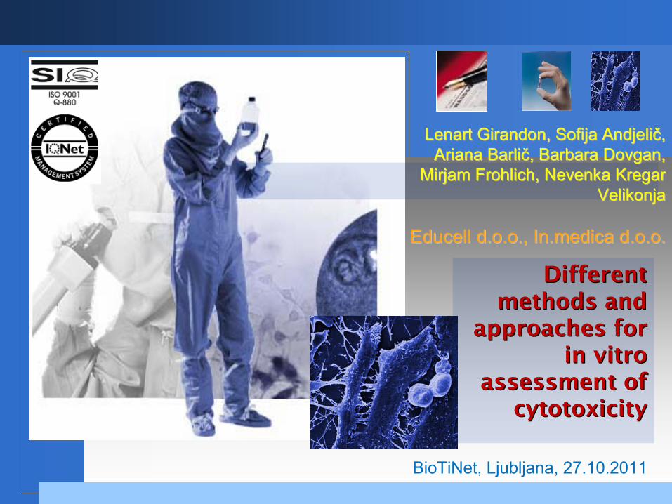
Different Different methods and methods and
approaches for approaches for in vitroin vitro
assessmentassessment
ofof cytotoxicitycytotoxicity
BioTiNet, Ljubljana,
27.10.2011
Lenart Lenart GirandonGirandon, Sofija , Sofija AndjeliAndjeličč, , ArianaAriana
BarliBarličč, Barbara Dovgan, , Barbara Dovgan,
Mirjam Mirjam FrohlichFrohlich, Nevenka Kregar , Nevenka Kregar VelikonjaVelikonja
EducellEducell
d.o.o., d.o.o., In.medicaIn.medica
d.o.o. d.o.o.
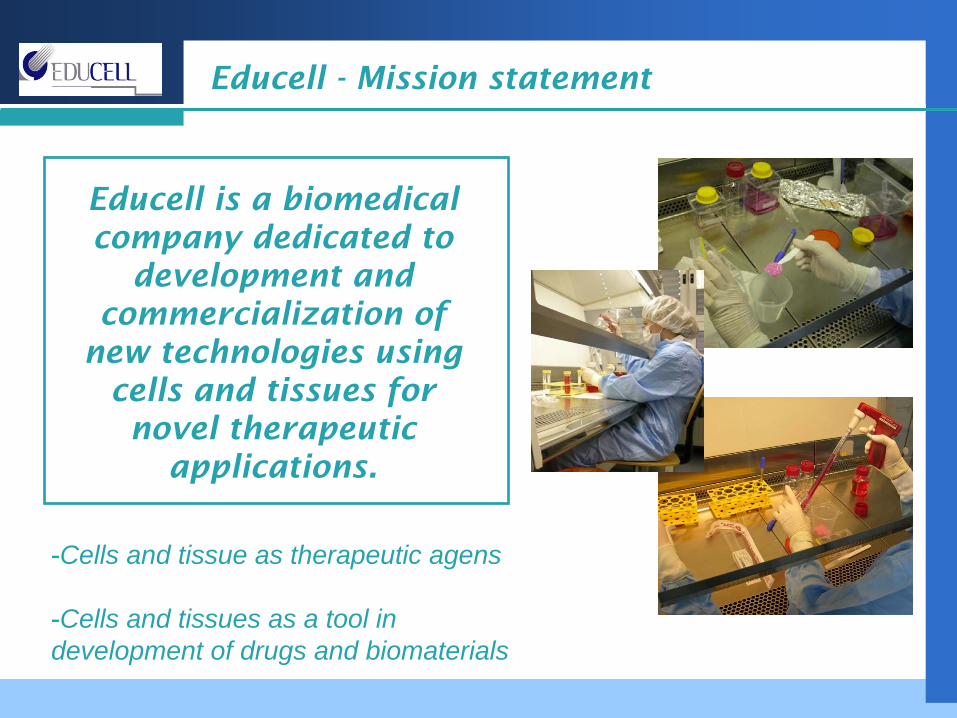
Educell
-
Mission
statement
Educell
is a biomedical company
dedicated
to
development
and commercialization
of
new
technologies
using cells
and
tissues
for
novel therapeutic applications.
-Cells and tissue as therapeutic agens
-Cells and tissues as a tool in development of drugs and biomaterials
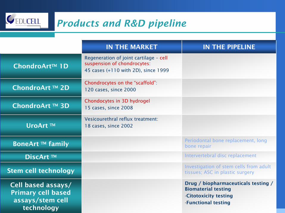
Products
and
R&D pipeline
IN THE MARKET IN THE PIPELINE
ChondroArt™
1DRegeneration of joint cartilage –
cell suspension of chondrocytes:45
cases
(+110 with 2D), since 1999
ChondroArt ™
2DChondrocytes on the “scaffold”:120 cases, since 2000
ChondroArt ™
3DChondocytes in 3D hydrogel15
cases, since 2008
UroArt ™Vesicourethral reflux treatment:18
cases, since 2002
BoneArt ™
familyPeriodontal bone replacement,
long bone repair
DiscArt ™ Intervertebral disc replacement
Stem cell technologyInvestigation of stem cells from
adult tissues; ASC in plastic surgery
Cell based assays/ Primary cell based assays/stem cell
technology
Drug / biopharmaceuticals testing / Biomaterial testing
-Citotoxicity testing
-Functional testing
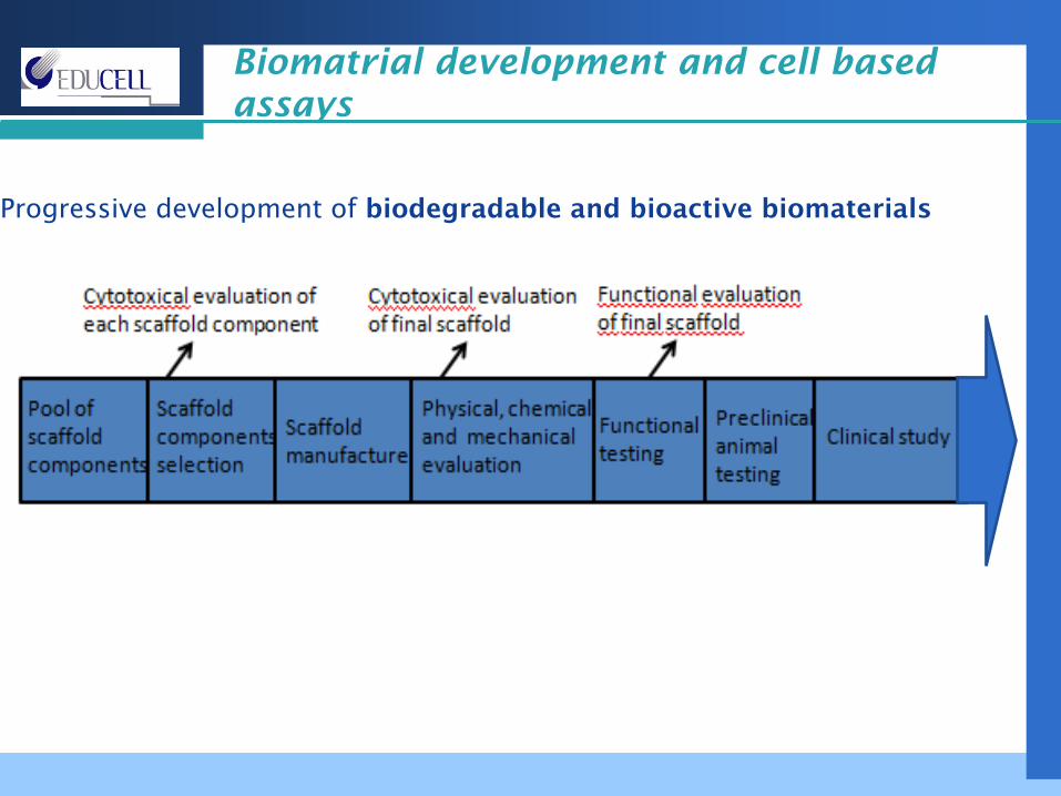
Biomatrial development and cell based assays
Progressive development of biodegradable and bioactive biomaterials
Citotoxicity
Cytotoxicity
is the quality of being toxic
to cells. Examples of toxic agents are:- a chemical substance, - an immune cell
or
- some types of venom
Treating cells with a cytotoxic compound can result in a variety
of cell fates:
-necrosis, in which they lose membrane integrity and die rapidly as a result of cell lysis
-citostatic effect: the cells can stop actively growing and dividing (a decrease in cell viability),
-Apoptosis:
the cells can activate a genetic program of controlled cell death.
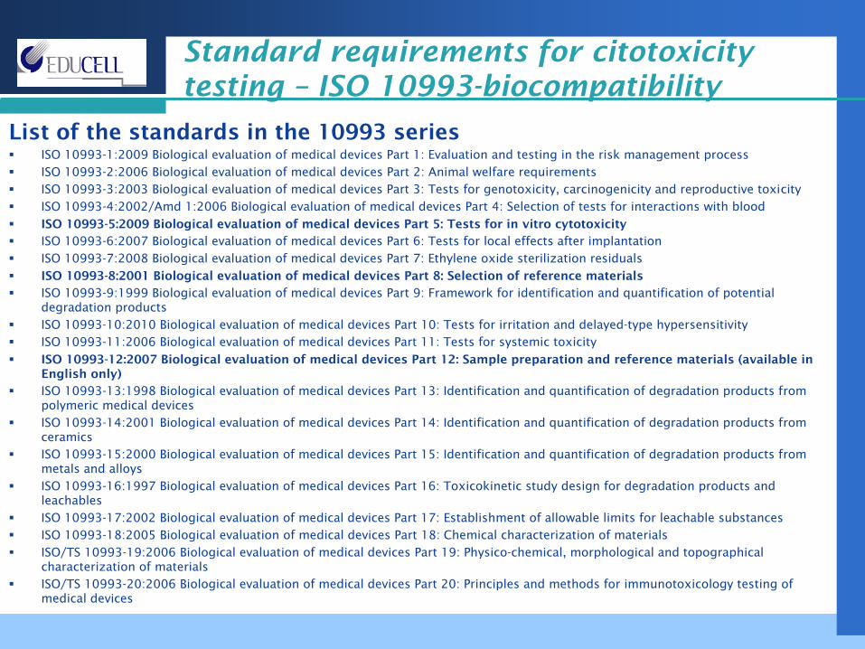
Standard requirements for citotoxicity testing –
ISO 10993-biocompatibility
List of the standards in the 10993 seriesISO 10993-1:2009 Biological evaluation of medical devices Part 1: Evaluation and testing in the risk management processISO 10993-2:2006 Biological evaluation of medical devices Part 2: Animal welfare requirementsISO 10993-3:2003 Biological evaluation of medical devices Part 3: Tests for genotoxicity, carcinogenicity and reproductive toxicityISO 10993-4:2002/Amd 1:2006 Biological evaluation of medical devices Part 4: Selection of tests for interactions with bloodISO 10993-5:2009 Biological evaluation of medical devices Part 5: Tests for in vitro cytotoxicityISO 10993-6:2007 Biological evaluation of medical devices Part 6: Tests for local effects after implantationISO 10993-7:2008 Biological evaluation of medical devices Part 7: Ethylene oxide sterilization residualsISO 10993-8:2001 Biological evaluation of medical devices Part 8: Selection of reference materials
ISO 10993-9:1999 Biological evaluation of medical devices Part 9: Framework for identification and quantification of potential degradation productsISO 10993-10:2010 Biological evaluation of medical devices Part 10: Tests for irritation and delayed-type hypersensitivityISO 10993-11:2006 Biological evaluation of medical devices Part 11: Tests for systemic toxicityISO 10993-12:2007 Biological evaluation of medical devices Part 12: Sample preparation and reference materials (available in English only)ISO 10993-13:1998 Biological evaluation of medical devices Part 13: Identification and quantification of degradation products from polymeric medical devicesISO 10993-14:2001 Biological evaluation of medical devices Part 14: Identification and quantification of degradation products from ceramicsISO 10993-15:2000 Biological evaluation of medical devices Part 15: Identification and quantification of degradation products from metals and alloysISO 10993-16:1997 Biological evaluation of medical devices Part 16: Toxicokinetic study design for degradation products and leachablesISO 10993-17:2002 Biological evaluation of medical devices Part 17: Establishment of allowable limits for leachable substancesISO 10993-18:2005 Biological evaluation of medical devices Part 18: Chemical characterization of materialsISO/TS 10993-19:2006 Biological evaluation of medical devices Part 19: Physico-chemical, morphological and topographical characterization of materialsISO/TS 10993-20:2006 Biological evaluation of medical devices Part 20: Principles and methods for immunotoxicology testing of medical devices
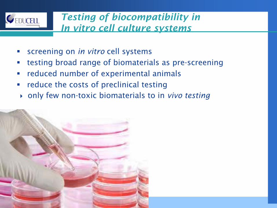
screening on in vitro cell systemstesting broad range of biomaterials as pre-screeningreduced number of experimental animalsreduce the costs of preclinical testingonly few non-toxic biomaterials to in vivo testing
Testing of biocompatibility in In vitro cell culture systems
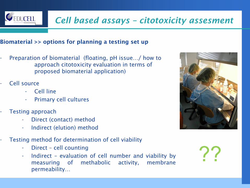
Cell based assays –
citotoxicity assesment
Biomaterial >> options for planning a testing set up
- Preparation of biomaterial (floating, pH issue…/ how to approach citotoxicity evaluation in terms of proposed biomaterial application)
- Cell source-
Cell line-
Primary cell cultures
- Testing approach-
Direct (contact) method-
Indirect (elution) method
- Testing method for determination of cell viability-
Direct –
cell counting-
Indirect –
evaluation of cell number and viability by measuring of methabolic activity, membrane permeability…
??
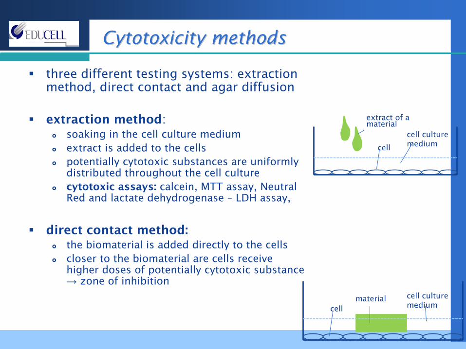
three different testing systems: extraction method, direct contact and agar diffusion
extraction method:soaking in the cell culture mediumextract is added to the cellspotentially cytotoxic substances are uniformly distributed throughout the cell culturecytotoxic assays: calcein, MTT assay, Neutral Red and lactate dehydrogenase – LDH assay,
direct contact method:the biomaterial is added directly to the cells closer to the biomaterial are cells receive higher doses of potentially cytotoxic substance→ zone of inhibition
CCytotoxicity methodsytotoxicity methods
extract of a material
cell culture mediumcell
cell
cell culture medium
material
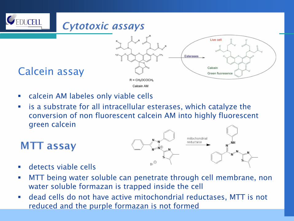
calcein AM labeles only viable cellsis a substrate for all intracellular esterases, which catalyze the conversion of non fluorescent calcein AM into highly fluorescentgreen calcein
detects viable cells MTT being water soluble can penetrate through cell membrane, nonwater soluble formazan is trapped inside the celldead cells do not have active mitochondrial reductases, MTT is not reduced and the purple formazan is not formed
Calcein assay
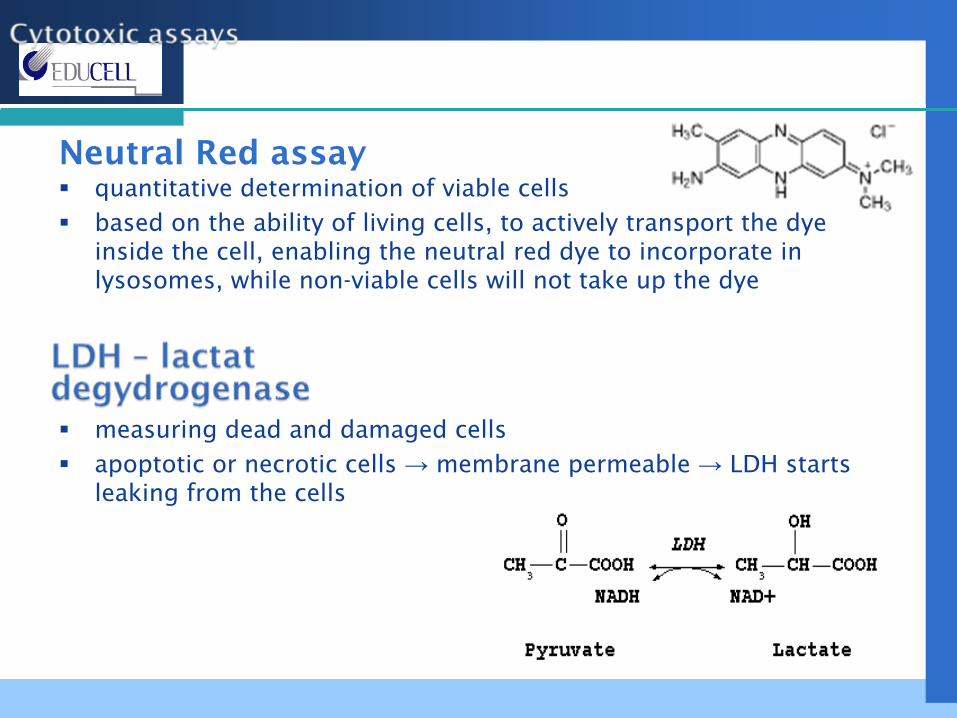
quantitative determination of viable cells based on the ability of living cells, to actively transport the dye inside the cell, enabling the neutral red dye to incorporate in lysosomes, while non-viable cells will not take up the dye
measuring dead and damaged cellsapoptotic or necrotic cells → membrane permeable → LDH starts leaking from the cells
Neutral Red
assay
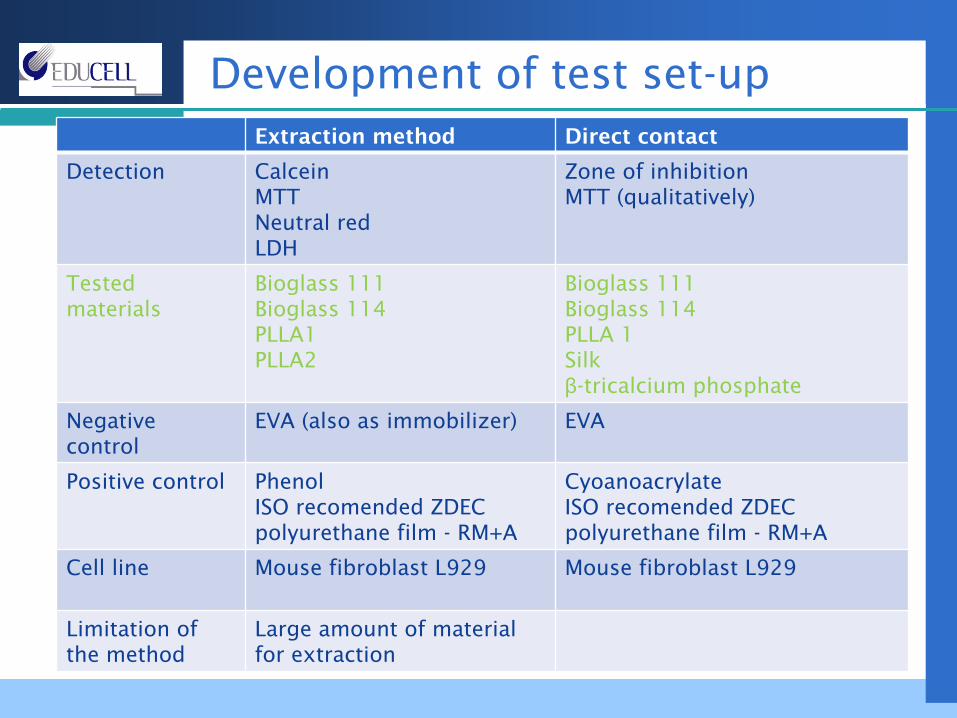
Development of test set-upExtraction method Direct contact
Detection CalceinMTTNeutral redLDH
Zone of inhibitionMTT (qualitatively)
Tested materials
Bioglass 111Bioglass 114PLLA1PLLA2
Bioglass 111Bioglass 114PLLA 1Silkβ-tricalcium phosphate
Negative control
EVA (also as immobilizer) EVA
Positive control Phenol ISO recomended ZDEC polyurethane film -
RM+A
CyoanoacrylateISO recomended ZDEC polyurethane film -
RM+A
Cell line Mouse fibroblast L929 Mouse fibroblast L929
Limitation of the method
Large amount of material for extraction
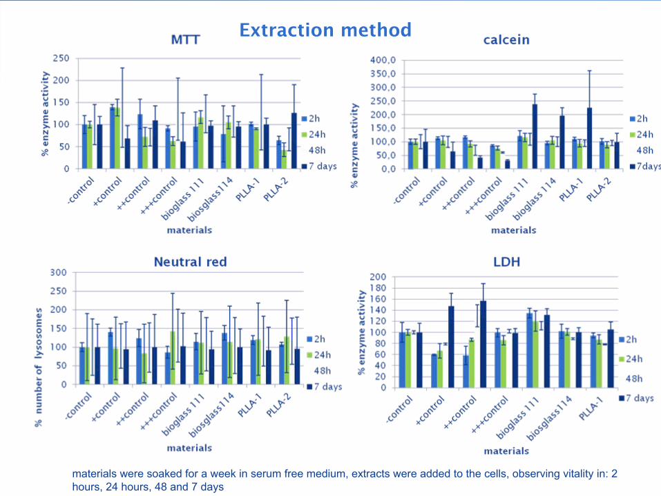
materials were soaked for a week in serum free medium, extracts were added to the cells, observing vitality in: 2 hours, 24 hours, 48 and 7 days
Extraction method
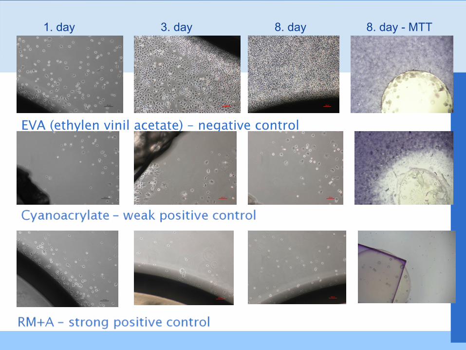
EVA (ethylen vinil acetate) –
negative control
1. day 3. day 8. day 8. day -
MTT
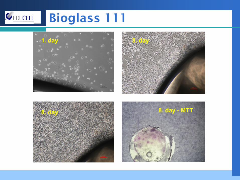
Bioglass 111
1. day 3. day3. day
8. day 8. day -
MTT
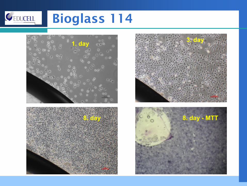
Bioglass 114
1. day 3. day3. day
8. day 8. day -
MTT
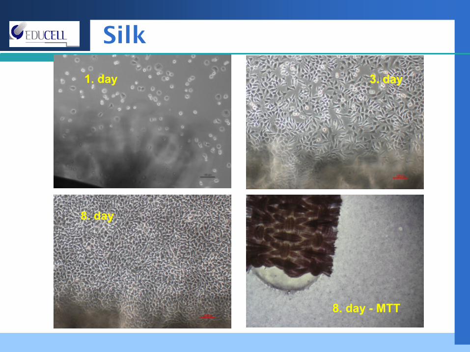
Silk
1. day 3. day3. day
8. day
8. day -
MTT
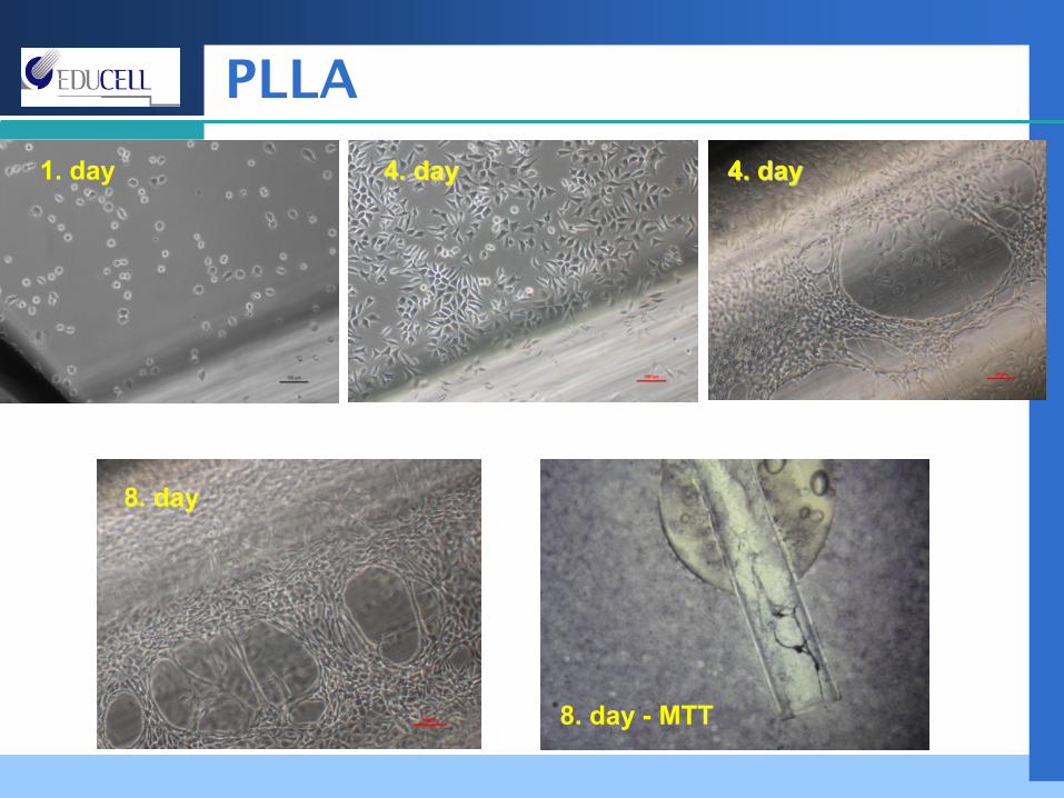
PLLA
1. day 4. day4. day
8. day
8. day -
MTT
4. day4. day
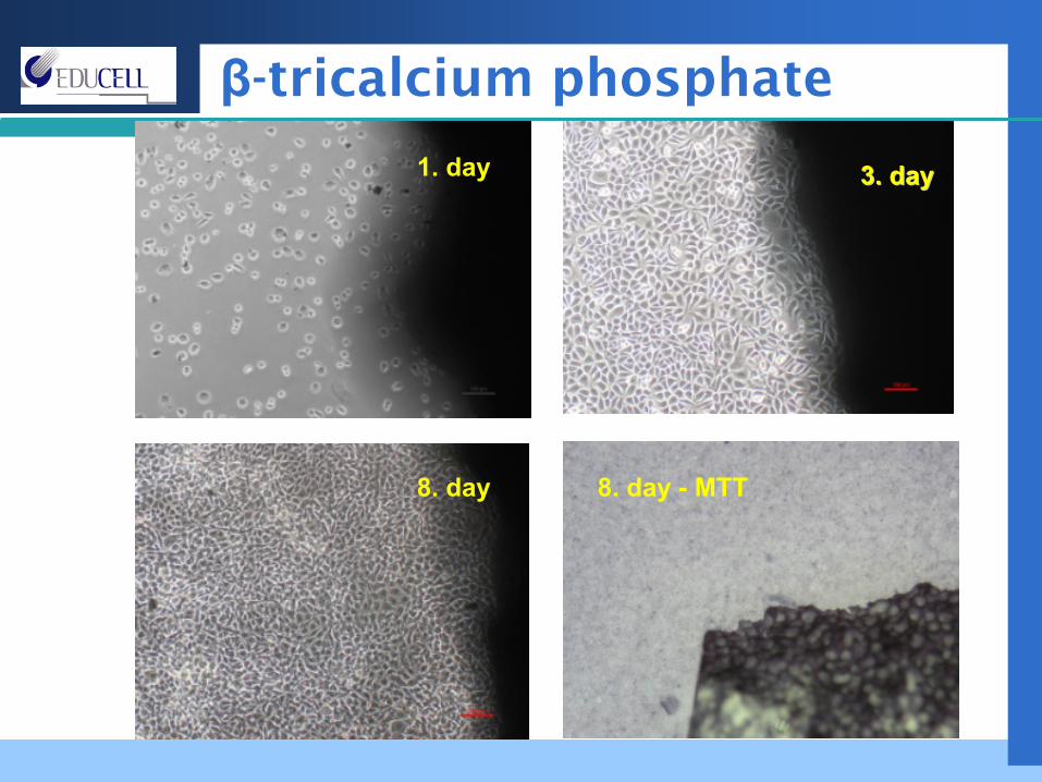
β-tricalcium phosphate
1. day 3. day3. day
8. day 8. day -
MTT
Functional tests and Primary cells
Functional tests→
evaluation of effectiveness
of the
compound on in vitro
level in a system that mimics in vivo situation (prior animal trials)
Primary cells:more relevant
preclinical data Primary cell phenotype reflects more the environment of the native tissue in comparison to immortalized cell lines.high biological variability results in diverse response of the cells on the tested agents
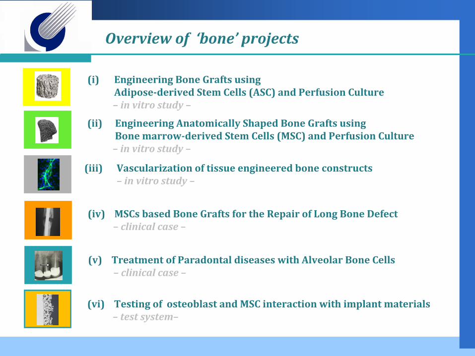
(iv) MSCs based Bone Grafts for the Repair of Long Bone Defect–
clinical case
–
Overview
of ‘bone’
projects
(ii)
Engineering Anatomically Shaped Bone Grafts usingBone marrowderived Stem Cells (MSC) and Perfusion Culture –
in vitro study –
(i)
Engineering Bone Grafts using Adiposederived Stem Cells (ASC) and Perfusion Culture –
in vitro study –
(vi) Testing of osteoblast and MSC interaction with implant materials–
test system–
(iii) Vascularization of tissue engineered bone constructs–
in vitro study –
(v) Treatment of Paradontal diseases with Alveolar Bone Cells–
clinical case
–
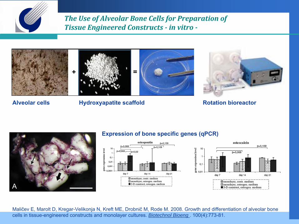
The Use of Alveolar Bone Cells for Preparation of Tissue Engineered Constructs
in vitro
Maličev E, Marolt D, Kregar-Velikonja N, Kreft ME, Drobnič
M, Rode M.
2008.
Growth and differentiation of alveolar bone cells in tissue-engineered constructs and monolayer cultures.
Biotechnol Bioeng . 100(4):773-81.
Alveolar cells Hydroxyapatite scaffold Rotation bioreactor
Expression of bone specific genes (qPCR)
Fröhlich M, Grayson WL, Marolt D, Gimble JM, Kregar-Velikonja N, Vunjak-Novakovic G. 2010.
Bone grafts engineered from human adipose-derived stem cells in perfusion bioreactor culture.
Tissue Eng Part A. 16(1):179-89.
decellularized bone matrix
Adipose-derived Stem Cells(ASC)
Engineering Bone Grafts using Adiposederived Stem Cells
(ASC) and Perfusion Culture
Peristaltic pump
perfusion bioreactor
control osteo
stat
icpe
rfus
ed
static perfused
stat
icpe
rfus
ed
osteopontin collagenVIABILITY CELL DISTRIBUTION BONE MATRIX PROTEINS
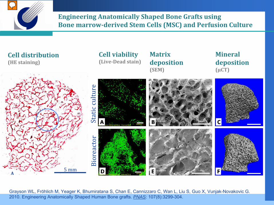
A5 mm
Static culture
Bioreactor
Grayson WL, Fröhlich M, Yeager K, Bhumiratana S, Chan E, Cannizzaro C, Wan L, Liu S, Guo X, Vunjak-Novakovic G. 2010. Engineering Anatomically Shaped Human Bone grafts. PNAS: 107(8):3299-304.
Cell distribution(HE staining)
Cell viability(LiveDead stain)
Matrix deposition (SEM)
Mineral deposition (µCT)
Engineering Anatomically Shaped Bone Grafts usingBone marrowderived Stem Cells (MSC) and Perfusion Culture
In vitro evaluation of citotoxicity is very important for preclinical development of biomaterials
Methods for measuring cytotoxicity must be carefuly validated
Functional tests are important for assesment of interaction between cell and materials and have to be adapted for a specific tested compound
Primary cells can show us more biologicaly relevant response in comparison to primary cell lines and also interindividual differences.
Conclusion
Ariana BarliAriana Barličč, , Lenart GirandonLenart GirandonMirjam FrohlichMirjam FrohlichSofija AndjeliSofija AndjeliččBarbara DovganBarbara Dovgan
Mirjam FrohlichMirjam Frohlich
Perfusion
bioreactor
studies:Columbia University, NY, USA, Gordana Vunjak Novakovic