Embed Size (px)
Citation preview

ORIGINAL RESEARCH ARTICLEpublished: 24 June 2014
doi: 10.3389/fnhum.2014.00464
Differences in time course activation of dorsolateralprefrontal cortex associated with low or high riskchoices in a gambling taskStefano Bembich1, Andrea Clarici2*, Cristina Vecchiet1, Giulio Baldassi1, Gabriele Cont1 and
Sergio Demarini1
1 Institute for Maternal and Child Health, IRCCS “Burlo Garofolo”, Trieste, Italy2 Psychiatric Clinic Unit, Department of Medical, Surgical and Health Sciences, University of Trieste, Trieste, Italy
Edited by:
John J. Foxe, Albert Einstein Collegeof Medicine, USA
Reviewed by:
Ippeita Dan, Jichi MedicalUniversity, JapanColm Gerard Connolly, University ofCalifornia San Francisco, USA
*Correspondence:
Andrea Clarici, Psychiatric ClinicUnit, Department of Medical,Surgical and Health Science,University of Trieste, Via Paolo deRalli 5, 34138 Trieste, Italye-mail: [email protected]
Prefrontal cortex plays an important role in decision making (DM), supporting choicesin the ordinary uncertainty of everyday life. To assess DM in an unpredictable situation,a playing card task, such as the Iowa Gambling Task (IGT), has been proposed. Thistask is supposed to specifically test emotion-based learning, linked to the integrity ofthe ventromedial prefrontal cortex (VMPFC). However, the dorsolateral prefrontal cortex(DLPFC) has demonstrated a role in IGT performance too. Our aim was to study, bymultichannel near-infrared spectroscopy, the contribution of DLPFC to the IGT executionover time. We tested the hypothesis that low and high risk choices would differentiallyactivate DLPFC, as IGT execution progressed. We enrolled 11 healthy adults. To identifyDLPFC activation associated with IGT choices, we compared regional differences inoxy-hemoglobin variation, from baseline to the event. The time course of task executionwas divided in four periods, each one consisting of 25 choices, and DLPFC activation wasdistinctly analyzed for low and high risk choices in each period. We found different timecourses in DLPFC activation, associated with low or high risk choices. During the firstperiod, a significant DLPFC activation emerged with low risk choices, whereas, duringthe second period, we found a cortical activation with high risk choices. Then, DLPFCactivation decreased to non-significant levels during the third and fourth period. This studyshows that DLPFC involvement in IGT execution is differentiated over time and accordingto choice risk level. DLPFC is activated only in the first half of the task, earlier by lowrisk and later by high risk choices. We speculate that DLPFC may sustain initial and morecognitive functions, such as attention shifting and response inhibition. The lack of DLPFCactivation, as the task progresses, may be due to VMPFC activation, not detectable byfNIRS, which takes over the IGT execution in its second half.
Keywords: multichannel NIRS, DLPFC, risk, Iowa Gambling Task, attention shifting, response inhibition
INTRODUCTIONThere is a general agreement in the role of the prefrontal cor-tex in decision-making (DM), under conditions of complexityand unpredictability (e.g., Bechara et al., 2000a; Bechara, 2001;Clark et al., 2003; Wise, 2008; Gläscher et al., 2012). Accordingto Damasio’s “somatic marker hypothesis” (Damasio, 1994),patients with ventromedial prefrontal cortex (VMPFC) dam-age develop severe impairments in personal and social DM,while other intellectual abilities are usually well preserved. Thereappears to be an important deficit in the emotional learning pro-cess: the somatic activation of an emotional response is no longerassociated with DM, in uncertain conditions, therefore impairingthe DM process itself.
In order to assess the complex nature of human DM underuncertainty, the Iowa Gambling Task (IGT) has been developed(Bechara et al., 1994). It is a playing card task which simu-lates real-life decisions. The IGT is supposed to specifically test
emotion-based learning, linked to DM under uncertainty. Forexample, during the task, participants show an anticipatory skinconductance response associated with risky choices (Becharaet al., 1999), while patients with a damage in VMPFC, both failthe test and do not show anticipatory skin conductance responses(Bechara et al., 1994, 2000b). Additionally, VMPFC may play arole in the continuous updating of expectations about reward andpunishment based on experience (Fellows and Farah, 2005; Oyaet al., 2005; Stocco and Fum, 2007).
Other areas of the prefrontal cortex may play a relevant role inDM processes associated with the IGT execution. Among these,the most important seems to be the dorsolateral prefrontal cortex(DLPFC), whose role in a gambling task has been demonstratedboth by clinical and neuroimaging studies. Patients with a lesionin the right DLPFC showed significantly worse performances thanpatients with focal lesions in other areas (with the exclusion ofVMPFC) or controls (e.g., Manes et al., 2002; Clark et al., 2003).
Frontiers in Human Neuroscience www.frontiersin.org June 2014 | Volume 8 | Article 464 | 1
HUMAN NEUROSCIENCE

Bembich et al. Cortical NIRS assessment during IGT
Moreover, Fellows and Farah (2005) found that eliminating theneed for a reversal learning from IGT improved the performanceof VMPFC patients, but not that of DLPFC patients. In addition,using positron emission tomography (PET), a significant activa-tion in right DLPFC was found during IGT execution (e.g., Ernstet al., 2002, 2003; Bolla et al., 2003, 2005).
In a recent functional magnetic resonance imaging (fMRI)research, a modified double deck version of IGT was used tostudy the role of prefrontal cortex in task rule learning (Hartstraet al., 2010). A differential involvement of DLPFC, anterior cin-gulated cortex (ACC)/pre-supplementary motor area (pre-SMA)and medial orbital frontal cortex (med-OFC) was observed, asIGT execution progressed. DLPFC and ACC/pre-SMA were moreactive during the first phase of IGT performance, while med-OFC was more active in later phases. However, differential corticalactivation according to different choice risk levels was not studied.
So far IGT has been studied only by fMRI (e.g., Lin et al.,2008; Lawrence et al., 2009) or PET (e.g., Ernst et al., 2002).Consequently, the execution of the original task had to be adaptedto the spatial and technical limitations of such devices (e.g.,implementing a computerized version of IGT or limiting cere-bral activity detection to a restricted part of the test). As apossible alternative, closer to real life situations, multichannelnear-infrared spectroscopy (NIRS) may be used (e.g., Cazzellet al., 2012). It permits detection of regional hemoglobin con-centration changes in the cortex induced by brain activity withfairly good spatial resolution and very high temporal resolution(Maki et al., 1995; Taga et al., 2003), even if detection is limitedto the cortical surface. NIRS cortical monitoring is non-invasive,does not require strict motion restriction during measurements(subjects are not put into a scanner), is safe (Ito et al., 2000) andresistant to movement artifacts. These characteristics make it par-ticularly suitable for research concerning also complex cognitivefunctions with awake healthy subjects in a relatively unrestrictedenvironment.
Although DLPFC activation during IGT has been observedmainly in the initial phases of the performance (Hartstra et al.,2010), there are no studies assessing if such activation is differen-tially associated with high- or low-risk choices as the task goes on.The aim of this pilot exploratory study was to further assess, bymultichannel NIRS, the dynamic contribution of DLPFC to theIGT execution over time, in healthy adults. We tested the hypoth-esis that DLPFC would be differentially activated by low- andhigh-risk choices as the IGT execution progressed.
MATERIALS AND METHODSPARTICIPANTSA total of 11 participants (seven females), aged between 21 and 38years (mean: 27.7 ± 5.7), participated to this study. They were allhealthy and there was no history of neurological, sensory, psychi-atric or addictive disorders. All were right handed, as assessed bythe Edinburgh Questionnaire (Oldfield, 1971). Written informedconsent was obtained from each participant after full proceduraland technical explanation of the experiment. The ethics com-mittee of the Institute for Maternal and Child Health IRCCS“Burlo Garofolo”—Trieste (Italy), where all tests were conducted,approved the research.
THE IOWA GAMBLING TASKWe administered the IGT as proposed in the original study ofBechara et al. (1994), using physical playing cards. Participantschose 100 cards from four decks (A, B, C, D) in any sequence theypreferred. There were two advantageous or low risk decks (C andD), and two disadvantageous or high risk decks (A and B). Theschedule of rewards and punishments differed in decks: in deck A(disadvantageous/high risk) and deck C (advantageous/low risk)there were smaller but more frequent unpredicted punishments,while in deck B (disadvantageous/high risk) and deck D (advan-tageous/low risk) punishments were greater but less frequent.The only difference from the original task consisted in the valueof rewards and punishments and in the currency (Euros ratherthan Dollars). Participants were actually given a total amount ofC 2000 (fake money) at the beginning of the task. When play-ing on disadvantageous decks, participants won C 100 for everycard turned, but incurred in losses between C 150 and C 1250.When playing with advantageous decks, they won C 50 for everycard turned and incurred in losses between C 25 and C 250. Ifsubjects chose all the cards in decks A and B they lost compre-hensively C 1000, if they chose all the cards in decks C and D theywon comprehensively C 1000. Every win or loss actually modi-fied the amount of money held by the participant, and he/she wasinvited to consider the amount of fake money possessed, won orlost as real and own money. The aim of the game was to win asmuch money as possible, and to avoid disadvantageous decks. Atotal net score of less than 50 advantageous choices is the cut-off value that indicates that participants are not choosing cardsadvantageously.
MULTICHANNEL NIRS RECORDINGIn order to detect cortical activity, we used the Hitachi ETG-100 OT device (Hitachi Medical Corporation, Tokyo, Japan),which is a multichannel NIRS system (Maki et al., 1995) record-ing simultaneously from 24 channels on the cortex. It emitsnear-infrared light at two wavelengths, 780 and 830 nm, and thereflected light is sampled once every 100 ms. This device estimateschanges in the concentration of oxy-hemoglobin (HbO2) deoxy-hemoglobin and total hemoglobin in response to stimulation inthe unit of mM•mm, that is the product of the hemoglobin con-centration changes expressed in millimolar and the optical pathlength expressed in millimeters.
For detecting DLPFC activation during the IGT, we used a4 × 4 plastic fibers holder containing 16 optical fibers (oroptodes) of 1 mm in diameter, 8 emitters and 8 detectors, whichwere placed 3 cm apart and allowed 24 detecting channels. Thefibers holder was positioned above the frontal lobes of par-ticipants using the international 10–20 EEG placement system(Jasper, 1958). The anterior row of channels (Figure 1) was puton the virtual line joining Fp1 and Fp2 points, placing channel 23on Fpz. When the holder was positioned, it was made sure that thefibers touched the scalp. The device automatically detects whetherthe contact is adequate to measure emerging photons.
Cortical areas covered by each channel were identified refer-ring to the methods developed by Tsuzuki et al. (2007) andby Singh et al. (2005), which allow fNIRS data to be prob-abilistically registered to the standard Montreal Neurological
Frontiers in Human Neuroscience www.frontiersin.org June 2014 | Volume 8 | Article 464 | 2

Bembich et al. Cortical NIRS assessment during IGT
FIGURE 1 | (A) Schematic representation of fibers holder positioning overthe DLPFC. Stripped circles represent near-infrared emitters and dottedcircles detectors. Squares are channels and their respective number.Reference points of the international 10–20 electroencephalography systemof electrode placement are indicated as well. (B) Channels’ projection onDLPFC region of a rendered brain.
Institute (MNI) space also without using a 3D digitizer, as inour case. The probabilistic localization for each channel is givenin Table 1. Cerebral labeling is based on the brain atlas con-structed by Tzourio-Mazoyer et al. (2002). To have an estimateof channel projections on a rendered brain, we used the “Spatialregistration of NIRS channels location” function of the NIRS-SPM version 4_r1 software (Ye et al., 2009), which is a SPM5and MATLAB based software package (http://bisp.kaist.ac.kr/NIRS-SPM). Using the “Stand alone” option, not needing MRIimages, we obtained a spatial representation of the channel loca-tions on a rendered brain, referring to the MNI coordinatesreported in Table 1.
PROCEDUREWe administered the IGT in a soft lighted and sound isolatedroom, and participants sat on a chair in front of a desk. In nor-mal adults, the cerebral vascular response takes about 10–12 sto be completed (Wobst et al., 2001; Meek, 2002). Thus, weadjusted the IGT test to detect DLPFC activation associated toeach choice: approximately 12 s were allowed between choosingcards. Specifically, the participant could make each choice onlyafter the presentation of a green slide on a computer screen, whichwas positioned in front of him or her on the same table where theIGT was performed, behind the four card packs. The slide waspresented for 6 s, then there was a break of other 6 s, when thecomputer screen turned black. Then, the green slide was shownagain, for the successive IGT choice and so it went on for the 100picks of the entire DM task (Figure 2). We hypothesized that, sep-arating each choice from the following one by 12 s, it would allowus to assess the DLPFC activation associated with each choice(Schroeter et al., 2004), with a limited interference on the IGTlearning processes (Gupta et al., 2009). The entire experimentalprocedure was completed in about 25 min.
DATA ANALYSISOur analyses focused on the increase of HbO2 concentration,which is considered an estimate of cerebral activation (e.g., Meek,2002). Possible components of the HbO2 signal related to slowfluctuations of cerebral blood flow and heartbeat noise were
Table 1 | Probabilistic cortical channels localization in MNI space and
the corresponding cortical labeling.
Channel Anatomical label MNI coordinates
estimation (mm)
x y z SD
1 R F sup gyrus 24 24 60 7.4
2 R/L* F sup medial gyrus 2 28 59 7.9
3 L F sup gyrus −22 23 61 7.7
4 R F middle gyrus 37 30 50 6.9
5 R F sup gyrus 13 40 54 6.9
6 L F sup gyrus −12 41 54 6.8
7 L F middle gyrus −35 30 50 7.2
8 R F sup gyrus 25 47 44 6.1
9 R/L* F sup medial gyrus 2 50 44 6.7
10 L F sup gyrus −22 47 43 6.7
11 R F middle gyrus 38 52 29 5.6
12 R F sup gyrus 14 61 34 6.1
13 L F sup gyrus −12 60 35 5.7
14 L F middle gyrus −36 51 29 6.4
15 R F sup gyrus 26 65 19 4.9
16 R/L* F sup medial gyrus 3 66 22 7.1
17 L F sup gyrus −24 65 20 5.6
18 R F middle gyrus 39 63 4 4.9
19 R F sup gyrus 15 71 9 4.5
20 L F sup gyrus −13 72 9 4.9
21 L F middle gyrus −37 63 4 5.1
22 R F sup orb gyrus 28 68 −5 4.1
23 R/L* F middle orb gyrus 3 68 −4 5.4
24 L F sup orb gyrus −24 68 −5 4.2
R, right; L, left; F, frontal; Orb, orbital; sup, superior.*Channels located on the nasion-Inion virtual line.
removed by bandpass filtering between 0.02 and 1 Hz and, inorder to prevent movement artifacts, a further filter was usedto remove detections with rapid changes in HbO2 concentration(signal variations > 0.1 mM•mm over two consecutive samples).We also visually checked the signals recorded in each channel ofall subjects, in order to detect low signal-to-noise ratio due to badtransmission of near-infrared light (e.g., due to hair obstruction).There was no need to exclude any registrations from data analysisdue to a bad signal-to-noise ratio.
An event design was used and the active channels were iden-tified by means of paired t-tests. In each participant, for everychannel, an arbitrary baseline was calculated as the mean inrelative changes of HbO2 in the 2 s before the onset of thegreen slide. The hemodynamic response associated with eachchoice was calculated as the mean change in HbO2 concen-tration over the 10 s after the onset. To identify the activatedchannels, e.g., those showing a HbO2 increase, we performedone-tailed paired t-tests and compared, for each channel, thebaseline and the choice associated hemodynamic response. Inorder to evidence possible changes in DLPFC activation associ-ated with different choice risk levels, as the performance wenton, the time course of task execution was divided in four
Frontiers in Human Neuroscience www.frontiersin.org June 2014 | Volume 8 | Article 464 | 3

Bembich et al. Cortical NIRS assessment during IGT
periods, each one consisting of 25 choices, and DLPFC activa-tion was distinctly analyzed for low and high risk choices in eachperiod.
To control Type I error in multiple testing situations, we used afalse discovery rate (FDR) approach (Genovese et al., 2002; Singhand Dan, 2006), that controls the proportion of false positivesamong channels that are significantly detected. We selected a qvalue of 0.05, so that there were no more than 5% false positives(on average) in the number of channels emerging with significantcontrasts. Statistical analyses were conducted using SPSS version13.0 for Windows (SPSS Inc., Chicago, IL, USA).
RESULTSAll but one participant in our sample chose advantageously,showing an IGT net score above the cut-off threshold of atleast 50 choices from advantageous decks. Table 2 reports singleperformances.
During the first task period (1st–25th choice), five channelspassed the FDR threshold (P < FDR 0.05) in association withlow risk choices (advantageous card decks C and D). They were
FIGURE 2 | Illustration of the experimental procedure concerning the
first two choices. Each choice could be made just after the presentation ofthe green slide, but within 12 s (e.g., the interval between choices weselected to allow the detection of a complete hemodynamic responseassociated with each decision). The black screen was inserted to fill the12 s inter-choice interval creating an alternation with the green slide, whichhad to be again presented to allow the next choice. This was repeated for100 times.
Table 2 | List of IGT participants’ performances (in bold it is reported
the one with a score under the cut-off threshold of at least 50
advantageous choices).
Participant id Sex Age Igt score
1 M 26 76
2 F 24 583 F 25 564 F 26 36
5 M 27 846 M 35 567 F 35 788 F 26 869 M 21 7810 F 38 6611 F 22 62
channel 13 (t10 = −2.818; P = 0.009), located on left superiorfrontal gyrus, channel 15 [t(10) = −6.471; P < 0.001], locatedon right superior frontal gyrus, channel 16 [t(10) = −3.454; P =0.003], located on right/left superior and medial frontal gyrus,channel 19 [t(10) = −3.042; P = 0.006], located on right supe-rior frontal gyrus, and channel 22 [t(10) = −3.222; P = 0.0045],located on right superior/orbital frontal gyrus (Figures 3, 4).Instead, in association with high risk choices (disadvanta-geous card decks A and B), no channel passed the FDRthreshold.
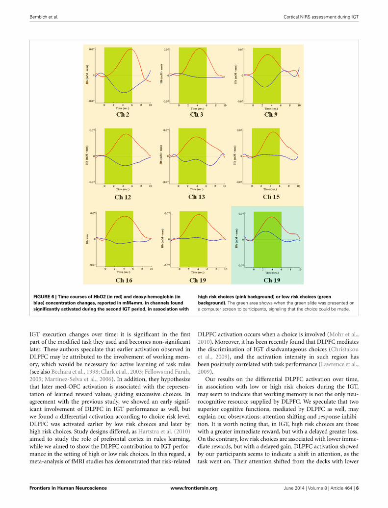
During the second task period (26th–50th choice), in asso-ciation with low risk choices, only channel 19 [t(10) = −3.910;P = 0.0015], located on right superior frontal gyrus, passedthe FDR threshold. In association with high risk choices,on the other hand, eight channels passed the FDR thresh-old (P < FDR 0.05). They were channel 2 [t(10) = −5.858;P = 0.006], located on right/left superior and medial frontalgyrus, channel 3 [t(10) = −4.300; P = 0.001], located on leftsuperior frontal gyrus, channel 9 [t(10) = −3.841; P = 0.0015],located on right/left superior and medial frontal gyrus, chan-nel 12 [t(10) = −3.441; P = 0.003], located on right superiorfrontal gyrus, channel 13 [t(10) = −3.349; P = 0.0035], locatedon left superior frontal gyrus, channel 15 [t(10) = −4.078;P = 0.001], located on right superior frontal gyrus, channel16 [t(10) = −2.688; P = 0.0115], located on right/left superiorand medial frontal gyrus, and channel 19 [t(10) = −4.382; P =0.0005], located on right superior frontal gyrus (Figures 5, 6).
During the third and fourth task period (51st–100th choice),no channel passed the FDR threshold (P < FDR 0.05), for eitherlow- or high-risk choices.
Four channels were activated in association with both low- orhigh-risk choices. They were channels 13, 15, 16, and 19, coveringleft, right and central portions of DLPFC. Additionally, in associa-tion with low-risk choices, also the right frontal pole was activated(channel 22), while, in association with high-risk choices, theDLPFC activation extended more posteriorly, to channels 2, 3,and 9.
FIGURE 3 | Cortical location, on a rendered brain, of channels found
significantly activated in association with low risk choices (evidence in
green), during the first IGT period.
Frontiers in Human Neuroscience www.frontiersin.org June 2014 | Volume 8 | Article 464 | 4

Bembich et al. Cortical NIRS assessment during IGT
FIGURE 4 | Time courses of HbO2 (in red) and deoxy-hemoglobin (in
blue) concentration changes, reported in mM•mm, in channels found
significantly activated during the first IGT period, in association with
low risk choices. The dark green area shows when the green slide waspresented on a computer screen to participants, signaling that the choicecould be made.
FIGURE 5 | Cortical location, on a rendered brain, of channels found
significantly activated in association with high risk choices (evidence
in pink), or in high and low risk choices (evidence in pink/green),
during the second task period.
DISCUSSIONWe used multichannel NIRS to study if DLPFC is differentiallyactivated, over time, by low or high risk choices during the exe-cution of the complete administration of IGT, a playing card taskdeveloped to study DM under uncertainty. We have found thatDLPFC significantly contributes to the IGT execution in the firsthalf of the task. Additionally, we found different time courses inits activation as the task went on, associated with low or high risk
choices. Specifically, DLPFC was shown to be activated by lowrisk choices in the first period (1st–25th choice) and by high riskchoices in the second period (26th–50th choice) of IGT perfor-mance. Activated DLPFC areas associated with low or high riskchoices partially overlapped in both left, right and central por-tions of the DLPFC region monitored. In association with low riskchoices (first period), along with activation of left/right superiorfrontal gyrus and left/right medial frontal gyrus (channels 13, 15,16, and 19), also the right frontal pole was activated (channel 22).In association with high risk choices (second period), the DLPFCactivation was more extensive and involved more posterior areas,possibly including the supplementary motor area (channels 2 and3). In both cases, activation was slightly lateralized on the rightside. No significant variations in DLPFC activity emerged duringthe second half of IGT execution, regardless of choice risk level.
The involvement of DLPFC, especially with a right lateral-ization, in IGT execution has already been shown. Clinically, abad IGT performance was observed in patients with right DLPFClesions (Clark et al., 2003; Fellows and Farah, 2005) and, by neu-roimaging, DLPFC cortical areas were found activated during theIGT test (Ernst et al., 2002, 2003; Bolla et al., 2003, 2005). BesidesDLPFC, other areas may play a role in DM, such as VMPFC(Bechara et al., 2000a), the amygdala (Bechara, 2001; Bar-Onet al., 2003) and the insula (Lin et al., 2008; Lawrence et al., 2009),but such areas are too deeply located to be detected by NIRSdevices. Primary and secondary sensory areas (Bechara, 2001;Bar-On et al., 2003) are involved in DM as well, but they were out-side our region of interest. More recently, Hartstra et al. (2010)have demonstrated, by fMRI, that activation of DLPFC during
Frontiers in Human Neuroscience www.frontiersin.org June 2014 | Volume 8 | Article 464 | 5
Bembich et al. Cortical NIRS assessment during IGT
FIGURE 6 | Time courses of HbO2 (in red) and deoxy-hemoglobin (in
blue) concentration changes, reported in mM•mm, in channels found
significantly activated during the second IGT period, in association with
high risk choices (pink background) or low risk choices (green
background). The green area shows when the green slide was presented ona computer screen to participants, signaling that the choice could be made.
IGT execution changes over time: it is significant in the firstpart of the modified task they used and becomes non-significantlater. These authors speculate that earlier activation observed inDLPFC may be attributed to the involvement of working mem-ory, which would be necessary for active learning of task rules(see also Bechara et al., 1998; Clark et al., 2003; Fellows and Farah,2005; Martinez-Selva et al., 2006). In addition, they hypothesizethat later med-OFC activation is associated with the represen-tation of learned reward values, guiding successive choices. Inagreement with the previous study, we showed an early signif-icant involvement of DLPFC in IGT performance as well, butwe found a differential activation according to choice risk level.DLPFC was activated earlier by low risk choices and later byhigh risk choices. Study designs differed, as Hartstra et al. (2010)aimed to study the role of prefrontal cortex in rules learning,while we aimed to show the DLPFC contribution to IGT perfor-mance in the setting of high or low risk choices. In this regard, ameta-analysis of fMRI studies has demonstrated that risk-related
DLPFC activation occurs when a choice is involved (Mohr et al.,2010). Moreover, it has been recently found that DLPFC mediatesthe discrimination of IGT disadvantageous choices (Christakouet al., 2009), and the activation intensity in such region hasbeen positively correlated with task performance (Lawrence et al.,2009).
Our results on the differential DLPFC activation over time,in association with low or high risk choices during the IGT,may seem to indicate that working memory is not the only neu-rocognitive resource supplied by DLPFC. We speculate that twosuperior cognitive functions, mediated by DLPFC as well, mayexplain our observations: attention shifting and response inhibi-tion. It is worth noting that, in IGT, high risk choices are thosewith a greater immediate reward, but with a delayed greater loss.On the contrary, low risk choices are associated with lower imme-diate rewards, but with a delayed gain. DLPFC activation showedby our participants seems to indicate a shift in attention, as thetask went on. Their attention shifted from the decks with lower
Frontiers in Human Neuroscience www.frontiersin.org June 2014 | Volume 8 | Article 464 | 6

Bembich et al. Cortical NIRS assessment during IGT
immediate rewards, in the first task period (1st–25th choice),to those with greater losses, in the second task period (26th–50th choice). In both cases, an attention response, mediated byDLPFC (e.g., Manes et al., 2002), was elicited by those decks (andrelated choices) that seemed to be more disadvantageous (see alsoChristakou et al., 2009; Lawrence et al., 2009). Moreover, in thesecond period of the task, when DLPFC was activated in associa-tion with high risk choices, frontocentral cortical areas (channels9 and 16) and the supplementary motor area (channels 2 and3) were also activated. Both regions have been associated withresponse inhibition and behavioral control (Elliott et al., 2000;van Gaal et al., 2008; Fassbender et al., 2009).
Our experimental procedure aimed to assess DLPFC activa-tion associated with low or high risk choices, as they were made.Consequently, the activation of cortical regions with a role inresponse inhibition was detected when the high-risk decision wasmade. We speculate that the extended DLPFC activation in thesecond IGT quarter, may reflect a conflict between previouslylearned and newer rules, in order to make the best decision.The activation involving the right frontal pole in the first quar-ter may indicate an attempt to elaborate more abstract task rules(e.g., Venkatraman and Huettel, 2012), as an initial approach toan unknown complex task. Concerning the absence of DLPFCactivation in the third and fourth period of the IGT, we spec-ulate that it may be due to VMPFC taking over the task, whichis undetectable by fNIRS. Indirectly, this hypothesis is supportedby Hartstra et al. (2010), who found only a later involvementof medial prefrontal cortex regions (med-OFC in their case) inIGT performance. Moreover, Lawrence et al. (2009) found a lin-ear decrease in lateral OFC and pre-supplementary motor areaactivation as IGT execution progressed.
Our study has some obvious limitations. First, we enrolled alimited number of subjects and it should be considered a pilotstudy. Although functional neuroimaging studies are typicallyperformed in small populations, our results need to be confirmedin a larger sample. Characterizing participants for personalitytraits and mood state would further improve such a study, since ithas been proved that these variables have an influence on IGTperformance (Buelow and Suhr, 2009). Second, multichannelNIRS has a poorer spatial resolution than fMRI. However, pre-vious research has shown a good correlation between these twotechniques (Toronov et al., 2001; Strangman et al., 2002). Third,NIRS measurement of hemoglobin variations are limited to thelateral surface of the cerebral cortex. Fourth, the experimentaldesign we chose did not allow us to discriminate among the spe-cific prefrontal functions involved in the early distinction betweenlow- and high-risk choices. Finally, some possible systemic effectswe have not directly checked, e.g., blood flow variation in thescalp during the experiment, could have had an influence onour results. However, this was probably not the case because anasymmetric pattern of cortical activation was observed.
In conclusion, this study has further deepened the complexrole of DLPFC to early DM under uncertainty process, as assessedby IGT. We have shown that, in the first half of the perfor-mance, DLPFC activation differs over time, according to the risklevel of the choice. We propose that, beside working memory,other complex functions, such as attention shifting and response
inhibition, are involved in sustaining the DLPFC role in discrimi-nating disadvantageous choices during the task. Thereafter, as rulelearning establishes, DM under uncertainty seems to be preva-lently mediated by VMPFC. Thus, it emerges a wide, complexand temporally dynamic involvement of the prefrontal cortex indecisions concerning the ordinary uncertainty of everyday life.
ACKNOWLEDGMENTThis study has been financed by the grant N. 50/11 of the Institutefor Maternal and Child Health—IRCCS “Burlo Garofolo”—Trieste, Italy.
REFERENCESBar-On, R., Tranel, D., Denburg, N. L., and Bechara, A. (2003). Exploring the neu-
rological substrate of emotional and social intelligence. Brain 126, 1790–1800.doi: 10.1093/brain/awg177
Bechara, A. (2001). Neurobiology of decision-making: risk and reward. Semin. Clin.Neuropsychiatry 6, 205–216. doi: 10.1053/scnp.2001.22927
Bechara, A., Damasio, A. R., Damasio, H., and Anderson, S. W. (1994). Insensitivityto the future consequences following damage to prefrontal cortex. Cognition 50,7–15. doi: 10.1016/0010-0277(94)90018-3
Bechara, A., Damasio, H., and Damasio, A. R. (2000b). Emotion, decision mak-ing and the orbitofrontal cortex. Cerb. Cortex 10, 295–307. doi: 10.1093/cer-cor/10.3.295
Bechara, A., Damasio, H., Damasio, A. R., and Lee, G. P. (1999). Different con-tributions of the human amygdala and ventromedial prefrontal cortex todecision-making. J. Neurosci. 19, 5473–5481.
Bechara, A., Damasio, H., Tranel, D., and Anderson, S. W. (1998). Dissociation ofworking memory from decision-making within the human prefrontal cortex.J. Neurosci. 18, 428–437.
Bechara, A., Tranel, D., and Damasio, H. (2000a). Charachterization of thedecision-making deficit of patients with ventromedial prefrontal cortex lesions.Brain 123, 2189–2202. doi: 10.1093/brain/123.11.2189
Bolla, K. I., Eldreth, D. A., London, E. D., Kiehl, K. A., Mouratidis, M.,Contoreggi, C., et al. (2003). Orbitofrontal cortex dysfunction in abstinentcocaine abusers performing a decision-making task. Neuroimage 19, 1085–1094.doi: 10.1016/S1053-8119(03)00113-7
Bolla, K. I., Eldreth, D. A., Matochik, J. A., and Cadet J. L. (2005). Neural sub-strates of faulty decision-making in abstinent marijuana users. Neuroimage 26,480–492. doi: 10.1016/j.neuroimage.2005.02.012
Buelow, M. T., and Suhr, J. A. (2009). Construct validity of the Iowa Gambling Task.Neurpsychol. Rev. 19, 102–114. doi: 10.1007/s11065-009-9083-4
Cazzell, M., Li, L., Lin, Z. J., Petel, S. J., and Liu, H. (2012). Comparison of neuronalcorrelates of risk decision making between genders: an exploratory fNIRS studyof the Balloon Analogue Risk Task (BART). Neuroimage 62, 1869–1911. doi:10.1016/j.neuroimage.2012.05.030
Christakou, A., Brammer, M., Giampietro, V., and Rubia, K. (2009). Right ven-tromedial and dorsolateral prefrontal cortices mediate adaptive decisions underambiguity by integrating choice utility and outcome evaluation. J. Neurosci. 29,11020–11028. doi: 10.1523/JNEUROSCI.1279-09.2009
Clark, L., Manes, F., Antoun, N., Sahakian, B. J., and Robbins, T. W. (2003).The contributions of lesion laterality and lesion volume to decision-makingimpairment following forntal lobe damage. Neuropsychologia 41, 1474–1483.doi: 10.1016/S0028-3932(03)00081-2
Damasio, A. R. (1994). Descartes’ Error: Emotion, Reason, and The Human Brain.New York, NY: Putnam.
Elliott, R., Dolan, R. J., and Frith C. D. (2000). Dissociable functions in the medialand lateral orbitofrontal cortex: evidence from human neuroimaging studies.Cereb. Cortex 10, 308–317. doi: 10.1093/cercor/10.3.308
Ernst, M., Bolla, K., Mouratidis, M., Contoreggi, C., Matochik, J. A., Kurian,V., et al. (2002). Decision-making in a risk-taking task: a PET study.Neuropsychopharmachology 26, 682–691. doi: 10.1016/S0893-133X(01)00414-6
Ernst, M., Kimes, A. S., London, E. D., Matochik, J. A., Eldreth, D., Tata, S.,et al. (2003). Neural substrates of decision making in adults with atten-tion deficit hyperactivity disorder. Am. J. Psychiatry 160, 1061–1070. doi:10.1176/appi.ajp.160.6.1061
Frontiers in Human Neuroscience www.frontiersin.org June 2014 | Volume 8 | Article 464 | 7
Bembich et al. Cortical NIRS assessment during IGT
Fassbender, C., Hester, R., Murphy, K., Foxe, J. J., Foxe, D. M., and Garavan, H.(2009). Prefrontal and midline interactions mediating behavioural control. Eur.J. Neurosci. 29, 181–187. doi: 10.1111/j.1460-9568.2008.06557.x
Fellows, L. K., and Farah, M. J. (2005). Different underlying impairments indecision-making following ventromedial and dorsolateral frontal lobe damagein humans. Cereb. Cortex 15, 58–63. doi: 10.1093/cercor/bhh108
Genovese, C. R., Lazar, N. A., and Nichols, T. (2002). Thresholding of statisticalmaps in functional neuroimaging using the false discovery rate. Neuroimage 15,870–878. doi: 10.1006/nimg.2001.1037
Gläscher, J., Adolphs, R., Damasio, H., Bechara, A., Rudrauf, D., Clamia, M., et al.(2012). Lesion mapping of cognitive control and value-based decision makingin the prefrontal cortex. Proc. Natl. Acad. Sci. U.S.A. 109, 14681–14686. doi:10.1073/pnas.1206608109
Gupta, R., Duff, M. C., Denburg, N. L., Cohen, N. J., Bechara, A., andTranel, D. (2009). Declarative memory is critical for sustained advan-tageous complex decision-making. Neuropsychologia 47, 1686–1693. doi:10.1016/j.neuropsychologia.2009.02.007
Hartstra, E., Oldenburg, J. F., Van Leijenhorst, L., Rombouts, S. A., and Crone,E. A. (2010). Brain regions involved in the learning and application of rewardrules in a two-deck gambling task. Neuropsychologia 48, 1438–1446. doi:10.1016/j.neuropsychologia.2010.01.012
Ito, Y., Kennan, R. P., Watanabe, E., and Koizumi, H. (2000). Assessment of heatingeffects in skin during continuous wave near infrared spectroscopy. J. Biomed.Opt. 5, 383–390. doi: 10.1117/1.1287730
Jasper, H. H. (1958). The ten– twenty electrode system of the InternationalFederation. Electroencephalogr. Clin. Neurophysiol. 10, 367–380.
Lawrence, N. S., Jollant, F., O’Daly, O., Zelaya, F., and Phillips, M. L. (2009). Distinctroles of prefrontal cortical subregions in the Iowa Gambling Task. Cereb. Cortex19, 1134–1143. doi: 10.1093/cercor/bhn154
Lin, C. H., Chiu, Y. C., Cheng, C. M., and Hsieh, J. C. (2008). Brain maps of Iowagambling task. BMC Neurosci. 9:72. doi: 10.1186/1471-2202-9-72
Maki, A., Yamashita, Y., Ito, Y., Watanabe, F., Mayanagi, Y., and Koizumi,H. (1995). Spatial and temporal analysis of human motor activity usingnon-invasive NIR topography. Med. Phys. 22, 1997–2005. doi: 10.1118/1.597496
Manes, F., Sahakian, B., Clark, L., Rogers, R., Antoun, N., Aitken, M., et al. (2002).Decision-making processes following damage to prefrontal cortex. Brain 125,624–639. doi: 10.1093/brain/awf049
Martinez-Selva, J. M., Sanchez-Navarro, J. P., Bechara, A., and Roman, F.(2006). Brain mechanisms involved in decision-making. Rev. Neurol. 42,411–418.
Meek, J. (2002). Basic principles of optical imaging and application to thestudy of infant development. Dev. Sci. 5, 371–380. doi: 10.1111/1467-7687.00376
Mohr, P. N. C., Biele, G., and Heekeren, H. R. (2010). Neural processing of risk.J. Neurosci. 30, 6613–6619. doi: 10.1523/JNEUROSCI.0003-10.2010
Oldfield, R. C. (1971). The assessment and analysis of handedness: the Endinburginventory. Neuropsychologia 9, 97–113. doi: 10.1016/0028-3932(71)90067-4
Oya, H., Adolphs, R., Kawasaki, H., Bechara, A., Damasio, A., and Howard, M. A.3rd. (2005). Electrophysiological correlates of reward prediction error recordedin the human prefrontal cortex. Proc. Natl. Acad. Sci. U.S.A. 102, 8351–8356.doi: 10.1073/pnas.0500899102
Schroeter, M. L., Zysset, S., and von Cramon, D. Y. (2004). Shortening inter-trial intervals in event-related cognitive studies with near-infrared spectroscopy.Neuroimage 22, 341–346. doi: 10.1016/j.neuroimage.2003.12.041
Singh, A. K., and Dan, I. (2006). Exploring the false discovery rate in multichannelNIRS. Neuroimage 33, 542–549. doi: 10.1016/j.neuroimage.2006.06.047
Singh, A., K., Okamoto, M., Dan, H., Jurcak, V., and Dan, I. (2005). Spatial regis-tration of multichannel multi-subject fNIRS data to MNI space without MRI.Neuroimage 27, 842–851. doi: 10.1016/j.neuroimage.2005.05.019
Strangman, G., Culver, J. B., Thompson, J. H., and Boas, D. A. (2002). Aquantitative comparison of simultaneous BOLD fMRI and NIRS record-ings during functional brain activation. Neuroimage 17, 719–731. doi:10.1006/nimg.2002.1227
Stocco, A., and Fum, D. (2007). Implicit emotional biases in decision mak-ing: the case of the Iowa gambling task. Brain Cogn. 66, 253–259. doi:10.1016/j.bandc.2007.09.002
Taga, G., Asakawa, K., Maki, A., Konishi, Y., and Koizumi, H. (2003). Brain imag-ing in awake infants by near-infrared optical topography. Proc. Natl. Acad. Sci.U.S.A. 19, 10722–10727. doi: 10.1073/pnas.1932552100
Toronov, V., Webb, A., Choi, J. H., Wolf, M., Michalos, A., Gratton, E., et al.(2001). Investigation of human brain hemodynamics by simultaneous near-infrared spectroscopy and functional magnetic resonance imaging. Med. Phys.28, 521–527. doi: 10.1118/1.1354627
Tsuzuki, D., Jurcak V., Singh, A. K., Okamoto M., Watanabe W., and Dan I. (2007).Virtual spatial registration of stand-alone fNIRS data to MNI space. Neuroimage34, 1506–1518. doi: 10.1016/j.neuroimage.2006.10.043
Tzourio-Mazoyer, N., Landeau, B., Papathanassiou, D., Crivello, F., Etard, O.,Delcroix, N., et al. (2002). Automated anatomical labelling of activation in SPMusing a macroscopic anatomical parcellation of the MNI MRI single-subjectbrain. Neuroimage 15, 273–289. doi: 10.1006/nimg.2001.0978
van Gaal, S., Ridderinkhof, K. R., Fahrenfort, J. J., Scholte, H. S., and Lamme, V. A.F. (2008). Frontal cortex mediates unconsciously triggered inhibitory control.J. Neurosci. 28, 8053–8062. doi: 10.1523/JNEUROSCI.1278-08.2008
Venkatraman, V., and Huettel, S. A. (2012). Strategic control in decision-making under uncertinity. Eur. J. Neurosci. 35, 1075–7082. doi: 10.1111/j.1460-9568.2012.08009.x
Wise, S. P. (2008). Forward frontal fields: phylogeny and fundamental function.Trends Neurosci. 31, 599–608. doi: 10.1016/j.tins.2008.08.008
Wobst, P., Wenzel, R., Kohl, M., Obrig, H., and Villringer, A. (2001). Linearaspects of changes in deoxygenated hemoglobin concentration and cytochromeoxidase oxidation during brain activation. Neuroimage 13, 520–530. doi:10.1006/nimg.2000.0706
Ye, J. C., Tak, S. H., Jang, K. E., Jung, J. W., and Jang, J. D. (2009). NIRS-SPM:statistical parametric mapping for near-infrared spectroscopy. Neuroimage 44,428–447. doi: 10.1016/j.neuroimage.2008.08.036
Conflict of Interest Statement: The authors declare that the research was con-ducted in the absence of any commercial or financial relationships that could beconstrued as a potential conflict of interest.
Received: 30 September 2013; accepted: 06 June 2014; published online: 24 June 2014.Citation: Bembich S, Clarici A, Vecchiet C, Baldassi G, Cont G and Demarini S (2014)Differences in time course activation of dorsolateral prefrontal cortex associated withlow or high risk choices in a gambling task. Front. Hum. Neurosci. 8:464. doi: 10.3389/fnhum.2014.00464This article was submitted to the journal Frontiers in Human Neuroscience.Copyright © 2014 Bembich, Clarici, Vecchiet, Baldassi, Cont and Demarini. Thisis an open-access article distributed under the terms of the Creative CommonsAttribution License (CC BY). The use, distribution or reproduction in other forumsis permitted, provided the original author(s) or licensor are credited and that the orig-inal publication in this journal is cited, in accordance with accepted academic practice.No use, distribution or reproduction is permitted which does not comply with theseterms.
Frontiers in Human Neuroscience www.frontiersin.org June 2014 | Volume 8 | Article 464 | 8