Embed Size (px)
Citation preview

Diets Rich in Saturated and Polyunsaturated Fatty AcidsInduce Morphological Alterations in the Rat VentralProstateAngelica Furriel, Pamella Campos-Silva, Paola Cariello Guedes Picarote Silva, Waldemar Silva Costa,
Francisco Jose Barcellos Sampaio, Bianca Martins Gregorio*
Department of Anatomy, State University of Rio de Janeiro, Urogenital Research Unit, Biomedical Center, Rio de Janeiro, Brazil
Abstract
Aim: To evaluate the influence of dietary lipid quality on the body mass, carbohydrate metabolism and morphology of therat ventral prostate.
Materials and Methods: Wistar rats were divided into four groups: SC (standard chow), HF-S (high-fat diet rich in saturatedfatty acids), HF-P (high-fat diet rich in polyunsaturated fatty acids) and HF-SP (high-fat diet rich in saturated andpolyunsaturated fatty acids). We analyzed body mass, fat mass deposits, plasma blood, insulin resistance and the ventralprostate structure.
Results: Groups that received high-fat diets were heavier and presented larger fat deposits than SC group. The HF-S and HF-SP groups had higher glucose, insulin and total cholesterol serum levels and insulin resistance compared with the SC. Theacinar area, epithelium height and area density of the lumen were higher in the HF-SP than in the other groups. Theepithelium area density and epithelial cell proliferation were greater in the HF-P and HF-SP than in the SC group. All of thegroups that received high-fat diets had greater area density of the stroma, area density of smooth muscle cells and stromalcell proliferation compared with the SC group.
Conclusion: Diets rich in saturated and/or polyunsaturated fatty acids induced overweight. Independently of insulinresistance, polyunsaturated fatty acids increased prostate stromal and epithelial cell proliferation. Saturated fatty acidsinfluenced only stromal cellular proliferation. These structural and morphometric alterations may be considered risk factorsfor the development of adverse remodeling process in the rat ventral prostate.
Citation: Furriel A, Campos-Silva P, Silva PCGP, Costa WS, Sampaio FJB, et al. (2014) Diets Rich in Saturated and Polyunsaturated Fatty Acids InduceMorphological Alterations in the Rat Ventral Prostate. PLoS ONE 9(7): e102876. doi:10.1371/journal.pone.0102876
Editor: Raul M. Luque, University of Cordoba, Spain
Received December 16, 2013; Accepted June 24, 2014; Published July 16, 2014
Copyright: � 2014 Furriel et al. This is an open-access article distributed under the terms of the Creative Commons Attribution License, which permitsunrestricted use, distribution, and reproduction in any medium, provided the original author and source are credited.
Funding: This work was supported by FAPERJ, CNPq and CAPES. The funders had no role in study design, data collection and analysis, decision to publish, orpreparation of the manuscript.
Competing Interests: The authors have declared that no competing interests exist.
* Email: [email protected]
Introduction
Obesity is the most common cause of insulin resistance (IR) in
peripheral tissue as well as adipose tissue [1]. Obesity, IR and type
2 diabetes mellitus are considered risk factors for the development
of benign prostatic hyperplasia (BPH) [2,3]. BPH is the fourth
most prevalent disease in the male population over the age of 50
years [4]; the etiology is multifactorial and may be affected by
genetic [5], nutritional [6] and hormonal [7] factors. Experimental
studies show that administering diets rich in lipids leads to the
enlargement of the prostate in rats [3,8]. Furthermore, obesity
itself also contributes to the onset of BPH and many cancers,
including prostate cancer [9,10].
Previous studies have reported that polyunsaturated fatty acids-
PUFAs (mainly eicosapentaenoic- EPA and docosahexaenoic-
DHA), which are highly unsaturated, are more susceptible to lipid
peroxidation. Lipid peroxides can increase the expression of the
enzyme 5-alpha-reductase and consequently the formation of
dihydrotestosterone (DHT), which could stimulate the growth of
prostatic epithelial and stromal cells [11]. However, Liang and
colleagues suggest that PUFAs such as alpha-linolenic acid and
linoleic acid may act as potential endogenous inhibitors of the
enzyme 5-alpha-reductase and thus as inhibitors of cell prolifer-
ation [12].
The action mechanism of saturated fatty acids (SFA) in prostate
tissue is still controversial. Increased consumption of SFA increases
the synthesis of total cholesterol and LDL-cholesterol and lowers
HDL-cholesterol, increasing the risk for the development of BPH
[2]. Van Kuilenburg and colleagues (2011) showed that dyslipi-
demia is associated with increased circulation of several growth
factors, including basic fibroblast growth factor (bFGF) [13]. This
growth factor acts as an important stimulator of fibroblast
proliferation and collagen synthesis and deposition in the
extracellular matrix and stimulates angiogenesis [14].
PLOS ONE | www.plosone.org 1 July 2014 | Volume 9 | Issue 7 | e102876

In light of these findings, it is important to evaluate the effects of
obesity induced by administering different types of hyperlipidemic
diets on the morphology of the rat ventral prostate.
Materials and Methods
The animal protocols were approved by the Animal Ethics
Committee of the State University of Rio de Janeiro (Protocol
Number CEA027/2012), and the procedures were conducted in
accordance with the guidelines for experimentation with animals
(NIH Publication Nu. 85-23, revised 1996). The animals were
housed at a controlled temperature (2162uC) on a 12 h light/dark
cycle with free access to food and water. Next, they were assigned
to receive a specific diet.
Experimental designThirty-nine 12-week-old male Wistar rats were divided into four
groups. One group received only the standard chow throughout
the entire experiment (SC group; n= 9), whereas the other groups
received a high-fat diet (HF) classified according to its lipid
content: HF-S (high-fat diet rich in saturated fatty acids; n = 10),
HF-P (high-fat diet rich in polyunsaturated fatty acids; n = 10),
HF-SP (high-fat diet rich in saturated and polyunsaturated fatty
acids; n = 10). The SC diet (14% protein, 76% carbohydrate, 10%
fat) and the high-fat diets (14% proteins, 36% carbohydrate, 50%
fat) were prepared in accordance with the recommendations of the
American Institute of Nutrition (AIN-93M) [15] (Table 1). Lard
and/or canola oil was the source lipid of the diets. The diets were
produced by Pragsolucoes (Jau, SP, Brazil- www.pragsolucoes.com.
br).
The diets were administered to the rats from three to seven
months of age. Each rat was weighed and measured weekly until
the end of the experiment. All of the study groups received water
and food ad libitum, and their food intake was assessed daily.
EuthanasiaThe animals were killed at 28-week-old. After 12 hours of
fasting, the animals were deeply anesthetized (sodium pentobar-
bital intraperitoneally, 100 mg/kg), and all efforts were made to
minimize suffering. Blood was collected directly from the left
atrium. The ventral prostate was dissected and fixed for structural
analyses. Epididymal, subcutaneous and retroperitoneal fat were
also dissected, weighed and fixed.
Serum biochemistry, hormone levels and carbohydratemetabolismAfter the blood was collected, the serum was separated by
centrifugation at room temperature (3000 rpm, 8 min) and stored
at 220uC. Glucose, total cholesterol and triglyceride (TG)
concentrations were measured using a colorimetric assay (Bioclin;
Belo Horizonte, Minas Gerais, Brazil). An automatic spectropho-
tometer was used following the instructions recommended by the
manufacturer of Bioclin commercial kits: glucose monoreagent-
K082, cholesterol monoreagent-K083 and triglycerides mono-
reagent-K117. Serum analyses for insulin (Rat/Mouse Insulin kit,
Millipore - Cat. EZRMI-13 k – St Charles, Missouri, USA) and
testosterone (General Testosterone kit, Uscn - Cat. E90458Ge –
Wuhan, China) were performed using commercially available
enzyme-linked immunosorbent assay (ELISA) kits.
IR was calculated using the HOMA-IR (homeostasis model
assessment for IR index): insulin*glucose/22.5.
ImmunohistochemistryThe immunohistochemical analyses were performed on the
prostatic tissue from the ventral lobe of the rat prostate. Slides
were prepared from 5-mm sections of the formalin-fixed, paraffin-
embedded tissues, subjected to antigen retrieval with Tris-EDTA
buffer (Proliferating Cell Nuclear Antigen- PCNA) and incubated
with trypsin for 15 minutes at 37uC (Alpha Smooth Muscle Actin).
Table 1. Composition of experimental diets (following the AIN-93M recommendations for rodents) [18].
SC HF-S HF-P HF-SP
Corn starch 465.70 192.60 192.60 192.60
Casein 140.00 175.00 175.00 175.00
Sucrose 100.00 100.00 100.00 100.00
Soybean oil 40.00 40.00 40.00 40.00
Rapeseed oil 0.00 0.00 238.00 119.00
Lard 0.00 238.00 0.00 119.00
Fibre 50.00 50.00 50.00 50.00
L-cistin 1.80 1.80 1.80 1.80
Colin 2.50 2.50 2.50 2.50
Antioxidants 0.06 0.06 0.06 0.06
Mixed minerals 35.00 35.00 35.00 35.00
Mixed vitamins 10.00 10.00 10.00 10.00
TOTAL (g) 1000.00 1000.00 1000.00 1000.00
Energy (KJ/Kg) 15925.80 20900.00 20900.00 20900.00
Carbohydrate (%) 76.00 36.00 36.00 36.00
Protein (%) 14.00 14.00 14.00 14.00
Lipid (%) 10.00 50.00 50.00 50.00
SC, standard chow diet; HF-S, high-fat diet rich in saturated fatty acid (lard); HF-P, high-fat diet rich in polyunsaturated fatty acid (rapeseed oil); HF-SP, high-fat diet rich insaturated and polyunsaturated fatty acids.doi:10.1371/journal.pone.0102876.t001
High-Fat Diets and the Rats Ventral Prostate
PLOS ONE | www.plosone.org 2 July 2014 | Volume 9 | Issue 7 | e102876

Endogenous peroxidase activity was blocked by incubating the
slides with 3% H2O2 in methanol for 15 minutes followed by
applying a protein block (phosphate-buffered saline/bovine serum
albumin- 5%). Mouse polyclonal primary antibodies to PCNA
(1:100; Invitrogen, 13-3900) and Alpha Smooth Muscle Actin
(Invitrogen, 08-0106) were added and incubated overnight. Next,
sections were treated with a biotinylated secondary antibody
(K0679; Universal DakoCytomation LSAB Kit, Peroxidase,
Glostrup, Denmark) and amplified with a biotin–streptavidin
system (K0679; Universal DakoCytomation LSAB + Kit, Perox-
idase, Glostrup, Denmark). 3, 3 diaminobenzidine tetrachloride
(K3466, DakoCytomation, Glostrup, Denmark) was used as the
chromogen. After incubation, the sections were counterstained
with Mayer hematoxylin.
Morphometric analysisThe prostate was dissected; fragments of the ventral lobe were
fixed with freshly prepared fixative (1.27 M formaldehyde in 0.1 M
phosphate buffer, pH 7.2) for 48 h at room temperature and
embedded in Paraplast Plus (Sigma-Aldrich, St Louis, MO, USA).
Next, the material was sectioned at a nominal thickness of 5 mmand stained with hematoxylin and eosin. For all of the
morphometric analyses, five slides from each animal were
obtained and five fields were evaluated for a total of 25 fields
per animal. By performing an image analysis with a digital camera
(Olympus DP71) on an Olympus BX51 microscope running
ImageJH software (Image Processing and Analysis in Java), the
acinar area and the epithelium height were estimated using a
minimum of 225 measures per group. For these analyses, we used
photomicrographs at 200x and 600x magnifications, respectively.
The area density of the epithelium, lumen, connective tissue and
smooth muscle cells (immunostained), expressed as a percentage,
was estimated using the point intercepts method with a grid of 100
points superimposed on the magnified images (200x).
A quantitative assessment of proliferating cells was performed
based on the anti-PCNA immunohistochemistry. Separately, the
ratios of the number of dividing cell nuclei (immunostained) to the
epithelial and stromal areas were calculated in each field we
evaluated.
Data analysisThe data were tested for normality and homogeneity of the
variances and then reported as the means 6 standard deviations
(SDs). The differences among the groups were analyzed using a
one-way analysis of variance (one-way ANOVA) followed by
Bonferroni’s post hoc test. A p-value #0.05 was considered
statistically significant (Prism version 5.00 for Windows; GraphPad
Software, San Diego, California, USA).
Results
Food intake, Body mass evolution and fat depositsThere were no difference in the food intake among the SC
(17.8961.48 g), HF-S (16.4262.59 g), HF-P (18.1061.36 g) and
HF-SP (16.4565.10 g) groups. Throughout the experiment, the
SC group was the lightest. At seven months of age, the body mass
values of the HF-S (529.30657.39 g), HF-P (546.40640.13 g) and
HF-SP groups (532.90648.27 g) were 25%, 29% and 26% higher
than the body mass of the SC group (424.22640.29 g, p,0.0001),
respectively (Table 2). It is important to mention that the animals
subjected to the various HF diets had larger epididymal,
retroperitoneal and subcutaneous fat deposits than the SC group.
The epididymal fat mass of the HF-S, HF-P and HF-SP groups
were 67%, 91% and 90% higher than that of the SC group
Table
2.Biometric
andmetabolic
param
eters
oftheexp
erimentalgroups.
Data
SC
HF-S
HF-P
HF-SP
ANOVA
Mean
SD
Mean
SD
Mean
SD
Mean
SD
pvalue
Bodymass,g
424.22
40.29
529.30
57.39a
546.40
40.13a
532.90
48.27a
,0.0001
Epididym
alfat,mg
7.65
2.69
12.79
4.05a
14.64
4.75a
14.54
2.12a
0.0004
Retroperitoneal
fat,mg
9.56
4.68
17.06
4.55a
17.56
2.73a
20.51
4.96a
0.0001
Subcutaneousfat,mg
2.48
1.37
6.96
1.61a
5.47
2.35a
7.55
1.42a
,0.0001
Totalcholesterol,mg/dL
80.56
11.75
104.80
12.95a
100.10
10.38
105.20
19.65a
0.002
Triglycerides,mg/dL
86.29
23.68
95.63
8.60
83.63
17.52
84.25
5.04
0.37
Testosterone,ng/m
L5.48
0.83
4.73
1.14
4.28
1.25
5.11
0.89
0.25
Insulin
,mlU/m
l1.49
0.41
2.75
0.45a
2.15
0.83
2.85
0.92a
0.003
Glucose,mmol/L
7.87
1.61
10.62
2.36a
9.85
1.61
11.09
1.65a
0.006
HOMA-IR
0.41
0.12
1.32
0.54a
0.91
0.37
1.36
0.50a
0.003
SC,standardchow
diet;HF-S,high-fat
dietrich
insaturatedfattyacid
(lard);HF-P,h
igh-fat
dietrich
inpolyunsaturatedfattyacid
(rap
eseedoil);H
F-SP,high-fat
dietrich
insaturatedan
dpolyunsaturatedfattyacids.Thevaluesare
presentedas
themean
san
dstan
darddeviations(SD).Thesymbol[a]indicatesaresultthat
isdifferentfrom
theSC
group(one-w
ayANOVAan
dBonferroni’s
post
hoctest,p,0.05).
doi:10.1371/journal.pone.0102876.t002
High-Fat Diets and the Rats Ventral Prostate
PLOS ONE | www.plosone.org 3 July 2014 | Volume 9 | Issue 7 | e102876
(p=0.0004), respectively. The retroperitoneal fat deposits of the
HF-S, HF-P and HF-SP groups were 78%, 84% and 115%
(p=0.0001) greater, respectively, than the SC group, whereas the
subcutaneous fat deposits were 181%, 121% and 204% (p,
0.0001) greater compared with the SC group (Table 2).
Lipid profile, hormone levels and the HOMA-IR indexTriglyceride and testosterone levels were not different among
the groups. However, the HF-S and HF-SP groups developed
hypercholesterolemia at the end of the experiment. These two
groups had plasma levels of total cholesterol that were 30% and
31% greater (p = 0.002), respectively, than the plasma levels of the
SC group (Table 2). Besides, the HF-S and HF-SP groups
developed hyperinsulinemia (p=0.003) and hyperglycemia at the
end of the experiment (p=0.006) (Table 2). Consequently, these
groups had higher HOMA-IR values than the SC group
(p=0.003) (Table 2). The HF-P group showed no differences in
values of cholesterol, insulin, glucose and HOMA-IR index when
compared to the SC, HF-S and HF-SP groups.
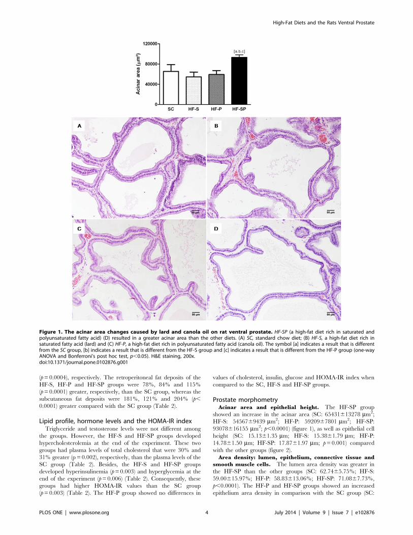
Prostate morphometryAcinar area and epithelial height. The HF-SP group
showed an increase in the acinar area (SC: 65431613278 mm2;
HF-S: 5456769439 mm2; HF-P: 5920967801 mm2; HF-SP:
93078616155 mm2; p,0.0001) (figure 1), as well as epithelial cell
height (SC: 15.1361.35 mm; HF-S: 15.3861.79 mm; HF-P:
14.7861.50 mm; HF-SP: 17.8761.97 mm; p=0.001) compared
with the other groups (figure 2).
Area density: lumen, epithelium, connective tissue and
smooth muscle cells. The lumen area density was greater in
the HF-SP than the other groups (SC: 62.7465.75%; HF-S:
59.00615.97%; HF-P: 58.83613.06%; HF-SP: 71.0867.73%,
p,0.0001). The HF-P and HF-SP groups showed an increased
epithelium area density in comparison with the SC group (SC:
Figure 1. The acinar area changes caused by lard and canola oil on rat ventral prostate. HF-SP (a high-fat diet rich in saturated andpolyunsaturated fatty acid) (D) resulted in a greater acinar area than the other diets. (A) SC, standard chow diet; (B) HF-S, a high-fat diet rich insaturated fatty acid (lard) and (C) HF-P, a high-fat diet rich in polyunsaturated fatty acid (canola oil). The symbol [a] indicates a result that is differentfrom the SC group, [b] indicates a result that is different from the HF-S group and [c] indicates a result that is different from the HF-P group (one-wayANOVA and Bonferroni’s post hoc test, p,0.05). H&E staining, 200x.doi:10.1371/journal.pone.0102876.g001
High-Fat Diets and the Rats Ventral Prostate
PLOS ONE | www.plosone.org 4 July 2014 | Volume 9 | Issue 7 | e102876

17.9863.61%; HF-S: 21.7269.13; HF-P: 24.59612.84%; HF-SP:
24.0165.48%, p,0.0001). In addition, all of the groups that
received a high-fat diet had greater area density in the connective
tissue (SC: 4.2662.50%; HF-S: 10.6965.87%; HF-P:
8.4264.36%; HF-SP: 8.0964.85%; p,0.0001) and in the smooth
muscle cells (SC: 5.3761.78%; HF-S: 7.7962.91%; HF-P:
7.6662.47%; HF-SP 7.0162.98%, p=0.0003) compared with
the SC group (figure 3).
Epithelial and stromal cell proliferation. The HF-P and
HF-SP groups had elevated epithelial cell proliferation in
comparison with the SC group (SC: 4.15* 102661.17* 1026 1/
mm2; HF-S: 4.58* 102666.59* 1027 1/mm2; HF-P: 5.62*
102661.56* 1026 1/mm2; HF-SP: 5.44* 102668.07* 1027 1/
mm2; p=0.0007). With regard to stromal cell proliferation, the
groups that consumed a diet high in saturated and/or polyunsat-
urated fatty acids had higher values compared with the SC group
(SC: 6.58* 102762.23* 1027 1/mm2; HF-S: 1.47* 102667.13*
1027 1/mm2; HF-P: 1.98* 102669.79* 1027 1/mm2; HF-SP:
1.45* 102668.05* 1027 1/mm2; p=0.0003) (figure 4).
Discussion
The present study evaluated the effects of obesity induced by
different types of high-fat diets on the morphology of the rat
ventral prostate. Our results show that diets rich in saturated fatty
acids (HF-S), polyunsaturated fatty acids (HF-P) and both types of
fatty acids (HF-SP) lead to overweight animals. The increase in
body mass was confirmed by the increase in retroperitoneal,
epididymal and subcutaneous fat deposits.
Recent studies have linked obesity with hyperinsulinemia and
IR, which are considered risk factors for the development of
metabolic syndrome [16]. Diets rich in SFA and cholesterol are
Figure 2. Epithelial height changes caused by lard and canola oil on rat ventral prostate. HF-SP, a high-fat diet rich in saturated andpolyunsaturated fatty acid) (D) resulted in higher epithelial cell height than the other diets. (A) SC, standard chow diet; (B) HF-S, a high-fat diet rich insaturated fatty acid (lard) and (C) HF-P, a high-fat diet rich in polyunsaturated fatty acid (canola oil). The symbol [a] indicates a result that is differentfrom the SC group, [b] indicates a result that is different from the HF-S group and [c] indicates a result that is different from the HF-P group (one-wayANOVA and Bonferroni’s post hoc test, p,0.05). H&E staining, 600x.doi:10.1371/journal.pone.0102876.g002
High-Fat Diets and the Rats Ventral Prostate
PLOS ONE | www.plosone.org 5 July 2014 | Volume 9 | Issue 7 | e102876

associated with factors that negatively affect metabolism and
predispose individuals to the development of IR and type 2
diabetes mellitus [17]. On the other hand, the consumption of
PUFAs has been considered a protective factor against the
development of these changes [17]. Our results align with these
works because the animals in the HF-S and HF-SP groups showed
IR, evidenced by the HOMA-IR values, as well as hyperinsulin-
emia and hyperglycemia. In this context, it is notable that
Figure 3. Morphological changes caused by different high-fat diets. (A) SC, standard chow diet produced no prostate alterations; (B) HF-S, ahigh-fat diet rich in saturated fatty acid (lard) caused an increase in the area density of the connective tissue and the smooth muscle cells; (C) HF-P, ahigh-fat diet rich in polyunsaturated fatty acid (canola oil) promoted an increase in the area density of the epithelium, the connective tissue and thesmooth muscle cells; (D) HF-SP, a high-fat diet rich in saturated and polyunsaturated fatty acids induced an increase in the area density of the lumen,the epithelium, the connective tissue and the smooth muscle cells. The symbol [a] indicates a result that is different from the SC group, [b] indicates aresult that is different from the HF-S group and [c] indicates a result that is different from the HF-P group (one-way ANOVA and Bonferroni’s post hoctest, p,0.05). H&E staining and immunostaining for Alpha Smooth Muscle Actin, 200x.doi:10.1371/journal.pone.0102876.g003
High-Fat Diets and the Rats Ventral Prostate
PLOS ONE | www.plosone.org 6 July 2014 | Volume 9 | Issue 7 | e102876

consuming saturated fat (lard) in different concentrations (50%
and 25% of the total energy of the diet) impaired carbohydrate
metabolism, maximizing the damage to the prostate. However, a
diet rich in PUFAs did not affect the glycemic response of the
animals.
On a related note, as we expected, PUFAs inhibited the increase
in serum cholesterol. Some studies recommend consuming PUFAs
and MUFAs to improve the lipid profile [18,19]. Inversely,
excessive consumption of SFAs resulted in hypercholesterolemia,
confirming the results previously reported in the literature [17].
Like Souza-Mello and colleagues (2007) [20] we did not find any
differences in the plasma triglyceride levels of the groups, although
foods rich in animal fat are associated with increased triglycerides
[21].
However, the mechanism of SFAs and PUFAs act on prostate
tissue is controversial and poorly understood. Our histomorpho-
metric and immunohistochemical analyses showed that all of the
animals that consumed a high-fat diet demonstrated a sharper
proliferation in the stromal compartment of the prostate, along
with increased area densities of the connective tissue and the
smooth muscle cells. In addition, only the HF-P and HF-SP groups
showed an increase in epithelial proliferation, which was
confirmed by the increase in the area density of the epithelium.
Numerous growth factors have been described as stimulators of
stromal and epithelial cell proliferation; some of them act
exclusively in the prostatic epithelium or stroma [22]. In our
study, we found that PUFAs appear to stimulate the pathways that
cause proliferation in both the epithelium and in the stroma, while
the diets based on animal fats appear to be more related to
proliferation in the stroma of the prostate.
Canola oil is rich in PUFAs of the n-3 series, which are
precursors of long-chain PUFAs such as EPA and DHA that are
more susceptible to lipid peroxidation. The lipid peroxides could
increase the expression of the enzyme 5-alpha-reductase and
consequently the formation of DHT [11]. Thus, they could
generate cellular proliferation in both the epithelium and the
Figure 4. PCNA-positive cells in the epithelium and stroma of the rat ventral prostate. (A) SC, standard chow diet, (B) HF-S, high-fat dietrich in saturated fatty acid (lard), (C) HF-P, high-fat diet rich in polyunsaturated fatty acid (canola oil) and (D) HF-SP, high-fat diet rich in saturated andpolyunsaturated fatty acids. The symbol [a] indicates a result that is different from the SC group (one-way ANOVA and Bonferroni’s post hoc test, p,0.05). Immunostaining for Proliferating Cell Nuclear Antigen- PCNA, 600x.doi:10.1371/journal.pone.0102876.g004
High-Fat Diets and the Rats Ventral Prostate
PLOS ONE | www.plosone.org 7 July 2014 | Volume 9 | Issue 7 | e102876

stroma of the prostate, thereby triggering the expansion of the
gland [10].
Hypercholesterolemic diets are associated with increased
synthesis of bFGF, which can increase collagen production in
the stromal compartment of the prostate. Van Kuilenburg and
colleagues (2011) [13] observed that the increase in serum total
cholesterol was positively correlated with an increase in circulating
bFGF. Therefore, a diet high in cholesterol may increase the
synthesis of bFGF and consequently induce proliferation solely in
the stroma of the gland, as seen in our study.
Moreover, the enlargement of the prostate could be related with
the increasing of testosterone serum levels [23]. However, like
Vikram and colleagues (2010) [3], there were no significant
changes in the circulating concentrations of testosterone in the
different experimental groups which attribute the prostate growth
to the diet administration.
Although the HF-P and HF-SP groups showed an increase in
epithelial cell proliferation and an increase in the area density of
the epithelium, only the HF-SP group showed an increase in the
acinar area. This result was corroborated by the high area density
of the lumen and the increase in the height of the epithelial cells.
The increase in the epithelial area density observed in the HF-P
group without a concomitant increase in the acinar area may
indicate that there was an increase in the number of prostatic
acini. Compared with the acini of the HF-SP group, the acini of
the HF-P group may be smaller but more numerous. In contrast,
the increase in the acinar area observed only in the HF-SP group
can be explained by the increase in the size of the acinar epithelial
cells. It has been suggested that hypertrophy of the prostatic
epithelial cells is an indicator of the secretory capacity of these cells
because the increase in secretion causes the dilation of organelles
such as the endoplasmic reticulum and the Golgi complex [24].
Thus, diets rich in cholesterol do not appear to influence the
proliferation of epithelial cells. However, they may be related to
the stimulation of these secretory cells when combined with
PUFAs, thus modifying the physiology of the gland.
It is noteworthy that the increase in cell proliferation in the
group fed a high-fat diet based on canola oil was independent of
the development of IR, showing the direct effect of PUFAs on the
prostate. However, we cannot rule out the possibility that insulin
stimulated cell proliferation, given that the HF-S and HF-SP
groups were hyperglycemic and hyperinsulinemic (IR). Insulin
may be associated with the pathogenesis of BPH through its
excitatory action on the sympathetic nervous system [25]. It
reduces the binding of globulin sex hormones, making these
hormones bioavailable [26]. Their effects extend pathways
mediated by insulin-like growth factor (IGF) [27]. Insulin can
also stimulate prostate growth by activating pathways that trigger
cellular proliferation, such as the IRS/PI3-K pathway which is
associated with glucose uptake and the MEK/ERK pathway,
which is responsible for mitogenic action. When IR develops, the
IRS/PI3-K pathway is impaired while the MEK/ERK pathway
remains unchanged [28].
The area density of smooth muscle increased in all of the
animals receiving a high-fat diet, regardless of the lipid quality.
The increase in this stromal component is closely associated with
the pathogenesis of BPH. Studies in humans have shown that
hyperplastic prostates had higher volume density of smooth muscle
than normal prostates. The increase in the density and tone of
these fibers may be related to the development of obstructive
symptoms in BPH [29].
Conclusions
Thus, the high-fat diet administration, independent of the lipid
quality, promoted an increase of body mass and insulin resistance
in animals. Polyunsaturated fatty acids increased stromal and
epithelial cell proliferation. In contrast, saturated fatty acids
influenced cellular proliferation in the stromal compartment only.
Certainly, more studies are needed to confirm these findings, given
the scarcity of the literature on the subject.
Acknowledgments
The authors thank Katia Sodre for her technical assistance.
Author Contributions
Conceived and designed the experiments: BMG. Performed the experi-
ments: AF PCS PCGPS. Analyzed the data: BMG WSC FJBS.
Contributed reagents/materials/analysis tools: WSC FJBS. Wrote the
paper: AF BMG.
References
1. Kapoor D, Malkin CJ, Channer KS, Jones TH (2005) Androgens, insulin
resistance and vascular disease in men. Clin Endocrinol (Oxf) 63: 239–250.doi:10.1111/j.1365-2265.2005.02299.x.
2. Nandeesha H, Koner BC, Dorairajan LN, Sen SK (2006) Hyperinsulinemia and
dyslipidemia in non-diabetic benign prostatic hyperplasia. Clin Chim Acta 370:
89–93. doi:10.1016/j.cca.2006.01.019.
3. Vikram A, Jena GB, Ramarao P (2010) Increased cell proliferation and
contractility of prostate in insulin resistant rats: linking hyperinsulinemia withbenign prostate hyperplasia. Prostate 70: 79–89. doi:10.1002/pros.21041.
4. Issa MM, Regan TS (2007) Medical therapy for benign prostatic hyperplasia-
present and future impact. Am J Manag Care 13 Suppl 1: S4–S9.
5. Sanda MG, Beaty TH, Stutzman RE, Childs B, Walsh PC (1994) Genetic
susceptibility of benign prostatic hyperplasia. J Urol 152: 115–119.
6. Bravi F, Bosetti C, Dal Maso L, Talamini R, Montella M, et al. (2006)
Macronutrients, fatty acids, cholesterol, and risk of benign prostatic hyperplasia.Urology 67: 1205–1211. doi:10.1016/j.urology.2006.01.007.
7. Marker PC, Donjacour AA, Dahiya R, Cunha GR (2003) Hormonal, cellular,and molecular control of prostatic development. Dev Biol 253: 165–174.
doi:10.1016/S0012-1606(02)00031-3.
8. Rahman NU, Phonsombat S, Bochinski D, Carrion RE, Nunes L, et al. (2007)
An animal model to study lower urinary tract symptoms and erectiledysfunction: the hyperlipidemic rat. BJU Int 100: 658–663. doi:10.1111/
j.1464-410X.2007.07069.x.
9. Hsing AW, Sakoda LC, Chua S Jr (2007) Obesity, metabolic syndrome, and
prostate cancer. Am J Clin Nutr 86: Suppl S843–S857.
10. Ribeiro DL, Pinto ME, Rafacho A, Bosqueiro JR, Maeda SY, et al. (2012) High-
fat diet obesity associated with insulin resistance increases cell proliferation,
estrogen receptor, and PI3K proteins in rat ventral prostate. J Androl 33: 854–865. doi:10.2164/jandrol.111.016089.
11. Suzuki S, Platz EA, Kawachi I, Willett WC, Giovannucci E (2002) Intakes of
energy and macronutrients and the risk of benign prostatic hyperplasia. Am J Clin
Nutr 75: 689–697.
12. Liang T, Liao S (1992) Inhibition of steroid 5 alpha-reductase by specificaliphatic unsaturated fatty acids. Biochem J 285: 557–562.
13. van Kuilenburg J, Lappegard KT, Sexton J, Plesiewicz I, Lap P, et al. (2011)Persisting thrombin activity in elderly patients with atrial fibrillation on oral
anticoagulation is decreased by anti-inflammatory therapy with intensivecholesterol-lowering treatment. J Clin Lipidol 5: 273–280. doi:10.1016/
j.jacl.2011.05.003.
14. Zhou J, Zhao Y, Wang J, Zhang S, Liu Z, et al. (2012) Therapeutic angiogenesis
using basic fibroblast growth factor in combination with a collagen matrix inchronic hindlimb ischemia. The Scientific World Journal. In press.
15. Reeves PG, Nielsen FH, Fahey GC Jr (1993) AIN-93 purified diets forlaboratory rodents: final report of the American Institute of Nutrition ad hoc
writing committee on the reformulation of the AIN-76A rodent diet. J Nutr 123:
1939–1951.
16. Gorden P, Zadeh ES, Cochran E, Brown RJ (2012) Syndromic insulinresistance: models for the therapeutic basis of the metabolic syndrome and other
targets of insulin resistance. Endocr Pract 18: 763–771. doi:10.4158/
EP12139.RA.
17. Coelho DF, Pereira-Lancha LO, Chaves DS, Diwan D, Ferraz R, et al. (2011)Effect of high-fat diets on body composition, lipid metabolism and insulin
sensitivity, and the role of exercise on these parameters. Braz J Med Biol Res 44:
966–972. doi:10.1590/S0100-879x2011007500107.
High-Fat Diets and the Rats Ventral Prostate
PLOS ONE | www.plosone.org 8 July 2014 | Volume 9 | Issue 7 | e102876

18. Montoya MT, Porres A, Serrano S, Fruchart JC, Mata P, et al. (2002) Fatty acid
saturation of the diet and plasma lipid concentrations, lipoprotein particle
concentrations, and cholesterol efflux capacity. Am J Clin Nutr 75: 484–491.
19. Rokling-Andersen MH, Rustan AC, Wensaas AJ, Kaalhus O, Wergedahl H, et
al. (2009) Marine n-3 fatty acids promote size reduction of visceral adipose
depots, without altering body weight and composition, in male Wistar rats fed a
high-fat diet. Br J Nutr 102: 995–1006. doi:10.1017/S0007114509353210.
20. Souza-Mello V, Mandarim-de-Lacerda CA, Aguila MB (2007) Hepatic
structural alteration in adult programmed offspring (severe maternal protein
restriction) is aggravated by post-weaning high-fat diet. Br J Nutr 98: 1159–
1169. doi:10.1017/S0007114507771878.
21. Hodson L, Skeaff CM, Chisholm WA (2001) The effect of replacing dietary
saturated fat with polyunsaturated or monounsaturated fat on plasma lipids in
free-living young adults. Eur J Clin Nutr 55: 908–915.
22. Gerdes MJ, Larsen M, Dang TD, Ressler SJ, Tuxhorn JA, et al. (2004)
Regulation of rat prostate stromal cell myodifferentiation by androgen and
TGF-beta1. Prostate 58: 299–307. doi 10.1002/pros.10327.
23. Grossmann M, Cheung AS, Zajac JD (2013) Androgens and prostate cancer;
pathogenesis and deprivation therapy. Best Pract Res Clin Endocrinol Metab.27: 603–616. doi:10.1016/j.beem.2013.05.001.
24. Gross SA, Didio LJ (1987) Comparative morphology of the prostate in adult
male and female Praomys (Mastomys) Natalensis studied with electronmicroscopy. J Submicrosc Cytol 19: 77–84.
25. Morgan DA, Balon TW, Ginsberg BH, Mark AL (1993) Nonuniform regionalsympathetic nerve responses to hyperinsulinemia in rats. Am J Physiol 264: 423–
427. doi:0165-1838(95)00108-5.
26. Hautanen A (2000) Synthesis and regulation of sex hormone-binding globulin inobesity. Int J Obes Relat Metab Disord 24 Suppl 2: S64–S70.
27. Giovannucci E (2003) Nutrition, insulin, insulin-like growth factors and cancer.Horm Metab Res 35: 694–704.
28. Vikram A, Jena G, Ramarao P (2010) Insulin-resistance and benign prostatichyperplasia: the connection. Eur J Pharmacol 641: 75–81. doi:10.1016/
j.ejphar.2010.05.042.
29. Morgan DA, Balon TW, Ginsberg BH, Mark AL (2001) Stereological analysis ofhistologic components in transition zone of normal and hyperplastic human
prostates. Brazilian Journal of Urology 27: 26–31.
High-Fat Diets and the Rats Ventral Prostate
PLOS ONE | www.plosone.org 9 July 2014 | Volume 9 | Issue 7 | e102876