Embed Size (px)
Citation preview
ORIGINAL RESEARCHpublished: 03 March 2016
doi: 10.3389/fphar.2016.00038
Frontiers in Pharmacology | www.frontiersin.org 1 March 2016 | Volume 7 | Article 38
Edited by:
Gabriella Aviello,
University College Dublin, Ireland
Reviewed by:
Sinead Corr,
Trinity College Dublin, Ireland
Malgorzata Kubica,
University College Dublin, Ireland
*Correspondence:
Enzo Spisni
Specialty section:
This article was submitted to
Gastrointestinal and Hepatic
Pharmacology,
a section of the journal
Frontiers in Pharmacology
Received: 30 November 2015
Accepted: 11 February 2016
Published: 03 March 2016
Citation:
De Fazio L, Spisni E, Cavazza E,
Strillacci A, Candela M, Centanni M,
Ricci C, Rizzello F, Campieri M and
Valerii MC (2016) Dietary Geraniol by
Oral or Enema Administration Strongly
Reduces Dysbiosis and Systemic
Inflammation in Dextran Sulfate
Sodium-Treated Mice.
Front. Pharmacol. 7:38.
doi: 10.3389/fphar.2016.00038
Dietary Geraniol by Oral or EnemaAdministration Strongly ReducesDysbiosis and SystemicInflammation in Dextran SulfateSodium-Treated MiceLuigia De Fazio 1, Enzo Spisni 1*, Elena Cavazza 1, Antonio Strillacci 1, Marco Candela 2,
Manuela Centanni 2, Chiara Ricci 3, Fernando Rizzello 4, Massimo Campieri 4 and
Maria C. Valerii 1
1 Biology Unit, Department of Biological, Geological and Environmental Sciences, University of Bologna, Bologna, Italy,2Department of Pharmacy and Biotechnology, University of Bologna, Bologna, Italy, 3Department of Clinical and
Experimental Sciences, University of Brescia, Brescia, Italy, 4Department of Medical and Surgical Sciences, University of
Bologna, Bologna, Italy
(Trans)-3,7-Dimethyl-2,6-octadien-1-ol, commonly called geraniol (Ge-OH), is an acyclic
monoterpene alcohol with well-known anti-inflammatory, antitumoral, and antimicrobial
properties. It is widely used as a preservative in the food industry and as an antimicrobial
agent in animal farming. The present study investigated the role of Ge-OH as an
anti-inflammatory and anti-dysbiotic agent in the dextran sulfate sodium (DSS)-induced
colitis mouse model. Ge-OH was orally administered to C57BL/6 mice at daily doses of
30 and 120mg kg(−1) body weight, starting 6 days before DSS treatment and ending
the day after DSS removal. Furthermore, Ge-OH 120mg kg(−1) dose body weight was
administered via enema during the acute phase of colitis to facilitate its on-site action. The
results show that orally or enema-administered Ge-OH is a powerful antimicrobial agent
able to prevent colitis-associated dysbiosis and decrease the inflammatory systemic
profile of colitic mice. As a whole, Ge-OH strongly improved the clinical signs of colitis
and significantly reduced cyclooxygenase-2 (COX-2) expression in colonocytes and in
the gut wall. Ge-OH could be a powerful drug for the treatment of intestinal inflammation
and dysbiosis.
Keywords: geraniol, inflammatory bowel disease (IBD), dextran sulfate sodium (DSS)-induced colitis,
cyclooxygenase-2 (COX-2), inflammation, dysbiosis
INTRODUCTION
More than 90% of the 100 trillion cells in the human body are microbes, most of which residein the digestive tract and are collectively known as the intestinal microbiota (Yaung et al., 2014).The bacterial flora is extremely dense and diverse and shapes fundamental physiological processessuch as digestion and the development of gut-associated lymphoid tissues and systemic immunity.The intestinal microbiota plays a crucial role in maintaining colonic homeostasis, while microbialdysbiosis can contribute to a wide spectrum of disease (Kamada et al., 2013).
Abbreviations:Ge-OH, geraniol; DSS, Dextran sulfate sodium; IBD, Inflammatory bowel disease; COX-2, Cyclooxygenase-2;
CRC, Colorectal cancer.
De Fazio et al. Geraniol Treatment of DSS Colitic Mice
Inflammatory bowel disease (IBD), which includes Crohn’sdisease (CD), and ulcerative colitis (UC), is a chronicinflammatory disorder of the intestinal tract associated withabdominal pain, intestinal bleeding, weight loss, and diarrhea(Koloski et al., 2008). The etiology of IBD is unknown but theone dominant hypothesis is that the inflammation results fromaltered or pathogenic microbiota in a genetically susceptiblehost. A growing body of literature implicates the abnormalovergrowth or dominance of particular bacterial species in thepathogenesis of IBD. Notably, mouse model studies of IBDhave shown protection against the development of IBD in agerm-free environment, corroborating the role of gut flora inthe pathogenesis of this spectrum of illnesses (Missaghi et al.,2014).
As in humans, the two most abundant bacterial phyla inC57BL6/J mice are the Firmicutes (60–80% of sequences) andthe Bacteroidetes (20–40%). Few bacteria are present in themouse gut soon after birth. The neonate is inoculated withmicroorganisms by the mother and the environment and themicrobiota is fully established when themouse reaches adulthoodat around 8 weeks, even if it is still susceptible to changes incomposition (Laukens et al., 2015). In healthy adults, diet changesremain the major player in microbiota dynamics.
Essential oil mixtures have been shown to play a significantrole in the modulation of animal gut microbiota (Oviedo-Rondón et al., 2006) but their mechanism(s) of action remainincompletely understood (Thompson et al., 2013).
Essential oils (EO) are volatile natural complex compoundscharacterized by a strong odor and synthesized by aromaticplants as secondary metabolites (Bakkali et al., 2008). They arehighly complex natural mixtures which may contain up to 60components at widely varying concentrations. In nature, EO playimportant roles in the protection of plants acting as antibacterial,antiviral, antifungal, and insecticidal agents (Bakkali et al., 2008;Fang et al., 2010). Recently, EO have been used in animal feedto treat infections, manipulate gut fermentation, and improveproductivity (Wallace et al., 2010).
Geraniol (Ge-OH) is a naturally acyclic monoterpenecomponent of EO extracted from lemongrass, rose, and otheraromatic plants. Several studies on the biological activities of Ge-OH have shown it to be a highly active antitumoral, antimicrobialcompound, with antioxidant and anti-inflammatory properties(Ahmad et al., 2011; Thapa et al., 2012; Khan et al., 2013).
Ge-OH’s antimicrobial activities do not seem to have specificcellular targets. Like other EO, Ge-OH is a hydrophobiccompound able to bind to the bacterial wall modifying itsdynamic organization, with a consequent loss of ions andATP depletion (Di Pasqua et al., 2006; Turina et al., 2006).In addition to bacterial growth inhibition, especially effectiveon Gram-positive bacteria (Thapa et al., 2012), Ge-OH alsodamages bacterial proteins, and lipids (Burt, 2004; Oussalahet al., 2007). Ge-OH effectively modulates the drug resistance ofseveral Gram-negative bacterial species such as E. aerogenes, E.coli, and P. aeruginosa by restoring drug susceptibility in strainsoverexpressing efflux pumps (Solórzano-Santos and Miranda-Novales, 2012). It is important to emphasize that humanpathogenic bacteria are more sensitive to Ge-OH than are
commensal species even if the nature of this selectivity remainsunsettled (Singh et al., 2012).
Ge-OH has antioxidant activities in eukaryotic cells (Khanet al., 2013). By reducing oxidative stress, Ge-OH may preventdrug-induced mitochondrial dysfunction in hepatocytes (Singhet al., 2012). In vivo, it proved able to enhance neurodegenerationin a mice model of Parkinson’s disease (Rekha et al., 2013).
In vitro and in vivo, Ge-OH inhibits the expression ofcyclooxygenase-2 (COX-2; Chaudhary et al., 2013), a key enzymein inflammation (Strillacci et al., 2010). The anti-inflammatoryproperties of Ge-OH have been assessed on different animalmodels and in this context it has been shown that its moleculartarget is not only COX-2 but also NF-kB (Marcuzzi et al., 2011;Khan et al., 2013; Medicherla et al., 2015).
Considering all its activities, Ge-OH seems to be an excellentcandidate for the treatment of gut and systemic inflammationsand for the control of gut dysbiosis. Medicherla et al. (2015) havealready proved that Ge-OH effectively modulates experimentalcolitis, but the possibility of using this molecule as a therapeuticagent has yet to be demonstrated. Their study did not considerthe chemical characteristics of Ge-OH that require specificformulations to be administered. They administered Ge-OHorally diluted in saline, forgetting that Ge-OH is insoluble inaqueous solutions in which it rapidly tends to separate from thewater.Moreover, once separated fromwater, Ge-OH reaches highconcentrations at which it could irritate the gut mucosa. Since,this substance rapidly crosses enterocyte monolayers (Heinleinet al., 2014), its site of action and its impact on the microbiotashould also be evaluated. To determine whether Ge-OH couldbecome a therapeutic option in humans, we administered Ge-OHin appropriate oral or enema formulations to dysbiotic mice andcompared its effects with one of the standard therapies currentlyused to manage gut inflammation in IBD patients.
MATERIALS AND METHODS
Ge-OH Oral FormulationGe-OH oral formulation was optimized for the administrationroute chosen and for a possible transition to use in humans asit has a strong smell and very unpleasant taste. In addition, Ge-OH is completely water insoluble and could irritate the mucosaeif administered pure. The oral formulation was then optimizedfor a slow release of Ge-OH using a patented soy lecithinincapsulation. Natural Ge-OH (analytical grade,>98% pure) andsoy lecithin were purchased from Prodasynth (Grasse, France).All the other reagents were purchased from SIGMA-Aldrich (StLouis, MO, USA). The stable suspension was prepared by CedaxSrl (Forlì, Italy) by adding Ge-OH (ρ = 0.899 g/cm3; 17%by weight) to a solution containing sucrose (16%), deionizedwater (22%) and soy lecithin (25%), and ethanol (20%) aspreservative (patent PCT WO 201 1/128597). The suspensionwas stored at 4◦C and administered by oral gavage to Ge-OH-treated mice (4, 5, or 18µl of Ge-OH suspension brought tothe final volume of 100µl with Ge-OH-free suspension). AGe-OH-free suspension containing sucrose (16%), soya lecithin(25%), and ethanol (20%) was administered to the controlgroup.
Frontiers in Pharmacology | www.frontiersin.org 2 March 2016 | Volume 7 | Article 38

De Fazio et al. Geraniol Treatment of DSS Colitic Mice
Ge-OH Enema FormulationThe Ge-OH formulation for enema administration was preparedusing glycerin to increase the viscosity of the solution and therebyfacilitate both intracolonic injection and colonic retention.Enema Ge-OH solution was prepared as follows: natural Ge-OH(4% v/v) was added to a solution containing PBS and glycerol(30%v/v). An amount of solution corresponding to 120mg kg(−1)
(body weight, die) was freshly prepared and administered byenema during the acute phase of colitis. A control solution mixedas previously described but without Ge-OH was also preparedand administered to the enema control group. Enema treatmentswere administered via a 16G venous catheter (diameter 2mm,length 48mm; BD Bioscience, Buccinasco, Italy) advancedthrough the rectum into the colon until the tip was 10mmproximal to the anus. A venous catheter was applied to a 1mlsyringe and the suspension was gently injected into the rectum.Animals were sedated using tiletamine 10mg kg(−1) plus xylazine2.5mg kg(−1) during enema administration.
Hydrocortisone Enema TreatmentEnema administrations were prepared as follows: hydrocortisone(0.08% w/v, Sigma) was added to a solution containing PBSand glycerol (30%v/v). An amount of solution correspondingto 2.5mg kg(−1) (body weight, die) was freshly prepared andadministered by enema during the acute phase of colitis. Enematreatments were administered via a 16G venous catheter aspreviously described.
Animal TreatmentSixty-four eight-week-old male C57BL/6 mice were purchasedfrom Charles River Laboratories (Lecco, Italy). Animals werehoused in collective cages with a controlled environmentcontaining two mice each, at 22± 2◦C and 50% humidity, undera 12-h light/dark cycle. Mice were allowed to acclimate to theseconditions for at least 7 days before inclusion in experiments andhad free access to food and water throughout the study.
Mice were randomized into eight experimental groups: thefirst (I) group called CTRL (n = 8) received only tap waterfor 37 days (1–37). Group II called SoySusp-or received only
tap water for 37 days (1–37) and mice were treated with oralGe-OH-free suspension for 17 days (days 8–24) to analyzepossible modifications of the microbiota induced by the soylecithin suspension in healthy mice. All the groups III–VIIIreceived tap water for 16 days (1–16), oral administration of 1.5%(w/v) dextran sulfate sodium (DSS for colitis, TdB Consultancy,Sweden) for 7 days in tap water (days 17–23), and tap water for14 days (25–37). DSS was freshly prepared every 7 days and theaverage amount of DSS taken was recorded daily. In addition toDSS, group III, called DSS (n = 8), received the control oralGe-OH-free suspension for 17 days (days 8–24), while group IV,called DSS+Ger30 or (n = 8), received Ge-OH orally [30mgkg(−1)] for 17 days (days 8–24). Group V, called DSS+Ger120 or(n = 8), received Ge-OH [120mg kg(−1)] for 17 days (days 8–24), and group VI (n = 8), called DSS+Ger120 En, received fourenema administrations of Ge-OH on experimental days 19, 21,23, and 25. Group VII (n = 8), called DSS+Susp enema, receivedfour enema administrations of glycerol-PBS suspension on days19, 21, 23, and 25.
The last group, VIII, called DSS+hydrocortisone enema,received four enema administrations of glycerol-PBS-hydrocortisone on days 19, 21, 23, and 25. This group wasused as a model to understand how colitis is clinically modulatedby a powerful drug. Hydrocortisone was administered by enemaat doses of 2.5mg kg(−1) body weight.
The experimental design is schematized in Figure 1. Theexperiments were carried out in accordance European and Italianguidelines. They were approved by the Institutional EthicalReview Board of the University of Bologna and by the ItalianMinistry for Research and were repeated twice.
Disease Activity Index (DAI)DAI was calculated by the combined score of weight loss, stoolconsistency and bleeding, as detailed in Table 1. All parameterswere scored from day 1 to day 37.
Histological Evaluation of ColitisMice (n = 2 for each experimental group) were anesthetizedusing Zoletil-100 [10mg kg(−1); Virbac, Carros, France], and
FIGURE 1 | Experimental design of the study. Animal treatment and the collection of feces, blood, and tissue are indicated (dark blue) in the grid.
Frontiers in Pharmacology | www.frontiersin.org 3 March 2016 | Volume 7 | Article 38
De Fazio et al. Geraniol Treatment of DSS Colitic Mice
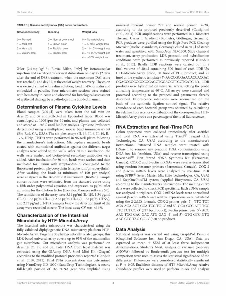
TABLE 1 | Disease activity index (DAI) score parameters.
Stool consistency Bleeding Weight loss
0 = Formed 0 = Normal color stool 0 = No weight loss
1 = Mild-soft 1 = Brown color 1 = 5–10% weight loss
2 = Very soft 2 = Reddish color 2 = 11–15% weight loss
3 = Watery stool 3 = Bloody stool 3 = 16–20% weight loss
4 = >20% weight loss
Xilor [2.5mg kg(−1); Bio98, Milan, Italy] by intramuscularinjection and sacrificed by cervical dislocation on day 25 (2 daysafter the end of DSS treatment, when the maximum DAI scorewas reached), and day 37, at the end of weight recovery. The colonwas excised, rinsed with saline solution, fixed in 4% formalin andembedded in paraffin. Four micrometer sections were stainedwith hematoxylin-eosin and observed for histological assessmentof epithelial damage by a pathologist in a blinded manner.
Determination of Plasma Cytokine LevelsBlood samples (200µl) were taken from the tail vein ondays 25 and 37 and collected in Eppendorf tubes. Blood wascentrifuged at 1000 rpm for 10min, and plasma was collectedand stored at −80◦C until BioPlex analysis. Cytokine levels weredetermined using a multiplexed mouse bead immunoassay kit(Bio-Rad, CA, USA). The six-plex assays (IL-1β, IL-6, IL-10, IL-17A, IFNγ, TNFα) were performed in 96-well plates followingthe manufacturer’s instructions. Microsphere magnetic beadscoated with monoclonal antibodies against the different targetanalytes were added to the wells. After 30min incubation, thewells were washed and biotinylated secondary antibodies wereadded. After incubation for 30min, beads were washed and thenincubated for 10min with streptavidin-PE conjugated to thefluorescent protein, phycoerythrin (streptavidin/phycoerythrin).After washing, the beads (a minimum of 100 per analyte)were analyzed in the BioPlex 200 instrument (BioRad). Sampleconcentrations were estimated from the standard curve usinga fifth-order polynomial equation and expressed as pg/ml afteradjusting for the dilution factor (Bio-Plex Manager software 5.0).The sensitivities of the assay were 3.14 pg/ml (IL-1β), 1.34 pg/ml(IL-6), 1.38 pg/ml (IL-10), 2.38 pg/ml (IL-17), 1.38 pg/ml (IFNγ),and 2.73 pg/ml (TNFα). Samples below the detection limit of theassay were recorded as zero. The intra-assay CV was <14%.
Characterization of the IntestinalMicrobiota by HTF-Microbi.ArrayThe intestinal mice microbiota was characterized using thefully validated diphylogenetic DNA microarray platform HTF-Microbi.Array. Targeting 33 phylogenetically related groups, thisLDR-based universal array covers up to 95% of the mammaliangut microbiota. Gut microbiota analysis was performed ondays 18, 25, 29, and 38. Total DNA from fecal material wasextracted using the QIAamp DNA Stool Mini Kit (Qiagen)according to the modified protocol previously reported (Candelaet al., 2010, 2012). Final DNA concentration was determinedusing NanoDrop ND-1000 (NanoDrop Technologies). A nearlyfull-length portion of 16S rDNA gene was amplified using
universal forward primer 27F and reverse primer 1492R,according to the protocol previously described (Castiglioniet al., 2004) PCR amplifications were performed in a BiometraThermal Cycler T Gradient (Biometra, Göttingen, Germany).PCR products were purified using the High Pure PCR CleanupMicrokit (Roche, Mannheim, Germany), eluted in 30µl of sterilewater and quantified with NanoDrop ND-1000. Slide chemicaltreatment, array production, LDR protocol, and hybridizationconditions were performed as previously reported (Candelaet al., 2012). Briefly, LDR reactions were carried out in afinal volume of 20µl containing 500 fmol of each LDR-UAHTF-Microbi.Array probe, 50 fmol of PCR product, and 25fmol of the synthetic template (5′-AGCCGCGAACACCACGATCGACCGGCGCGCGCAGCTGCAGCTTGCTCATG-3′). LDRproducts were hybridized on universal arrays, setting the probeannealing temperature at 60◦C. All arrays were scanned andprocessed according to the protocol and parameters alreadydescribed. Fluorescence intensities were normalized on thebasis of the synthetic ligation control signal. The relativeabundance of each bacterial group was obtained by calculatingthe relative fluorescence contribution of the corresponding HTF-Microbi.Array probe as a percentage of the total fluorescence.
RNA Extraction and Real-Time PCRColon specimens were collected immediately after sacrificeand total RNA was extracted using Trizol R© reagent (LifeTechnologies, CA, USA) according to the manufacturer’sinstructions. Extracted RNA samples were treated withDNase I to remove any genomic DNA contamination usingDNA-free kit (Ambion, USA) and reverse-transcripted usingRevertAidTM First Strand cDNA Synthesis Kit (Fermentas,Canada). COX-2 and β-actin mRNAs were reverse-transcribedusing random hexamer primers (Fermentas, Canada). COX-2and β-actin mRNA levels were analyzed by real-time PCRusing SYBR R© Select Master Mix (Life Technologies, CA, USA)and StepOnePlusTM system (Applied Biosystems, CA, USA)according to the manufacturers’ instructions. The melting curvedata were collected to check PCR specificity. Each cDNA samplewas analyzed in triplicate. COX-2 mRNA levels were normalizedagainst β-actin mRNA and relative expressions were calculatedusing the 2-21Ct formula. COX-2 primer pair: 5′- TTC TCTACA ACA ACT CCA TCC TC -3′ and 5′- GCA GCC ATT TCCTTC TCT CC -3′ (247 bp product); β-actin primer pair: 5′- ACCAAC TGG GAC GAC ATG GAG -3′ and 5′- GTG GTG GTGAAG CTG TAG CC -3′ (380 bp product).
Data AnalysisStatistical analysis was carried out using GraphPad Prism 6(GraphPad Software Inc., San Diego, CA, USA). Data areexpressed as mean ± SEM of at least three independentdeterminations. Student’s t-test, analysis of variance (one-wayANOVA) followed by Bonferroni’s post-hoc test for multiplecomparison were used to assess the statistical significance of thedifferences. Differences were considered statistically significantat P < 0.05. Euclidean distance of HTF-Microbi.Array relativeabundance profiles were used to perform PCoA and analysis
Frontiers in Pharmacology | www.frontiersin.org 4 March 2016 | Volume 7 | Article 38
De Fazio et al. Geraniol Treatment of DSS Colitic Mice
was accomplished using the R packages Made4, Vegan, and Stats(www.cran.org).
RESULTS
Clinical Colitis ActivityThe effect of DSS and DSS-Ge-OH treatments was evaluatedconsidering the DAI calculated as the sum of weight loss,stool consistency, and blending scores (Table 1). All DSS-treatedmice started to show mild clinical signs of disease 2 daysbefore the end of the 1.5% DSS treatment (day 21) due to thesimultaneous increase in stool consistency index and bleedingindex (maximum DAI score = 2.3). The most evident clinicalsigns of each group were recorded between days 25 and 27(Figure 2) with a maximum DAI score of 9.1 for the DSS groupand with severe weight loss that peaked between days 25 and28 (Figure 2A). Ge-OH at 30mg kg(−1) reduced the DAI scoreof colitis during the acute phase but did not affect this indexduring the recovery phase (Figure 2B). At this Ge-OH dose,the DAI score maintained the same trend observed in DSS-treated mice. At the higher oral dose, Ge-OH reduced the DAIscore for almost the entire duration of colitis and especiallyduring the recovery phase. Statistical analysis of data in Figure 2Bare provided in Supplementary Table 1. These positive Ge-OHeffects were further enhanced when colitic mice were treatedwith enema-administered Ge-OH, resulting in a very low weightloss and a strongly reduced DAI score for the whole duration ofcolitis.
Inflammatory Cytokine Profile of ColitisPlasma levels of IL-1β, IL-6, IL-10, IL-17, TNFα, and IFNγ weredetected in blood samples from all experimental mice group attwo different time points, one corresponding to the acute phase ofcolitis (day 25), and one at the end of the recovery phase (day 37).DSS treatment significantly increased (P < 0.05) all the cytokinesmeasured, both at day 25 and day 37 (Figure 3). At day 25, oraladministration of Ge-OH at the lower dose of 30 mg/mg kg(−1)
did not modify the inflammatory profile of DSS-treated mice.Oral administration of the higher Ge-OH dose of 120mg kg(−1)
and Ge-OH 120mg kg(−1) enema administration significantlydecreased IL-10, IL-17, TNFα, and IFNγ ( P < 0.05), but neitherIL-1β nor IL-6. At day 37 when colitis tended to become chronic,Ge-OH-treated mice showed a better inflammatory profile thanDSS-treated mice. In particular, the lower dose of oral Ge-OHsignificantly reduced all the measured cytokines (P < 0.05).The higher oral dose and enema administration of Ge-OHsignificantly decreased IL-1β, IL-17, IFNγ, and TNFα (P < 0.05)but neither IL-6 nor IL-10.
Histological Evaluation of ColitisHistological evaluation of the colon was made from the colocecaljunction to the anus. Overall, the tissue damage tended tobe limited to the terminal colon and rectum regions, andcan be classified as moderate colitis (Figure 4). At day 25(Figures 4A–C), the colon mucosa in the DSS-treated miceshowed a diffuse loss of goblet cells, focal crypt abscesses, diffusehyperemia, moderate cellular infiltration in themucosa, and focal
epithelial erosions. Diffuse hyperemia, mild loss of goblet cells,mild cellular infiltration but no crypt abscesses, or epithelialerosions were also present in the mucosa of oral Ge-OH-treatedmice at both doses administered [see Supplementary Figure 1 forGe-OH 30mg kg(−1)]. The colon in the Ge-OH enema-treatedmice was characterized by a lowermucosa distortion (elongation)and showed moderate loss of epithelium, and low leukocyteinfiltrations.
After weight recovery (day 37), the colon mucosa in theDSS-treated mice showed a diffuse loss of goblet cells, focalcrypt abscesses, diffuse hyperemia, and mild cellular infiltration(Figure 4D), while the mucosa of oral Ge-OH-treated micepresented diffuse hyperemia but a milder loss of goblet cells,a milder cellular infiltration, and no crypt abscesses at withdose administered (Figures 4E,F). Colon mucosa in the enemaGe-OH-treated mice showed a normal architecture similar tothat of healthy controls. In conclusion, histological and clinicalimprovements were evident in the Ge-OH-treated mice andparticularly in the enema-treated animals.
Ge-OH-Induced Microbiota ModificationsSince the Ge-OH-free suspension itself did not inducemicrobiotaalterations, we investigated the impact of Ge-OH treatment onDSS-induced microbiota dysbiosis in mice. Mice stools werecollected on days 18, 25, 29, and 37. Figure 5 shows thephylogenetic structure of the intestinal microbiota characterizedusing the HTF-Microbi.Array universal platform. DSS treatmentprompted profound, progressive, and transient changes in micemicrobiota composition, compared to colitis-negative controls(group I), defining a peculiar microbiota trajectory during theinduced colitis. In particular, on day 18, after 1 day of DSStreatment, the overall microbiota structure of DSS mice stillresembled that of control mice. At day 25, after seven days of DSS,we observed a global temporary restructuring of the intestinalmicrobiota composition. At day 29 a transitory reduction ofBacteroidetes associated with an increase in Firmicutes wasrecorded. However, on day 37, DSS-treated mice recovered amicrobiota structure similar to that of healthy controls.
While oral Ge-OH treatment at 30mg kg(−1) exerted onlya mild impact on the temporal dynamics of DSS-inducedmicrobiota dysbiosis, oral and enema treatment at a dose of120mg kg(−1) resulted in considerable protection against thetransient DSS-dependent reduction of Bacteroidetes, favoring afaster recovery of a community profile similar to that of healthycontrols. In particular, on day 25, Ge-OH at 120mg kg(−1) (bothenema and orally administered) triggered a Lactobacillaceaeincrease that reached a relative abundance of 11.2 and 9.7%respectively, notably higher than the corresponding value incontrol mice. This Ge-OH-dependent high relative abundanceof Lactobacillaceae was maintained until day 29 after which Ge-OH-treated mice permanently recovered from the DSS-inducedreduction of Bacteroidetes 8 days earlier with respect to thecorresponding DSS-treated mice. These effects are certainlyrelated to the antibacterial action of Ge-OH, evidenced by itslow minimal inhibitory concentration (MIC) on model bacteriaspecies (see Supplementary Table 2). Differently from whatobserved in DSS treated mice, in healthy mice Ge-OH treatment,
Frontiers in Pharmacology | www.frontiersin.org 5 March 2016 | Volume 7 | Article 38

De Fazio et al. Geraniol Treatment of DSS Colitic Mice
FIGURE 2 | Weight change percentage (A) and disease activity index (DAI) score of colitis (B) in different mice experimental groups. Maximum DAI score
was reached between days 25 and 27. Maximum weight loss (22%) was recorded between days 22 and 27. Weight recovery ends at days 37. Data are expressed as
mean ± SD. Analysis of variance (one way-ANOVA) was performed (for weight changes only at days 26, 29, and 32) to assess the statistical significance of the
differences. *P < 0.05 if compared to DSS group mean values. Statistical significance for DAI score differences (analysis of variance, one way-ANOVA) are reported in
Supplementary Table 1.
even at the dose of 120mg kg(−1) (orally administered), didnot produced the same marked changes in the microbiota.Indeed, the microbiota composition of mice treated with Ge-OH
120mg kg(−1) showed a slight increase in Lactobacillaceae,Bacillaceae and Bacteroidetes families (see SupplementaryFigure 2).
Frontiers in Pharmacology | www.frontiersin.org 6 March 2016 | Volume 7 | Article 38

De Fazio et al. Geraniol Treatment of DSS Colitic Mice
FIGURE 3 | Plasma cytokine variations during experimental colitis, measured at days 25 and 37. Cytokines were determined using a 6-plex mouse bead
immunoassay kit. Levels of IL-1β (A), IL-6 (B), IL-10 (C), IL-17A (D), IFN-γ (E), and TNFα (F) are shown. Data are expressed as mean ± SEM of at least three
replicates (n = 9). #P < 0.05 in the comparison between to Ge-OH 30 and Ge-OH 120 or groups. *P < 0.05 if compared to DSS group.
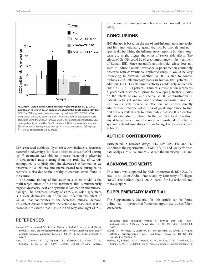
Down-Regulation of COX-2 through Ge-OHTreatmentSince COX-2 plays a crucial role in the production of many lipidmediators involved in intestinal inflammation and is one of themajor targets of IBD pharmacological therapy, we analyzed COX-2 mRNA expression in colon tissues during DSS-induced colitis(Figure 6). Our data support the previously reported findingthat COX-2 mRNA significantly increases in the gut wall ofDSS-treated mice (De Fazio et al., 2014). At day 25, we observed
a significant increase (1.8-fold, P < 0.05) in COX-2 expressionin the gut wall of DSS-treated mice. Ge-OH decrease the COX-2expression in DSS treated mice returning it to values comparableto those of the control.
DISCUSSION
Inflammatory bowel disease (IBD) comprises a group ofchronic inflammatory conditions affecting the gastrointestinal
Frontiers in Pharmacology | www.frontiersin.org 7 March 2016 | Volume 7 | Article 38

De Fazio et al. Geraniol Treatment of DSS Colitic Mice
FIGURE 4 | Differences in histological architecture induced by Ge-OH 120mg kg(−1) and hydrocortisone 2.5mg kg(−1) during the experimental colitis.
Colon specimens were collected from mice on days 25 (A–D) and 37 (E–H). Histopathological changes in individual crypts are shown in representative hematoxylin
and eosin-stained sections. Red arrows indicate loss of crypt architecture associated with epithelial damage and flattened villi while black arrows indicate leukocyte
infiltration (Magnification: 10X; bar = 100µm).
tract. The mucosal immune system of IBD patients has lostthe ability to self-regulate and remains chronically activated.
IBD is a well-established risk factor for colon cancer (CRC)
development, with an increasing incidence linked to youngerage at IBD diagnosis, longer IBD duration, and more severeintestinal inflammation. Conventional IBD therapies includeCOX-2 inhibitors (aminosalicylates and their derivatives),corticosteroids, immunomodulatory drugs, antibiotics, andbiologic drugs such as the monoclonal antibody against tumornecrosis factor alpha (TNFα), a pivotal pro-inflammatorycytokine able to start and maintain the inflammatory process inthe gut. Besides antibiotics, probiotics have also been used in thetreatment of ulcerative colitis to counteract dysbiosis (Bibiloniet al., 2005). Since, IBD usually relapses, all these therapies requirelong-term administration.
Ge-OH is a non-toxic compound, classified as GenerallyRecognized As Safe (GRAS) by the US Food and DrugAdministration. The European Food Security Agency (EFSA)hazard assessment conclusion for Ge-OH established a DerivedNo Effect Level (DNEL) of 13.5mg kg(−1) for humans(General Population—Hazard via oral route), correspondingto 100–120mg kg(−1) in mice. Ge-OH is currently receivingsubstantial attention for its antitumorigenic, anti-inflammatory,and antimicrobial effects that have been clearly demonstratedin vitro. Nevertheless, its role as an anti-dysbiotic agent in coloninflammation has never been investigated. Our study adopteda mouse model of DSS-induced moderate to severe colitis toevaluate the antimicrobial and anti-inflammatory therapeuticactivity of Ge-OH doses considered safe.
Ge-OH, orally administered at 30 and 120mg kg(−1) halvedthe mice weight loss and reduced the disease activity index (DAI)of colitis. At histological level, Ge-OH was able to preserve cryptarchitecture and decrease leukocyte infiltration, with a muchmore evident effect at the higher dose (both enema or orallyadministered). Moreover, enema-administered Ge-OH stronglyimproved signs of colitis maintaining a lower DAI and preservingcolon mucosa integrity. These clinical observations are furthersupported by a significant reduction of COX-2mRNA expressionin the colonic mucosa of Ge-OH-treated mice.
Circulating cytokine levels are indicative of the overallinflammatory status of animals, with IL-1, IL-6, IL-17, and TNFαplaying a key role in the pathogenesis of IBD (Muzes et al.,2012). TNFα is a master cytokine in IBD pathogenesis and itsorchestrating role in colonic inflammation is confirmed by theefficacy of anti-TNFα therapy in IBD patients (Chaparro et al.,2012). The circulating TNFα level correlates with clinical activityboth in ulcerative colitis and Crohn’s disease (Bibiloni et al., 2005)and increases in acute phases of DSS colitis (Alex et al., 2009).So, while circulating TNFα and IL-17 levels seem to correlatewith the DSS colitis clinical course, IL-1β, and IL-10 mainlycorrelate with the histological damage that tends to becomechronic (Alex et al., 2009; De Fazio et al., 2014). The higher oraldose of Ge-OH significantly reduced circulating TNFα and IL-17 in Ge-OH-treated mice after weight recovery at the end ofthe experiments. This decrease was equally evident after Ge-OHenema administration. These results are in agreement with thoseobtained by Medicherla et al. (2015) who found a significantlyreduced expression of the major pro-inflammatory cytokines
Frontiers in Pharmacology | www.frontiersin.org 8 March 2016 | Volume 7 | Article 38

De Fazio et al. Geraniol Treatment of DSS Colitic Mice
FIGURE 5 | Temporal dynamics at the family level of the fecal microbial community of dextran sulfate sodium (DSS)-treated mice. The microbiota
composition of healthy mice (CTRL), colitic mice (DSS), colitic geraniol orally treated mice [Ge-OH 30mg kg(−1), 120mg kg(−1)], and colitic geraniol enema-treated
mice [120mg kg(−1)] is shown. Other Bacteriodetes and Firmicutes families that are not are listed separately have been combined into a single group. The microbiota
composition of the mice group treated with Ge-OH-free oral suspension or Ge-OH-free enema suspension showed no differences from those of the healthy mice
group.
in the colon specimens (TNF-α, IL-1β, and IL-6), associatedwith reduced total and nuclear amounts of NF-κB (p65) afteroral administration of Ge-OH [50 and 100mg Kg (−1)]. Theyalso identified an antioxidant activity of Ge-OH at colon level,evaluated as a decrease in lipid peroxidation marker.
DSS treatment compromises gut microbiota homeostasis,resulting in a dysbiosis characterized by a transient reductionof dominant mutualistic microbiota components such asBacteroidetes, confirming previous findings (Nagalingam et al.,
2011). Ge-OH oral and enema treatment at 120mg kg(−1)
protects DSS-treated mice against this transient reductionof Bacteroidetes, boosting a faster recovery of a healthymicrobiota profile. Interestingly, 120mg kg(−1) geraniol-treatedmice presented a transient increase in the relative abundanceof Lactobacillaceae from day 25 to day 29. This raises thequestion of whether the transient Ge-OH-dependent increasein Lactobacillaceae, heralding the recovery of a healthy profile,is somehow involved in promoting a faster recovery from
Frontiers in Pharmacology | www.frontiersin.org 9 March 2016 | Volume 7 | Article 38
De Fazio et al. Geraniol Treatment of DSS Colitic Mice
FIGURE 6 | Geraniol (Ge-OH) modulates cyclooxygenase-2 (COX-2)
expression in vivo in colon specimens during the acute phase (day 25).
COX-2 mRNA expression was evaluated by real-time PCR. COX-2 mRNA
levels were normalized against β-actin mRNA and relative expressions were
calculated using the 2-21Ct formula. COX-2 overexpression induced by DSS
was significantly reduced by Ge-OH treatment. Data are expressed as mean ±
SEM of at least three replicates (n = 6). *P < 0.05 compared to DSS group.#P < 0.05 compared to CTRL group.
DSS-associated dysbiosis. Dysbiosis always includes a decreasedbacterial biodiversity (Honda and Littman, 2012). GeOH 120mgkg(−1) treatment was able to increase bacterial biodiversityin DSS-treated mice starting from the 10th day of Ge-OHassumption. It is likely that the decreased inflammation weobserved in Ge-OH oral and enema-treated mice during colitisrecovery is also due to the healthy microbiota status found inthese mice.
The central finding of this study in a colitis model is themulti-target effect of Ge-OH treatment that simultaneouslytargeted dysbiosis, local, and systemic inflammation andmucosaldamage. The decreased activity of COX-2 in colon specimensis a clear demonstration of the anti-inflammatory effect ofGe-OH that contributes to the decreased mucosal damage.This effect certainly involves the colonic mucosa, even if it isreasonable to assume that in vivo Ge-OH may also target COX-2
expression in immune system cells inside the colon wall (Su et al.,2010).
CONCLUSIONS
IBD therapy is based on the use of anti-inflammatory moleculesand immunomodulatory agents that act by strongly and non-specifically inhibiting the inflammatory response but their long-term use might trigger the onset of severe side-effects. Theeffects of Ge-OH could be of great importance in the treatmentof human IBD. Since geraniol’s antimicrobial effect does not
seem to induce bacterial resistance, a phenomenon commonlyobserved with conventional antibiotic drugs, it would be veryinteresting to ascertain whether Ge-OH is able to controldysbiosis and inflammatory status in human IBD patients. Inaddition, Ge-OH’s anti-tumor activities could help reduce therisk of CRC in IBD patients. Thus, this investigation representsa preclinical assessment prior to developing further studieson the effects of oral and enema Ge-OH administration inpatients with gut inflammation and/or dysbiosis. Since Ge-OH has its peak therapeutic effect on colitis when directlyadministered into the colon, it is of great importance to findoral delivery systems able to inhibit intestinal Ge-OH absorptionafter its oral administration. On the contrary, Ge-OH withoutany delivery system may be orally administered to obtain asystemic anti-inflammatory effect or to target other organs, suchas brain.
AUTHOR CONTRIBUTIONS
Participated in research design: LD, MV, MC, FR, and ES.Conducted the experiments: LD,MV, AS, EC, and CR. Performeddata analysis: MC, ES, and MC. Wrote the manuscript: LD andES.
ACKNOWLEDGMENTS
This study was supported by Xeda international, RD7 Z.A. Lacrau, 13670 Saint Andiol, France and by University of Bologna(RFO). The authors thank Dr. A. Sardo for his technical andmoral support.
SUPPLEMENTARY MATERIAL
The Supplementary Material for this article can be foundonline at: http://journal.frontiersin.org/article/10.3389/fphar.2016.00038
REFERENCES
Ahmad, S. T., Arjumand, W., Seth, A., Nafees, S., Rashid, S., Ali, N., et al. (2011).
Preclinical renal cancer chemopreventive efficacy of geraniol by modulation of
multiple molecular pathways. Toxicology 290, 69–81. doi: 10.1016/j.tox.2011.
08.020
Alex, P., Zachos, N. C., Nguyen, T., Gonzales, L., Chen, T. E.,
Conklin, L. S., et al. (2009). Centola Distinct cytokine patterns
identified from multiplex profiles of murine DSS and TNBS-
induced colitis. Inflamm. Bowel Dis. 15, 341–352. doi: 10.1002/ibd.
20753
Bakkali, F., Averbeck, S., Averbeck, D., and Idaomar, M. (2008). Biological
effects of essential oils–a review. Food Chem. Toxicol. 46, 446–475. doi:
10.1016/j.fct.2007.09.106
Bibiloni, R., Fedorak, R. N., Tannock, G. W., Madsen, K. L., Gionchetti, P.,
Campieri, M., et al. (2005). VSL#3 probiotic-mixture induces remission in
Frontiers in Pharmacology | www.frontiersin.org 10 March 2016 | Volume 7 | Article 38

De Fazio et al. Geraniol Treatment of DSS Colitic Mice
patients with active ulcerative colitis. Am. J. Gastroenterol. 100, 1539–1546. doi:
10.1111/j.1572-0241.2005.41794.x
Burt, S. (2004). Essential oils: their antibacterial properties and potential
applications in foods–a review. Int. J. Food Microbiol. 94, 223–253. doi:
10.1016/j.ijfoodmicro.2004.03.022
Candela, M., Consolandi, C., Severgnini, M., Biagi, E., Castiglioni., B., Vitali,
B., et al. (2010). High taxonomic level fingerprint of the human intestinal
microbiota by ligase detection reaction–universal array approach. BMC
Microbiol. 10:116. doi: 10.1186/1471-2180-10-116
Candela, M., Rampelli, S., Turroni, S., Severgnini, M., Consolandi, C., De Bellis,
et al. (2012). Unbalance of intestinal microbiota in atopic children. BMC
Microbiol. 12:95. doi: 10.1186/1471-2180-12-95
Castiglioni, B., Rizzi, E., Frosini, A., Sivonen, K., Rajaniemi, P., Rantala, A., et al.
(2004). Development of a universal microarray based on the ligation detection
reaction and 16S rRNA gene polymorphism to target diversity of cyanobacteria.
Appl. Environ. Microbiol. 70, 7161–7172. doi: 10.1128/AEM.70.12.7161-
7172.2004
Chaparro, M., Guerra, I., Muñoz-Linares, P., and Gisbert, J. P. (2012).
Systematic review: antibodies and anti-TNF-alpha levels in inflammatory bowel
disease. Aliment. Pharmacol. Ther. 35, 971–986. doi: 10.1111/j.1365-2036.2012.
05057.x
Chaudhary, S. C., Siddiqui, M. S., Athar, M., and Alam, M. S. (2013). Geraniol
inhibits murine skin tumorigenesis by modulating COX-2 expression, Ras-
ERK1/2 signaling pathway and apoptosis. J. Appl. Toxicol. 2013, 828–837. doi:
10.1002/jat.2739
De Fazio, L., Cavazza, E., Spisni, E., Strillacci, A., Centanni, M., Candela, M., et al.
(2014). Longitudinal analysis of inflammation and microbiota dynamics in a
model of mild chronic dextran sulphate sodium-induced colitis in mice.World
J. Gastroenterol. 20, 2051–2061. doi: 10.3748/wjg.v20.i8.2051
Di Pasqua, R., Hoskins, N., Betts, G., and Mauriello, G. (2006).Changes
in membrane fatty acids composition of microbial cells induced by
addition of thymol, carvacrol, limonene, cinnamaldehyde, and eugenol in
the growing media. J. Agric. Food Chem. 54, 2745–2749. doi: 10.1021/jf0
52722l
Fang, R., Jiang, C. H., Wang, X. Y., Zhang, H. M., Liu, Z. L., Zhou, L., et al. (2010).
Insecticidal activity of essential oil of Carum Carvi fruits from China and its
main components against two grain storage insects. Molecules 15, 9391–9402.
doi: 10.3390/molecules15129391
Heinlein, A., Metzger, M., Walles, H., and Buettner, A. (2014). Transport of hop
aroma compounds across Caco-2 monolayers. Food Funct. 5, 2719–2730. doi:
10.1039/C3FO60675A
Honda, K., and Littman, D. R. (2012). The microbiome in infectious disease
and inflammation. Annu. Rev. Immunol. 30, 759–795. doi: 10.1146/annurev-
immunol-020711-074937
Kamada, N., Seo, S. U., Chen, G. Y., and Núñez, G. (2013). Role of the gut
microbiota in immunity and inflammatory disease. Nat. Rev. Immunol. 13,
321–335. doi: 10.1038/nri3430
Khan, A. Q., Khan, R., Qamar, W., Lateef, A., Rehman, M. U., Tahir, M.,
et al. (2013). Geraniol attenuates 12-O-tetradecanoylphorbol-13-acetate (TPA)-
induced oxidative stress and inflammation in mouse skin: possible role
of p38 MAP Kinase and NF-κB. Exp. Mol. Pathol. 94, 419–429. doi:
10.1016/j.yexmp.2013.01.006
Koloski, N. A., Bret, L., and Radford-Smith, G. (2008). Hygiene: hypothesis
in inflammatory bowel disease: a critical review of the literature. World J.
Gastroenterol. 14, 165–173. doi: 10.3748/wjg.14.165
Laukens, D., Brigitta, M., Brinkman, R. J., De Vos, M., and Vandenabeele,
P. (2015). Heterogeneity of the gut microbiome in mice: guidelines for
optimizing experimental design. FEMS Microbiol. Rev. 40, 117–132. doi:
10.1093/femsre/fuv036
Marcuzzi, A., Crovella, S., and Pontillo, A. (2011). Geraniol rescues inflammation
in cellular and animal models of mevalonate kinase deficiency. In Vivo 25,
87–92.
Medicherla, K., Sahu, B. D., Kuncha, M., Kumar, J. M., and Sudhakar, G., Sistla, R.
(2015). Oral administration of geraniol ameliorates acute experimental murine
colitis by inhibiting pro-inflammatory cytokines and NF-κB signaling. Food
Funct. 6, 2984–2995. doi: 10.1039/c5fo00405e
Missaghi, B., Herman, W., Barkema, K. L., and Ghosh, M. S. (2014). Perturbation
of the human microbiome as a contributor to inflammatory bowel disease.
Pathogens 3, 510–527. doi: 10.3390/pathogens3030510
Muzes, G., Molnár, B., Tulassay, Z., and Sipos, F. (2012). Changes of the cytokine
profile in inflammatory bowel diseases. World J. Gastroenterol. 18, 5848–5861.
doi: 10.3748/wjg.v18.i41.5848
Nagalingam, N. A., Kao, J. Y., and Young, V. B. (2011). Microbial ecology of the
murine gut associated with the development of dextran sodium sulfate-induced
colitis. Inflamm. Bowel Dis. 17, 917–926. doi: 10.1002/ibd.21462
Oussalah, M., Caillet, S., Salmiéri, S., Saucier, L., and Lacroix, M. (2007).
Antimicrobial effects of alginate-based films containing essential oils on Listeria
monocytogenes and Salmonella typhimurium present in bologna and ham.
J. Food Prot. 70, 901–908.
Oviedo-Rondón, E. O., Hume, M. E., Hernández, C., and Clemente-Hernández, S.
(2006). Intestinal microbial ecology of broilers vaccinated and challenged with
mixed Eimeria species, and supplemented with essential oil blends 2006. Poult
Sci. 85, 854–860. doi: 10.1093/ps/85.5.854
Rekha, K. R., Selvakumar, G. P., Santha, K., and Inmozhi Sivakamasundari,
R. (2013). Geraniol attenuates α-synuclein expression and neuromuscular
impairment through increase dopamine content in MPTP intoxicated mice
by dose dependent manner. Biochem Biophys Res Commun. 440, 664–670. doi:
10.1016/j.bbrc.2013.09.122
Singh, B. K., Tripathi, M., Chaudhari, B. P., Pandey, P. K., and Kakkar, P.
(2012). Natural terpenes prevent mitochondrial dysfunction, oxidative stress
and release of apoptotic proteins during nimesulide-hepatotoxicity in rats.
PLoS ONE 7:e34200. doi: 10.1371/journal.pone.0034200
Solórzano-Santos, F., and Miranda-Novales, M. G. (2012). Essential oils from
aromatic herbs as antimicrobial agents. Curr. Opin. Biotechnol. 23, 136–141.
doi: 10.1016/j.copbio.2011.08.005
Strillacci, A., Griffoni, C., Lazzarini, G., Valerii, M. C., DiMolfetta, Rizzello, F., et al.
(2010). Selective cyclooxygenase-2 silencing mediated by engineered E. coli and
RNA interference induces anti-tumour effects in human colon cancer cells. Br.
J. Cancer 103, 975–986. doi: 10.1038/sj.bjc.6605859
Su, Y. W., Chao, S. H., Lee, M. H., Ou, T. Y., and Tsai, Y. C. (2010).
Inhibitory effects of citronellol and geraniol on nitric oxide and prostaglandin
E(2)production in macrophages. Planta Med. 76, 1666–1671. doi: 10.1055/s-
0030-1249947
Thapa, D., Losa, R., Zweifel, B., and Wallace, R. J. (2012). Sensitivity of pathogenic
and commensal bacteria from the human colon to essential oils. Microbiology
158, 2870–2877. doi: 10.1099/mic.0.061127-0
Thompson, A., Meah, D., Ahmed, N., Conniff-Jenkins, R., Chileshe, E., Phillips,
C. O., et al. (2013). Comparison of the antibacterial activity of essential oils and
extracts of medicinal and culinary herbs to investigate potential new treatments
for irritable bowel syndrome. BMC Complement. Altern. Med. 13:338. doi:
10.1186/1472-6882-13-338
Turina, A. V., Nolan, M. V., Zygadlo, J. A., and Perillo, M. A. (2006). Natural
terpenes: self-assembly and membrane partitioning. Biophys. Chem. 122,
101–113. doi: 10.1016/j.bpc.2006.02.007
Wallace, R. J., Oleszek,W., Franz, C., Hahn, I., Baser, K. H., Mathe, A., et al. (2010).
Dietary plant bioactives for poultry health and productivity. Br. Poult. Sci. 51,
461–487. doi: 10.1080/00071668.2010.506908
Yaung, S. J., Church, G. M., and Wang, H. H. (2014). Recent progress in
engineering human-associated microbiomes. Methods Mol. Biol. 1151, 3–25.
doi: 10.1007/978-1-4939-0554-6_1
Conflict of Interest Statement: The authors declare that the research was
conducted in the absence of any commercial or financial relationships that could
be construed as a potential conflict of interest.
Copyright © 2016 De Fazio, Spisni, Cavazza, Strillacci, Candela, Centanni, Ricci,
Rizzello, Campieri and Valerii. This is an open-access article distributed under the
terms of the Creative Commons Attribution License (CC BY). The use, distribution or
reproduction in other forums is permitted, provided the original author(s) or licensor
are credited and that the original publication in this journal is cited, in accordance
with accepted academic practice. No use, distribution or reproduction is permitted
which does not comply with these terms.
Frontiers in Pharmacology | www.frontiersin.org 11 March 2016 | Volume 7 | Article 38