Embed Size (px)
Citation preview

Diet-Induced Effects and the Potential for Cardiovascular Risk
Olivia Hanna Koury
A Thesis in
The Department of
Exercise Science
Presented in Fulfillment of the Requirements
for the Degree of Master of Science (Exercise Science) at
Concordia University
Montreal, Quebec, Canada
April 2015
©Olivia Hanna Koury, 2015

CONCORDIA UNIVERSITYSchool of Graduate Studies
This is to certify that the thesis prepared
By:
Entitled:
and submitted in partial fulfillment of the requirements for the degree of
complies with the regulations of the University and meets the accepted standards withrespect to originality and quality.
Signed by the final examining committee:
______________________________________ Chair
______________________________________ Examiner
______________________________________ Examiner
______________________________________ Supervisor
Approved by ________________________________________________Chair of Department or Graduate Program Director
________________________________________________Dean of Faculty
Date ________________________________________________
Master of Science (Exercise Science)
Dr. Robert Kilgour
Dr. Peter Bogaty
Dr. Richard Courtemanche
Dr. Andreas Bergdahl

! iii!
Abstract Diet-Induced Effects and the Potential for Cardiovascular Risk
Olivia Hanna Koury
! Cardiovascular disease remains the leading cause of death in America impacting
the lives of millions. There are many modifiable risk factors that may help in the
prevention and treatment of cardiovascular disease such as coronary artery disease or
congestive heart failure. Proper diet and healthy weight management is a crucial factor in
preventing disease. Dieting is a large component of American living, and the low-
carbohydrate high-protein diet remains the most common form of weight loss. This thesis
will investigate different types of diets and eating habits that can affect risk of developing
cardiovascular disease.
An apolipoprotein E-deficient mouse model was used to investigate different diets
and how they may develop cardiovascular disease, more specifically, atherosclerosis in
the aorta and altered mitochondrial functioning in the heart. The diet of interest for this
project, was a low-carbohydrate high-protein diet. This diet promotes an environment
that causes a shift in cellular metabolism due to macronutrient imbalances. Past research
has yet to address a possible link between low-carbohydrate high-protein diets and
atherosclerosis and vascular smooth muscle cells, and mitochondrial functioning. This
thesis contains data that will be of interest to both clinicians and patients, in terms of safe
dieting and the potential for cardiovascular risk.
!

iv
Table of Contents
Chapter I Introduction...................................................................................................................................1 Theoretical Context………………………………………………………………………..2 The Atherosclerotic Process and Pathway………………………………………………....2 Pathways Leading to Atherosclerosis……………………………………………………...7 Vascular Smooth Muscle Cells & Their Involvement in Lesion Development…………...8
Structure and Morphology………………………………………………………………....8 VSMCs: Function and Roles……………………………………………………………....9 The Diseased Intima: Phenotype Alteration, Migration & Proliferation…………………10 Low-Carbohydrate Diets and Cellular Metabolism.…………………………………..….12 Project Overview…………………………………………………………………………14
Chapter II The role of casein in the development of hypercholesterolemia………………..…….15 Chapter III A low-carbohydrate high-protein diet induces vascular smooth muscle cell dedifferentiation in an ApoE-/- murine model……………………………………..…….25
Chapter IV Altered Mitochondrial Functioning in Apolipoprotein E-Deficient Mice Induced by a Low-Carbohydrate High-Protein Diet…………………………………………………...37 Chapter V Concluding Remarks……………………………………………………………………....….49 Chapter VI References………………………………………………………………………………………52 Appendix……………………………………………………………………………………….57

v
List of Figures
Figure 1: Layers of an artery……………………………………………………….2
Figure 2: Pathophysiological factors of atherosclerosis…………………………....3
Figure 3: Schematic illustration of cellular events………………………………....6
Figure 4: Phenotype shift of vascular smooth muscle cell……………………..…..9
Figure 5: Schematic of plaque development ……………………………………..12

1
I
Introduction

2
Theoretical Context Cardiovascular Disease (CVD) is now the second leading cause of death in Canada [1];
the main contributor being ischemic heart disease and thus atherosclerosis [2]. With the rise of an
overweight and obese population, proper weight maintenance is crucial for controlling risk
factors for CVD and other conditions. Many people resort to exercise and strict dieting for weight
loss and disease prevention, however not all diets have proven to be safe. Although effective for
weight loss, the popular low-carbohydrate high-protein diets (LCHP) may be counterproductive
for CVD prevention in that they may impose damaging effects to blood vessels. The implications
of a low-carbohydrate high-protein diet will be explored in regards to potential effects on the
development of atherosclerosis and vascular smooth muscle cell activity.
The Atherosclerotic Process and Pathway
Atherosclerosis is best described as a chronic
inflammatory response to a vascular injury, involving
many inflammatory cells, cytokines and adhesion
molecules to form an atheroma [3-5]. An atheroma
involves the deposition of cholesterol and other fatty
substances as well as necrotic tissue within the intimal
layer of an artery, rendering the lumen stenotic [6]. The
artery is composed of three main layers: the intima,
media, and adventitia (externa) as seen in Figure 1. On
the innermost layer, the intima, lies a continuous layer
of cells termed the “endothelium” that essentially separates the blood in the lumen from the rest
Figure 1: Layers of an artery [7]

3
of the vessel [8]. The endothelial layer composed of endothelial cells is involved in coagulation,
inflammatory responses, and vascular tone via phosphorylation of endothelial nitric oxide
synthase (eNOS) into nitric oxide (NO) [8,9]. The vascular injury present in this disease may be
of physical or a biochemical origin that effectively alters the endothelium [10].
The traditional “response-to-injury” theory states that endothelial stripping absolutely
caused atherosclerosis [12] however we now understand that there is a spectrum of possible
insults that can contribute to the morphological changes in endothelium [13]. Endothelial cell
dysfunction occurs when the normal homeostatic balance of the cell is no longer maintained,
leading to either impairment in vasorelaxation or up-regulation of adhesiveness on the endothelial
lining for circulating inflammatory cells [9]. Risk factors such as high amounts of low-density
lipoprotein cholesterol (LDL-c), hypertension, diabetes, and cigarette smoke can lead to
Figure 2: Pathophysiological factors that may play a role in atherosclerosis [11]
478 Harjot K. Saini et al.
of adhesion molecules such as vascular cell adhesion molecule-1 (VCAM-1),intercellular adhesion molecule-1 (ICAM-1), E-selectin, and endothelialleukocyte adhesion molecule is regulated by proinflammatory cytokines suchas tumor necrosis factor-α (TNF-α), interleukin (IL)-1, IL-4, IL-6, and inter-feron-γ [19]. However, in the presence of endothelial dysfunction, the levels ofthese cytokines have been shown to be elevated, which in turn cause moreproduction of adhesion molecules and thus favor the monocyte recruitmentand adhesion to the endothelium [20]. The recruited monocytes differentiateto macrophages in the subendothelial space and further aggravate the develop-ment of atherosclerosis [21].
Formation of Fatty StreakThe alteration in endothelial permeability triggers the transmigration ofLDL particles into the intima through the endothelial layer, where it is modi-fied by oxidative stress to oxidized LDL (Ox-LDL) [22]. Macrophage lipoxy-genase, myeloperoxidase, and NADPH oxidase are the major sources ofreactive oxygen species production [23]. Oxidation of polyunsaturated fattyacids within the LDL causes the release of aldehyde and ketones such asmalondialdehyde and 4-hydroxynonenal, which can alter the lysine residueson apolipoproteins B-100 (apoB-100), the major protein of LDL [24]. The
FIGURE 22.1. Different biochemical and cellular processes participating in the devel-opment of atherosclerosis.
Hypercholesterolemia
Homocysteine
Oxidative stress Leukocyte adhesionand migration
Plateletdysfunction
Inflammation, Ox LDL, smooth musclecell migration and proliferation,
foam cell formation
Atherosclerosis
Endothelialdysfunction
Smooth muscledysfunction
Hypertension
Pathophysiological factors
Diabetes Geneticalterations
Infectiousmicroorganisms

4
endothelial dysfunction; whether it is denudation, the alteration of permeability, or the change of
cytokine or growth factor secretion [13]. Regardless of the insult ensued; the common factor is
the damage to the endothelium [13].
Atherosclerosis typically develops in steps that may regress or progress towards an
unstable atheroma. As abovementioned, disturbance of the endothelial begins the sequence of
events and the body’s initial reaction is to thicken the vessel wall as a “compensatory
mechanism” to the damage [13]. A common cause for inflammation in the medium or large sized
arteries is the state of hypercholesterolemia (14). The arterial inflammatory response begins when
excess low-density lipoproteins (LDL) infiltrate the intima layer of the artery, and through
oxidation, the now oxidized LDL (oxLDL) release phospholipids that activate the endothelial
cells [15,16]. The oxLDL residing in the intima layer is what activates the endothelial cells and
causes an increased expression of adhesion molecules and inflammatory genes [17]. With
increased expression of leukocyte adhesion molecules, cells that roll on the surface will adhere to
the site of activation [18,19]. The two key adhesion molecules that are up-regulated are
intercellular adhesionmolecule-1 (ICAM-1) and vascular-cell adhesion molecule-1 (VCAM-1)
[8]. These adhesion molecules will facilitate attachment and transendothelial migration through
the inter-endothelial junction of leukocytes, such as monocytes and lymphocytes, into the
subendothelial area [8,20]. Chemokines found in the intima layer will incite the monocytes to
perform such a migration [14]. Once in the subendothelial region, macrophage colony-
stimulating factor (M-CSF) induces the differentiation of monocytes into macrophages [14]. With
the help of scavenger receptors located on the macrophage, a broad range of particles, including
the oxLDL, are “eaten up” by the macrophage, who later transform into foam cells [14,21]. In
addition to scavenger receptors, toll-like receptors that also bind pathogen-like molecules, but

5
will as well activate the macrophage to produce inflammatory cytokines, proteases, and cytotoxic
nitrogen and oxygen radical molecules [22].
In a pathological state as such, many pro-inflammatory or pro-oxidative stimuli imposed
by risk factors can generate reactive oxygen species (ROS) via vascular cells [8]. The production
of ROS is harmful because they can serve as second-messenger coupling molecules that transmit
signals to elevate the expression of pro-atherogenic products [8]. A protection against ROS can
be the antioxidant defense mechanisms. However, when the rate of ROS production exceeds the
performance of the antioxidant system, the condition of oxidative stress ensues [8].
At this stage in the development, fatty streaks or xanthomas are formed due to the large
lipid core. Most xanthomas can regress by means of lifestyle modifications, and will not fully
develop into atherosclerotic lesions [23]. However, in the later stages of development, intimal
hyperplasia occurs in which the arterial wall becomes thickened due to the migration and
proliferation of vascular smooth muscle cells (VSMC) and the irregular accumulation of
extracellular matrix [13]. The hyperactivity of the VSMCs will be discussed in greater detail in a
later section.
With established lipid deposition and intimal thickening, the plaque can be deemed either
‘stable’ or ‘unstable’ depending on a variety of factors that may render the plaque vulnerable to
rupture [24]. Many key players such as T-cells, macrophages, and mast cells [25-27] produce
molecules that can negatively destabilize a lesion: inflammatory cytokines, proteases, coagulation
factors, radicals, and vasoactive molecules [14]. ‘Destabilizing’ a lesion implies the destruction
of collagen in the extracellular matrix, inhibiting the formation of a stable fibrous cap over the
lesion, or beginning a thrombus formation [28-31]. Destabilization depends entirely on the
crucial balance between collagen synthesis and collagen breakdown [8]. In one respect, VSMCs
produce interstitial collagen that offers tensile strength and stability to the maintenance of the

6
fibrous cap [8]. However, due to the presence of T cells, macrophages are induced to produce
MMPs and cysteine proteases, 2 proteases that play a key role in collagen breakdown [8, 14]. T
cells may also slow down the collagen production by VSMCs by producing interferon-γ, which
send signals to VSMCs to halt production [8]. The main alarming consequence of atherosclerosis
is the risk that a plaque can occlude an artery, or rupture resulting in thrombus formation [13].
Figure 3: Schematic illustration of cellular events in atherosclerosis [32]

7
Pathways Leading to Atherosclerosis
It is crucial to acknowledge that atherosclerosis is not solely a ‘lipid-burdened’ disease.
There are 3 circumstances that may lead to atherosclerosis: inflammation, autoimmunity and
infection [33]. Many triggers for inflammation occur hand-in-hand with the risk of developing
atherosclerosis such as oxidized LDL, dyslipidemia, hypertension, diabetes and obesity. These
conditions may indirectly increase the risk for atherosclerosis by elevating expression of pro-
inflammatory cytokines, chemokines, or adhesion molecules, all of which lead to a pro-
inflammatory atherogenic pathway [33].
Atherosclerosis has also been speculated to be an infectious disease as some infectious
agents may generate inflammatory stimuli [34]. Although infectious viruses alone may not be a
predominant cause, a study showed that patients with coronary artery disease (CAD), had
elevated levels of antibodies against viruses chlamydia pneumonia, helicobacter pylori, herpes
simplex, or cytomegalovirus [33,35]. Extravascular infections, such as gingivitis may increase
inflammatory cytokines in remote atherosclerotic lesions, while intravascular infections may
provide local inflammatory stimuli [33].
Many cells of the immune system are involved in the pathologic processes occurring in
the subendothelial region. Some of these immune cells include macrophages, T-cells,
autoantibodies, autoantigens, and cytokines (interleukins, tumor-necrosis factor, interferon-γ,
platelet-derived growth factor) [36]. Systemically, the inflammatory response involves acute-
phase reactants serum amyloid-A, fibrinogen, and C-reactive protein [36]. Heat-shock proteins
(HSPs), proteins found in most species induced by heat shock, have also been shown to promote
atherogenesis [37]. Patients with early lesions, expressed higher levels of anti-HSP65 antibodies
[38].
8
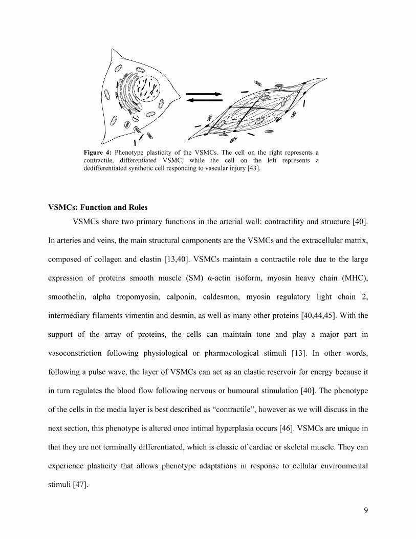
Vascular Smooth Muscle Cells & Their Involvement in Lesion Development
The VSMC is a highly specialized cell found in mature animals that expresses a unique
collection of ion channels, signaling molecules, and contractile proteins to perform cell
contraction [39]. These cells found in blood vessels may proliferate at a very slow rate, and
express minimal synthetic activity in a healthy environment [39].
Structure and Morphology
The VSMCs essentially act as the bulk of the vessel wall, found mainly in the media layer
bound together by internal and external elastic lamina [40]. In pathological conditions, VSMCs
can be found in high numbers in the intimal region, in which they normally represent only a
minor population. VSMCs will never reside in the adventitia layer, as it is only populated with fat
cells, fibroblasts, and nerves [40]. These VSMCs have been suggested to express two main
phenotypes: contractile and synthetic [40, 41]. Smooth muscle cells exhibiting a contractile
phenotype are found in the media layer and are common in healthy, differentiated arteries
containing many microfilament bundles [42]. These contractile cells are spindle-shaped, as seen
in figure 4, and contain copious amounts of contractile fibers such as actin and myosin [40].
Upon migration to the intimal layer during atherosclerosis, the VSMCs experience a phenotype
switch to the synthetic, which is typical of a diseased artery [42]. These dedifferentiated synthetic
cells are more rhomboid-shaped and contain cytoplasm that have a prevalence of rough
endoplasmic reticulum and matured Golgi apparatus [42].
9
VSMCs: Function and Roles
VSMCs share two primary functions in the arterial wall: contractility and structure [40].
In arteries and veins, the main structural components are the VSMCs and the extracellular matrix,
composed of collagen and elastin [13,40]. VSMCs maintain a contractile role due to the large
expression of proteins smooth muscle (SM) α-actin isoform, myosin heavy chain (MHC),
smoothelin, alpha tropomyosin, calponin, caldesmon, myosin regulatory light chain 2,
intermediary filaments vimentin and desmin, as well as many other proteins [40,44,45]. With the
support of the array of proteins, the cells can maintain tone and play a major part in
vasoconstriction following physiological or pharmacological stimuli [13]. In other words,
following a pulse wave, the layer of VSMCs can act as an elastic reservoir for energy because it
in turn regulates the blood flow following nervous or humoural stimulation [40]. The phenotype
of the cells in the media layer is best described as “contractile”, however as we will discuss in the
next section, this phenotype is altered once intimal hyperplasia occurs [46]. VSMCs are unique in
that they are not terminally differentiated, which is classic of cardiac or skeletal muscle. They can
experience plasticity that allows phenotype adaptations in response to cellular environmental
stimuli [47].
Figure 4: Phenotype plasticity of the VSMCs. The cell on the right represents a contractile, differentiated VSMC, while the cell on the left represents a dedifferentiated synthetic cell responding to vascular injury [43]. !

10
The Diseased Intima: Phenotype Alteration, Migration & Proliferation
The proliferation of smooth muscle cells is a response-to-injury and a necessary process
to occur at the onset of inflammation [48]. There are three main phases in the response that cause
movement of VSMCs and intimal hyperplasia. During the initial onset, replication of VSMCs
occurs, typically stimulated by the release of basic fibroblast growth factor (bFGF) from the dead
and damaged cells [40]. Once having multiplied, the smooth muscle cells will migrate towards
the intima from the internal elastic lamina of the medial layer. During the migration of the
VSMCs, a change of phenotype occurs from contractile to fibroblast-like synthetic phenotype
[47]. Many molecules mediate this travelling action; however platelet-derived growth factor
(PDGF) plays a crucial role and has been debated to be mitogenic for VSMCs [40]. Once having
reached the intimal region, the third phase entails proliferation of the cells and secretion of
extracellular matrix with the ability to form a stable “fibrous cap” between the lipid lesion and
arterial lumen [49].
The proliferatory phase for the VSMCs will occur under the influence of an assortment of
growth factors and cytokines, and only upon completion, will the extracellular matrix be released
and accumulated in abnormally large amounts [23]. Factors that can act as chemotactic agents for
the smooth muscle cells are; PDGF, acidic fibroblast growth factor (aFGF), bFGF, α-thrombin,
and insulin-like growth factor 1 (IGF-1) [13,50-53]. These compounds are termed ‘chemotactic’
because they will affect the normal activity of the VSMCs in the media and favor changes
towards migration and proliferation of the cells [13]. In contrast, there are substances produced
that inhibit VSMC proliferation and intimal growth in response to the vascular stimuli: heparin,
TGF-β, and nitric oxide (NO) [54]. In effect, these chemotactic growth factors along with

11
infiltrating leukocytes and adhesion molecules allow for a positive feedback loop that continue
the vicious cycle of VSMC hyperactivity and increased intimal thickening [13,55].
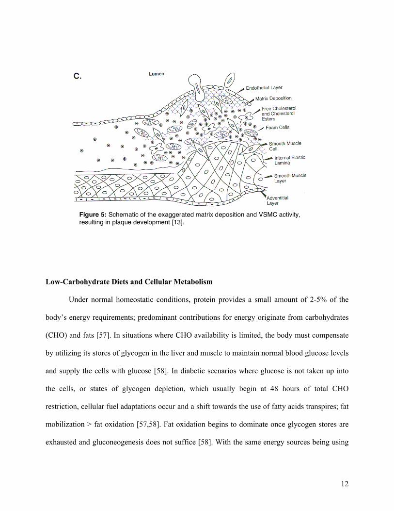
Aside from the increased VSMC activity towards the intima, the extracellular matrix
plays a perilous role in plaque development. The synthetic VSMCs in the intima are different
from the contractile VSMCs in that they secrete proteins of the extracellular matrix, such as
collagen I and II [23]. The components of the extracellular matrix offer tensile strength for the
arterial wall; due to the VSMC’s production of collagen fibers (types I, III, V), small
proteoglycans, and the elastic membranes [56,23]. In an atherosclerotic plaque, there is
exaggerated matrix deposition that severely narrows the vessel lumen [23]. Moreover, the
distribution of collagen and elastin is distinct, since the intimal extracellular matrix exhibits much
more collagen and much less elastin than does the medial extracellular matrix [23]. It has recently
been speculated that perhaps macrophages may also be able to produce some components of the
extracellular matrix by means of secretion of TGF- β [23]. With excessive extracellular matrix,
the fibrous cap atheroma will begin to form. The atheroma entails the accumulation of partially
necrotic foam cells from the earlier inflammatory phase, which is separated from the vessel
lumen by VSMC-derived fibrotic tissue [23].
12
Low-Carbohydrate Diets and Cellular Metabolism
Under normal homeostatic conditions, protein provides a small amount of 2-5% of the
body’s energy requirements; predominant contributions for energy originate from carbohydrates
(CHO) and fats [57]. In situations where CHO availability is limited, the body must compensate
by utilizing its stores of glycogen in the liver and muscle to maintain normal blood glucose levels
and supply the cells with glucose [58]. In diabetic scenarios where glucose is not taken up into
the cells, or states of glycogen depletion, which usually begin at 48 hours of total CHO
restriction, cellular fuel adaptations occur and a shift towards the use of fatty acids transpires; fat
mobilization > fat oxidation [57,58]. Fat oxidation begins to dominate once glycogen stores are
exhausted and gluconeogenesis does not suffice [58]. With the same energy sources being using
Figure 5: Schematic of the exaggerated matrix deposition and VSMC activity, resulting in plaque development [13].
!
13
but in different proportions, the fuel source shifts from being “glucocentric” to “adipocentric”
[59].
When triglycerides are forced to be broken into fatty acids and glycerol for energy use, a
process called β-oxidation occurs in the mitochondria to convert fatty acids into a readily useful
form [60]. The fatty acids are converted into acetyl-CoA and can enter the Krebs Cycle in the
same fashion as glucose. In conditions lacking CHO and fat for energy, protein catabolism offers
an important source of energy in a few ways. Before any amino acids (AA) can be oxidized,
removal of the amino group must occur. In one instance, the detached amino group can get
transferred and given to another AA (ketoacid), to form a new AA [57]. This process is called
transamination. Otherwise, oxidative deamination can occur when the amino group may be
removed and become free ammonia (NH3) [61]. Nevertheless, when the amino group is removed,
a carbon skeleton is remaining and may become oxidized or converted to form different
substances capable of entering the Krebs Cycle: CO2, H2O, acetyl-CoA, oxaloacetate, α-
ketoglutarate [61].

14
Project Overview
Dieting trends and use of supplementation is practiced widely in North America for
various reason, many of which focus on weight loss or disease prevention. The goal of this thesis
is to better understand how manipulating macronutrient distribution in the diet can have profound
effects on a cellular level both beneficial and harmful. Animal source protein, such as casein or
whey is commonly used for protein supplementation with the goal of gaining muscle mass.
Casein protein is frequently used for custom-made animal diets for research purposes. Animal
proteins are of interest to researchers due to their potential hypercholesterolemic effect in humans
and animals. This matter will be investigated in depth in the published review paper to follow.
Another common use of dieting is a low-carbohydrate high-protein diet, generally
referred to as the “Atkin’s diet”. This carbohydrate-eliminating method of weight loss has yet to
be studied in regards to cardiovascular health, more specifically vascular smooth muscle cells in
atherosclerosis and mitochondria functioning in congestive heart failure. Reduction of
carbohydrates has a direct implication in the normal cellular metabolism, whereby a shift from a
carbohydrate energy source to a fat energy source occurs. These projects aim to assess whether
this shift can affect the vascular smooth muscle cells and their role in development in
atherosclerosis in the thoracic aorta, more specifically their phenotype shift from contractile to
synthetic phenotype. As well, the mitochondrial respiration will be assessed in the cardiac tissue
whether they function better in a lipid-rich environment. Information from this thesis can provide
ample information to health care professionals and individuals engaging in dieting practices, to
better understand the risks and safety.

15
II
The role of casein in the development of hypercholesterolemia Olivia Hanna Koury, Celena Scheede-Bergdahl, Andreas Bergdahl

16
Contribution of Authors Olivia Hanna Koury: preparation of manuscript. Celena Scheede-Bergdahl: editing of manuscript. Andreas Bergdahl: preparation of manuscript. !

MINI REVIEW
The role of casein in the development of hypercholesterolemia
Olivia Hanna Koury & Celena Scheede-Bergdahl &Andreas Bergdahl
Received: 27 February 2014 /Accepted: 6 October 2014 /Published online: 15 October 2014# University of Navarra 2014
Abstract Atherosclerosis remains the leading cause ofsevere cardiovascular complications such as cardio- andcerebrovascular events. Given that prevention and earlyintervention play important roles in the reduction ofcardiovascular complications associated with athero-sclerosis, it is critical to better understand how to targetthe modifiable risk factors, such as diet, in order to bestminimize their contributions to the development of thedisease. Studies have shown that various dietary sourcesof protein can affect blood lipid levels, a modifiable riskfactor for atherosclerosis, either positively or negatively.This clearly highlights that not all proteins are “createdequal.” For example, consumption of diets high in eitheranimal- or vegetable-based sources of protein have re-sulted in varied and inconsistent effects on blood cho-lesterol levels, often depending on the amino acid com-position of the protein and the species investigated.Careful consideration of the source of dietary proteinmay play an important role in the prevention of athero-sclerosis and subsequent cardiovascular complications.Given the recent focus on high protein diets, an empha-sis on controlled studies in the area is warranted. Thegoal of this review is to present the current state of the
literature that examines the effects of casein, a common-ly utilized animal-based protein, on blood cholesterollevels and the varying effects noted in both animals andhumans.
Keywords Casein protein . Hypercholesterolemia . Soyprotein . Lipoprotein . Cardiovascular disease
Cholesterol and the development of atherosclerosis
Atherosclerosis lies at the root of many serious cardio-vascular complications such as myocardial infarction,stroke, gangrene, intermittent claudication, and limbamputation [39]. The initiation of the atheroscleroticprocess depends mainly on the state and function ofthe endothelial layer, which represents the demarcationbetween the vessel wall and the blood [15]. Endothelialdysfunction is characterized by two aspects: a reductionof the bioavailability of nitric oxide, which leads toimpaired vasoreactivity, and the activation of the endo-thelial cells [7, 51]. Taken together, these features inducea pro-inflammatory, proliferative, and pro-coagulatorystate, all of which contribute to the progression of ath-erogenesis [1]. Factors associated with endothelial dys-function include smoking, oxidative stress, diabetes,metabolic dysfunction, obesity, hypercholesterolemia,and hypertension [20] (Fig. 1).
High-density lipoproteins (HDL) are considered tobe a negative risk factor for the development of cardio-vascular disease and have been casually referred to as“good” cholesterol. The cardioprotective effects of HDL
J Physiol Biochem (2014) 70:1021–1028DOI 10.1007/s13105-014-0365-9
O. H. Koury :A. Bergdahl (*)Department of Exercise Science, Concordia University,7141 Sherbrooke Street West, Montreal, QC H4B 1R6,Canadae-mail: [email protected]
C. Scheede-BergdahlDepartment of Kinesiology & Physical Education,McGill University,475 Pine Avenue West, Montreal, QC H2W 1S4, Canada

are due to its role in reverse cholesterol transport (RCT),a key process that regulates cholesterol clearance fromthe systemic circulation. The purpose of RCT is theremoval of excess (free) cholesterol from peripheralcells and reuptake by the liver for eventual bile saltsynthesis and excretion [8]. In the early stages of ath-erosclerosis, a higher than normal concentration of cir-culating low-density lipoproteins (LDL) results in theirpenetration into the subendothelial layer of the bloodvessel. LDL, transported by apolipoprotein B (ApoB)into the vascular wall, becomes biochemically modifiedand, subsequently, triggers an inflammatory response[21]. There are three histological features of the unstableatheroma: (1) the large lipid core which is present whenthe abovementioned RCT is incapable of regulatingblood cholesterol levels, (2) the abundance of inflam-matory cells, and (3) a thin fibrous cap [17, 22]. Theatheroma is not only a collection of cholesterol, waste,and fibrotic tissue but is also a lesion composed ofendothelial and smooth muscle cells with infiltratingleukocytes and other inflammatory cells [32]. Essential-ly, the initial endothelial dysfunction leads to the fattystreak formation and, ultimately, fibrous cap formation[39].
The risk of developing atherosclerosis and conse-quent ischemic heart disease increases with the presenceof pro-atherogenic substances such as intermediate den-sity lipoproteins (IDL), low-density lipoproteins (LDL),
and very low-density lipoproteins (VLDL) [33]. Evi-dence in the literature has suggested that certain proteinsappear to exert a greater effect on blood cholesterollevels than others [50]. Given that the recent trendtowards high protein diets in the pursuit of weight lossand reduction of chronic disease risk, it remains imper-ative to fully appreciate the associations between certainprotein types and the potential for increased cardiovas-cular disease risk. This increased risk may occur despitesuccessful weight loss. The goal of this review paper isto examine whether casein, a dietary source of protein,has an effect on blood cholesterol and whether it can beconsidered a positive risk factor for the development ofatherosclerosis.
What is casein and does it have a rolein the atherosclerotic process?
Over the last five decades, there has been a steadyinterest in the abilities of certain proteins to promoteeither a pro- or anti-atherogenic effect. In particular,casein has often been included in studies as an animalsource of protein [50]. Milk products contain two mainprotein components: whey and casein. Whey proteinrepresents approximately 18 to 20 % of mammalianmilk protein, while casein represents the remaining80–82 % [4]. Casein is regarded as one of the most
Fig. 1 Schematic view of the arterial wall and the steps in ather-oma formation. Risk factors such as a biochemical imbalance(high LDL) trigger an endothelial activation beyond the normal
noxious stimuli. LDL enters the subendothelial layer and becomeoxidized to allow attraction of monocytes, cytokines, and otherinflammatory cells to support the inflammatory process
1022 O.H. Koury et al.

nutritive milk proteins, as it contains all common aminoacids and is rich in essential amino acids (EAA) [45].Purified casein is produced from skim milk by a pro-cessing technique where the protein is separated fromthe whey, dried, and then resolubilized [9]. Isolated milkcasein forms micelle complexes when dispersed in thewater phase of milk. The micelle structures have fivedifferent subunits of the casein subtype: α-casein, α-2casein, β-casein, κ-casein, and γ-casein. Commonamong these five structures are the calcium-phosphatebonds that hold them together and that they all containsalt and water [9].
The primary difference between casein, whey pro-tein, and other high-quality proteins is the rate of digest-ibility. Casein is considered to be a slow-digesting pro-tein because it curdles or gels in the stomach, thusdelaying release in the intestines. This results in a grad-ual but steady rise in blood amino acid concentrationfollowing ingestion [9]. Since the blood amino acidconcentrations are kept relatively low, it slows but ex-tends the rate of protein synthesis [16]. Casein alsodemonstrates anti-catabolic properties, which simulta-neously inhibits protein breakdown [4, 6]. In situationswhere weight loss is desired, the anti-catabolic proper-ties of casein result in it being the preferred source ofprotein for hypocaloric diets [5]. In light of these char-acteristics, casein is attractive for many weight lossprograms that include high protein intake.
As early as the 1970s and 1980s, animal studiesreported that casein increased serum cholesterol levels,thus playing a role in the development of atherosclero-sis. Previously, it was believed that cardiovascular dis-ease and atherosclerosis were a result of the amount offat in the diet and, in particular, the cholesterol andsaturated fats. In light of results seen in animal studies,a high casein diet may also be considered a risk factor inthe development of atherosclerotic plaque. Casein-mediated hypercholesterolemia has been shown to de-velop independently of exogenous cholesterol and sat-urated fat consumption [28]. Studies have been per-formed in order to compare lipid profiles upon adher-ence to diets that vary in amount of cholesterol,cholesterol-free semi-purified diets, and various proteinsources. Early studies have shown that soy protein, asource of vegetable protein, appears to play a protectiverole in the vasculature and reduces the concentration oftotal cholesterol and LDL, contrary to the detrimentaleffects of casein [10]. A negative association betweensoy protein intake and the development of coronary
heart disease and nonfatal myocardial infarctions wasalso shown in a group of middle-aged and older Chinesewomen [52], although these protective effects of soy arenot detected in all studies [24]. The association betweenanimal-based protein and cardiovascular disease is alsosupported by a meta-analysis of five prospective studiesthat compared mortality in vegetarians and non-vegetar-ians. This analysis showed that subjects who consumedanimal protein had a 24 % higher mortality from ische-mic heart disease, even after controlling for potentialconfounding factors such as age, sex, smoking status,alcohol, habitual exercise, education, and body massindex [29]. Given the potential detrimental effects ofcasein and other animal-based proteins in the develop-ment of vascular disease, as suggested in studies such asthese, it is important to consider dietary protein sourcewhen recommending high protein diets rather than con-sidering all proteins equal.
The effects of casein in the animal model
Research conducted in male New Zealand white rabbitsby Huff and colleagues clearly demonstrated that chang-es in body weight, plasma cholesterol, triglyceridelevels, as well as liver cholesterol occurred upon varyingthe dietary protein source (i.e., either animal or plantprotein) [26]. The diet used in the Huff study was termeda “low-fat semi-purified diet”, consisting of either 27 %casein or soy isolate, along with 60 % dextrose, 5 %celluflour, 4 % salt mix, 3 %molasses, and 1 % corn oil.The 16 animals were divided into 2 groups: 8 received adiet rich in casein and 8 received a diet rich in soyprotein isolate. This low-fat semi-purified diet was giv-en to both groups for 10 months, resulting in signifi-cantly lower levels of mean plasma cholesterol in thesoy isolate group as compared to the casein semi-purified group. After 10 months, the casein-fed animalsended up with higher mean triglyceride levels, higherliver cholesterol (Table 1), and developed atherosclerot-ic lesions, particularly in the aortic arch region. Despitethe negative effects on plasma lipids and cholesterol,casein-fed animals gained less weight than the soy-fedanimals (2.9 and 3.3 kg, respectively).
Plasma VLDL, LDL, and IDL levels were all signif-icantly higher in the casein-fed versus the soy-fed ani-mals (Table 2). Huff and colleagues concluded that thishypercholesterolemic state must be the result of choles-terol that is endogenously produced by the liver or
Effects of casein on blood lipid profile 1023

intestine in response to the manipulation of dietaryprotein source [26]. This significant rise in IDL, themain transporter of cholesterol, has been observed undersimilar conditions since although the underlying causeremains speculative [44].
In the Huff study, it is interesting that both the caseinand soy diets lacked exogenous cholesterol and werelow in saturated fats. From these, it is evident that thesource of protein directly affects the distribution patternand concentration of cholesterol being transported in theblood [26]. The amino acid composition of the protein isas important as the protein source [11]. It is known thatamino acids vary in their effects on serum cholesterolconcentrations, as well as further variations when
ingested as proteins [11]. Illustrating this concept, theamino acids lysine and methionine contribute to thedevelopment of hypercholesterolemia, whereas arginineis able to counteract this effect [30, 31]. The essentialamino acids found in casein have been hypothesized tobe responsible for the observed increase of total choles-terol and LDL [11]. By replacing casein with isolatedsoy protein, which has a different amino acid composi-tion, the increases in total and LDL cholesterol contentin the serum associated with casein can be avoided [3].
Terpstra and colleagues reported similar results, buttheir data also demonstrated the dose effect of casein fedto Zucker strain rats [46]. Their study included sixgroups of animals fed with either a commercial diet withno cholesterol, a commercial diet with 1.2 % cholester-ol, or four types of semi-purified cholesterol enricheddiets (20 % casein, 50 % casein, 20 % soybean, or 50 %soybean (g/100 g of feed)). The results revealed a moreprominent hypercholesterolemic effect occurring in di-ets with a higher percentage of casein. The same effectwas observed in rabbits [25, 46], as well as in pigeons[34, 35].
Evidence in the literature supports the notion that thecasein-mediated cholesterol increases may be biphasicin nature: extremely low and extremely high amounts ofcasein in the diet appear to have the greatest impact onblood cholesterol levels [27, 40]. Increases in cholester-ol were most apparent with either diets containing rela-tively small amounts (5 %) or large amounts (40 to60 %) of casein. Diets consisting of moderate amountsof casein (i.e., 20 %) appeared to produce the smallesteffects [19]. Gender also appears to play a role: Femalerats were more predisposed to developing hypercholes-terolemia in response to casein ingestion [47]. Thisobservation had also been previously reported by Filiosand colleagues [19] with blood cholesterol levels dou-bling in magnitude in female versus male rats.
Research conducted by Hermus and colleagues fo-cused on demonstrating the different combinations ofprotein with gelatin on serum cholesterol levels andbody weight gain in rabbits [23]. The team set up fourexperimental groups: (1) semi-purified diet containingstrictly casein as the protein source; (2) casein andgelatin; (3) casein, gelatin, and fish protein; and (4)casein, gelatin, fish protein, and soy protein. After58 weeks of diet adherence, the group fed the dietconsisting of casein only demonstrated a growth-retarding effect compared to the other groups whoachieved normal growth. Additionally, the casein group
Table 1 The condition of the rabbits following the observance ofa casein- or soy-rich diet. Cholesterol and triglyceride contents inthe plasma and liver were all significantly elevated, excluding theliver triglyceride (adapted from [26] with permission)
Casein(mg/dl)
Soy protein(mg/dl)
Mean plasma cholesterola (mg/dl) 247±12 66±3b
Liver cholesterol (mg/g wet wt) 6.6±1 3.3±0.2b
Mean plasma triglyceridea (mg/dl) 95±4 58±3b
Liver triglyceride (mg/g wet wt) 7.0±0.7 6.6±0.3
Lipid profile in rabbits fed either casein or soy protein diet for10 months. Results are expressed as a mean ± SE for 8 rabbits ineach dietary groupa The overall mean ± SE for the entire 10-month periodb Significantly different from the casein-fed group P<0.01 fromStudent’s t test
Table 2 The varying distribution of plasma cholesterol that de-veloped among the four lipoprotein classes: VLDL, IDL, LDL,and HDL, as well as the total plasma concentration. Differenceswith both diet groups in all lipoprotein classes are significant,excluding the HDL (adapted from [26] with permission)
Density class Casein (mg/dl) Soy protein (mg/dl)
VLDL d<1.006 76±6 11±3a
IDL 1.006<d>1.019 132±14 25±6a
LDL 1.019<d>1.063 46±6 10±2a
HDL 1.063<d>1.21 19±4 12±3
Total plasma concentration 275±25 58±6a
Plasma cholesterol distribution among lipoprotein classes. Resultsare expressed as mean ± SE for 6 rabbits in each dietary groupa Significantly different from the casein-fed rabbits (P<0.01) fromStudent’s t test
1024 O.H. Koury et al.
also achieved a hypercholesterolemic state that wasunseen in the other three experimental groups. Thisstudy demonstrated that the addition of alternate proteinsources to the diet was able to blunt the hypercholester-olemic effects of casein.
Overall, it is understood that the atherosclerotic pro-cess can be initiated by hypercholesterolemia, with se-rum total cholesterol and lipoproteins being well-established risk factors for the disease [21]. Althoughelevated concentrations of serum cholesterol contributeto the development of fatty streaks, this is not the solereason for disease susceptibility [28]. Other known riskfactors, such as family history, obesity, chronic hyper-glycemia, and physical and/or biochemical injuries,have also been implicated [21, 28]. To fully appreciatethe origins and mechanisms involved in the progressionof atherosclerosis, what has been learned from studiesinvolving possible dietary sources of cholesterol mustalso be considered. When certain animals are fed with acasein-rich diet, there is a higher correlation with lipo-philic plaques and high serum cholesterol content than adiet consisting of plant protein [28]. The lipoproteindensity concentration is also altered, depending onwhether a plant or animal protein source is considered[37]. Theories suggest that soybean protein contain sa-ponins, which are protective against hypercholesterol-emia [42]. It is also thought that dietary fiber increasesabsorption of bile acids in the intestine. This will resultin loss of bile acid through fecal excretion which will becompensated by stimulation of hepatic conversion ofcholesterol into bile acids [41]. The main question ishow casein protein causes an elevation in cholesterol,such that the protein source is as detrimental as thesource of fat [26]. What provokes this mechanism toincrease cholesterol to such a level as to induce athero-genic plaque?
Many speculations and theories have been put forthover the past 30 years as to how and why casein inges-tion raises cholesterol levels and why certain species aremore susceptible to the effects of casein. The activityand concentration of enzyme alkaline phosphatase playan important role since it has the potential to dephos-phorylate casein and prevent accumulation ofphosphopeptides [36]. In 1988, Van Der Meer and col-leagues conducted studies that involved feeding bothrabbits and rats a similar diet that included elevatedcasein content in order to investigate intestinal absorp-tion and bile acid excretion. They reported that caseininduces a hypercholesterolemic effect in rabbits due to
low intestinal phosphatase activity and with a highglycine conjugation of bile acids, whereas in the rat,where little effect of casein was noted, the conjugationof bile acids occurs primarily via other amino acids,such as taurine [48].
Another potential factor that may contribute to theeffects of casein is the LDL receptors. It has beenreported that animals fed casein-enriched diet had adownregulation of hepatic LDL receptors preceded byan increase in plasma cholesterol [12]. Other studieshave also shown that casein stimulates LDL ApoBsynthesis, therefore increasing the circulating LDL [11].
What effect does casein have in a human model?
Contrary to the observations seen in animals, the major-ity of intervention studies that have investigated theeffects of plant and animal protein on serum cholesterollevels in humans have reported inconsistent effects. In1983, Sacks and colleagues tested whether dairy protein(casein) or soy protein would have an effect on plasmacholesterol in 13 strict vegetarians. The study designconsisted of a 1-week pre-intervention period, duringwhich, baseline measurements such as body weight,cholesterol profile, triglycerides, and VLDL-c/TG ratiowere measured. Following the baseline period, all 13subjects were split into groups that followed two phases:a diet enriched in casein for 20 days and then soy for20 days or vice versa. Results yielded no significantchanges in LDL or protective HDL cholesterol frombaseline. As well, there was no difference of lipid profileor lipoproteins in the soy and casein groups during their40-day intervention [43].
Another study conducted by Van Raaij and col-leagues looked at similar aspects, but in healthy non-vegetarian subjects eating a “western” simulated diet.All 69 participants began by eating a casein-soy or“cassoy” diet for a control period of 10 days in orderto establish baseline measurements for the remainder ofthe experimental design. In this cassoy diet, 65 % of theprotein content was a 2:1 mixture of casein and soy,respectively. Following the 10-day control period, indi-viduals were divided into three groups for 28 days:maintenance cassoy diet, casein diet, or soy diet. Inter-estingly, there were barely any changes seen betweenthe experimental casein and soy groups in regard to totalcholesterol. Subjects adhering to the casein-enricheddiet did not demonstrate any significant changes in
Effects of casein on blood lipid profile 1025

lipoprotein fractions. On the other hand, the soy groupdemonstrated improvements in the LDL and HDL con-centrations as compared to the casein group, and thereductions in LDL were also significant within the soygroup from their baseline measurements [49]. The im-provements in LDL-c and HDL-c were also seen inanother study with participants eating a cholesterol-enriched casein or soy diet [38]. VLDL concentrationsremained unchanged in both study groups.
Crouse and colleagues conducted a similar studywith healthy individuals having elevated LDL concen-trations between 3.62 and 5.17 mmol/L. Subjects main-tained a casein-rich diet or a soy-rich diet with varyingamounts of isoflavones for 9 weeks. Following thisdietary intervention, LDL-c and total cholesterollowered significantly from baseline in individuals inthe soy group, with a dose-dependent relationship be-tween cholesterol improvements and isoflavoneamount. There was no significant improvement inHDL-c or TG concentration between the groups [14],suggesting that casein supplementation is not an effec-tive intervention for individuals with pre-existing highcholesterol.
From these studies performed on humans, the resultssuggest that casein protein may not have such a pro-nounced effect on cholesterol levels as seen in certainanimal models, such as rabbits. The studies share mixedresults and conclusions. A soy diet would be morebeneficial for lowering cholesterol levels in hypercho-lesterolemic patients since casein-enriched diets demon-strate few advantages in lowering LDL-c or TG. Inhealthy subjects, on the other hand, it seems as thoughcasein and soy diets lack influence on cholesterol levels[43]. However, it should be pointed out that, in order tobetter mimic the study design in animals, the humanexperiments would need to be conducted for longerperiods of time and/or administered at earlier periodsin their lives [49]. The important point here is to con-sider whether the lack of effect is indeed a phenomenonin the human model or whether it is due to limitations instudy design. Further work is needed in order to objec-tively determine the safety of diets high in casein.
Clinical implications of a high casein diet
Meal replacements (MR) containing elevated amountsof plant or animal protein have proven successful inpromoting weight loss in obese patients. In a
randomized controlled trial by Anderson and colleagues[2], patients consuming a MR with high casein for16 weeks had a tendency towards more weight lossand greater fat loss than individuals on high soy diets.Although associated with only modest weight loss, theindividuals consuming soy MR showed promising im-provements in their cardiovascular risk profile (i.e.,LDL, TC, visceral fat, and systolic blood pressure).The period following weight loss is crucial to success-fully sustain weight reduction, and for this, high caseindiets have been found to be useful [13].
Attention must also be drawn to the popular use ofprotein supplementation following exercise training.Postprandial protein synthesis has been studied, andthe data indicates that amino acids dictate future proteinsynthesis, breakdown, and oxidation [6]. Protein shakesof whey and casein are frequently used to maintain andincrease muscle mass, hence protein synthesis or anab-olism following exercise. As compared to whey protein,ingestion of casein protein in a meal or drink may onlyinduce a small increase in protein synthesis, but isassociated with a substantial decrease in protein break-down [6]. These physiological processes are possibledue to the retarded increase in plasma AA concentra-tions. Without the pronounced hyperaminoacidemicpeak associated with whey, casein offers a “longer last-ing” rise in AA levels and sustains protein breakdowninhibition. Casein supplementation, compared to whey,has been shown to improve body composition resultingin decreased percent body fat; an increase of lean mass;and greater muscle strength in legs, chest, and shoulders[18]. These effects are most likely due to casein’s anti-catabolic property. Depending on individual aims ormotives, athletes may opt for casein as their choice ofprotein supplementation for the aforementionedreasons.
Conclusion
Casein is not only naturally present in foods containingdairy such as milks and cheeses but is also extensivelyutilized in its purified form as a powdered protein sup-plement. For decades, saturated fats, and cholesterolhave been deemed responsible for the development ofcardiovascular disease and conditions such as athero-sclerosis and the “clogging of arteries.” Studies havealso found that casein protein is just as aversive as fatsfor some animal species. Casein can have a negative
1026 O.H. Koury et al.
impact on the serum cholesterol concentration and raiseit to levels that pose a severe danger to the lipid profile.There have been speculations that casein is responsiblefor the disruption of bile acid binding in the smallintestine, leaving elevated levels of free bile acids tobe re-absorbed. It is key to clearly understand whetherhypercholesterolemia is enough to induce atherosclero-sis and simulate endothelial dysfunction and to whatextent does it raise cholesterol and cause atherosclerosis.Another important aspect that would require furtherinvestigation is the possibility that humans may mani-fest the same hypercholesterolemic dangers of casein ifgiven for longer periods of time comparable to animalstudies. Although healthy humans are assumed to beless sensitive to dietary modifications than animals,there may be changes noted if the human studies wouldparallel animal studies in terms of time periods andadministration [49]. Would a diet low in fats but highin animal protein be harmful to individuals and posecardiovascular risks? The potential dangers of caseinshould serve as a red flag for dieters, trainers, physi-cians, or nutritionists alike, especially when consideredfor populations who may already be considered at riskfor cardiovascular complications. In regard to supple-mentation, patients in a rehabilitation setting due toconditions such as cancer cachexia or sarcopenia mayalso be administered large doses of soy, whey, or casein.Also of concern are people who exercise train andconsume exceptionally high concentrations ofwhey or casein protein supplementation in orderto increase protein synthesis and muscle mass.Understanding the implications of a high caseinprotein diet is vital in order to assess the healthstatus and long-term lipid profile of an individualadhering to such a diet.
References
1. Anderson TJ (1999) Assessment and treatment of endothelialdysfunction in coronary artery disease and implications fortherapy. J Am Coll Cardiol 34:631–638
2. Anderson JW, Fuller J, Patterson K, Blair R, Tabor A (2006)Soy compared to casein meal replacement shakes with highenergy-restricted diets for obese women: randomized con-trolled trial. Metabolism 56:280–288
3. Anthony MS, Clarkson TB, Bullock BC, Wagner JD (1997)Soy protein versus soy phytoestrogens in the prevention ofdiet-induced coronary artery atherosclerosis of male cynomol-gus monkeys. Arterioscler Thromb Vasc Biol 17:2524–2531
4. Antonio J, Incledon T (2001) The anticatabolics. In: AntonioJ, Stout J (eds) Sports supplements. Lipincott Williams &Wilkins, Philadelphia, pp 111–136
5. Bendtsen LQ, Lorenzen JK, Bendsen NT, Rasmussen C,Astrup A (2013) Effect of dairy proteins on appetite, energyexpenditure, body weight, and composition: a review of theevidence from controlled clinical trials. Adv Nutr 4(4):418–438
6. Boirie YM, Dangin P, Gachon P, Vasson MP, Maubois JL,Beaufrere B (1997) Slow and fast dietary proteins differentlymodulate postprandial protein accretion. Proc Natl Acad Sci US A 94(26):14930–14935
7. Bonetti PO, Lerman LO, Lerman A (2003) Endothelial dys-function—a marker for atherosclerotic risk. ArteriosclerThromb Vasc Biol 23:168–175
8. Burges JW, Sinclair PA, Chretien CM, Boucher J, Sparks DL(2006) Reverse cholesterol transport. In: Cheema SK (ed)Biochemistry of atherosclerosis. Springer, New York, pp 3–22
9. Campbell B (2012) Dietary protein efficiency: dietary proteintypes. In: Lowery LM, Antonio J (eds) Dietary protein andresistance exercise. CRC, Boca Raton, pp 95–114
10. Carroll KK (1991) Review of clinical studies on cholesterol-lowering response to soy protein. J AmDiet Assoc 91(7):820–827
11. Carroll KK, Kurowska EM (1995) Soy consumption andcholesterol reduction: review of animal and human studies. JNutr 125:594S–597S
12. Chao YS, Yasmin TT, Alberts AW (1982) Effects of chole-styramine on low density lipoprotein binding sites on livermembranes from rabbits with endogenous hypercholesterol-emia induced by a wheat starch-casein diet. J Biol Chem 257:3623–3627
13. Claessens M, Van Baak MA, Monsheimer S, Saris WHM(2009) The effect of a low-fat, high-protein or high-carbohydrate ad libitum diet on weight loss maintenance andmetabolic risk factors. Int J Obes 33:296–304
14. Crouse JR, Morgan T, Terry JG, Ellis J, Vitolins M, Burke GL(1999) A randomized trial comparing the effect of casein withthat of soy protein containing varying amounts of isoflavoneson plasma concentrations of lipids and lipoproteins. ArchIntern Med 159:2070–2076
15. Cullen P, Rauterberg J, Lorkowski S (2005) The pathogenesisof atherosclerosis. In: Von Eckardstein A (ed) Atherosclerosis:diet and drugs. Springer, New York, pp 3–70
16. Dangin M, Guillet C, Garcia-Rodenas C, Gachon P,Bouteloup-Demange C, Reiffers-Magnani K, Fauquant J,Ballevre O (2003) The rate of protein digestion affects proteingain differently during aging in humans. J Physiol 549(2):635–644
17. Davies MJ (1997) The composition of coronary-arteryplaques. N Engl J Med 336(18):1312–1314
18. Demling RH, DeSanti L (2000) Effects of a hypocaloric diet,increased protein intake and resistance training on lean massgains and fat mass loss in overweight police officers. AnnNutrMetab 44(1):21–29
19. Filios LC, Naito C, Andrus SB, Portman OW, Martin RS(1958) Variations in cardiovascular sudanophilia with changesin the dietary level of protein. Am J Physiol 194:275–279
20. Forrester JS (2008) The pathogenesis of atherosclerosis andplaque instability. In: Holtzman JL (ed) Atherosclerosis andoxidant stress. Springer, New York, pp 1–10
Effects of casein on blood lipid profile 1027

21. Gross MD (2008) Lipids, oxidation, and cardiovascular dis-ease. In: Holtzman JL (ed) Atherosclerosis and oxidant stress.Springer, New York, pp 79–96
22. Hansson GK (2005) Inflammation, atherosclerosis, and coro-nary artery disease. N Engl J Med 352(16):1685–1695
23. Hermus RJJ, West CJ, Van Weerden EJ (1983) Failure ofdietary-casein-induced acidosis to explain the hypercholester-olaemia of casein-fed rabbits. J Nutr 113(3):618–629
24. Hu FB, Stampfer MJ, Manson JE et al (1999) Dietary proteinand risk of ischemic heart disease in women. Am J Clin Nutr70:221–227
25. Huff MW, Hamilton RMG, Carroll KK (1977) Plasma cho-lesterol levels in rabbits fed low fat, cholesterol-free,semipurified diets: effects of dietary proteins, protein hydro-lysates and amino acid mixtures. Atherosclerosis 28(2):187–195
26. Huff MW, Roberts DCK, Carroll KK (1982) Long-term ef-fects of semipurified diets containing casein or soy proteinisolate on atherosclerosis and plasma lipoproteins in rabbits.Atherosclerosis 41:327–336
27. Jones RJ, Huffman S (1956) Chronic effect of dietary proteinon hypercholesterolaemia in the rat. Proc Soc Exp Biol Med93:519–522
28. Katan MJ, Louis HM, Vroomen LH, Hermus RJJ (1982)Reduction of casein-induced hypercholesterolaemia and ath-erosclerosis in rabbits and rats by dietary glycine, arginine andalanine. Atherosclerosis 43:381–391
29. Key TJ, Fraser GE, Thorogood M et al (1999) Mortality invegetarians and nonvegetarians: detailed findings from a col-laborative analysis of 5 prospective studies. Am J Clin Nutr70:516S–524S
30. Kurowska EM, Carroll KK (1992) Effect of high levels ofselected dietary essential amino acids on hypercholesterolae-mia and down-regulation of hepatic LDL receptors in rabbits.Biochim Biophys Acta 1126(2):185–191
31. Kurowska EM, Carroll KK (1994) Hypercholesterolemic re-sponses in rabbits to selected groups of dietary essential aminoacids. J Nutr 124:364–370
32. Libby P (2002) Inflammation in atherosclerosis. Nature 420:868–874
33. Libby P, Ridker PM, Maseri A (2002) Inflammation andatherosclerosis. Circulation 105:1135–1143
34. Little JM, Angell EA (1977) Dietary protein level and exper-imental aortic atherosclerosis. Atherosclerosis 26(2):173–179
35. Lofland HB, Clarkson TB, Goodman HO (1961) Interactionsamong dietary fat, protein, and cholesterol in atherosclerosis-susceptible pigeons: effect on serum cholesterol and aorticatherosclerosis. Circ Res 9:919–924
36. Lorient D, Linden G (1976) Dephosphorylation of bovinecasein by milk alkaline phosphatase. J Dairy Res 43:19–26
37. Mahley RW, Holcombe KS (1977) Alterations of the plasmalipoproteins and apoproteins following cholesterol feeding inthe rat. J Lipid Res 18:314–324
38. Meinhertz H, Nilausen K, Faergemen O (1990) Effects ofdietary proteins on plasma lipoprotein levels in normal sub-jects: interaction with dietary cholesterol. J Nutr Sci Vitaminol36(2):S157–S164
39. Moreno JJ, MitjavilaMT (2003) The degree of unsaturation ofdietary fatty acids and the development of atherosclerosis. JNutr Biochem 14(4):182–195
40. Nath N, Harper AE, Elvehjem CA (1959) Diet andcholesteremia: part 3 effect of dietary proteins with particularreference to the lipids in wheat gluten. Can J Biochem Physiol37:1375–1384
41. Oakenfull DG, Fenwick DE, Hood RL (1979) Effects ofsaponins on bile acids and plasma lipids in the rat. Br J Nutr42:209–216
42. Oakenfull DG, Sidhu GS (1990) Could saponins be a usefultreatment for hypercholesterolemia? Eur J Clin Nutr 44:79–88
43. Sacks FM, Breslow JL,Wood PG, Kass EH (1983) Lack of aneffect of dairy protein (casein) and soy protein on plasmacholesterol of strict vegetarians. An experiment and a criticalreview. J Lipid Res 24:1012–1020
44. Samman S, Khosla KK, Carroll KK (1990) Intermediatedensity lipoprotein-apolipoprotein B turnover in rabbits fedsemipurified diets containing casein or soy protein. Ann NutrMetab 34(2):98–103
45. Sindayikengera S, Xia W (2006) Nutritional evaluation ofcaseins and whey proteins and their hydrolysates fromProtamex. J Zhejiang Univ (Sci) 7(2):90–98
46. Terpstra AHM, Harkes L, Van der Veen FH (1981) The effectof different proportions of casein in semipurified diets on theconcentration of serum cholesterol and the lipoprotein com-position in rabbits. Lipids 16:114–119
47. Terpstra AHM, Van Tintelin G, West CE (1981) The effect ofsemipurified diets containing different proportions of eithercasein or soybean protein on the concentrations of cholesterolin whole serum, serum lipoproteins and liver in male andfemale rats. Atherosclerosis 42:85–95
48. Van Der Meer R, De Vries H, Van Tintelen G (1988) Thephosphorylation state of casein and the species-dependency ofits hypercholesterolaemic effect. Br J Nutr 59:467–473
49. Van Raaij JMA, Katan MB, Hautvast JGAJ, Hermus RJJ(1981) Effects of casein versus soy protein diets on serumcholesterol and lipoproteins in young healthy volunteers. AmJ Clin Nutr 34:1261–1271
50. Vega-Lopez S, Lichtenstein AH (2005) Dietary protein typeand cardiovascular disease risk factors. Prev Cardiol 8(1):31–40
51. Xian CJ, Shoubridge CA, Read LC (1995) Degradation ofIGF-1 in the adult rat gastrointestinal tract is limited by aspecific antiserum or the dietary protein casein. J Endocrinol146:215–225
52. Zhang X, Shu XO, Fao Y-T et al (2003) Soy food consump-tion is associated with lower risk of coronary heart disease inChinese women. J Nutr 133:2874–2878
1028 O.H. Koury et al.

! ! !25
III
A low-carbohydrate high-protein diet induces vascular smooth muscle cell dedifferentiation in an ApoE-/- murine model Olivia Hanna Koury and Andreas Bergdahl

! ! !26
Contribution of Authors Olivia Hanna Koury: animal handling, surgeries, immunoblotting, measurements of contractility, analysis of data, preparation of manuscript. Andreas Berdgahl: animal handling, concept development, student supervision, statistics, analysis of data, preparation of manuscript.
! 27
Obesity has been associated with a host of adverse health conditions, such as insulin resistance, systemic inflammation and cardiovascular disease risk factors [1, 2]. Due to the deleterious effects of obesity on both society and the individual, the search for an efficient and safe means of weight loss remains a priority. Diets that are rich in protein and/or fat, with limited carbohydrates (i.e.: the Zone, Dr. Atkins) have earned attention due to their obvious potential for weight reduction. Despite their apparent “success”, it remains to elucidate all potential side effects associated with
these methods of weight loss. Recent data obtained in a murine model, highlights the possibility that not all diets are created “equal” and that the traditional serum risk markers used to assess weight loss programs may not capture the full picture. In the study by Foo et al., (2009), Apolipoprotein E-knockout (ApoE-/-) mice given a low-carbohydrate high-protein (LCHP) diet (12% carbohydrate, 43% fat, 45% protein and 0.15% cholesterol) developed more aortic atherosclerotic lesions and impaired ability to generate new vessels in response to tissue ischemia when compared to both a
A low-carbohydrate high-protein diet induces vascular smooth muscle cell dedifferentiation in an ApoE-/- murine model Olivia H. Koury1 and Andreas Bergdahl1
1Department of Exercise Science, Concordia University, Montreal, QC, Canada Correspondence to Andreas Bergdahl, PhD., Department of Exercise Science, Concordia University, 7141 Sherbrooke West, Montreal, QC H4B 1R6 Email: [email protected] !Summary: Low-carbohydrate high-protein (LCHP) diets remain the leading weight loss regimen proving to be quite rapid and highly effective. These diets have been praised for the health benefits associated with insulin resistance, hypertension, weight loss, and lipid control. However, a link between these diets and altered vascular smooth muscle cell (VSMC) phenotype has yet to be addressed. With the rise in obesity, and frequent use of LCHP diets, this project aimed to investigate LCHP induced effects on the VSMCs. We hypothesized that the LCHP diets would experience a more significant shift to synthetic phenotype than a control (CON) or western diet (WD). Male Apolipoprotein E-deficient mice were randomly assigned to one of three diets: CON, WD, or LCHP diet. Following 6 weeks on the diet, animals were euthanized and their thoracic aortas were removed. Oil Red O staining, immunoblotting, and wire myography were performed on these aorta to assess the state of dedifferentiation of the VSMCs. Immunoblotting revealed a significant decrease of contractile proteins α-actin and calponin, and a significant increase of TRPC-1 indicating a shift from contractile to synthetic phenotype. This property did not translate to functional measures, as the wire myograph did not reveal a loss in force-generating capacity of the cell. Oil Red O staining did not show a major difference in lipid accumulation in the 3 diets. Data from this study revealed an evident dedifferentiation occurring in the VSMCs however this change did not manifest a change in the contractile ability of the cell. !Keywords: atherosclerosis!! dedifferentiation! vascular smooth muscle cell !phenotype! low-carbohydrate high-protein
! 28
control group (regular chow diet of 65% carbohydrate, 15% fat and 20% protein) and, more interestingly, mice put on a representation of a western-type diet (43% carbohydrate, 42% fat, 15% protein) [3]. Although the data presented in the Foo et al. study is interesting in terms of the presence of atherosclerosis in the aorta, the link that they present between the low carbohydrate/high protein diet and the formation of lesions primarily rests on the evaluation of endothelial progenitor cells. These observations, however, do not take into consideration the stages in the development of atherosclerosis itself. Early atherosclerotic progression consists primarily of the infiltration of fat into the vascular wall and formation of fatty streaks and is reversible through dietary modifications, pharmacological interventions and exercise [4].
The following stages of atherosclerosis involve the development of the lipid core and stabilization of a fibrous cap, involving largely vascular smooth muscle cells (VSMCs) [5]. Differentiated VSMCs are highly specialized units, located in the media layer of arteries. Their primary function is to contract and relax, consequently controlling blood pressure and blood flow distribution [6-10]. In a healthy adult vessel, the contractile phenotype is characterized by markers such as smooth muscle (SM) α-actin, calponin, SM22-α, SM-myosin heavy chain, h-caldesmon [9]. Unique to VSMCs is their remarkable plasticity as they, unlike cardiomyocytes or skeletal muscle cells, are not terminally differentiated [9,11] and can thus modify their phenotype according to physiological provocations [11]. This shift from contractile to synthetic state serves great importance during vascular development and pathology as it allows for malleability and thus reversible adaptations [6, 10-12]. Following vascular injury, contractile VSMCs go through a transient modification of phenotype, which involves a reduced expression of contractile genes [10]. This plasticity seems to be necessary and may confer a survival advantage since it provides means for the smooth muscle to respond to altered conditions in its surroundings. The dedifferentiation of the VSMCs leads to migration and proliferation to the intimal layer, followed by the formation of connective tissue matrix that accumulates lipids and both free and esterified cholesterol [13]. At the point where vascular smooth
muscle cells actually dedifferentiate and migrate into the vessel wall, the atherosclerotic lesions are considered no longer reversible and may present a serious concern in terms of cardiovascular risk [14]. However, the solid formation of extracellular matrix by VSMCs is also thought to serve as a protective component in plaque stabilization [13, 15] subsequently preventing rupture [16]. The situation is exacerbated by the continuous accumulation of macrophages that ingest lipids and become foam cells [17, 18]. At this stage, the response is thought to be chronic and the lesion progresses to a more advanced stage.
Foo et al as well as many other studies have looked at the effects of a low-carbohydrate diet in terms of insulin resistance, hypertension, ketosis, however, to date nothing has been produced about the VSMC phenotype switching [19-24]. The goal of this study was to determine whether the altered physiological environments induced by a low-carbohydrate diet would favor a VSMC phenotype shift. Methods Animals
Apolipoprotein E knockout (ApoE-/-) mice were obtained from Jackson Laboratories (Bar Harbor, Maine, USA) and used for breeding. The resulting litters were weaned and separated based on sex at 21-28 days. The males were housed individually and randomly assigned to one of three diets for 6 weeks: control (CON), western (WD), and low-carbohydrate high-protein (LCHP) diet. All procedures were approved by the Animal Ethics Committee of Concordia University (protocol ID: #30000259) and were conducted in accordance with guidelines of the Canadian Council on Animal Care. Diet Specifications
All 3 animal diets were iso-caloric. Table 1 displays the diet specifications in terms of percentage of caloric intake from fat, carbohydrates, and protein. The CON diet, 5075 Charles River Autoclavable Rodent Diet, reflects a healthy, standard macronutrient distribution. The WD, obtained from Harlan Laboratories (TD.110229) replicates a high-fat high-
To be submitted to Journal of Cardiovascular Medicine!! April 2015
! 29
carbohydrate ‘American’ diet, consisting of 42% from fat, 42% from CHO, and 16% from protein. The LCHP diet, obtained from Harlan Laboratories (TD.04524) simulates an Atkin’s diet used for weight loss and contains 43% from fat, 11% CHO, and 46% protein. Both WD and LCHP diets are modifications of TD.88137 (Harlan Laboratories), used for studies of atherosclerosis, and contain comparable amounts of cholesterol (1.5 g/kg).
Control Western LCHP
Carbohydrates 63% 42.2% 11%
Fats 14% 42.1% 43.2%
Proteins 24% 15.7% 45.8%
Experimental Techniques Oil Red O Staining Oil Red O (ORO) staining, quantifying the lipid deposits within the vessel, was performed according to Nunnari et al. [25] on fresh tissue from the thoracic aorta. A 3% stock solution was prepared (10 ml isopropanol, 0.3g Oil Red O Powder from Sigma-Aldrich), as well as a working stain consisting of stock solution and water (3:2 parts respectively) filtered through a Whatman No. 1 Filter. The aortas were cleaned of adventitial fat and then soaked in the working stain for 30min, rinsed in distilled water, cleaned again of excess adventitial fat, and split longitudinally to measure for area. Samples were then individually placed in chloroform: methanol (1:1) solution for 2 min to extract the stain before the absorbance was measured at 520 nm using a spectrophotometer. Immunoblotting The thoracic aorta was removed from the sacrificed animal cleaned of fat and connective tissue, and stored at -80°C. The tissue was then pulverized/homogenized using liquid nitrogen with ~70µl lysis buffer containing (in mM) 250 NaCl, 50 HEPES (pH 7.5), 10% glycerol, 1% triton X-100, 1.5 MgCl2 1 EGTA, 10 Na4P2O7 NaF, 800 µM Na3VO4.
After 1 hour on ice, the cell slurry was centrifuged at 13,000 rpm for 10 min, and the supernatant was collected. 10µl of lysate was mixed with 2µl DTT and 2µl sample buffer and loaded on a 10% acrylamide-SDS gel followed by a transfer onto a 0.45µm nitrocellulose membrane (162-0115 Bio-Rad) in 10mM sodium tetraborate buffer. Ponceau staining was done as loading control before the membranes were blocked in 3% bovine serum albumin (BSA) in 0.1% Tween 20 in Tris-buffered saline (TBS) (10mM Tris-HCl, pH 7.5, 150 mm NaCl) for 1 hour at room temperature. This was followed by overnight incubation 4°C with Abcam primary antibodies: α-actin ab5694 (1:2000), calponin ab46794 (1:2000), TRPC1 ab88182 (1:2000). The membranes were then washed, and incubated with secondary antibodies: TRPC1 and calponin ab6721 (1:15000), α-actin ab6728 (1:15000). Membranes were exposed with ECL chemiluminescence and developed bands were analyzed with Image J Software. Isometric Force Vessels were dissected free of surrounding tissue and sliced into sections ~3mm in length. Sectioned vessels were mounted onto the Radnoti wire myograph working chamber with a 25µm Tungsten wire. Once securely mounted, the vessels were stretched to identical basal tonus in a Ca2+-free Kreb’s solution containing (in mM) 117.9 NaCl, 4.7 KCl, 1.2 MgCl2, 25 NaHCO3, 1.2 NaH2PO4, 0.0027 EDTA, 0.1 ascorbic acid, 11 glucose. Once stable, 6 ml of HEPES buffered Kreb’s solution containing (in mM) 135.5 NaCl, 5.9 KCl, 1.2 MgCl2, 11.6 glucose, 11.6 HEPES, pH 7.4 (NaOH) was added. When a stable baseline was attained the solution was changed to High K+ (HK) for approx. 6 min to induce maximal membrane depolarization and thus highest contraction. This process was repeated for a second High K+ contraction after sufficient relaxation. Following the second HK contraction and relaxation, endothelin-1 (ET-1) (Sigma Aldrich E7764) was added to the chamber giving final concentration of 10nM for approx. 5 min to generate a contraction. Data was analyzed using LabChart Reader version 8.0.
To be submitted to Journal of Cardiovascular Medicine!! April 2015
Table 1. Rodent diet specifications.!
! 30
Statistical Analysis Quantitative data are mean ± SEM. Statistical analysis was determined with 2-tailed Student’s t-test or single-factor ANOVA. p < 0.05 was considered significant. n represents the sample number. In the figures presented, *p< 0.05, ** p< 0.01.
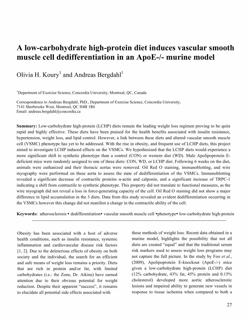
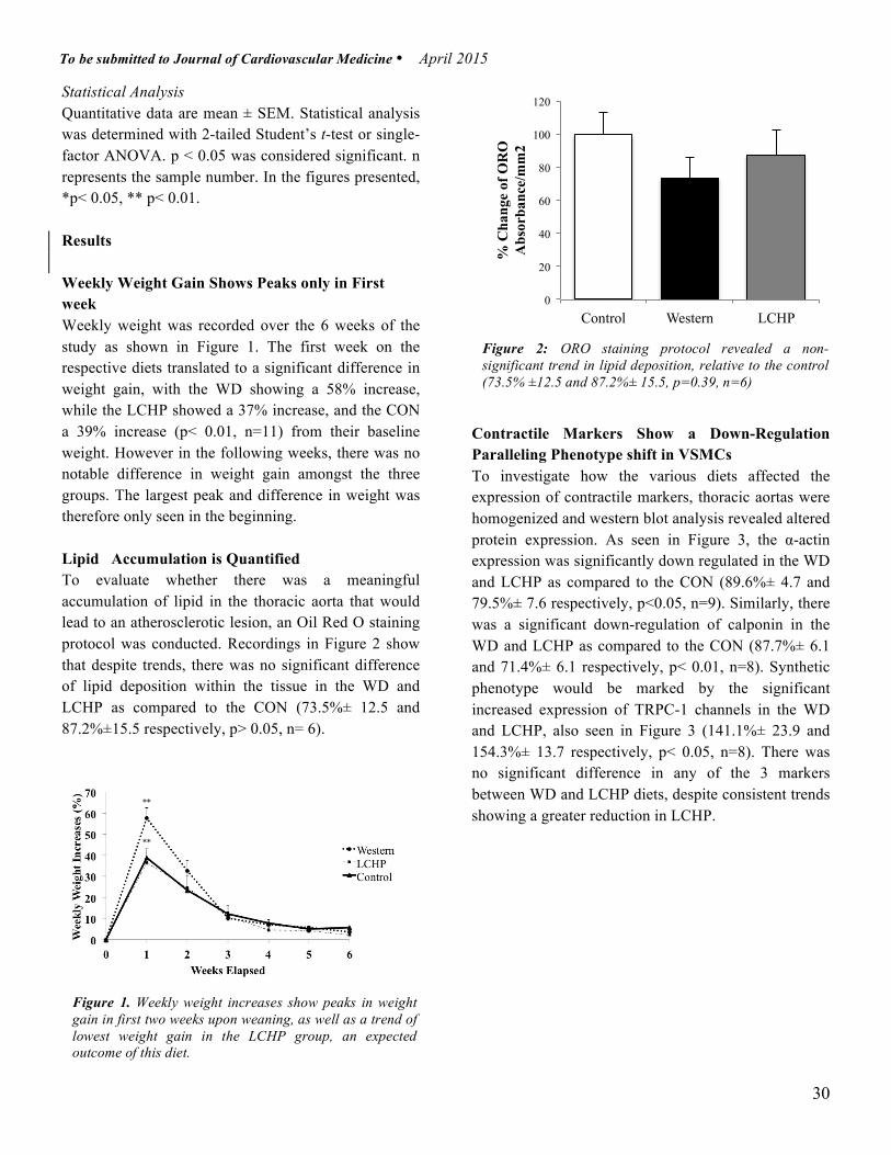
Results Weekly Weight Gain Shows Peaks only in First week Weekly weight was recorded over the 6 weeks of the study as shown in Figure 1. The first week on the respective diets translated to a significant difference in weight gain, with the WD showing a 58% increase, while the LCHP showed a 37% increase, and the CON a 39% increase (p< 0.01, n=11) from their baseline weight. However in the following weeks, there was no notable difference in weight gain amongst the three groups. The largest peak and difference in weight was therefore only seen in the beginning. Lipid Accumulation is Quantified To evaluate whether there was a meaningful accumulation of lipid in the thoracic aorta that would lead to an atherosclerotic lesion, an Oil Red O staining protocol was conducted. Recordings in Figure 2 show that despite trends, there was no significant difference of lipid deposition within the tissue in the WD and LCHP as compared to the CON (73.5%± 12.5 and 87.2%±15.5 respectively, p> 0.05, n= 6).
Contractile Markers Show a Down-Regulation Paralleling Phenotype shift in VSMCs To investigate how the various diets affected the expression of contractile markers, thoracic aortas were homogenized and western blot analysis revealed altered protein expression. As seen in Figure 3, the α-actin expression was significantly down regulated in the WD and LCHP as compared to the CON (89.6%± 4.7 and 79.5%± 7.6 respectively, p<0.05, n=9). Similarly, there was a significant down-regulation of calponin in the WD and LCHP as compared to the CON (87.7%± 6.1 and 71.4%± 6.1 respectively, p< 0.01, n=8). Synthetic phenotype would be marked by the significant increased expression of TRPC-1 channels in the WD and LCHP, also seen in Figure 3 (141.1%± 23.9 and 154.3%± 13.7 respectively, p< 0.05, n=8). There was no significant difference in any of the 3 markers between WD and LCHP diets, despite consistent trends showing a greater reduction in LCHP.
0
20
40
60
80
100
120
Control Western LCHP
% C
hang
e of
OR
O
Abs
orba
nce/
mm
2
Figure 1. Weekly weight increases show peaks in weight gain in first two weeks upon weaning, as well as a trend of lowest weight gain in the LCHP group, an expected outcome of this diet.
Figure 2: ORO staining protocol revealed a non-significant trend in lipid deposition, relative to the control (73.5% ±12.5 and 87.2%± 15.5, p=0.39, n=6)
To be submitted to Journal of Cardiovascular Medicine!! April 2015
!!**!
!!**!
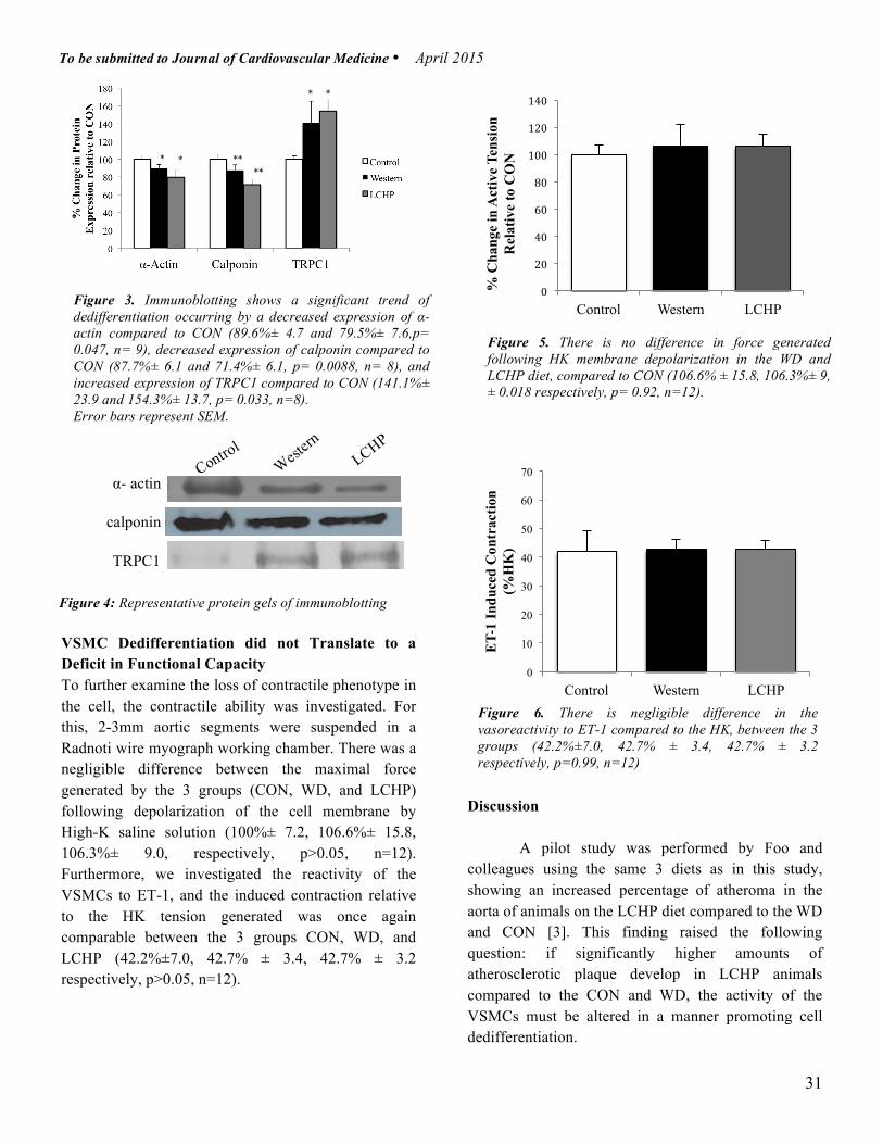
! 31
VSMC Dedifferentiation did not Translate to a Deficit in Functional Capacity To further examine the loss of contractile phenotype in the cell, the contractile ability was investigated. For this, 2-3mm aortic segments were suspended in a Radnoti wire myograph working chamber. There was a negligible difference between the maximal force generated by the 3 groups (CON, WD, and LCHP) following depolarization of the cell membrane by High-K saline solution (100%± 7.2, 106.6%± 15.8, 106.3%± 9.0, respectively, p>0.05, n=12). Furthermore, we investigated the reactivity of the VSMCs to ET-1, and the induced contraction relative to the HK tension generated was once again comparable between the 3 groups CON, WD, and LCHP (42.2%±7.0, 42.7% ± 3.4, 42.7% ± 3.2 respectively, p>0.05, n=12).
!
!!!!!Discussion
A pilot study was performed by Foo and colleagues using the same 3 diets as in this study, showing an increased percentage of atheroma in the aorta of animals on the LCHP diet compared to the WD and CON [3]. This finding raised the following question: if significantly higher amounts of atherosclerotic plaque develop in LCHP animals compared to the CON and WD, the activity of the VSMCs must be altered in a manner promoting cell dedifferentiation.
0!
20!
40!
60!
80!
100!
120!
140!
Control Western LCHP
% C
hang
e in
Act
ive
Tens
ion
Rel
ativ
e to
CO
N
0
10
20
30
40
50
60
70
Control Western LCHP
ET-
1 In
duce
d C
ontr
actio
n (%
HK
)
Figure 3. Immunoblotting shows a significant trend of dedifferentiation occurring by a decreased expression of α-actin compared to CON (89.6%± 4.7 and 79.5%± 7.6,p= 0.047, n= 9), decreased expression of calponin compared to CON (87.7%± 6.1 and 71.4%± 6.1, p= 0.0088, n= 8), and increased expression of TRPC1 compared to CON (141.1%± 23.9 and 154.3%± 13.7, p= 0.033, n=8). Error bars represent SEM.
Control
West
ern
LCHP
α- actin
calponin
TRPC1
Figure 5. There is no difference in force generated following HK membrane depolarization in the WD and LCHP diet, compared to CON (106.6% ± 15.8, 106.3%± 9, ± 0.018 respectively, p= 0.92, n=12). Error bars represent SEM.
Figure 6. There is negligible difference in the vasoreactivity to ET-1 compared to the HK, between the 3 groups (42.2%±7.0, 42.7% ± 3.4, 42.7% ± 3.2 respectively, p=0.99, n=12) Error bars represent SEM.
Figure 4: Representative protein gels of immunoblotting
To be submitted to Journal of Cardiovascular Medicine!! April 2015
!!!!*!!
!!!!!!!!*!!
!!!!!!!!**!!
!!!!!!!!!!!!!!!!!!!!!!!**!!
!!!!!!*!!!!!*!!

! 32
The main finding from this study was the evident shift of phenotype from contractile to synthetic within the VSMCs. Evaluating the state of the mature vessel depends on the relative presence of a variety of contractile apparatus proteins such as SM α-actin [26-28] and calponin [29-31], amongst others. It is evident that α-actin plays a vital role in cell contraction and is required for force-generating capacity of the VSMC [9, 32, 33] In murine aortic SM, 94% of the SM-actin is found in the α-type [34]. Similarly α-actin comprises >25% of the total protein in differentiated VSMCs proving to be the single most abundant protein in these matured cells [34-36]. It is well accepted that this predominant form SM α-actin, is greatly reduced following endothelial injury, and could represent as low as only 10% of the total SM-actin protein, formerly 94% [26, 37]. Our data demonstrated an approximate 20% reduction of α-actin in the LCHP from the control group, and 10% reduction in the WD. In cell differentiation, SM α-actin gene is activated early in embryonic development, and expressed early in the SM lineage [36, 38-42].
Another traditional contractile marker is calponin, first discovered in 1986 by Takahashi and colleagues [43]. Calponin, a calcium regulatory protein, is involved in regulation of SM contraction [10]. Calponin interacts with actin and tropomyosin in a Ca2+-independent manner, and calmodulin in a Ca2+-dependent manner [43, 44]. The amounts of calponin were decreased by approximately 30% in the LCHP and around 12% in the WD relative to the control group, conceptualizing the idea of loss of contractile property. Calponin is known to be expressed as an intermediate marker in the differentiation and dedifferentiation of SMC [29].
In addition to contractile proteins to mark an altered phenotype, other changes such as membrane properties, receptor population, and Ca2+ control might ensue prior to phenotype modulation [45]. Ca2+
handling and manipulation can have a gross impact on resultant cellular contraction, wherein addition of extracellular Ca2+ channels can have a profound proliferative effect [46, 47]. T-type calcium channels, more specifically the TRPC-1 type [48, 49], have been linked to store-operated Ca2+ entry (SOCE), which has been found to be up-regulated in proliferating SMCs of
the pulmonary artery [50]. Kumar and colleagues established that calcium channel TRPC-1 is commonly increased in blood vessels as a compensatory protective response following vascular injury [51]. This fact parallels the shift to synthetic previously described, since there was a 54% increase of TRPC1 in the LCHP and 41% increase in the WD compared to the CON group.
Since we observed a modulation of contractile proteins in the VSMCs, we questioned whether this alteration was provoked by an increase in lipid or some other endogenous factor. Although atherosclerosis is now understood to be more of an inflammatory disease, rather than a lipid-burdened disease [52], we nevertheless wanted to assess whether an excess of lipid may have played a role. An ORO staining protocol was used to quantify the concentration of lipid residing in the thoracic aorta. Interestingly enough, there was less lipid content in the WD and LCHP compared to the CON, however these trends were not significant.
Following evidence of a phenotype shift, the functional capacity of the cell was examined by means of wire myography. Whereby the pressure myography is favorable for small vessels with significant vasoreactivity, the wire myograph is a sensitive method preferred for larger vessels and records the tension produced under isometric conditions [53]. The aorta was sectioned and tested using High-K+ saline and endothelin-1 (ET-1) solution, predicted to demonstrate a decrease in tension or force produced. ET-1 is a potent vasoconstrictor and pro-inflammatory peptide [54]. Contrary to our expected outcome, the data revealed no difference in either case of induced contraction from the High-K solution or vasoconstrictor ET-1. The explanation for this observation may be two-fold. From one perspective, the 6-week length of study may have been premature to witness an impairment of vascular contractility. Contractile proteins α-actin and calponin are early and intermediate markers of dedifferentiation respectively, and only provide grounds to confirm events occurring at a certain point in time: early to intermediate dedifferentiation [29, 38]. Perhaps, functional capacity is only affected during late dedifferentiation. Lesions containing foam cells and VSMCs are typically seen at
To be submitted to Journal of Cardiovascular Medicine!! April 2015

! 33
8-10 weeks old in an ApoE -/- mouse given normal chow [55, 56] however, we noted a change in VSMC after 6 weeks of diet (approximately 9 weeks old). Fibrous plaques tend to appear at 15-20 weeks of age [55, 56]. It may have been an option to extend the 6 weeks, and see if the increased time translated to a subsequent loss in contractile response. On the other hand, it could be speculated that the thoracic aorta may never exhibit impressive contraction, as it is not physiologically responsible for maintenance of vascular tone and high vasoreactivity. Nonetheless, this is not a sound argument since there are studies that investigate vascular contractility in the aorta [36, 57]. Complete loss in contractile response following depolarization with KCl is possible, and has been shown using control and α-actin null mice [36], displaying a fundamental dysfunction in the contractile system with alteration in SM α-actin genes.
Data from this study show that a particular diet low in carbohydrates and high in protein, offers an imbalanced environment that favors a synthetic phenotype in VSMCs. Macronutrient composition is altered in a LCHP diet, consequently affecting the fuel sources and cellular metabolism. Although the same energy sources are utilized, the majority of the energy is now derived from fatty acids and ketones, hence an ‘adipocentric’ energy source as opposed to a ‘glucocentric’ source [58]. LCHP diets are frequently used for rapid and efficient weight loss, and this is conceptualized by the fact that LCHP animals did not gain as much weight as the WD (37% and 58%, respectively) in the beginning weeks, as seen in Figure 1. In a LCHP diet, approximately 70% of caloric requirements come from fatty acids via dietary fat or lipolysis, 20% come from ketone bodies acetoacetate and β-hydroxybutyrate, and 10% from glucose via gluconeogenesis or glycogenolysis [59]. This adaptation is of no surprise; it is thought that a substrate or by-product of using such energy sources plays an indirect role in mediating phenotype shift in VSMCs.
Platelet-derived growth factor-BB (PDGF-BB) is well documented as being responsible for some phenotype regulation, since it is a chemotactic agent meaning, a molecule that favors migration and proliferation [60-63]. Although there is a link with
PDGF and suppressed marker genes, another very influential factor is the gene known for maintenance of pluripotent embryonic stem cells: Kruppel-like Factor 4 (KLF4) [64]. Not typically expressed in adult mesenchymal cells [65], KLF4 becomes re-expressed and plays a role in mediating SMC phenotype switching in vivo [60, 66], since it strongly represses expression of SMC genes [60]. Following vascular injury, ApoE -/- mice on a WD showed increased expression of KLF4 [67, 68] and this abundance of KLF4 is accompanied by an inhibition of expression of known SMC marker genes [69]. KLF4 also has the ability to inhibit the increase of SMC differentiation mediated by TGF-β [14]. Taken together, this provides reason to speculate whether KLF4 can serve as a plausible endogenous factor indirectly involved in the VSMC activity through growth factors however, this theory lies beyond the scope of this study.
Conclusion
This study aimed to assess the effects of a low-carbohydrate high-protein diet on the role of vascular smooth muscle cells in atherosclerosis. Prior to our study, a possible link between VSMC phenotype and LCHP diets had never been made. This study emphasized how such a topic does indeed merit further enquiry, as there is a clear dedifferentiation occurring in VSMCs of ApoE -/- mice on a LCHP diet, potentially providing intriguing or relevant data for clinicians and patients both interested in this type of diet. !References
1. 1. Grundy SM (2004) Atherosclerosis imaging and the future of lipid management. Circulation. Vol 110(23): 3509-3511.
2. Dandona P, Aljada A, Chaudhuri A, et al. (2005) Metabolic syndrome: a comprehensive perspective based on interactions between obesity, diabetes, and inflammation. Circulation. Vol 111(11) 1448-1454.
3. Foo SY, Heller ER, Wykrzykowska J, Sullivan CJ, Manning-Tobin JJ, Moore KJ, […] Rosenzweig A (2009) Vascular effects of a low-carbohydrate high-protein diet. Procl Nat Acad Sci. Vol 106(36)15418-15423.
To be submitted to Journal of Cardiovascular Medicine!! April 2015
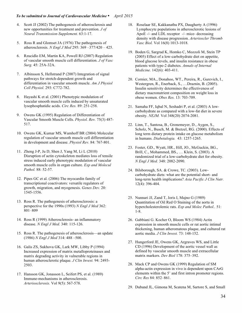
! 34
4. Scott JJ (2002) The pathogenesis of atherosclerosis and new opportunities for treatment and prevention. J of Neural Transmission-Supplement. 63:1-17.
5. Ross R and Glomset JA (1976) The pathogenesis of
atherosclerosis. N Engl J Med 295: 369 –377/420 – 425.
6. Rzucidlo EM, Martin KA, Powell RJ (2007) Regulation of vascular smooth muscle cell differentiation. J of Vasc Surg. 45: 25A-32A.
7. Albinsson S, Hellstrand P (2007) Integration of signal
pathways for stretch-dependent growth and differentiation in vascular smooth muscle. Am J Physiol Cell Physiol. 293: C772-782.
8. Hayashi K et al. (2001) Phenotypic modulation of
vascular smooth muscle cells induced by unsaturated lysophosphatidic acids. Circ Res. 89: 251-258.
9. Owens GK (1995) Regulation of Differentiation of
Vascular Smooth Muscle Cells. Physiol. Rev. 75(3) 487-517.
10. Owens GK, Kumar MS, Wamhoff BR (2004) Molecular
regulation of vascular smooth muscle cell differentiation in development and disease. Physiol Rev. 84: 767-801.
11. Zheng J-P, Ju D, Shen J, Yang M, Li L (2010)
Disruption of actin cytoskeleton mediates loss of tensile stress induced early phenotypic modulation of vascular smooth muscle cells in organ culture. Exp and Molecul Pathol. 88: 52-57.
12. Pipes GC et al. (2006) The myocardin family of
transcriptional coactivators: versatile regulators of growth, migration, and myogenesis. Genes Dev. 20: 1545-1556.
13. Ross R. The pathogenesis of atherosclerosis: a
perspective for the 1990s (1993) N Engl J Med 362: 801–809
14. Ross R (1999) Atherosclerosis- an inflammatory disease. N Engl J Med. 340: 115-126.
15. Ross R. The pathogenesis of atherosclerosis—an update (1986) N Engl J Med 314: 488 –500.
16. Galis ZS, Sukhova GK, Lark MW, Libby P (1994) Increased expression of matrix metalloproteinases and matrix degrading activity in vulnerable regions in human atherosclerotic plaque. J Clin Invest. 94: 2493- 2503.
17. Hansson GK, Jonasson L, Seifert PS, et al. (1989) Immune-mechanisms in atherosclerosis. Arteriosclerosis. Vol 9(5): 567-578.
18. Roselaar SE, Kakkanathu PX, Daugherty A (1996) Lymphocyte populations in atherosclerotic lesions of ApoE -/- and LDL receptor -/- mice- decreasing density with disease progression. Arterioscler Thromb Vasc Biol. Vol 16(8) 1013-1018.
19. Boden G, Sargrad K, Homko C, Mozzoli M, Stein TP
(2005) Effect of a low-carbohydrate diet on appetite, blood glucose levels, and insulin resistance in obese patients with type-2 diabetes. Annals of Internal Medicine. 142(6): 403-411.
20. Cornier, MA., Donahoo, WT., Pereira, R., Gurevich, I., Westergren, R., Enerback, S., …Draznin, B. (2005). Insulin sensitivity determines the effectiveness of dietary macronutrient composition on weight loss in obese women. Obes Res. 13: 703-709.
21. Samaha FF, Iqbal N, Seshadri P, et al. (2003) A low-
carbohydrate as compared with a low-fat diet in severe obesity. NEJM. Vol 348(20) 2074-2081.
22. Linn, T., Santosa, B., Gronemeyer, D., Aygen, S., Scholz, N., Busch, M. & Bretzel, RG. (2000). Effects of long term dietary protein intake on glucose metabolism in humans. Diabetalogia. 43: 1257-1265.
23. Foster, GD., Wyatt, HR., Hill, JO., McGuckin, BG., Brill, C., Mohammed, BS., … Klein, S. (2003). A randomized trial of a low-carbohydrate diet for obesity. N Engl J Med. 348: 2082-2090.
24. Bilsborough, SA. & Crowe, TC. (2003). Low-carbohydrate diets: what are the potential short- and long-term health implications? Asia Pacific J Clin Nutr. 12(4): 396-404.
25. Nunnari JJ, Zand T, Joris I, Majno G (1989) Quantitation of Oil Red O Staining of the aorta in hypercholesterolemic rats. Exp and Molec Pathol.. 51: 1-8.
26. Gabbiani G. Kocher O, Bloom WS (1984) Actin expression in smooth muscle cells or rat aortic intimal thickening, human atheromatous plaque, and cultured rat aortic media. J Clin Invest. 73: 148-152.
27. Hungerford JE, Owens GK, Argraves WS, and Little CD (1996) Development of the aortic vessel wall as defined by vascular smooth muscle and extracellular matrix markers. Dev Biol 178: 375–392.
28. Mack CP and Owens GK (1999) Regulation of SM alpha-actin expression in vivo is dependent upon CArG elements within the 5’ and first intron promoter regions. Circ Res 84: 852–861.
29. Duband JL, Gimona M, Scatena M, Sartore S, and Small
To be submitted to Journal of Cardiovascular Medicine!! April 2015
! 35
JV (1993) Calponin and SM 22 as differentiation markers of smooth muscle: spatiotemporal distribution during avian embryonic development. Differentiation 55: 1–11.
30. Miano JM, Cserjesi P, Ligon K, Perisamy M, and Olson EN (1994) Smooth muscle myosin heavy chain marks exclusively the smooth muscle lineage during mouse embryogenesis. Circ Res 75: 803– 812.
31. Strasser P, Gimona M, Moessler H, Herzog M, and Small JV (1993) Mammalian calponin: identification and expression of genetic variants. FEBS Lett 330: 13–18.
32. Kim HR, Gallant C, Leavis PC, Gunst SJ, Morgan KG (2008) Cytoskeletal remodeling in differential vascular smooth muscle is actin isoform dependent and stimulus dependent. Am J Physiol Cell Physiol. 295:C768-C778.
33. Owens GK, Thompson MM (1986) Developmental changes in isoactin expression in rat aortic smooth muscle cells in vivo. Relationship between growth and cytodifferentiation. J Biol Chem. 261(28): 13373-13380.
34. Vandekerckhove J and Weber K (1981) Actin typing on total cellular extracts. Eur J Biochem. 113: 595-603.
35. Fatigati V and Murphy RA (1984) Actin and tropomyosin variants in smooth muscles: dependence on tissue type. J Biol. Chem. 259:14383-14388.
36. Schildmeyer LA, Braun R, Taffet G, Debiasi M, Burns AE, Bradley A, Schwartz RJ (2000) Impaired vascular contractility and blood pressure homeostasis in the smooth muscle α-actin null mouse. FASEB Journal. 14: 2213-2220.
37. Kocher O, Skalli O, Bloom WS, Gabbiani G (1984) Cytoskeleton of rat aortic smooth muscle cells. Normal conditions and experimental intimal thickening. Lab Invest. 50(6): 645-652.
38. Owens GK, Loeb A, Gordon D, Thompson MM (1986) Expression of smooth muscle specific α-isoactin in cultured vascular smooth muscle cells: relationship between growth and cytodifferentiation. J Cell Biol. 102(2): 343-352.
39. Ruzicka DL & Schwartz RJ (1988) Sequential activation of α-actin genes during avian cardiogenesis: vascular smooth muscle α-actin gene transcripts mark the onset of cardiomyocyte differentiation. J. Cell. Biol. 107: 2575-2586.
40. McHugh KM, Crawford K, Lessard JL (1991) A comprehensive analysis of the developmental and tissue specific expression of the isoactin multigene family. Dev. Biol. 148: 442-458.
41. Sugi Y and Lough J (1992) Onset of expression and
regional deposition of alpha-smooth and sarcometric actin during avian heart development. Dev. Dynamics. 193: 116-124.
42. Babai F, Musevi-Aghdam J, Schurch W, Royal A, Gabbiani G (1990) Coexpression of α-sarcometric actin, α-smooth muscle actin and desmin during myogenesis in rat and mouse embryos, in skeletal muscle. Differentiation. 44:132-142..
43. Takahashi K, Hiwada K, Kokubu T (1986) Isolation and characterization of a 34000Da calmodulin- and F-actin binding protein from chicken gizzard smooth muscle. Biochem Biophys Res Commun. 141:20-26.
44. Winder SJ & Walsh MP (1990) Smooth muscle calponin. Inhibition of actomyosin MgATPase and regulation by phosphorylation. J Biolog Chem. Vol 265(17): 10148-10155.
45. Bergdahl A, Gomez MF, Wihlborg A-K, Erlinge D, Eyjolfson A, Shang-Zhong X, Beech DJ, Dreja K, Hellstrand P (2005) Plasticity of TRPC expression in arterial smooth muscle: correlation with store-operated Ca2+ entry. Am J Physiol Cell Physiol. 288: C872-880.
46. Magnier-Gaubil C, Hebert JM, Quarck R, Papp B, Corvazier E, Wuytack F, Levy-Toledano S, Enouf J (1996) Smooth muscle cell cycle and proliferation. Relationship between calcium influx and sarco-endoplasmic reticulum Ca2+ ATPase regulation. J Biol Chem. 271:27788-27794.
47. Munaron L, Antoniotti S, Fiorio Pla A, Lovisolo D (2004) Blocking Ca2+ entry: a way to control cell proliferation. Curr Med Chem. 11: 1533- 1543.
48. Wes PD, Chevesich J, Jeromin A, Rosenberg C, Stetten G, Montell C (1995) TRPC1, a human homolog of Drosophila store-operated channel. Proc Natl Acad Sci USA. 92: 9652-9656.
49. Xu SZ, Beech DJ (2001) TRPC1 is a membrane-spanning subunit of store-operated Ca2+ channels in native vascular smooth muscle cells. Circ Res. 88:84-87.
50. Golovina VA, Platoshyn O, Bailey CI, Wang J, Limsuwan A, Sweeney M, Rubin LJ, Yuan JX (2001) Upregulated TRP and enhanced capacitative Ca2+ entry in human pulmonary artery myocytes during proliferation. Am J Physiol Heart Circ Physiol. 280: H746-H755.
51. Kumar B, Dreja K, Shah SS, Cheong A, Xu S-Z, Sukumar P […] Beech DJ (2006) Upregulated TRPC1 channel in vascular injury in vivo and its role in human
To be submitted to Journal of Cardiovascular Medicine!! April 2015

! 36
neointimal hyperplasia. Circ Res. 98: 557-563.
52. Libby P, Ridker PM, Maseri A (2002) Inflammation and atherosclerosis. Circulation. 105:1135-1143.
53. Lu X & Kassab GS (2007) Vasoreactivity of blood vessels using a novel isovolumic myograph. An Biomed Eng. 35: 356-366.
54. Bohm F & Pernow J (2007) The importance of endothelin-1 for vascular dysfunction in cardiovascular disease. Cardiovascular Research. 76: 8-18.
55. Plump AS, Smith JD, Hayek T, Aalto-Setala K, Walsh A, Verstuyft JG, Rubin EM, Breslow JL (1992) Severe hypercholesterolemia and atherosclerosis in apolipoprotein A-deficient mice created by homologous recombination in ES cells. Cell. 71(2) 343-353.
56. Nakashima Y, Plump AS, Raines EW, Breslow JL, Ross R (1994) ApoE-deficient mice develop lesions of all phases of atherosclerosis throughout the arterial tree. Arterioscler Thromb. 14(1) 133-140.
57. Moltzer E, Riet L, Swagemakers SMA, Heijningen PM, Vermeij M, van Vegnel R […] Essers J (2011) Impaired vascular contractility and aortic wall degeneration in Fibulin-4 deficient mice: effect of angiotensin II type 1 (AT1) receptor blockade. PLOS one. Vol 6(8): 1-12.
58. Westman EC, Mavropoulos J, Yancy WS, Volek JS (2003) A review of low-carbohydrate ketogenic diets. Current Atherosclerosis Reports. 5:476-483.
59. Westman EC, Feinman RD, Mavropoulos JC, Vernon MC, Volek JS, Wortman JA, Yancy WS, Phinney SD (2007) Low-carbohydrate nutrition and metabolism. Am J Clin Nutr. 86:276-284.
60. Yan F-F, Liu Y-F, Liu Y, Zhao Z-X (2007) KLF4: a novel target for the treatment of atherosclerosis. Medical Hypotheses. 70: 845-847.
61. Berman, JW, Kazimi, M., Ma, H. (2002). Development of the Atherosclerotic Plaque. In D.L. Brown (Ed.), Cardiovascular Plaque Rupture (pp. 1-50). New York, NY: Marcel Dekker.
62. Holycross BJ, Blank RS, Thompson MM, Peach MJ, and Owens GK (1992) Platelet-derived growth factor-BB-induced suppression of smooth muscle cell differentiation. Circ Res 71: 1525–1532.
63. Wang Z, Wang DZ, Hockemeyer D, McAnallyJ, Norheim A, Olsen EN (2004) Myocardin and ternary complex factors compete for SRF to control smooth muscle gene expression. Nature. 428: 185-189.
64. Jiang J, Chan YS, Loh YH, Cai J, Tong GQ, Lim CA, et al. (2008) A core Klf circuitry regulates self-renewal of
embryonic stem cells. Nat Cell Biol. 10:353-360.
65. Gimbrone MA, Topper JN, Nagel T, et al. (2000) Endothelial dysfunction, hemodynamic forces, and atherogenesis. Ann NY Acad Sci. 902:230-239.
66. Gomez D and Owens GK (2012) Smooth muscle cell phenotypic switching in atherosclerosis. Cardiovascular Research. 95:156-164.
67. Cherepanova OA, Pidkovka NA, Yoshida T, Gan Q,
Adiguzel E, Bendeck MP, et al. (2009) Oxidized phospholipids induce type VIII collagen expression and vascular smooth muscle cell migration. Circ Res. 104: 609-618.
68. Yoshida T, Kaestner KH, Owens GK (2008) Conditional deletion of Kruppel-like factor 4 delays downregulation of smooth muscle cell differentiation markers but accelerates neointimal formation following vascular injury. Circ Res. 102: 1548-1557.
69. Liu Y, Sinha S, McDonald OG, Shang Y, Hoofnagle MH, Owens GK (2005) Kruppel-like factor 4 abrogates myocardin-induced activation of smooth muscle gene expression. J Biol Chem. 280:9719-9727.
!
To be submitted to Journal of Cardiovascular Medicine!! April 2015

! 37
IV Altered mitochondrial functioning in apolipoprotein E-deficient mice induced by a low-carbohydrate high-protein diet Olivia Hanna Koury*, Cynthia Rocha*, Andreas Bergdahl !!!!!!!!!!!!!!!!!!!!!!!!!!!!!!!!!

! 38
! Contribution of Authors *Olivia Hanna Koury: animal handling, surgeries, immunoblotting, analysis of data, preparation of manuscript. *Cynthia Rocha: high-resolution respirometry measurements, analysis of data, preparation of manuscript. Andreas Berdgahl: animal handling, concept development, student supervision, statistics, analysis of data, preparation of manuscript. !!* Both primary authorship

! 39
The recent rise in obesity has been associated with a number of adverse health effects such as heart disease, certain types of cancer, type-2 diabetes mellitus, and respiratory complications [1]. In efforts to reduce associated health risks, overweight and obese patients have been advised to maintain a healthy BMI. One of the most efficient means of weight loss is through dieting, more specifically a low-carbohydrate high-protein diet (LCHP). This diet has gained much popularity with the American public [2], and advocates claim it ‘burns more fat’ and leads to quicker weight
loss without adverse long-term effects [3]. Macronutrient composition is altered in a LCHP diet, drastically affecting the cellular metabolism. Although the same energy sources are utilized, majority of the energy is now derived from fatty acids and ketones due to the extreme reduction of carbohydrate. This is shifts metabolism from a ‘glucocentric’ source to an ‘adipocentric’ energy source [4]. In LCHP metabolism, 70% of caloric requirements come from fatty acids via dietary fat or lipolysis, 20% come from ketone bodies acetoacetate and β-hydroxybutyrate, and 10% from
Altered mitochondrial functioning in apolipoprotein E-deficient mice induced by a low-carbohydrate high-protein diet Olivia H. Koury1*, Cynthia Rocha1*, and Andreas Bergdahl
1Department of Exercise Science, Concordia University, Montreal, QC, Canada *Equal contribution towards manuscript Correspondence to Andreas Bergdahl, PhD., Department of Exercise Science, Concordia University, 7141 Sherbrooke West, Montreal, QC H4B 1R6 Email: [email protected] ! Summary: Low-carbohydrate high-protein (LCHP) diets have been growing in popularity in America for decades, proving to be one of the most popular and efficient methods of weight loss. Despite the superficial health benefits this type of diet may contribute in the case insulin resistance, hypertension, and lipid control for example, its implications on mitochondrial oxidative capacity and respiration have yet to be evaluated. The goal of this study was to evaluate the safety of LCHP diets by determining whether they foster an environment altering mitochondrial functioning. Male Apolipoprotein E-deficient mice were randomly assigned to either a control diet or low-carbohydrate high-protein diet for 6 weeks. Heart tissue was used for immunoblotting, and high-resolution respirometry (HRR) to quantify mitochondrial complexes and measure oxidative phosphorylation (OXPHOS) capacity. Our results indicate increased expression of all five mitochondrial complexes in the LCHP group compared to the control. Furthermore, HRR revealed increased efficiency of substrate utilization implying improved mitochondrial respiration. These findings correlate well with the understanding that cardiomyocytes prefer lipid as a fuel source, and the shift in metabolism caused by a LCHP diet provides such a favourable environment. Keywords: Apolipoprotein E!! cardiac mitochondria! oxidative capacity !bioenergetics! low-carbohydrate high-protein

! 40
glucose via gluconeogenesis or glycogenolysis [5]. LCHP diets have been studied extensively in the past decades regarding weight loss, lipid markers, insulin sensitivity, hypertension, and endothelial dysfunction [6-11]. However, no study has addressed a possible link with LCHP diets and heart failure due to cardiac mitochondrial dysfunction. Despite a general decrease in the number of deaths from cardiovascular disease (CVD) in the United States, congestive heart failure (CHF) remains as prevalent today as it was two decades ago [12]. Although very broadly defined, CHF can be best described as a multifactorial syndrome that renders the myocardium unable to pump blood efficiently to sustain demand [13]. Oxidative stress is now understood to be a key factor in heart failure, and a main reason for oxidative stress is the imbalance and negative impact of reactive oxygen species (ROS) produced by the mitochondria [14-18]. In recent years, studies have linked characteristics of heart failure to defective mitochondrial energetics and abnormal substrate metabolism [19- 21]. There is significant evidence showing mitochondrial dysfunction not only in cardiomyocytes, but also skeletal muscle of heart failure patients [22]. Aside from mitochondrial dysfunction, other problems that may contribute to the noticeable energetic defects are the number of mitochondria, substrate of choice, or oxidative capacity [23]. These facts imply that preserving cardiac mitochondrial functioning can become a future therapeutic aim for CHF [24-26].
Mitochondria are potentially significant sources of ROS especially when damaged (i.e. by long-term FA accumulation,) where they exhibit decreased oxidative based energy and thus less fuel for cells [27]. This is a central concern since the mitochondrion is the organelle in which energy (ATP) production and cellular respiration occur [28, 29]. Considering the maintenance of ATP drives all cellular processes, this process is imperative for a high-level workload organ such as the heart due to its high-energy demands [25, 30-33]. The capacity of the mitochondria relies heavily on substrate availability, notably lipids and carbohydrates. These substrates contribute to energy metabolism depending on their utilization through β-oxidation and the Kreb’s cycle. β-oxidation produces
acetyl-CoA from FAs that enters the Kreb’s cycle, generating electrons that are successively transported to the electron transport system (ETS) by NADH and FADH2 to amplify ATP production [34]. The Kreb’s cycle is the focal common aerobic metabolic pathway for carbohydrates, lipids and proteins [35]. Acetyl-CoA oxidation by this cycle is responsible for two thirds of the sum of ATP production and oxygen consumption [36].
Despite glucose and FAs both being oxidized in the mitochondria to ATP production in the cardiomyocytes, the latter is the preferred substrate (generating 70% of total ATP) [37-39]. Therefore, the uptake of FA from the plasma is vital for cardiac viability [21, 37- 40]. Furthermore, lipids have also been deemed necessary for the maintenance and re-initiation involved with the pure beating of heart cells, further highlighting their significance [41]. The heart has limited substrate storage capacity, thus the uptake of nutrients needs to be finely balanced since the underlying pathways have to respond both appropriately and competently to the continuous flux in energy demand and substrate availability [25]. Under normal conditions the mitochondria maintain a fine equilibrium between glucose and FAs, however, during physiological stress, the mitochondria shifts towards an increased carbohydrate metabolism. This shift adds stress to the heart and shunts the lipids into non-oxidative pathways creating more ROS than energy [42]. The activity of respiratory complex I in the mitochondria is especially prone to the effects of oxidative damage [43]. The same detrimental phenomenon occurs when there is a reduced oxygen supply as a result of the occlusion of the coronary vessels, a major cause of cardiovascular distress [44]. The mitochondria are an important health research target considering their role in generating cellular energy, by means of oxidative phosphorylation, a process affected by uptake of cholesterol and lipids from the plasma. The subsequently altered role of mitochondria may potentially be implicated in the pathophysiology of heart failure [45] and overall cardiovascular health. The apolipoprotein E deficient (ApoE-/-) murine model is a well-established model as it allows for the investigation of atherosclerosis due to its susceptibility to develop lesions rapidly, and
To be submitted to Journal of Lipid Research ! April 2015
! 41
similarly to humans [46-48]. ApoE is a 34 kDa glycoprotein made in the liver and brain, responsible for lipoprotein metabolism [48]. It helps to clear circulating cholesterol by mediating the binding of ApoE-containing lipoproteins and LDL receptors [49- 52].
This study examined whether ApoE-/- mice exhibit altered cardiac mitochondrial oxidative capacity when exposed to a low-carbohydrate high-protein environment. We quantified cardiac mitochondrial consumption in a 6-week old ApoE-/- murine model in comparison to age-matched controls. The results determined if alterations in energy metabolism had occurred by examining the oxidative capacity of the mitochondrial respiratory complexes. We hypothesized that the mitochondria from the ApoE-/- cardiac tissue would experience increased respiratory function and disturbed energy metabolism as compared to controls. Methods Animal Care
Apolipoprotein E knockout (ApoE-/-) mice were obtained from Jackson Laboratories (Bar Harbor, Maine, USA) and used for breeding. The resulting litters were weaned and separated based on sex at 21-28 days. Only the tissue from male mice was used for the purpose of this study. The males were housed individually in a thermo-neutral environment (22ºC), on a 12:12 h photoperiod, and randomly assigned to either a control diet (CON) or a low-carbohydrate high-protein (LCHP) diet for 6 weeks. Both groups of mice had access to water and their respective diets ad libitum. All procedures were approved by the Animal Ethics Committee of Concordia University (protocol ID: #30000259) and were conducted in accordance with guidelines of the Canadian Council on Animal Care. Diet Specifications
Both CON and LCHP animal diets were iso-caloric. Table 1 displays the diet specifications in terms of percentage of caloric intake from fat, carbohydrates (CHO) and protein. The CON diet, 5075 Charles River Autoclavable Rodent Diet, reflects a healthy, standard macronutrient distribution. Whereas, the LCHP diet, obtained from Harlan Laboratories (TD.04524)
simulates an Atkin’s diet used for weight loss and contains 43% from fat, 11% CHO, and 46% protein. The LCHP diet is a modification of TD.88137 (Harlan Laboratories), used for studies on atherosclerosis. Experimental protocol
The beating heart was removed immediately after euthanasia with CO2 according to the approved animal protocol and split into two different portions. One portion was snap frozen in liquid nitrogen, and stored at -80ºC for biochemical analysis; the other portion (the apex) was placed in an ice cold relaxing buffer (BIOPS) and used immediately to measure mitochondrial respiration. The BIOPS contains (in mM): CaK2EGTA 2.77, K2EGTA 7.23, Na2ATP 5.77, MgCl2·6H2O 6.56, Taurine 20, Na2Phosphocreatine 15, Imidazole 20 mM, Dithiothreitol 0.5, MES 50, pH 7.1 Preparation of permeabilized cardiac fibers
The apex was dissected for preparation of permeabilized myofibers. This was done by gentle dissection during which the fiber bundles were separated using sharp forceps. The fibers were then incubated in 3 ml BIOPS buffer containing 50 µg/ml saponin for 30 minutes and subsequently washed in ice-cold buffer (MiR05) for 2 x 10 min. MiR05 contains (mM): EGTA 0.5, MgCl2·6H2O 3.0, K-lactonionate 60, Taurine 20, KH2PO4 10, HEPES 20, Sucrose 110, BSA 1g/l, pH 7.1. Mitochondrial respiratory measurements
Measurements of oxygen consumption were performed in MiR05 at 37ºC using a polarographic oxygen sensor (Oxygraph-2k, Oroboros Instruments, Innsbruck, Austria). Approximately 2.0 to 2.5 mg of muscle tissue (wet weight) was place in either chamber in a cross-sectional design. O2 flux was resolved by DatLab and all experiments were carried out in hyperoxygenated levels to avoid O2 diffusion limitations. A sequential substrate addition protocol was used to allow functional dissection of the electron transport system: state 2 respiration (absence of adenylates) was assessed by addition of malate (2 mM) and octanoyl carnitine (1.5 mM), by adding ADP (5 mM) we could reach state 3 respiration for complex I. This was followed by addition of glutamate (10 mM)
To be submitted to Journal of Lipid Research ! April 2015

! 42
and succinate (10 mM) achieving maximal coupled state 3 respiration with parallel electron input to complex I and II. Oligomycin (2 µg/ml) was then added to block complex V and thereafter antimycin A (2.5 µM) to inhibit complex III. Finally ascorbate (2 mM) and TMPD (500 µM) were added to evaluate Complex IV respiration. These mitochondrial respiratory measurements were recorded for both the control group and the experimental group fed the LCHP diet. Mitochondrial Uncoupling
There were 6 different protocols, some of which were variations of one another, used to test for the presence of uncoupling as we wanted to ensure that the results obtained would not be from a potential uncoupling effect. A portion of the apex tissue mentioned previously was also studied using high-resolution respirometry (Oxygraph-2k, Oroboros Instruments, Innsbruck, Austria) to test for uncoupling, running simultaneously along side the mitochondrial respiratory experiments. The first protocol began with the addition of oligomycin (2 µg/ml) followed by succinate (10 mM), then FCCP step titrations (1 µl/step) and finally antimycin A (2.5 µM). The second protocol was the same as the first except GDP (10mM) was added in between the addition of succinate and FCCP. Therefore, uncoupling protein 1 (UCP1)-mediated uncoupling could be studied through the titrations of oligomycin, GDP and antimycin A. The third protocol consisted of the sequential addition of succinate (10 mM), ADP (5 mM) and then glutamate (10 mM) and malate (2 mM) simultaneously. The fourth protocol included the addition of malate (2 mM), octanoyl carnitine (1.5 mM), ADP (5 mM), glutamate (10 mM), oligomycin (2 µg/ml) and then finally multiple additions of GDP (10mM). The fifth protocol began with the addition of malate (2 mM) then octanoyl carnitine (1.5 mM), glutamate (10 mM), oligomycin (2 µg/ml) and again multiple additions of GDP (10mM). The sixth an final protocol consisted of adding malate (2 mM) then octanoyl carnitine (1.5 mM), oligomycin (2 µg/ml) and then multiple additions of GDP (10mM).
Protein Extraction, Immunoblotting, and Immunofluorescence
Tissue from the cross-section excised from right above the apex was pulverized/homogenized using liquid nitrogen with ~150µl lysis buffer containing (in mM) 250 NaCl, 50 HEPES (pH 7.5), 10% glycerol, 1% triton X-100, 1.5 MgCl2 1 EGTA, 10 Na4P2O7 NaF, 800 µM Na3VO4. After 1 h on ice the cell slurry was centrifuged at 13,000 rpm for 10 min, and the supernatant was collected. 10µl of lysate was mixed with 2µl DTT and 2µl sample buffer and loaded on a 12.5% acrylamide-SDS gel followed by a transfer onto a 0.45µm nitrocellulose membrane (162-0115 Bio-Rad) in 10mM sodium tetraborate buffer. Ponceau staining was done as loading control before the membranes were blocked in 3% bovine serum albumin (BSA) in 0.1% Tween in Tris-buffered saline (TBS) (10 mm Tris-HCl, pH 7.5, 150 mm NaCl) for 1 hour at room temperature, followed by overnight incubation at 4°C with primary antibody, total OXPHOS rodent antibody cocktail (1:2000, MS604 MitoSciences). The membranes were washed, and incubated with secondary antibody (1:15000, anti-mouse, ab6728 Abcam). Membranes were exposed with ECL chemiluminescence (Immun-Star Chemiluminescent; 1705070; Bio-Rad) and developed bands were analyzed with Image J Software. Statistical Analysis Summarized data are presented as means ± standard error of the mean (SEM) for immunoblotting, and standard estimate. Statistical comparisons were done using a two-tailed Student’s t-test. n represents the sample number. For all statistical evaluations, In the figures presented, *p< 0.05, ** p< 0.01. Results Relative Expression of Complexes I-V
Immunoblotting revealed an increase in all five complexes in the LCHP diet, normalized to the CON. These trends can be seen in Figure 1 however, only CIV showed a statistically significant difference in expression (CON: 100% ±5.7, LCHP: 122% ±4.8, p< 0.05 n= 6). Therefore, the only significant change in
To be submitted to Journal of Lipid Research ! April 2015

! 43
protein expression was in CIV when running samples from CON and LCHP hearts in parallel.
Mitochondrial respiration We examined oxygen consumption rates of saponin-permeabilized fibres to determine oxidative capacities of mitochondria in the cardiac muscles in situ along with control of the respiratory activity by ADP, the principal regulator. Mitochondrial leak, estimated as antimycin A flux rates subtracted from oligomycin flux rates, indicated a significant increase in the LCHP group as compared to the control group as seen in Figure 3 (52.4 ± 16.1 and 102.2 ± 12.1pmol/s/mg, respectively, p<0.03). There was also a significant difference between these two groups in terms of substrate control ratio for succinate (GM3/GMS3) (0.62 ± 0.08 and 0.44 ± 0.02, respectively, p<0.05) as illustrated in Figure 4. Neither the respiratory control ratio (RCR, state 3 over state 4 respiration so succinate divided by oligomycin respiration) (2.2 ± 0.8 and 3.0 ± 0.6 ) or the acceptor control ratio (ACR, maximal, ADP stimulated respiration divided by basal, ADP restricted respiration in other words ADP divided by malate respiration) (8.5 ± 1.4 and 12.0 ± 1.4), respectively, a ratio representing the degree of coupling between oxidation and phosphorylation showed any significant differences as seen in Figures 5 and 6. Therefore there was no indication of a likely more efficient oxidation
of substrates, nor a significant change within the OXPHOS process. Lipid OXPHOS capacity shown in Figure 7 shows the absence of a significant difference in the lipid coupling control ratio (L/P) between these two same groups (0.33 ± 0.04 and 0.26 ± 0.03). However, there was a significant difference in substrate utilization, as seen in Figure 2. Data show a significant increase in the utilization of octanoyl carnitine (28.3 ± 2.8 and 38.4pmol/s/mg ± 2.9, p<0.03), ADP (75.0 ± 5.6 and 140.0pmol/s/mg ±25.1, p<0.03), glutamate (87.2 ± 5.1 and 161.8pmol/s/mg ± 25.9, p<0.02), succinate (155.5 ± 27.0 and 362.8pmol/s/mg ± 47.9, p<0.01), oligomycin (74.8 ± 14.8 and 122.1pmol/s/mg ± 13.5, p<0.04) and ascorbate+TMPD (212.5 ± 24.8 and 292.9pmol/s/mg ± 19.5, p<0.03) by the LCHP group compared to controls (n=6). Malate and antimycin A utilization were not significant (9.7 ± 1.2 and 11.6pmol/s/mg ± 1.34; 22.4 ± 4.3 and 20.0 pmol/s/mg ± 3.5, respectively). The O2 flux showed an increased response in the LCHP group as the substrates were titrated as compared to controls (data not shown). Residual oxygen consumption (ROX) was similar in both groups (data not shown).
Figure 2: Substrate utilization by the mitochondria of permeabilized cardiac myofibers assessed through high-resolution respirometry. Data show a significant increase in the utilization of octanoyl carnitine (28.3 ± 2.8 and 38.4pmol/s/mg ± 2.9, p<0.03), ADP (75.0 ± 5.6 and 140.0pmol/s/mg ±25.1, p<0.03), glutamate (87.2 ± 5.1 and 161.8pmol/s/mg ± 25.9, p<0.02), succinate (155.5 ± 27.0 and 362.8pmol/s/mg ± 47.9, p<0.01), oligomycin (74.8 ± 14.8 and 122.1pmol/s/mg ± 13.5, p<0.04) and ascorbate+TMPD (212.5 ± 24.8 and 292.9pmol/s/mg ± 19.5, p<0.03) by the LCHP group compared to controls (n=6). Malate and antimycin A utilization were not significant (9.7 ± 1.2 and 11.6pmol/s/mg ± 1.34; 22.4 ± 4.3 and 20.0 pmol/s/mg ± 3.5, respectively). !
Figure 1: Immunoblotting of Complex I to Complex V using total OXPHOS rodent antibody cocktail from MitoSciences (MS604). Trends show an increase in all 5 complexes in the LCHP diet relative to the CON diet (CI 100%± 21.4 and 132.7%± 13.1, CII 100%± 8 and 114.8%± 8.5, CIII 100%± 13.3 and 144.9± 20.8, CIV 100%± 5.7 and 122.1%± 5.7 p<0.02, CV 100%± 8.4 and 118.5%± 7.3, respectively, n=6).
*!
!!!*!
!!*!
!!!!**!
*!
!!*!
To be submitted to Journal of Lipid Research ! April 2015

! 44
!!!!!!!
!!!!!!
!!!!
!!!!!!!
!!!!!!!!Discussion The main finding of this study is that animals assigned to a LCHP diet experience a drastic shift in cellular metabolism due to altered macronutrient distribution, favoring an increase in the efficiency of mitochondrial respiration. Taken together, the significant decrease in SCR and increase substrate utilization, specifically of ADP, indicate an increase in the efficiency of the mitochondria to metabolize different substrates. This observation was paralleled by protein expression using immunoblotting, as complexes 1 to 5 were all increased
0
20
40
60
80
100
120
Control LCHP
O2 F
lux
[pm
ol/s
/mg]
!!**!
0 0.1 0.2 0.3 0.4 0.5 0.6 0.7 0.8
Control LCHP
*!
0 0.05 0.1
0.15 0.2
0.25 0.3
0.35 0.4
Control LCHP
0 2 4 6 8
10 12 14 16
Control LCHP
0 0.5
1 1.5
2 2.5
3 3.5
Control LCHP
Figure 3:Mitochodnrial leak assessed by the difference between oligomycin and anitmycin a average rates of respiration. The data reflect a significantly increased mitochondrial leak in the LCHP group compared to controls (102.2 ± 12.1 vs 52.4 ± 16.1pmol/s/mg, respectively, p<0.03, n=6). !
Figure 4: Substrate control ratio (SCR) representing how the mitochondria handle different substrates expressed by dividing glutamate by succinate average rates of respiration. Data show a significant decrease in this ratio from control to the LCHP group (0.62 ± 0.08 and 0.44 ± 0.02, respectively, p<0.05, n=6). !
Figure 5: Lipid coupling control ratio (L/P) reflecting the efficiency to utilize lipids yet under similar respiratory levels, obtained by dividing octanoyl carnitine by glutamate average rates of respiration. The data show a non-significant decrease in the L/P from the control to the LCHP group (0.33 ± 0.04 and 0.26 ± 0.03, respectively, n=6). !
Figure 6: The acceptor control ratio (ACR) assesses the relative quality of phosphorylation determined by dividing ADP by malate average rates of respiration. The data illustrate a non-significant increase in the ACR of the LCHP group compared to the control group (12.0 ± 1.4 and 8.5 ± 1.4, respectively, n=6). !
Figure 7: The respiratory control ratio (RCR) represents state 3 respiration over state 4 respiration obtained by diving succinate by oligomycin average rates of respiration. The data demonstrate a non-significant increase in the RCR of the control group in comparison to the LCHP group (2.2 ± 0.3 and 3.0 ± 0.3, respectively, n=6). !
To be submitted to Journal of Lipid Research ! April 2015
! 45
in the LCHP diet compared to the CON, with a significant increase in complex 4. The relatively high lipid content in the LCHP diet is most probably what impacts the mitochondria in this study rather than the elevated protein content, considering the two main fuel sources for cardiac mitochondria are carbohydrates and lipids. The low-carbohydrate content shifts mitochondrial metabolism in the heart towards its preferred fuel, lipids [53]. Due to the lack of ApoE in the CON and LCHP groups, there is a baseline increase in serum fatty acid levels, which is only supplemented with additional lipid substrate availability in the LCHP group due to the altered macronutrient composition. This may lead to an even more prominent substrate metabolism shift as previously mentioned, that would promote a greater dependency on fatty acids as the source of energy as seen in previous studies [54]. One possible hypothesis to explain our results would be that it may be in fact cytochrome c that is affected by the high lipid environment induced by the LCHP diet and not directly complex IV. The extremely basic and minute hemoprotein, cytochrome c, is plentiful in the mitochondrial intermembrane space. Its main two roles are transporting electrons in mitochondrial respiration and by being released into the cytosol from the mitochondria, it subsequently activates the apoptotic pathway [55-58]. The upregulation of complex IV may be a compensation for how the relatively high lipid content in this diet affects cytochrome c. The second possible hypothesis that could contribute to better interpreting our results would be an upregulation in β-oxidation responding to the elevated fatty acid availability. Complex I and II are less important in creating a proton gradient however, β-oxidation likely provides an extra stimulus somewhere between complexes I or II and complex IV promoting the extra surge of shuttled electrons into complex IV. Cytochrome c’s role would also be somewhat implicated in this potential explanation as well. Fatty acid metabolism has a higher oxygen cost as compared to carbohydrate metabolism which could lead to ischemic-like conditions when the mitochondria experience a prolonged exposure to a high lipid environment. Studies have shown that ischemia leads
to a loss of cytochrome c [59]. Therefore in the LCHP group, there may have been a change in the presence of cytochrome c thus in turn affecting complex IV and the electron leak from the cardiac mitochondria, altering overall mitochondrial respiration. All in all, these speculations are in accordance with our findings since there was an increase in substrate utilization yet a decrease in SCR, implying a decrease in complex I. ADP being significantly increased in the LCHP group along with the significantly increased expression of complex IV could translate into an increased electron flow somewhere in the path towards complex IV, a critical component in removing electrons from the system. This is typically evaluated through effects on cytochrome c. The significant increase in mitochondrial leak despite no presence of an uncoupling effect, nor a change in the degree of coupling between oxidation and phosphorylation, as seen with the L/P ratio, would have to be further evaluated for thorough understanding of this effect. In addition, variations in mitochondrial density should be assessed with expression of voltage-dependent anion channel (VDAC) using immunoblotting. To further evaluate the hypothetical interpretations of our data, we could monitor changes when using cytochrome c during high-resolution respirometry titrations using the Oxygraph in supplemental experiments, to confirm whether it is indeed affected by this high lipid environment. References 1. Kopelman PG (2000) Obesity as a medical problem.
Nature. Vol 6(4): 635-643.
2. Bravata DM, Sanders L, Huang J, Krumholz HM, Olkin I, Gardner CD, Bravata DM (2003) Efficacy and safety of low-carbohydrate diets. A systematic review. JAMA. 289(14): 1837-1850.
3. Atkins RC (1998) Dr. Atkins’ new diet revolution: New
York, NY: Avon Books.
4. Westman EC, Mavropoulos J, Yancy WS, Volek JS (2003) A review of low-carbohydrate ketogenic diets. Current Atherosclerosis Reports. 5:476-483.
To be submitted to Journal of Lipid Research ! April 2015

! 46
5. Westman EC, Feinman RD, Mavropoulos JC, Vernon MC, Volek JS, Wortman JA, Yancy WS, Phinney SD (2007) Low-carbohydrate nutrition and metabolism. Am J Clin Nutr. 86:276-284
6. Kennedy, ET., Bowman, SA., Spence, JT., Freedman,
M. & King, J. (2001). Popular diets: Correlation to health, nutrition, and obesity. J Am Diet Assoc. 101:411-420.
7. Krauss, RM., Blanche, PJ., Rawlings, RS., Fernstrom,
HS. & Williams, PT. (2006). Separate effects of reduced carbohydrate intake and weight loss on atherogenic dyslipidemia. Am J Clin Nutr. 83:1025-1031.
8. Boden G, Sargrad K, Homko C, Mozzoli M, Stein TP
(2005) Effect of a low-carbohydrate diet on appetite, blood glucose levels, and insulin resistance in obese patients with type-2 diabetes. Annals of Internal Medicine. 142(6): 403-411.
9. Gannon, MC. & Nuttall, FQ. (2003). An increase in
dietary protein improves the blood glucose response in persons with type 2 diabetes. Am J Clin Nutr. 78: 734-741.
10. Foster, GD., Wyatt, HR., Hill, JO., McGuckin, BG.,
Brill, C., Mohammed, BS., … Klein, S. (2003). A randomized trial of a low-carbohydrate diet for obesity. N Engl J Med. 348: 2082-2090
11. Phillips, SA., Jurva, JW., Syed, AQ., Syed, AQ.,
Kulinksi, JP., Pleuss, J., … Gutteman, DD. (2008). Benefit of low-fat over low-carbohydrate diet on endothelial healthy in obesity. Hypertension. 51: 376-382.
12. Go AS, Mozaffarian D, Roger VL, Benjamin EJ, Berry
JD, Blaha MJ […] Turner MB (2014) Heart disease and stroke statistics – 2014 update. A report from the American Heart Association. Circulation. 128: 1-286.
13. Heart and Stroke Foundation (2013) Heart Failure.
Retrieved from http://www.heartandstroke.com/site/c.ikIQLcMWJtE/b.3484065/k.C530/Heart_disease__Heart_failure.htm on March 10, 2015.
14. Tsutsui H, Ide T, Kinugawa S (2006) Mitochondrial
oxidative stress, DNA damage, and heart failure. Antioxidants & Redox Signaling. Vol 8 (9&10):1737-1744.
15. Belch JJ, Bridges AB, Scott N, Chopra M (1991)
Oxygen free radicals and congestive heart failure. Br Heart J. 65: 245-248.
16. Hill MF and Singal PK (1996) Antioxidant and oxidative stress changes during heart failure subsequent to myocardial infarction in rats. Am J Pathol. 148: 291-300.
17. Hill MF and Singal PK (1997) Right and left myocardial
antioxidant responses during heart failure subsequent to myocardial infarction. Circulation. 96: 2414-2420.
18. Mallat Z, Philip I, Lebret M, Chatel D, Maclouf J, and
Tedgui A. (1998). Elevated levels of 8-iso-prostaglandin F2alpha in pericardial fluid of patients with heart failure: a potential role for in vivo oxidant stress in ventricular dilation and progression to heart failure. Circulation. 97: 1536-1539.
19. Ingwall JS, Weiss RG (2004) Is the failing heart energy
starved? ON using chemical energy to support cardiac function. Circ Res. 95: 135- 145.
20. Neubauer S (2007) The failing heart: an engine out of
fuel. N Eng J Med. 356: 1140-1151. 21. Stanley WC, Chandler MP (2002) Energy metabolism in
the normal and failing heart: potential for therapeutic interventions. Heart Fail Rev. 7:115-130.
22. Mettauer B, Zoll J, Garnier A, Ventura-Clapier R (2006)
Heart failure: a model of cardiac and skeletal muscle energetic failure. Pflugers Arch. 452: 653- 666.
23. Murray AJ, Edwards LM, Clarke K (2007)
Mitochondria and heart failure. Current Opinion in Clinical Nutrition and Metabolic Care. Vol 10(6): 704-711.
24. Lee L, Campbell R, Scheuermann-Freestone M, et al
(2005) Metabolic modulation with perhexiline in chronic heart failure: a randomized, controlled trail of short-term use of a novel treatment. Circulation. 112:3280-3288
25. Huss JM, Kelly DP (2005) Mitochondrial energy
metabolism in heart failure: a question of balance. J Clin Invest. 115: 547-555.
26. Abozguia K, Clarke K, Lee L, Frenneaux M (2006)
Modification of myocardial substrate use as a therapy for heart failure. Nat Clin Pract Cardiovasc Med. 3: 490-498.
27. Elahi M, Kong Y, Matata B (2009) Oxidative stress as a
mediator of cardiovascular disease . Oxid Med Cell Longev;2:259-269.
28. Stanley WC, Recchia FA, Lopaschuk GD (2005) Myocardial substrate metabolism in the normal and failing heart. Physiol Rev. 85:1093-1129.
To be submitted to Journal of Lipid Research ! April 2015

! 47
29. Saks V et al. (2006) Molecular system bioenergetics: regulation of substrate supply in response to heart energy demands. J Physiol. 577:769-777.
30. Goldberg IJ, Trent CM, Schulze C (2012) Lipid
Metabolism and Toxicity in the Heart. Cell Metabolism. 15:805-812.
31. Giordano FJ. Oxygen, oxidative stress, hypoxia and
heart failure (2005) J Clin Invest. 115:500-8. 32. Barth E, Stammler G, Speiser B, Schaper J. (1992)
Ultrastructural quantitation of mitochondria and myofilaments in cardiac muscle from 10 different animal species including man. J Mol Cell Cardiol. 24:669-681.
33. Bers DM (2001) Excitation-contraction coupling and
cardiac contractile force. 2nd edn. Dordrecht/Boston/London:Kluwer Academic Publishers.
34. Lopaschuk GD et al. (1994) Regulation of fatty acid
oxidation in the mammalian heart in health and disease. Biochim Biophys Acta. 1213:263-276.
35. Akram M (2013) Citric Acid Cycle and Role of its
Intermediates in Metabolism. Cell Biochem Biophys.1-4. 36. Baldwin JE, Krebs H (1981) The evolution of metabolic
cycles. Nature. 291:381–2. 37. Bing RJ, Siegel A, Ungar I, Gilbert M. (1954)
Metabolism of the human heart II: Studies on fat, ketone and amino acid metabolism. Am J Med.16:504-515.
38. Shipp JC, Opie LH, Challoner D. (1961) Fatty acid and
glucose metabolism in the perfused heart. Nature.189:1018-9.
39. Wisnecki JA, Gertz EQ, Neese RA, Mayr M. (1987)
Myocardial metabolism of free fatty acids: studies with 14C labelled substrates in humans. J Clin Invest. 79:359-366.
40. Taegtmeyer H (1994) Energy metabolism of the heart:
from basic concepts to clinical applications. Curr Probl Cardiol. 19:59-113.
41. Fujimoto A, Harary I (1965) The Effects of Lipids on
Enzyme Levels in Beating Rat Heart Cells. Biochem Bioph Res Co. 20(4): 456-462.
42. Kolwicz SC, Tian R (2011) Glucose metabolism and
cardiac hypertrophy. Cardiovasc Res. 90:194-201.
43. Mercer JR et al. (2012). The mitochondria-targeted antioxidant MitoQ decreases features of the metabolic syndrome in ATM+/-/ApoE-/-mice. Free Radical Biology & Medicine. 52: 841-849.
44. Clayton DA, Wibom R, Larsson NG (2004) A switch in
metabolism precedes increased mitochondrial biogenesis in respiratory chain-deficient mouse hearts. Proc Natl Acad Sci USA. 101(9):3136-3141.
45. Sheeran FL, Pepe S. (2006) Energy deficiency in the
failing heart: Linking increased reactive oxygen species and disruption of oxidative phosphorylation rate. Biochim Biophys Acta. 1757:543-552.
46. Jawien J, NastaNek P & Korbut R. (2004). Mouse
models of atherosclerosis. J Physiol Pharmacol 55(3): 503-517.
47. Meir KS & Leitersdorf E. (2004). Atherosclerosis in the
apolipoprotein-E-deficient mouse: a decade of progress. Arterioscler Thromb Vasc Biol 24(6): 1006-1014.
48. Meyrelles SS, Peotta VA, Pereira TMC & Vasquez EC.
(2011). Endothelial Dysfunction in the Apolipoprotein E-deficient Mouse:insights into the influence of diet, gender and aging. Lipids in Health and Disease 10(211): 1-18.
49. Breslow JL (1993) Trangenic mouse models of
lipoprotein metabolism and atherosclerosis. Proc Natl Acad Sci USA. 90(18) 8314-8318.
50. Plump AS, Smith JD, Hayek T, Aalto-Setala K, Walsh
A, Verstuyft JG, Rubin EM, Breslow JL (1992) Severe hypercholesterolemia and atherosclerosis in apolipoprotein A-deficient mice created by homologous recombination in ES cells. Cell. 71(2) 343-353
51. Kuhlencordt PJ, Gyurko R, Han F, Scherrer-Crosbie M,
Aretz TH, Hajjar R, Picard MH, Huang PL (2001) Accelerated atherosclerosis, aortic aneurysm formation, and ischemic heart disease in apolipoprotein A/endothelial nitric oxide synthase double-knockout mice. Circulation. 104(4): 448-454.
52. Zhang SH, Reddick RL, Piedrahita JA, Maeda N (1992)
Spontaneous hypercholesterolemia and arterial lesions in mice lacking apolipoprotein E. Science. 258(5081): 468-471.
53. Berg JM, Tymoczko JL, Stryer L (2002) Biochemistry,
5th Ed. New York: WH Freeman. 54. Rocha C, Scheede-Bergdahl C, Whitesell T & Bergdahl
A. (2014) Implications of Apolipoprotein E Deficiency on Cardiac Mitochondrial Oxygen Consumption in a
To be submitted to Journal of Lipid Research ! April 2015
! 48
Young Mouse Model. European Journal of Cardiovascular Medicine. 3(1): 394-400.
55. Trusova, VM, Gorbenko GP, Molotkovsky,JG &
Kinnunen PKJ.!(2010)!Cytochrome c-Lipid Interactions: New Insights from Resonance Energy Transfer. Biophysical Journal. 99(6): 1754–1763.
56. Pinheiro TJT, et al. (2003) Direct evidence for the
cooperative unfolding of cytochrome c in lipid membranes from H-2 H exchange kinetics. J. Mol. Biol. 303(2): 617–626.
57. Shidoji, YK, et al. (1999) Loss of molecular interaction
between cytochrome c and cardiolipin due to lipid peroxidation. Biochem. Biophys. Res. Commun, 264(3): 343–347.
58. Schug, ZT, & Gottlieb, E. (2009) Cardiolipin acts as a
mitochondrial signalling platform to launch apoptosis. Biochim. Biophys. Acta. 1788:2022–2031.
59. Borutaite V, Toleikis A & Brown GC. (2013) In the eye
of the storm: mitochondrial damage during heart and brain ischaemia. FEBS Journal. 280: 4999–5014.
To be submitted to Journal of Lipid Research ! April 2015

! 49
V
Concluding Remarks !

! 50
Concluding Remarks The goal of this thesis was to better understand the role diet plays in terms of
cardiovascular disease. In particular, my focus was on low-carbohydrate high-protein diets and
their safety or risks involved. These diets contain negligible amounts of carbohydrates and high
amounts of protein and fat. The lack of carbohydrates induces a shift in metabolism towards more
fat oxidation rather than glucose utilization. This shift does not happen without consequence, and
data from my project demonstrate that cellular and biochemical mechanisms are indeed being
affected. Vascular smooth muscle cells demonstrated a loss in contractile proteins supporting the
notion that the cells are undergoing a phenotype shift from contractile to synthetic, however this
effect was not translated to a compromised functional capacity. This led us to believe that
patients more susceptible to endothelial damage, with history of vessel occlusion, or stent
implantation should practice a certain degree of caution when engaging on low-carbohydrate
diets. However, in the project assessing mitochondrial function, we saw a beneficial effect
induced by the use of fatty acids and a lipid-rich environment. The cardiac mitochondria prefer
lipid to produce energy and demonstrated a favorable outcome, utilizing substrates and lipids
more efficiently using lipids for ATP production.
To further expand this project, the following suggestions are recommended: measure
additional contractile proteins with immunoblotting for dedifferentiation evaluation, incorporate
another experimental group 12 weeks of age to re-assess functional capacity by wire myography
and mitochondrial functioning for an aging effect, include cytochrome c to analyze electron flow
in cardiac mitochondria, and lastly measure reversibility of damage by adding and removing the
diet for equal amounts of time. This will bring vital information for both clinicians and patients

! 51
alike to make a more thorough assessment of the safety or risks associated with these diets on our
cardiovascular system.

! 52
VI
References !

! 53
References 1. Statistics Canada. (2012). Leading Causes of Death in Canada.
2. Statistics Canada. (2011). Mortality, summary list of causes 2008. 3. Davies, MG. & Hagen, PO. (1994). Pathobiology of intimal hyperplasia. Br J Surg. 81: 1254-1269. 4. Watanabe, T., Haraoka, S., Shimokama, T. (1996). Inflammatory and immunological nature of
atherosclerosis. Int J Cardiol; 54 (suppl): S51-60. 5. Jonasson, L., Holm, J., Skalli, O., Bondjers, G., Hansson, GK. (1986). Regional accumulations of T
cells, macrophages, and smooth muscle cells in the human atherosclerotic plaque. Arteriosclerosis; 6: 131-138.
6. Mendis, S., Puska, P., Norrving, B., editors. (2011). Geneva: World Health Organization; 2011. Global
Atlas on Cardiovascular Disease Prevention and Control. 7. Promocell (n.d.). Endothelial Cells (Large Vessels). Promocell. Retrieved March 6, 2014, from
http://www.promocell.com/products/human-primary-cells/endothelial-cells-large-vessels/ 8. Cipollone, F., Fazia, ML. & Mezzetti, A. (2007). Oxidative stress, inflammation, and atherosclerotic
plaque development. International Congress Series. 1303: 35-40. 9. Gonzalez, AM. & Selwyn, AP. (2003). Endothelial function, inflammation, and prognosis in
cardiovascular disease. Am J Med. 115 (8A): 99S-106S. 10. Ross, R. (1993). The pathogenesis of atherosclerosis. Nature. 362; 801-809. 11. Saini, HK. (2006). Modification of biochemical and cellular processes. In SK Cheema (Ed.),
Biochemistry of Atherosclerosis (pp. 473-494). New York, NY: Springer. 12. Ross, R., Glomset, J., Harker, L. (1977). Response to injury and atherogenesis. Am J Pathol. 86: 675-
684. 13. Berman, JW., Kazimi, M., Ma, H. (2002). Development of the Atherosclerotic Plaque. In D.L. Brown
(Ed.), Cardiovascular Plaque Rupture (pp. 1-50). New York, NY: Marcel Dekker. 14. Hansson GK (2005) Mechanisms of disease- inflammation, atherosclerosis, and coronary artery
disease. NEJM. Vol 352(16) 1685-1695. 15. Skalen, K., Gustafsson, M., Rydberg, EK., et al. (2002). Subendothelial retention of Atherogenic
lipoproteins in early atherosclerosis. Nature. 417: 750-754. 16. Leitinger, N. (2002). Oxidized phospholipids as modulators of inflammation in atherosclerosis. Curr
Opin Lipidol. 14: 421-430. 17. Dai, G., Kaazempur-Mofrad, MR., Natarajan, S, et al. (2004). Distinct endothelial phenotypes evoked
by arterial waveforms derived from atherosclerosis-susceptible and –resistant regions of human vasculature. Proc Natl Acad Sci USA. 101: 14871-14876.
18. Massberg, S., Brand, K., Gruner, S. et al. (2002). A critical role of platelet adhesion in the initiation of
atherosclerotic lesion formation. J Exp Med. 196: 887-896.

! 54
19. Eriksson, EE., Xie, X., Werr, J., Thoren, P. & Lindbom, L. (2001). Importance of primary capture and L-selectin-dependent secondary capture in leukocyte accumulation in inflammation and atherosclerosis in vivo. J Exp Med. 194: 205-218.
20. Cybulsky, MI. & Gimbrone, MA Jr. (1991). Endothelial expression of a mononuclear leukocyte
adhesion during atherogenesis. Science. 251: 788-791. 21. Peiser, L., Mukhopadhyay, S. & Gordon, S. (2002). Scavenger receptors in innate immunity. Curr
Opin Immunol. 14: 123-128. 22. Janeway, CA Jr. & Medzhitov, R. (2002). Innate immune recognition. Annu Rev Immunol. 20: 197-
216. 23. Cullen, P., Rauterberg, J., Lorkowski, S. (2005). The Pathogenesis of Atherosclerosis. In A. Von
Eckardstein (Ed.), Atherosclerosis: Diet and Drugs (pp. 3-70). New York, NY: Springer. 24. Braganza, DM & Bennett, MR. (2001). New insights into atherosclerotic plaque rupture. Postgrad
Med J. 77: 94-98. 25. Kovanen, PT., Kaartinen, M. & Paavonen, T. (1995). Infiltrates of activated mast cells at the site of
coronary atheromatous erosion or rupture in myocardial infarction. Circulation. 92:1084-1088. 26. Van der Wal, AC., Becker, AE., Van der Loos., CM. & Das, PK. (1994). Site of intimal rupture or
erosion of thrombosed coronary atherosclerotic plaques is characterized by an inflammatory process irrespective of the dominant plaque morphology. Circulation. 89:36-44.
27. Moreno, PR., Falk, E., Palacios, IF., Newell, JB., Fuster, V. & Fallon, JT. (1994). Macrophage
infiltration in acute coronary syndromes: implications for plaque rupture. Circulation. 90:775-778. 28. Hansson, GK., Hellstrand, M., Rymo, L., Rubbia, L. & Gabbiani, G. (1989). Interferon-gamma inhibits
both proliferation and expression of differentiation-specific alpha-smooth muscle cells. J Exp Med. 170: 1595-1608.
29. Amento, EP, Ehsani, N., Palmer, H. & Libby, P. (1991). Cytokines and growth factors positively and
negatively regulate interstitial collagen gene expression in human vascular smooth muscle cells. Arterioscler Thromb. 11: 1223-1230.
30. Saren, P., Welgus, HG. & Kovanen, PT. (1996). TNF-alpha and IL-1beta selectively induce expresson
of 92-kDa gelatinase by human macrophages. J Immunol. 157:4159-4165. 31. Mach, F., Schonbeck, U., Bonnefoy, JY., Pober, JS. & Libby, P. (1997). Activation of
monocyte/macrophage functions related to acute atheroma complication by ligation of CD40: induction of collagenase, stromelysin, and tissue factor. Circulation. 96:396-399.
32. Schloen, FJ. (2005). Blood vessels. In: Kumar, V., Abbas, AK., Kausto, N (Eds.). Robbins and Cotran
Pathologic Basis of Disease (pp. 525). 7th ed. Philedelphia, PA: Elsevier Saunders. 33. Libby, P., Ridker, PM. & Maseri, A. (2002). Inflammation and atherosclerosis. Circulation. 105: 1135-
1143 34. Danesh, J., Collins, R., Peto, R. (1997). Chronic infections and coronary heart disease: is there a link?
Lancet. 350: 430-436. 35. Saikku, P., Leinonen, M., Mattila, K, et al. (1988). Serological evidence of an association of a novel
chlamydia, TWAR, with chronic coronary heart disease and acute myocardial infarction. Lancet. 2: 93-986.

! 55
36. Sherer Y and Shoenfeld Y (2006) Mechanisms of disease: atherosclerosis in autoimmune disease.
Nature Clinical Practice Rheumatology. Vol 2(2): 99-106. 37. George J, Greenberg S, Barshack I, et al. (2001) Accelerated intimal thickening in carotid arteries of
balloon injured rats after immunization against heat shock protein 70. J Am Coll Cardiol. Vol38(5): 1564-1569.
38. Xu QB, Willeit J, Marosi M, et al. (1993) Association of serum antibodies to heat-shock protein-65
with carotid atherosclerosis. Lancet. Vol 341(8840): 255-259. 39. Owens, GK., Kumar, MS. & Wamhoff, BR. (2004). Molecular regulation of vascular smooth muscle
cell dedifferentiation in development and disease. Physio Rev. 84: 767-801. 40. Chan, Philip. (2007). Vascular smooth muscle cells: structure and function. In R. Fitridge & M.
Thompson (Eds.), Mechanisms of Vascular Disease: a textbook for vascular surgeons (pp. 14-21). New York, NY: Cambridge University Press.
41. Campbell, G & Campbell G. (1990). The phenotypes of smooth muscle expressed in human atheroma.
Ann NY Acad Sci. 598: 143-158. 42. Bochaton-Piallat, M-L. & Gabbiani, G. (2005). Modulation of Smooth Muscle Cell Proliferation and
Migration: Role of Smooth Muscle Cell Heterogeneity. In A. Von Eckardstein (Ed.), Atherosclerosis: Diet and Drugs (pp. 645-663). New York, NY: Springer.
43. Milewicz, DM., Kwartler, CS., Papke, CL., Regalado, ES., Cao, J. & Reid, AJ. (2010). Genetic
variants promoting smooth muscle cell proliferation can result in diffuse and diverse vascular diseases: Evidence for a hyperplastic vasculopathy. Genetics in Medicine. 12:196-203.
44. Kocher, O., Skalli, O., Bloom, WS., Gabbiani, G. (1984). Cytoskeleton of rat aortic smooth muscle
cells. Normal conditions and experimental intimal thickening. Lab Invest. 50: 645-652. 45. Gabbiani, G., Rungger-Brandle, E., de Castonay, C., Franke, WW. (1982). Vimentin-containing
smooth muscle cells in aortic intimal thickening after endothelial injury. Lab Invest. 47: 265-269. 46. Kocher, O., Gabbiani, F., Gabbiani, G., et al. (1991). Phenotypic features of smooth muscle cells
during the evolution of experimental carotid artery intimal thickening. Biochemical and morphologic studies. Lab Invest. 65: 459-170.
47. Owens, GK. (1995). Regulation of differentiation of vascular smooth muscle cells. Physiol Rev. 75:
487-517. 48. Clowes, AW., Reidy, MA., Clowes, MM. (1983). Kinetics of cellular proliferation after arterial injury.
I. Smooth muscle growth in the absence of endothelium. Lab Invest. 49: 327- 333. 49. Dzau, VJ., Braun-Dullaeus, RC., Sedding, DC. (2002). Vascular proliferation and atherosclerosis: new
perspectives and therapeutic strategies. Nature Medicine. 8: 1249-1256. 50. Klagsbrun, M. & Edelman, ER. (1989). Biological and biochemical properties of fibroblast growth
factors. Implications for the pathogenesis of atherosclerosis. Arteriosclerosis. 9: 269-278. 51. Bjornsson, TD., Dryjski, M., Tluczek, J., et al. (1991). Acidic fibroblast growth factor promotes
vascular repair. Proc Natl Acad Sci USA. 88: 8651-8655. 52. Sara, VR. & Hall, K. (1990). Insulin-like growth factors and their binding proteins. Physiol Rev. 70:
591-614.

! 56
53. Okazaki, H., Majesky, MW., Harker, LA., Schwartz, SM. (1992). Regulation of platelet-derived
growth factor ligand and receptor gene expression by alpha-thrombin in vascular smooth muscle cells. Circ Res. 71: 1285-1293.
54. Davies, MG. & Hagen, PO. (1994). Pathobiology of intimal hyperplasia. Br J Surg. 81: 1254-1269. 55. Rogers, C., Edelman, ER., Simon, DI. (1998). A mAb to the beta2-leukocyte integrin Mac-1
(CD11b/CD18) reduces intimal thickening after angioplasty or stent implantation in rabbits. Proc Natl Acad Sci USA. 95:10134-10139.
56. Becker, AE. & van der Wal, AC. (2002). The Role of Inflammation in Plaque Rupture. In D.L. Brown
(Ed.), Cardiovascular Plaque Rupture (pp. 63-78). New York, NY: Marcel Dekker. 57. McCardle, WD., Katch, FI. & Katch, VL. (2010). Nutrition: The base for human performance. In
McCardle, WD., Katch, FI. & Katch, VL (Eds.), Exercise Physiology; Nutrition, Energy and Human Performance (7th ed.) Baltimore, MD: Lipincott Williams and Wilkins.
58. Adam-Perrot, A., Clifton, P. & Brouns, F. (2006). Low-carbohydrate diets: nutritional and
physiological aspects. Obesity Review. 7, 49-58. 59. Westman EC, Mavropoulos J, Yancy WS, Volek JS (2003) A review of low-carbohydrate ketogenic
diets. Current Atherosclerosis Reports. 5:476-483. 60. Bartlett, K. & Eaton, S. (2004). Mitochondrial β-oxidation. Eur J Biochem. 271:462-469. 61. Jeukendrup, A. & Gleeson, M. (2010). Fuel sources for muscle and exercise metabolism. In
Jeukendrup, A. & Gleeson, M (Eds.), Sport Nutrition (2nd ed) Champaign, IL: Human Kinetics.

! 57
Appendix !!

SPRINGER LICENSETERMS AND CONDITIONS
Mar 31, 2015
This is a License Agreement between Olivia Koury ("You") and Springer ("Springer")provided by Copyright Clearance Center ("CCC"). The license consists of your order details,the terms and conditions provided by Springer, and the payment terms and conditions.
All payments must be made in full to CCC. For payment instructions, please seeinformation listed at the bottom of this form.
License Number 3599420165191
License date Mar 31, 2015
Licensed content publisher Springer
Licensed content publication Journal of Physiology and Biochemistry
Licensed content title The role of casein in the development of hypercholesterolemia
Licensed content author Olivia Hanna Koury
Licensed content date Jan 1, 2014
Volume number 70
Issue number 4
Type of Use Thesis/Dissertation
Portion Full text
Number of copies 4
Author of this Springerarticle
Yes and you are the sole author of the new work
Order reference number None
Title of your thesis /dissertation
Diet-Induced Effects and the Potential for Cardiovascular Risk
Expected completion date Jun 2015
Estimated size(pages) 60
Total 0.00 CAD
Terms and Conditions
IntroductionThe publisher for this copyrighted material is Springer Science + Business Media. Byclicking "accept" in connection with completing this licensing transaction, you agree that thefollowing terms and conditions apply to this transaction (along with the Billing and Paymentterms and conditions established by Copyright Clearance Center, Inc. ("CCC"), at the time
Rightslink Printable License https://s100.copyright.com/App/PrintableLicenseFrame.jsp?pub...
1 of 4 15-03-31 11:41 AM

that you opened your Rightslink account and that are available at any time athttp://myaccount.copyright.com).
Limited LicenseWith reference to your request to reprint in your thesis material on which Springer Scienceand Business Media control the copyright, permission is granted, free of charge, for the useindicated in your enquiry.
Licenses are for one-time use only with a maximum distribution equal to the number thatyou identified in the licensing process.
This License includes use in an electronic form, provided its password protected or on theuniversity’s intranet or repository, including UMI (according to the definition at the Sherpawebsite: http://www.sherpa.ac.uk/romeo/). For any other electronic use, please contactSpringer at ([email protected] or [email protected]).
The material can only be used for the purpose of defending your thesis limited touniversity-use only. If the thesis is going to be published, permission needs to be re-obtained(selecting "book/textbook" as the type of use).
Although Springer holds copyright to the material and is entitled to negotiate on rights, thislicense is only valid, subject to a courtesy information to the author (address is given withthe article/chapter) and provided it concerns original material which does not carryreferences to other sources (if material in question appears with credit to another source,authorization from that source is required as well).
Permission free of charge on this occasion does not prejudice any rights we might have tocharge for reproduction of our copyrighted material in the future.
Altering/Modifying Material: Not PermittedYou may not alter or modify the material in any manner. Abbreviations, additions, deletionsand/or any other alterations shall be made only with prior written authorization of theauthor(s) and/or Springer Science + Business Media. (Please contact Springer at([email protected] or [email protected])
Reservation of RightsSpringer Science + Business Media reserves all rights not specifically granted in thecombination of (i) the license details provided by you and accepted in the course of thislicensing transaction, (ii) these terms and conditions and (iii) CCC's Billing and Paymentterms and conditions.
Copyright Notice:DisclaimerYou must include the following copyright and permission notice in connection with anyreproduction of the licensed material: "Springer and the original publisher /journal title,volume, year of publication, page, chapter/article title, name(s) of author(s), figurenumber(s), original copyright notice) is given to the publication in which the material wasoriginally published, by adding; with kind permission from Springer Science and Business
Rightslink Printable License https://s100.copyright.com/App/PrintableLicenseFrame.jsp?pub...
2 of 4 15-03-31 11:41 AM

Media"
Warranties: None
Example 1: Springer Science + Business Media makes no representations or warranties withrespect to the licensed material.
Example 2: Springer Science + Business Media makes no representations or warranties withrespect to the licensed material and adopts on its own behalf the limitations and disclaimersestablished by CCC on its behalf in its Billing and Payment terms and conditions for thislicensing transaction.
IndemnityYou hereby indemnify and agree to hold harmless Springer Science + Business Media andCCC, and their respective officers, directors, employees and agents, from and against anyand all claims arising out of your use of the licensed material other than as specificallyauthorized pursuant to this license.
No Transfer of LicenseThis license is personal to you and may not be sublicensed, assigned, or transferred by youto any other person without Springer Science + Business Media's written permission.
No Amendment Except in WritingThis license may not be amended except in a writing signed by both parties (or, in the caseof Springer Science + Business Media, by CCC on Springer Science + Business Media'sbehalf).
Objection to Contrary TermsSpringer Science + Business Media hereby objects to any terms contained in any purchaseorder, acknowledgment, check endorsement or other writing prepared by you, which termsare inconsistent with these terms and conditions or CCC's Billing and Payment terms andconditions. These terms and conditions, together with CCC's Billing and Payment terms andconditions (which are incorporated herein), comprise the entire agreement between you andSpringer Science + Business Media (and CCC) concerning this licensing transaction. In theevent of any conflict between your obligations established by these terms and conditions andthose established by CCC's Billing and Payment terms and conditions, these terms andconditions shall control.
JurisdictionAll disputes that may arise in connection with this present License, or the breach thereof,shall be settled exclusively by arbitration, to be held in The Netherlands, in accordance withDutch law, and to be conducted under the Rules of the 'Netherlands Arbitrage Instituut'(Netherlands Institute of Arbitration).OR:
All disputes that may arise in connection with this present License, or the breachthereof, shall be settled exclusively by arbitration, to be held in the Federal Republic ofGermany, in accordance with German law.
Rightslink Printable License https://s100.copyright.com/App/PrintableLicenseFrame.jsp?pub...
3 of 4 15-03-31 11:41 AM

Other terms and conditions:
v1.3
Questions? [email protected] or +1-855-239-3415 (toll free in the US) or+1-978-646-2777.
Gratis licenses (referencing $0 in the Total field) are free. Please retain this printablelicense for your reference. No payment is required.
Rightslink Printable License https://s100.copyright.com/App/PrintableLicenseFrame.jsp?pub...
4 of 4 15-03-31 11:41 AM