Embed Size (px)
Citation preview

Dielectric Breakdown Strength of Regenerated Silk Fibroin Films as aFunction of Protein ConformationMatthew B. Dickerson,† Scott P. Fillery,† Hilmar Koerner,† Kristi M. Singh,† Katie Martinick,‡
Lawrence F. Drummy,† Michael F. Durstock,† Richard A. Vaia,† Fiorenzo G. Omenetto,‡
David L. Kaplan,‡ and Rajesh R. Naik*,†
†Materials and Manufacturing Directorate, Air Force Research Laboratory, Wright-Patterson Air Force Base, Dayton, Ohio 45433‡Biomedical Engineering Department, Tufts University, Medford, Massachusetts 02155
*S Supporting Information
ABSTRACT: Derived from Bombyx mori cocoons, regenerated silk fibroin (RSF)exhibits excellent biocompatibility, high toughness, and tailorable biodegradability.Additionally, RSF materials are flexible, optically clear, easily patterned with nanoscalefeatures, and may be doped with a variety bioactive species. This unique combination ofproperties has led to increased interest in the use of RSF in sustainable andbiocompatible electronic devices. In order to explore the applicability of this biopolymerto the development of future bioelectronics, the dielectric breakdown strength (Ebd) ofRSF thin films was quantified as a function of protein conformation. The application ofprocessing conditions that increased β-sheet content (as determined by FTIR analysis)and produced films in the silk II structure resulted in RSF materials with improved Ebdwith values reaching up to 400 V/μm.
■ INTRODUCTION
Regenerated silk fibroin (RSF) is a mechanically robustbiopolymer that is extracted and purified from silkworm(Bombyx mori) cocoons.1−3 This biologically derived material issustainable, nontoxic, biodegradable, bioresorbable, biocompat-ible, and induces minimal immune and inflammatory responseswhen implanted in the body.1−4 The aqueous-based processingof fibroin facilitates the incorporation of cells, biomolecules,and small molecules into RSF materials where these labileelements remain active and are protected from harshenvironments.4 Additionally, fibroin-based materials are highlyamenable to modification (i.e., both genetic and chemical), maybe patterned by soft-lithography or nanoimprinting techniques,are visibly transparent, and possess tailorable/programmableenvironmental stability and mechanical characteristics.4−6 Thisunique combination of attractive properties has positioned RSFas a leading material for the generation of bioresponsive,bioactive, and biocompatible electronic devices.4,7 For example,RSF has been utilized as a biocompatible, surface-conformingsubstrate that facilitated the interfacing of electrode arrays withbrain tissue.8 Organic field effect and light emitting transistors(OFETs and OLETs) featuring RSF gate dielectrics have beenobserved to possess charge mobilities, on/off ratios andoptoelectronic characteristics similar to devices featuringpolymethyl-methacrylate (PMMA) or SiO2 gates.9 RSF hasalso been utilized as a gate dielectric in an organic thin filmtransistor (OTFT), where its presence promoted theorthorhombic phase of pentacene, resulting in high chargemobility values.10 The success of RSF as a dielectric media in
these applications, and the interest in utilizing flexible RSFmaterials as insulating substrates, offers immense potential forthe development of biocompatible and sustainable bioelectronicdevices.4,7 However, there is a critical need to understand andoptimize the dielectric properties of fibroin in order to exploreand maximize the potential of RSF films to operate as insulatingmaterials in high electric fields.The high electric field insulation properties of dielectric
materials are defined by their dielectric breakdown fieldstrength, which is determined by dividing the voltage atinsulation breakdown (failure) by the material thickness.Amorphous polymers (e.g., polystyrene (PS) and PMMA)and semicrystalline polymers (e.g., biaxially oriented poly-propylene (BOPP), polyethylene (PE) and poly(vinylidenefluoride) (PVDF)) typically exhibit breakdown strengths of200−400 and 600−800 V/μm, respectively.11−14 The break-down strength of semicrystalline polymers is affected by thecrystalline content, morphology, habit, and orientation withinthe sample.14−17 RSF materials are semicrystalline and, thus,offer a biologically derived and biocompatible analog to thesynthetic polymer dielectrics traditionally used in high electricfield environments.18 Significantly, processing conditionscontrolling the crystalline structure and content of RSF arewell established.19−21 Given the range of possible proteinconformations and crystallite fractions (up to 60%) possible for
Received: June 10, 2013Revised: August 26, 2013Published: August 29, 2013
Article
pubs.acs.org/Biomac
© 2013 American Chemical Society 3509 dx.doi.org/10.1021/bm4008452 | Biomacromolecules 2013, 14, 3509−3514

fibroin, there is substantial opportunity and need to explorestructure−dielectric property relationships for RSF materials.19
Herein, we demonstrate the dependence of the dielectricbreakdown strength of RSF films on crystalline structure and β-sheet content of this protein. Utilizing water- and methanol-based processing treatments to facilitate a broad range ofstructural motifs, the breakdown strength of RSF was exploredand a structure−property relationship based on β-sheet content(as determined by FTIR analysis) and protein conformationwas defined. The resultant relationship between RSF structureand breakdown strength offers a pathway to the optimization offilm processing conditions and dielectric properties. Weanticipate that establishing this structure−property relationshipwill facilitate the further development of RSF-based electronicand bioelectronic devices.
■ MATERIALS AND METHODSFibroin solutions were prepared from Bombyx mori cocoons (MulberryFarms, Fallbrook, CA) following Na2CO3-based degumming, LiBr-based dissolution, purification, and concentration processes previouslydetailed in the literature.3 An important modification from thisstandard process was the extensive dialysis (10 water changes, dialysisbath to sample ratio of ∼200:1, over a period of 7−10 days) conductedat 4 °C against 18.2 MΩ water, utilizing dialysis tubing that had beenpreviously prepared according to the manufacturer’s instructions.Following dialysis, insoluble materials were removed from the fibroinsolution by repeated centrifugation at 5000 rpm and filtration througha 5 μm syringe filter. The silk fibroin solution was concentrated byreverse dialysis at 4 °C, against an aqueous, 20 wt % PEG (8000average MW) solution utilizing 3,500 kDa MWCO tubing (FisherScientific Inc.) prepared according to the manufacturer’s instructions.Regenerated fibroin dopes containing 10−14 wt % protein werespread onto substrates by spin coating, using a 500 rpm/15 s spreadstep followed by a 2000 rpm/60 s thinning and solidification step.Films were subjected to various post spin coating treatment steps: anoncrystallization procedure (films isolated under vacuum); watervapor exposure; water vapor exposure with post-thermal dehydrationat 180 °C; methanol vapor exposure; methanol vapor exposure withpost-thermal dehydration at 180 °C; a 5 min 50 vol % methanol/5 min90 vol % methanol sequential immersion; and a single step 5 min 90vol % methanol immersion. Water vapor annealing was conducted byplacing films within a glass desiccator vessel (5.7 L volume) containing100 mL of 18.2 MΩ water bath separated from the samples by a raisedplatform.20 A 100% RH atmosphere was created by evacuating the airfrom the glass vessel and sealing the vessel to the ambient atmosphereand vacuum line. Samples were exposed to water vapor for 16 h atambient laboratory temperature (20−25 °C). Methanol vaporannealing was conducted by sealing samples within a 100 mLpolypropylene jar containing Kimwipes saturated with 7 mL ofmethanol that were separated from the samples by a raised platform.The samples were exposed to methanol vapor at 37 °C for 24 h.Sequential methanol bath treatments were conducted by submergingsubstrate-supported regenerated silk fibroin films in a 50 vol %methanol/18.2 MΩ water bath for 5 min, the films were thentransferred to a 90 vol % methanol/18.2 MΩ water bath for 5 min,removed and blown dry with N2 gas. Select silk fibroin films wereexposed to a single step 90 vol % methanol/18.2 MΩ water bath for 5min, removed and blown dry with N2 gas. Where appropriate, silkfibroin films were thermally annealed for 1 h at 180 °C under a 25mbar pressure within a vacuum oven (Memmert GmbH).Cross-sectional images of the silk fibroin films were obtained from
scanning electron microscopy (FEI Quanta II ESEM), utilizing a Auconductive coating. Silk fibroin film thickness was measured usingcontact profilometry (P-15, KLA-Tencor). Residual H2O weight wasdetermined through a Q500 Thermogravimetric Analyzer (TGA; TAInstruments), carried out under a dry flowing N2 gas environment.Wide angle X-ray experiments were carried out on a Statton Boxcamera at a 53 mm sample to image plate distance in transmission
mode using Cu Kα, generated by a Rigaku Ultrax 18 system. Silkfibroin films were delaminated from the Si(100) substrate andtransmission X-ray of the samples was conducted in-plane. 2D imagereduction/analysis was carried out using the software package Fit2D.FTIR analysis was achieved using a FTIR-ATR attachment (Bruker α-P) with silk fibroin films deposited on Si (100) substrates. Five (5)spectra per treatment were captured, Fourier self-deconvolution(FSD) of the infrared spectra in the amide I region was performedaccording to previously published procedures.19
Grazing incidence small angle scattering experiments were carriedout at the SAXS/WAXS beamline 7.3.3 of the Advanced Light Sourceat Lawrence Berkeley National Laboratory at 10 keV (1.24 Å) from abend magnet and focused via a Mo/B4C double multilayermonochromator. A Dectris Pilatus 1 M detector was used to collect2D SAX patterns at a detector to sample distance of 4m. 2D imageswere reduced using Nika 1 macros for Igor Pro.22 Images werecorrected for transmission, background, dark current, and initial beamintensity fluctuations.
Dielectric characterization, including impedance spectroscopy,dielectric breakdown strength and displacement-energy character-ization measurements were performed on silk fibroin films depositedon ITO/glass substrates, using a metal−insulator−metal (MIM)geometry. The top aluminum electrodes were deposited throughthermal evaporation (Edwards Auto 306) with an approximatethickness of 200 nm, as determined by profilometry. A total of ten(10) top finger-shaped electrodes were deposited per sample.Individual electrodes featured an area of 16.45 mm2, with a 6 mm2
contact pad at the substrate edge, overlapping the glass portion of thesubstrate. Electrical contact was facilitated using micromanipulatorprobes to a colloidal Ag dot on the Al contact pad. A flowing dry N2gas environment was maintained for all dielectric measurements.Impedance measurements were made on silk fibroin films using aNovocontrol Alpha Analyzer using a frequency sweep of 0.1 Hz to 1MHz, at an AC driving voltage of 1 V. The dielectric constant and losswere determined from phase-sensitive measurements of current andvoltage. Dielectric breakdown strength was measured using a Keithley6517B 1 kV high voltage source (Keithley Instruments), undercomputer control to enable current/voltage logging during the testprocedure. Voltage was applied to silk fibroin films under DCconditions at 25 V/sec up to the point of catastrophic failure, asevidenced from large changes to the logged current (>1 μA) and visualsparking.
■ RESULTS AND DISCUSSION
High purity, aqueous-based fibroin solutions (preparedaccording to standard methods, slightly modified to eliminateionic impurities) were utilized to produce RSF films for thisstudy.3 The careful removal of ionic impurities from fibroinsolutions was accomplished via the use of pretreated dialysistubing and extensive dialysis. This high-purity, aqueous solutionof fibroin was spin-coated onto substrates to create RSF thinfilms. Following spin-casting, RSF materials were exposed to avariety of water- and methanol-based treatment regimes inorder to develop samples with a broad range of proteinconformations and β-sheet contents.19,20 The primary structureof B. mori fibroin is composed of repetitive amino acid blocksthat form crystalline domains (consensus repeat Gly-Ala-Gly-Ser-Gly-Ala) and less-crystalline/amorphous regions (consen-sus repeat Gly-Ala-Gly-(Tyr/Val)-Gly-Ala) within the solidifiedmaterial.23 Depending on the processing of RSF films (e.g.,exposure to mechanical stress, organic solvents or thermaldehydration) the fibroin crystalline blocks may take on one ofthree structure types.19−21 These three distinct forms of fibroinare: amorphous/random coil (water-soluble, disorderedstructure), silk I (water-soluble, type II β-turns), and silk II(water insoluble, antiparallel β-sheet structure).21 Seventreatment procedures utilizing water, methanol and thermal
Biomacromolecules Article
dx.doi.org/10.1021/bm4008452 | Biomacromolecules 2013, 14, 3509−35143510
annealing were designed to produce RSF materials that exhibita range of β-sheet content and silk structures. These seven post-spin-cast processing procedures included: no treatment (i.e., anoncrystallization procedure in which the films were isolatedunder vacuum); water vapor exposure; water vapor exposurefollowed by heat treatment at 180 °C; methanol vaporexposure; methanol vapor exposure followed by heat treatmentat 180 °C; a 5 min 50 vol % methanol/5 min 90 vol %methanol sequential immersion; and a single step 5 min 90 vol% methanol immersion. Following post-spin-cast processingand parallel plate capacitor fabrication, RSF thin films wereisolated under a N2 environment to keep the materials dry(RSF has a propensity to absorb water, SupportingInformation, Figure S2).The device geometry utilized in this study is illustrated in
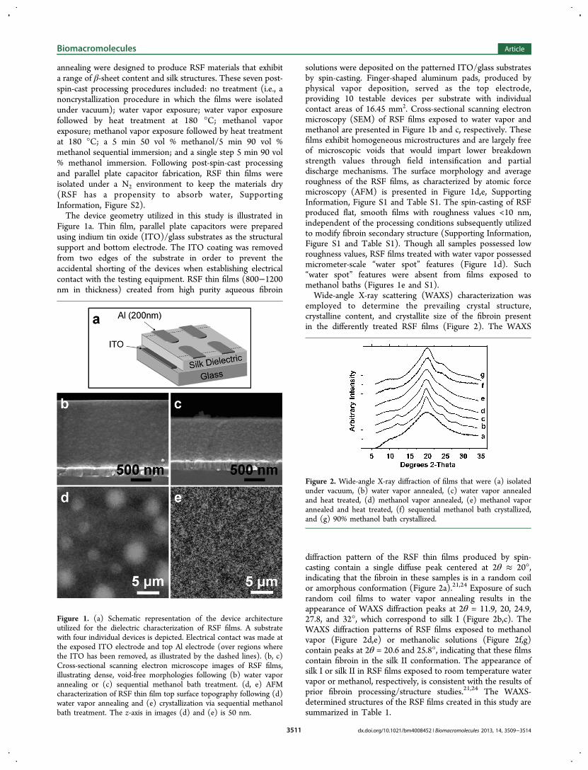
Figure 1a. Thin film, parallel plate capacitors were preparedusing indium tin oxide (ITO)/glass substrates as the structuralsupport and bottom electrode. The ITO coating was removedfrom two edges of the substrate in order to prevent theaccidental shorting of the devices when establishing electricalcontact with the testing equipment. RSF thin films (800−1200nm in thickness) created from high purity aqueous fibroin
solutions were deposited on the patterned ITO/glass substratesby spin-casting. Finger-shaped aluminum pads, produced byphysical vapor deposition, served as the top electrode,providing 10 testable devices per substrate with individualcontact areas of 16.45 mm2. Cross-sectional scanning electronmicroscopy (SEM) of RSF films exposed to water vapor andmethanol are presented in Figure 1b and c, respectively. Thesefilms exhibit homogeneous microstructures and are largely freeof microscopic voids that would impart lower breakdownstrength values through field intensification and partialdischarge mechanisms. The surface morphology and averageroughness of the RSF films, as characterized by atomic forcemicroscopy (AFM) is presented in Figure 1d,e, SupportingInformation, Figure S1 and Table S1. The spin-casting of RSFproduced flat, smooth films with roughness values <10 nm,independent of the processing conditions subsequently utilizedto modify fibroin secondary structure (Supporting Information,Figure S1 and Table S1). Though all samples possessed lowroughness values, RSF films treated with water vapor possessedmicrometer-scale “water spot” features (Figure 1d). Such“water spot” features were absent from films exposed tomethanol baths (Figures 1e and S1).Wide-angle X-ray scattering (WAXS) characterization was
employed to determine the prevailing crystal structure,crystalline content, and crystallite size of the fibroin presentin the differently treated RSF films (Figure 2). The WAXS
diffraction pattern of the RSF thin films produced by spin-casting contain a single diffuse peak centered at 2θ ≈ 20°,indicating that the fibroin in these samples is in a random coilor amorphous conformation (Figure 2a).21,24 Exposure of suchrandom coil films to water vapor annealing results in theappearance of WAXS diffraction peaks at 2θ = 11.9, 20, 24.9,27.8, and 32°, which correspond to silk I (Figure 2b,c). TheWAXS diffraction patterns of RSF films exposed to methanolvapor (Figure 2d,e) or methanolic solutions (Figure 2f,g)contain peaks at 2θ = 20.6 and 25.8°, indicating that these filmscontain fibroin in the silk II conformation. The appearance ofsilk I or silk II in RSF films exposed to room temperature watervapor or methanol, respectively, is consistent with the results ofprior fibroin processing/structure studies.21,24 The WAXS-determined structures of the RSF films created in this study aresummarized in Table 1.
Figure 1. (a) Schematic representation of the device architectureutilized for the dielectric characterization of RSF films. A substratewith four individual devices is depicted. Electrical contact was made atthe exposed ITO electrode and top Al electrode (over regions wherethe ITO has been removed, as illustrated by the dashed lines). (b, c)Cross-sectional scanning electron microscope images of RSF films,illustrating dense, void-free morphologies following (b) water vaporannealing or (c) sequential methanol bath treatment. (d, e) AFMcharacterization of RSF thin film top surface topography following (d)water vapor annealing and (e) crystallization via sequential methanolbath treatment. The z-axis in images (d) and (e) is 50 nm.
Figure 2. Wide-angle X-ray diffraction of films that were (a) isolatedunder vacuum, (b) water vapor annealed, (c) water vapor annealedand heat treated, (d) methanol vapor annealed, (e) methanol vaporannealed and heat treated, (f) sequential methanol bath crystallized,and (g) 90% methanol bath crystallized.
Biomacromolecules Article
dx.doi.org/10.1021/bm4008452 | Biomacromolecules 2013, 14, 3509−35143511

The WAXS diffraction patterns (Figure 2) of the RSF filmsproduced in this study were deconvoluted, using standardmethods and diffraction peak positions from the literature, todetermine the crystallinity of these materials.25,26 Thecrystallinity index of the RSF films was estimated utilizing thearea fractions of amorphous and crystalline (i.e., silk I or silk II)peaks in the WAXS data. An example of deconvolution (of awater vapor treated sample) is shown in SupportingInformation, Figure S4. To determine the crystallinity index,the area under the amorphous peak (i.e., gray curve inSupporting Information, Figure S4) is subtracted from the totalintegrated area of the WAXS pattern (100%), the remainingfraction is assigned to the crystalline phases of fibroin. Theresults of this analysis are presented in Supporting Information,Table S2, where the amorphous, silk I, and silk II structuredfilms possess crystallinity indexes of ∼16, 38, and 30%,respectively. The higher crystallinity index of the water vaporannealed samples is qualitatively consistent with the appearanceof the WAXS patterns of these films. The silk I peaks in thesediffraction patterns are sharper than the silk II peaks in theWAXS data of the methanol-exposed samples.The crystallite or domain size of fibroin in the various RSF
films can also be obtained from the full width at half-maximum(FWHM) of WAXS diffraction peaks according to the Scherrerequation. As expected from the visual appearance of the WAXSpatterns, the domain/crystallite size calculated from theFWHM of the peaks is larger for water vapor treated samples(∼5 nm) compared to methanol vapor treated samples (∼1−2nm). Note that crystallite size obtained via the Scherrerequation typically underestimates the size of the actual domainsin semicrystalline polymers (or materials with larger degree ofdisorder), such as these silk fibroin samples, by a factor of up to4.27 Further attempts to determine the fibroin crystallite sizeand possible preferred orientation in the RSF films were carriedout using grazing incidence small-angle X-ray scattering(GISAXS). As presented in Supporting Information, FigureS5, the small angle scattering curves from all RSF filmsexhibited a Guinier knee at values of q = 0.025 Å, whichcorresponds to scatterers of ∼25 nm. We assume thesescatterers are crystallites formed in the processing of the RSFfilms. This GISAXS-derived crystallite size agrees well with thecorrected values (4× corrected, as noted above) obtained fromthe Scherrer analysis of the water annealed RSF film WAXSdata (∼20 nm). Preferential orientation of fibroin crystalliteswas not noted in either 2D GISAXS data or WAXS conductedin plane and normal to the film, for any RSF film.In addition to WAXS characterization, Fourier transform
infrared-attenuated total reflection (FTIR-ATR) was utilized toprovide a quantitative description of the secondary proteinstructures present in the RSF films. Application of Fourier self-
deconvolution (FSD) to the FTIR spectrum in the amide Iregion (1575−1725 cm−1; Supporting Information, Figure S3b)has previously been used to accurately assess β-sheet content.19
Based on spectrum deconvolution within the amide I regionand assignment of vibration modes referenced in the literature,the structural conformation was quantitatively assessed and ispresented in Table 1 and Supporting Information, Table S1.The results show a progressively increasing fraction of β-sheetcontent, from 18 to 54%, mirroring a decrease in the randomcoil content of the films. This inverse relationship between β-sheet and random coil content during the crystallization of RSFhas been previously reported in the literature.19 It should benoted that, although the structure of silk I is defined as type IIβ-turn, the presence of this structure is interpreted as β-sheetcontent by FTIR analysis.19
Characterization of the dielectric breakdown strength wasinitiated using the RSF films deposited on ITO/glass substratesand exposed to the processing conditions outlined above.Measurements were performed under a dry N2 atmosphere andutilized a 25 Vdc s
−1 linear voltage ramp up to the point ofcatastrophic failure. The results from between 20 and 30individual tests per postspin-casting treatment condition wereanalyzed using Weibull statistics, an empirical failure probabilitydistribution described by:
γ α= − − − βP E1 exp[ {( )/ } ]F (1)
where PF is the probability of failure at a field of E; α is thecharacteristic breakdown strength (Ebd) associated with 63.2%probability of failure; β is the shape parameter describing thedistribution of E; γ is the threshold parameter, a value of E witha 0% failure probability (typically assigned a value of 0).28 Thecumulative probability distribution, based on a log{−ln(1 − p)}versus log E implementation of eq 1, is shown in Figure 3a.Using a linear fit to the cumulative probability function, the Ebdand the scatter parameter (β) were determined and are shownin Table 1. Based on the values detailed in Table 1, a structure−property relationship can be formed, linking Ebd with FTIR-determined β-sheet content (Figure 2b).The results for Ebd, shown in Figure 3, illustrate a link
between subtle changes to the RSF processing regime(designed to modify protein conformation and increase β-sheet content) and improved high voltage insulation potential.The Ebd (∼250 V/μm) of random coil RSF films is similar inmagnitude to the Ebd of amorphous synthetic polymers (e.g.,PMMA).11 Significant changes occur through methanol-basedexposure, resulting in films in the silk II structure with β-sheetcontents above 50% and Ebd values approaching 400 V/μm.Notably, the Ebd of RSF films matches or exceeds that of severalbiodegradable synthetic polymers, including polylactic acid andpolycaprolactone.29 Inspection of Figure 3 reveals that the
Table 1. Secondary Structure and Dielectric Breakdown Characteristics (Ebd and Scatter) of RSF Films As a Function of Post-Spin-Cast Processing Conditionsa
post-spin-cast processing condition
no treatment water vaporwater vapor+180 °C
methanolvapor
methanol vapor+180 °C
sequential methanolbath
90% methanolbath
silk structure amorphous silk I silk I silk II silk II silk II silk IIβ-sheet content (%) 18.0 ± 0.3 41.3 ± 0.5 44.7 ± 0.3 45.3 ± 0.3 43.7 ± 0.3 49.0 ± 1.0 54.0 ± 0.9characteristic breakdown(V/μm)
250.4 265.4 264.1 335.7 319.2 409.91 392.2
scatter (β) 13.52 20.71 19.56 5.86 6.44 8.58 14.43aFourier transform self-deconvolution of the FTIR amide I absorbance spectrum was utilized to calculate β-sheet content.
Biomacromolecules Article
dx.doi.org/10.1021/bm4008452 | Biomacromolecules 2013, 14, 3509−35143512

noncrystallized films and samples crystallized in methanol bathsrepresent the current bounds of RSF Ebd (i.e., within thelimitations of a substrate supported thin film format exploredhere). Of particular interest is the structural attributessupporting higher breakdown strengths. Though the electricalproperties of RSF films are affected by hydration in humidenvironments, the residual water content of the RSF samplestested under a protective atmosphere did not appear toinfluence Ebd (Tables 1 and S2). Based on the structure−property relationship represented in Figure 3b, increasing β-sheet content (as determined from FTIR analysis) offers adirect association to improved Ebd. However, significantimprovements to Ebd only occur after RSF films were exposedto methanol. Such methanol-treated films take on a silk IIstructure and possess β-sheet contents above 40%. Thedielectric permittivity of a methanol bath treated RSF wasdetermined to be 4.6 ± 0.2 at 1 kHz. Conversely to the resultsobserved for methanol treatment, exposure of RSF films towater vapor at room temperature and the conversion of fibrointo the silk I conformation offered only limited improvements tothe Ebd of the films. Analysis of the Weibull distribution formethanol vapor exposed films (Figure 3a) suggests adistribution of failure modes that bridge the distributions forRSF materials exposed to water vapor and methanolimmersion. From FTIR-ATR, the β-sheet content of themethanol vapor treated films is similar to that of water vaportreated RSF, while the WAXS pattern points to a transition of
fibroin from silk I to silk II. The exposure of RSF films tomethanol vapor and methanol bath processing conditionsresulted in approximately a 34 and 64% increase in the Ebd ofthese materials relative to uncrystallized (i.e., random coil)films, respectively. Considering these results, the breakdownstrength of RSF is most improved when the films are exposedto processing conditions that result in the production of fibroinfilms that have adopted the silk II conformation and possessappreciable (∼50%) β-sheet content (as determined by FTIRanalysis).The impact of structural parameters (e.g., chain conforma-
tion and crystalline fraction) on Ebd has previously beenexplored in semicrystalline polymers such as PVDF, PE, andpolypropylene (PP).14−17 Studies utilizing PP and PE haveidentified key structural parameters, such as crystal fraction,phase, habit, orientation, and size that impart improved highfield properties and lifetime stability.14,16,17 Application of theserelationships to the RSF material system further reinforces thetrends illustrated in Figure 3b. Inspection of Figure 3 and Table1 clearly reveals that the processing of RSF films, andspecifically the adoption of a highly developed silk II structure(as produced by exposure to a methanol bath) offers thegreatest improvement in Ebd.
■ CONCLUSIONSIn conclusion, increases in the FTIR-detected β-sheet contentand the presence of the silk II conformation in RSF thin filmsresults in significant improvements to the Ebd of this material,with Ebd values reaching 400 V/μm. Considering the uniquetailorability of the dielectric performance, surface character-istics, biofunctional doping, biodegradability, and inexpensivenature of RSF, this biopolymer offers immense potential as anelectrically insulating material for bioelectronics. Furtherimprovements in the dielectric performance of neat RSF filmsmay be possible through the mechanical stretching of RSF filmsto impart higher β-sheet content and chain orientation.30,31
Additional opportunities for silk-based materials as biodielectricsubstrates/devices may emerge through further advances inRSF processing, the production of hybrid materials, chemical/genetic manipulation of fibroin, or the use of silks from otherorganisms.2,5,32−37
■ ASSOCIATED CONTENT*S Supporting InformationAdditional RSF thin film characterization including AFM,dielectric storage, and loss of uncrystallized films followingexposure to atmospheric humidity, example WAXS patterndeconvolution, GISAXS and FTIR patterns, crystallinityindexes, RSF secondary structure (as determined by FTIR),and residual H2O by TGA. This material is available free ofcharge via the Internet at http://pubs.acs.org.
■ AUTHOR INFORMATIONCorresponding Author*E-mail: [email protected].
NotesThe authors declare no competing financial interest.
■ ACKNOWLEDGMENTSWe would like to thank Dr. Alexander Hexemer and Dr. EricSchaible for guidance, setup, and data collection at beamline
Figure 3. (a) Probability of dielectric failure (% probability of samplefailure) for regenerated silk fibroin films processed with variousfinishing procedures. (b) Ebd of regenerated silk fibroin films, as afunction of β-sheet content, determined at 63.2% failure probabilityfrom a linear regressive fit of the two-parameter Weibull failureanalysis.
Biomacromolecules Article
dx.doi.org/10.1021/bm4008452 | Biomacromolecules 2013, 14, 3509−35143513

7.3.3 at ALS/LBNL. This research was supported by the AirForce Office of Scientific Research.
■ REFERENCES(1) Omenetto, F. G.; Kaplan, D. L. Science 2010, 329, 528−531.(2) Hardy, J. G.; Romer, L. M.; Scheibel, T. R. Polymer 2008, 49,4309−4327.(3) Rockwood, D. N.; Preda, R. C.; Yucel, T.; Wang, X.; Lovett, M.L.; Kaplan, D. L. Nat. Protoc. 2011, 6, 1612−1631.(4) Tao, H.; Kaplan, D. L.; Omenetto, F. G. Adv. Mater. 2012, 24,2824−2837.(5) Murphy, A. R.; Kaplan, D. L. J. Mater. Chem. 2009, 19, 6443−6450.(6) Hakimi, O.; Knight, D. P.; Vollrath, F.; Vadgama, P. Composites,Part B 2007, 38, 324−337.(7) Irimia-Vladu, M.; Glowacki, E. D.; Voss, G.; Bauer, S.; Sariciftci,N. S. Mater. Today 2012, 15, 340−346.(8) Kim, D.-H.; Viventi, J.; Amsden, J. J.; Xiao, J.; Vigeland, L.; Kim,Y.-S.; Blanco, J. A.; Panilaitis, B.; Frechette, E. S.; Contreras, D.;Kaplan, D. L.; Omenetto, F. G.; Huang, Y.; Hwang, K.-C.; Zakin, M.R.; Litt, B.; Rogers, J. A. Nat. Mater. 2010, 9, 511−517.(9) Capelli, R.; Amsden, J. J.; Generali, G.; Toffanin, S.; Benfenati, V.;Muccini, M.; Kaplan, D. L.; Omenetto, F. G.; Zamboni, R. Org.Electron. 2011, 12, 1146−1151.(10) Wang, C. H.; Hsieh, C. Y.; Hwang, J. C. Adv. Mater. 2011, 23,1630−4.(11) Ieda, M.; Nagao, M.; Hikita, m. IEEE Trans. Dielectr. Electr. Insul.1994, 1, 934−945.(12) Laihonen, S. J.; Gafvert, U.; Schutte, T.; Gedde, U. W. IEEETrans. Dielectr. Electr. Insul. 2007, 14, 275−286.(13) Rabuffi, M.; Picci, G. IEEE Trans. Plasma Sci. 2002, 30, 1939−1942.(14) Yahagi, K. IEEE Trans. Electr. Insul. 1980, EI-15, 241−250.(15) Ceres, B. V.; Schultz, J. M. J. Appl. Polym. Sci., A 1984, 29, 4183.(16) Claude, J.; Lu, Y.; Li, K.; Wang, Q. Chem. Mater. 2008, 20,2078−2080.(17) Gao, L. Y.; Tu, D. M.; Zhou, S. C.; Zhang, Z. L. IEEE Trans.Electr.l Insul. 1990, 25, 535−540.(18) Kaplan, D. L.; Mello, C. M.; Arcidiacono, S.; Fossey, S.; Senecal,K.; Muller, W. In Protein-Based Materials; McGrath, K., Kaplan, D. L.,Eds.; Birkhauser Boston: Boston, MA, 1997.(19) Hu, X.; Kaplan, D.; Cebe, P. Macromolecules 2006, 39, 6161−6170.(20) Jin, H. J.; Park, J.; Karageorgiou, V.; Kim, U. J.; Valluzzi, R.;Cebe, P.; Kaplan, D. L. Adv. Funct. Mater. 2005, 15, 1241−1247.(21) Asakura, T.; Suzuki, Y.; Nakazawa, Y.; Yazawa, K.; Holland, H.P.; Yarger, J. L. Prog. Nucl. Magn. Reson. Spectrosc. 2013, 69, 23−68.(22) Ilavsky, J. J. Appl. Crystallogr. 2012, 45, 324−328.(23) Zhou, C. Z.; Confalonieri, F.; Medina, N.; Zivanovic, Y.;Esnault, C.; Yang, T.; Jacquet, M.; Janin, J.; Duguet, M.; Perasso, R.;Li, Z.-G. Nucl. Acid Res. 2000, 28, 2413−2419.(24) Drummy, L. F.; Farmer, B. L.; Naik, R. R. Soft Matter 2007, 3,877−882.(25) Alexander, L. E. X-ray Diffraction Methods in Polymer Science;Robert E. Kereger Publishing Co.: New York, 1979.(26) Lotz, B.; Keith, H. D. J. Mol. Biol. 1971, 61, 201−215.(27) Bunning, T. J.; Koerner, H.; Tsukruk, V. V.; McHugh, C. M.;Ober, C. K.; Adams, W. W. Macromolecules 1996, 29, 8717−8725.(28) Abernethy, R. B. The New Weibull Handbook, 5th ed.;Abernethy: North Palm Beach, FL, 2006.(29) Hirai, N.; Ishikawa, H.; Ohki, Y. IEEE Trans. Dielectr. Electr.Insul. 2007, 592−595.(30) Yao, J.; Masuda, H.; Zhao, C.; Asakura, T. Macromolecules 2002,35, 6−9.(31) Asakura, T.; Yao, J. Protein Sci. 2002, 11, 2706−2713.(32) Fillery, S. P.; Koerner, H.; Drummy, L.; Dunkerley, E.;Durstock, M. F.; Schmidt, D. F.; Vaia, R. A. ACS Appl. Mater. Interfaces2012, 4, 1388−1396.
(33) Kharlampieva, E.; Kozlovskaya, V.; Wallet, B.; Shevchenko, V.V.; Naik, R. R.; Vaia, R.; Kaplan, D. L.; Tsukruk, V. V. ACS Nano2010, 4, 7053−63.(34) Vollrath, F.; Porter, D. Soft Matter 2006, 2, 377−385.(35) Vollrath, F.; Porter, D.; Holland, C. Soft Matter 2011, 7, 9595−9600.(36) Lewis, R. V. Chem. Rev. 2006, 106, 3762−3774.(37) Drummy, L. F.; Koerner, H.; Phillips, D. M.; McAuliffe, J. C.;Kumar, M.; Farmer, B. L.; Vaia, R. A.; Naik, R. R. Mater. Sci. Eng., C2009, 29, 1266−1272.
Biomacromolecules Article
dx.doi.org/10.1021/bm4008452 | Biomacromolecules 2013, 14, 3509−35143514