Embed Size (px)
Citation preview
RESEARCH AND EDUCATION
Supported byResearch FuaResident, GbAssistant PrcAssistant PrMilwaukee, WdAssociate PreAssistant Pr
398
Die spacer thickness reproduction for central incisor crownfabrication with combined computer-aided design and 3D
printing technology: An in vitro study
Lisa N. Hoang, DMD, MS,a Geoffrey A. Thompson, DDS, MS,b Seok-Hwan Cho, DDS, MS, MS,cDavid W. Berzins, BS, PhD,d and Kwang Woo Ahn, PhDe
ABSTRACTStatement of problem. The inability to control die spacer thickness has been reported. However,little information is available on the congruency between the computer-aided design parametersfor die spacer thickness and the actual printout.
Purpose. The purpose of this study was to evaluate the accuracy and precision of the die spacerthickness achieved by combining computer-aided design and 3-dimensional printing technology.
Material and methods. An ivorine maxillary central incisor was prepared for a ceramic crown. Theprepared tooth was duplicated by using polyvinyl siloxane duplicating silicone, and 80 die-stonemodels were produced from Type IV dental stone. The dies were randomly divided into 5 groupswith assigned die spacer thicknesses of 25 mm, 45 mm, 65 mm, 85 mm, and 105 mm (n=16). Theprinted resin copings, obtained from a printer (ProJet DP 3000; 3D Systems), were cemented ontotheir respective die-stone models with self-adhesive resin cement and stored at room temperatureuntil sectioning into halves in a buccolingual direction. The internal gap was measured at 5defined locations per side of the sectioned die. Images of the printed resin coping/die-stone modelinternal gap dimensions were obtained with an inverted bright field metallurgical microscopeat ×100 magnification. The acquired digital image was calibrated, and measurements were madeusing image analysis software. Mixed models (a=.05) were used to evaluate accuracy. A falsediscovery rate at 5% was used to adjust for multiple testing. Coefficient of variation was used todetermine the precision for each group and was evaluated statistically with the Wald test (a=.05).
Results. The accuracy, expressed in terms of themean differences between the prescribed die spacerthickness and the measured internal gap (standard deviation), was 50 mm (11) for the 25 mm groupsimulated die spacer thickness, 30 mm (10) for the 45 mmgroup, 15 mm (14) for the 65 mmgroup, 3 mm(23) for the 85 mm group, and -10 mm (32) for the 105 mm group. The precision mean of the mea-surements, expressed as a coefficient of variation, ranged between 14% and 33% for the 5 groups.
Conclusions. For the accuracy evaluation, statistically significant differences were found for all thegroups, except the group of 85 mm. For the precision assessment, the coefficient of variation wasabove 10% for all groups, showing the printer’s inability to reproduce the uniform internal gapwithin the same group. (J Prosthet Dent 2015;113:398-404)
Clearance is needed between afixed restoration and the pre-pared tooth to provide space forthe luting agent. Methods suchas venting, axial grooves, andprovision of axial cement spacehave been used to reduce thehydraulic pressure between thecement and cast restorationand therefore improve seating,decrease seating time, and allowthe escape of excess cement.1-8
The application of paint-ondie spacer before waxing is oneof the most popular methodsfor providing this space.3,9 Diespacers have been used in fixeddental prostheses for manyyears.3,10-13
Die spacer has been shownto improve the marginal fitbetween the restoration andtooth preparation, decreasingthe risk of cement dissolution,plaque accumulation, recur-rent caries, and periodontalproblems.14 The thickness of
an American Academy of Fixed Prosthodontics Stanley D. Tylman research grant (02600-74716) and the Marquette University School of Dentistry Studentnd.raduate Prosthodontics, Marquette University School of Dentistry, Milwaukee, Wis.ofessor and Director, Department of General Dental Sciences, Graduate Prosthodontics, Marquette University School of Dentistry, Milwaukee, Wis.ofessor and Director, Predoctoral Prosthodontics and Biomaterials, Department of General Dental Sciences, Marquette University School of Dentistry,is.ofessor and Director, Department of General Dental Sciences, Graduate Dental Biomaterials, Marquette University School of Dentistry, Milwaukee, Wis.ofessor, Division of Biostatistics, Medical College of Wisconsin, Milwaukee, Wis.
THE JOURNAL OF PROSTHETIC DENTISTRY
Clinical ImplicationsThe incongruence between the computer-aideddesign program setting and the actual copingprintout negatively affect the clinical fit of thedefinitive restoration.
May 2015 399
this die spacer affects the fracture strength of a ceramicrestoration, its retention, and the marginal gap.1,2,15,16
According to Tuntiprawon and Wilson,15 ceramiccrowns displayed a greater fracture strength when themean internal gap at the axial wall was less than 73 mm.A lower failure strength was reported when the meaninternal gap was greater than 122 mm without any sig-nificant improvement in seating. Conflicting results havebeen reported on the influence of die spacer thickness onretention. Eames et al1 reported a 25% increase inretention when comparing 0 mm to 25 mm die spacerthickness. This improved retention was supported byCarter and Wilson,2 who reported that retentionincreased from 250 N to 375 N as die spacer thicknesschanged from 0 to 8 layers. In contrast, Jorgensen andEsbensen16 reported a moderate association betweenfilm thickness and retention, while Vermilyea et al17
reported a 32% reduction in retention when comparing0 with 2 layers of Tru-Fit paint on die spacer (GeorgeTaub Products and Fusion Co Inc). There is greaterconsistency when evaluating the cause and effect rela-tionship of die spacer thickness and marginal gap.2-4 Themagnitude of the marginal gap observed without the useof die spacer was as high as 649 mm3. The marginal gapwas significantly improved from 479 mm to 38 mm as thedie spacer thickness was increased from 1 to 8 layers.1-3
Marginal gap can also be influenced by the marginaldesign and total occlusal convergence of the prepara-tion.18,19 McLean and von Fraunhofer18 showed that thebest marginal gap was obtained with chamfer orrounded shoulder marginal design when compared withstraight shoulder marginal design. The maximum toler-ated marginal gap was 120 mm for ceramic crowns.
Hollenback20 suggested an ideal die spacer thicknessof 25 mm, which corresponds to the film thickness ofType I zinc phosphate cements. However, studies withcommercially available paint-on die spacer products haveshown inconsistent results.5,13,21,22 Computer-aideddesign (CAD) technology provides the ability to virtu-ally design restorations and program the die spacerthickness. The virtual coping can be produced by mill-ing23,24 or rapid prototyping. Rapid prototyping uses thelayer-to-layer fabrication of 3-dimensional physicalmodels directly from CAD data.25-27 Rapid prototyping isfurther subdivided into stereolithography, selective layersintering, fused deposition modeling, laminated object
Hoang et al
manufacturing, and 3D printing technology (3DPT).28
3DPT is used for copings, partial fixed dental prosthe-ses, anatomic contour pressable patterns, and partialremovable dental prosthesis frameworks, which areformed in either wax or acrylic resin.29
Accuracy is defined to assess the degree of closenessof the internal gap measurements to the reference values(programmed die spacer thicknesses). Therefore, thepurpose of this study was to evaluate the accuracy andprecision of the die spacer thickness achieved bycombining CAD and 3DPT to test the accuracy of CAD/3DPT by comparing various measured internal gapthicknesses with the prescribed values of 25 mm, 45 mm,65 mm, 85 mm, and 105 mm. The accuracy null hypothesiswas that no overall difference would be found betweenthe CAD/3DPT achieved internal gap thicknesses and theprescribed die spacer thicknesses. The secondary objec-tive of the research was to measure the printer’s ability toreproduce the uniform die spacer thickness for all thespecimens within an assigned die spacer thickness group.A statistical analysis was performed to test whether therewas difference among the varying die spacer thicknessesand the reproducibility of the printer. The coefficient ofvariation (CV) was adopted to determine the precision ofthe measurements. Ten percent of CV has been widelyused as cutoff to define high precision.30-32 Therefore, thenull hypothesis for precision was that the CV was equalto 10% because a CV less than 10% was consideredreasonable.30-32
MATERIAL AND METHODS
An ivorine maxillary central incisor (T1560; ColumbiaDentoform Corp) was prepared to receive a ceramiccrown restoration with the following features: a totalconvergence angle of 12 degrees, an incisal reduction of2 mm, a uniform axial reduction of 1.5 mm, and a deepchamfer.28 Before duplication, the prepared tooth wasattached to a circular base with 3 rectangular extensionsfabricated from a light-polymerized urethane dimetha-crylate tray material (Triad; Dentsply Intl). The materialincreased the diameter of the ivorine tooth shaft toprevent tooth breakage during separation from the sil-icone mold and to allow sectioning with a low-speedsaw (IsoMet speed saw; Buehler Ltd). Polyvinylsiloxane duplicating material (Double Take; IvoclarVivodent) was used to make 16 molds of the preparedtooth in a duplicating flask. Type IV dental stone (ResinRock; Whip Mix Corp) was used to fabricate 80 stonedies from the 16 silicone molds. The stone dies weredivided into 5 groups for the 5 die spacer thicknesslevels: 25 mm, 45 mm, 64 mm, 85 mm, and 105 mm(N=80). A power analysis was completed before theexperiment using the mixed models. A statistical anal-ysis was assumed to have been performed for each die
THE JOURNAL OF PROSTHETIC DENTISTRY
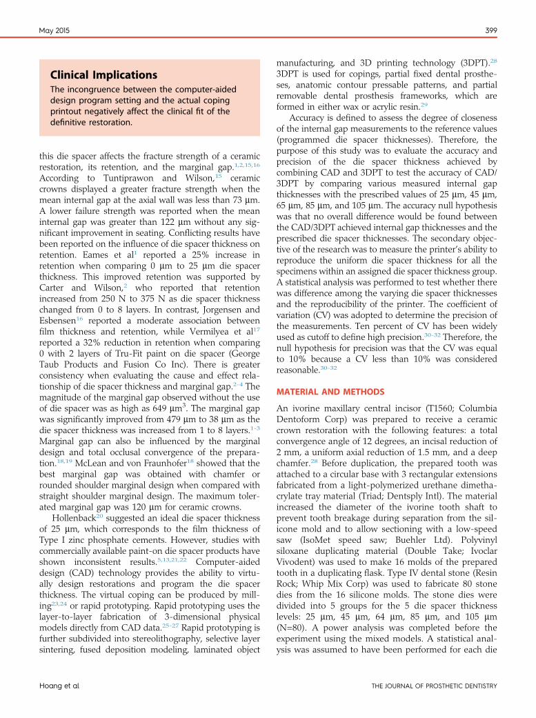
Figure 1. A, Cemented coping-die assembly placed under static load of 49 N. B, Low-speed diamond saw with diamond wafering blade. C, Specimensectioned into halves.
400 Volume 113 Issue 5
spacer thickness level. Given the die spacer thicknesslevel, 5 measurements for each specimen were treatedas repeated measurements to account for within indi-vidual correlation. The standard deviation of eachmeasurement was assumed to be 15 and the correlationamong the 5 measurements from each specimen to be0.8. A sample size of 80 (5 locations×16 teeth for eachdie spacer thickness level) was determined to be suffi-cient to detect a mean difference of 11 between overallmean (mean of the 5 locations) and the mean of the nullhypothesis with 80% power and at a 5% significancelevel.
The stone dies were shipped to a commercial labo-ratory (Nu-Art Dental Inc) for the fabrication of the resincopings. Each die was scanned (D700 3D scanner;3Shape) according to the manufacturer’s instructions torecord the external die form. The 3Shape CAD designsystem was used to locate the margin and assign the diespacer thickness, which was uniform throughout andterminated 0.5 mm from the finish line and copingthickness of 1.0 mm.2,5,33 Each resin coping was digitallymarked so that it could be paired with its correspondingstone die. The acrylic resin copings were printed (ProJetDP 3000; 3D Systems). The same dental technician at the
THE JOURNAL OF PROSTHETIC DENTISTRY
commercial laboratory completed all of the laboratoryprocesses.
One dispenser increment volume of self-adhesiveresin cement (RelyX Unicem 2; 3M ESPE) was usedto cement each printed resin coping to its corre-sponding die. The cement film thickness was deter-mined based on the American Dental AssociationSpecification No. 8 protocol, section 4.3.4.34 However,the recommended seating force was reduced to repli-cate the load used in this experiment (49 N). For thetest specimen, the coping was seated with a rockingmotion until it was completely seated onto the die. Thecemented coping-die assembly was placed under anapparatus capable of maintaining a static deadweightload of 49 N (Fig. 1A), and excess cement was removedwith a fine microbrush.35 The mesial, distal, buccal, andlingual surfaces were light polymerized (Demi Plus;Kerr) for 20 seconds each for a total of 80 seconds toensure complete polymerization. The cementedcoping-die assembly was kept under the weighted-base apparatus for 6 minutes. The specimens werestored at room temperature until sectioning.
Each specimen was sectioned in a buccolingual di-rection by using a low-speed diamond saw (IsoMet speed
Hoang et al
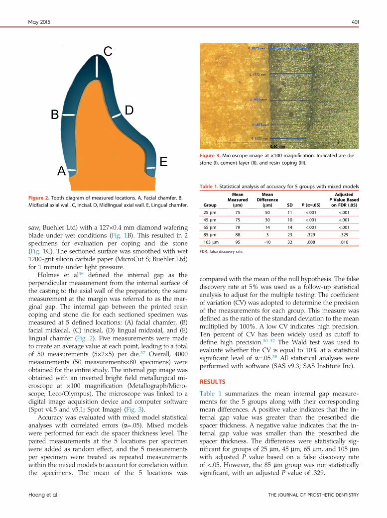
Figure 2. Tooth diagram of measured locations. A, Facial chamfer. B,Midfacial axial wall. C, Incisal. D, Midlingual axial wall. E, Lingual chamfer.
Figure 3. Microscope image at ×100 magnification. Indicated are diestone (I), cement layer (II), and resin coping (III).
Table 1. Statistical analysis of accuracy for 5 groups with mixed models
Group
MeanMeasured
(mm)
MeanDifference
(mm) SD P (a=.05)
AdjustedP Value Basedon FDR (.05)
25 mm 75 50 11 <.001 <.001
45 mm 75 30 10 <.001 <.001
65 mm 79 14 14 <.001 <.001
85 mm 88 3 23 .329 .329
105 mm 95 -10 32 .008 .016
FDR, false discovery rate.
May 2015 401
saw; Buehler Ltd) with a 127×0.4 mm diamond waferingblade under wet conditions (Fig. 1B). This resulted in 2specimens for evaluation per coping and die stone(Fig. 1C). The sectioned surface was smoothed with wet1200-grit silicon carbide paper (MicroCut S; Buehler Ltd)for 1 minute under light pressure.
Holmes et al36 defined the internal gap as theperpendicular measurement from the internal surface ofthe casting to the axial wall of the preparation; the samemeasurement at the margin was referred to as the mar-ginal gap. The internal gap between the printed resincoping and stone die for each sectioned specimen wasmeasured at 5 defined locations: (A) facial chamfer, (B)facial midaxial, (C) incisal, (D) lingual midaxial, and (E)lingual chamfer (Fig. 2). Five measurements were madeto create an average value at each point, leading to a totalof 50 measurements (5×2×5) per die.37 Overall, 4000measurements (50 measurements×80 specimens) wereobtained for the entire study. The internal gap image wasobtained with an inverted bright field metallurgical mi-croscope at ×100 magnification (Metallograph/Micro-scope; Leco/Olympus). The microscope was linked to adigital image acquisition device and computer software(Spot v4.5 and v5.1; Spot Image) (Fig. 3).
Accuracy was evaluated with mixed model statisticalanalyses with correlated errors (a=.05). Mixed modelswere performed for each die spacer thickness level. Thepaired measurements at the 5 locations per specimenwere added as random effect, and the 5 measurementsper specimen were treated as repeated measurementswithin the mixed models to account for correlation withinthe specimens. The mean of the 5 locations was
Hoang et al
compared with the mean of the null hypothesis. The falsediscovery rate at 5% was used as a follow-up statisticalanalysis to adjust for the multiple testing. The coefficientof variation (CV) was adopted to determine the precisionof the measurements for each group. This measure wasdefined as the ratio of the standard deviation to the meanmultiplied by 100%. A low CV indicates high precision.Ten percent of CV has been widely used as cutoff todefine high precision.30-32 The Wald test was used toevaluate whether the CV is equal to 10% at a statisticalsignificant level of a=.05.38 All statistical analyses wereperformed with software (SAS v9.3; SAS Institute Inc).
RESULTS
Table 1 summarizes the mean internal gap measure-ments for the 5 groups along with their correspondingmean differences. A positive value indicates that the in-ternal gap value was greater than the prescribed diespacer thickness. A negative value indicates that the in-ternal gap value was smaller than the prescribed diespacer thickness. The differences were statistically sig-nificant for groups of 25 mm, 45 mm, 65 mm, and 105 mmwith adjusted P value based on a false discovery rateof <.05. However, the 85 mm group was not statisticallysignificant, with an adjusted P value of .329.
THE JOURNAL OF PROSTHETIC DENTISTRY
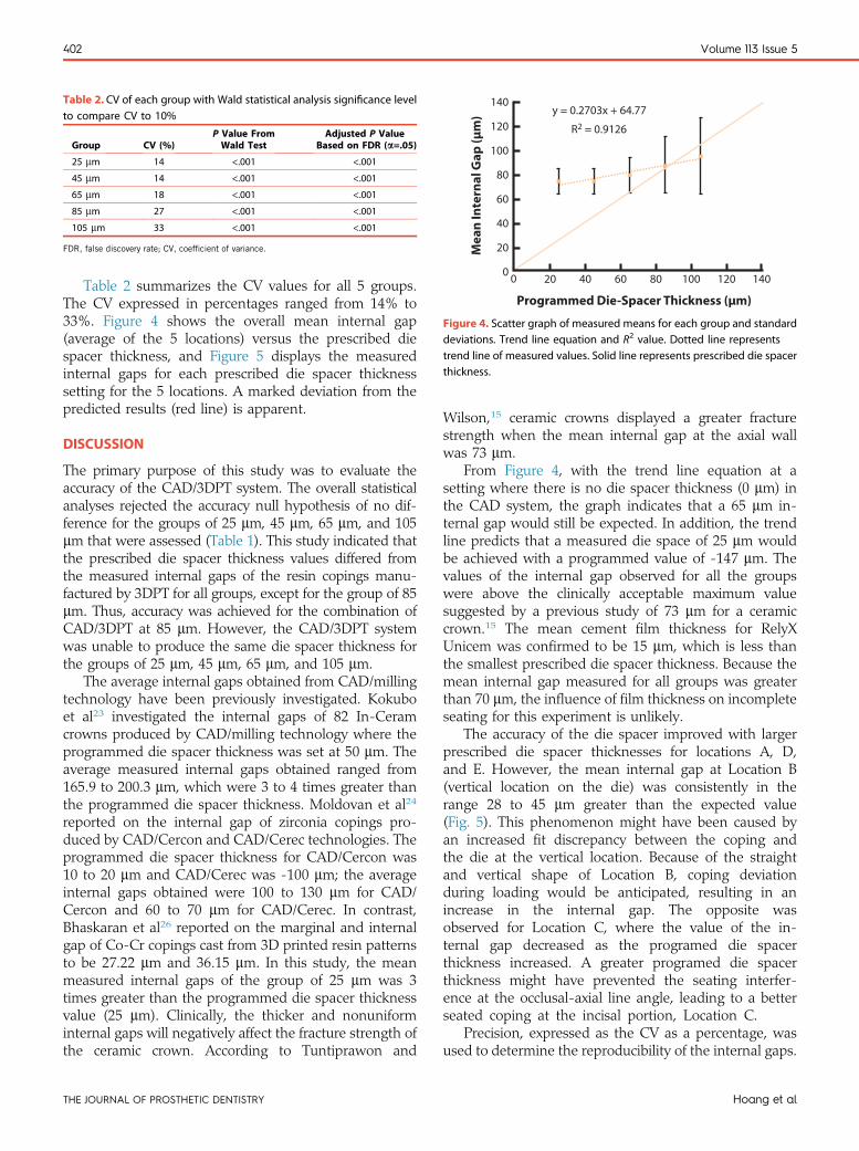
Table 2. CV of each group with Wald statistical analysis significance levelto compare CV to 10%
Group CV (%)P Value FromWald Test
Adjusted P ValueBased on FDR (a=.05)
25 mm 14 <.001 <.001
45 mm 14 <.001 <.001
65 mm 18 <.001 <.001
85 mm 27 <.001 <.001
105 mm 33 <.001 <.001
FDR, false discovery rate; CV, coefficient of variance.
140
120
100
80
60
40
20
20 40 60 80 100 120 14000
Programmed Die-Spacer Thickness (µm)
Me
an
In
tern
al
Ga
p (
µm
)
y = 0.2703x + 64.77
R2 = 0.9126
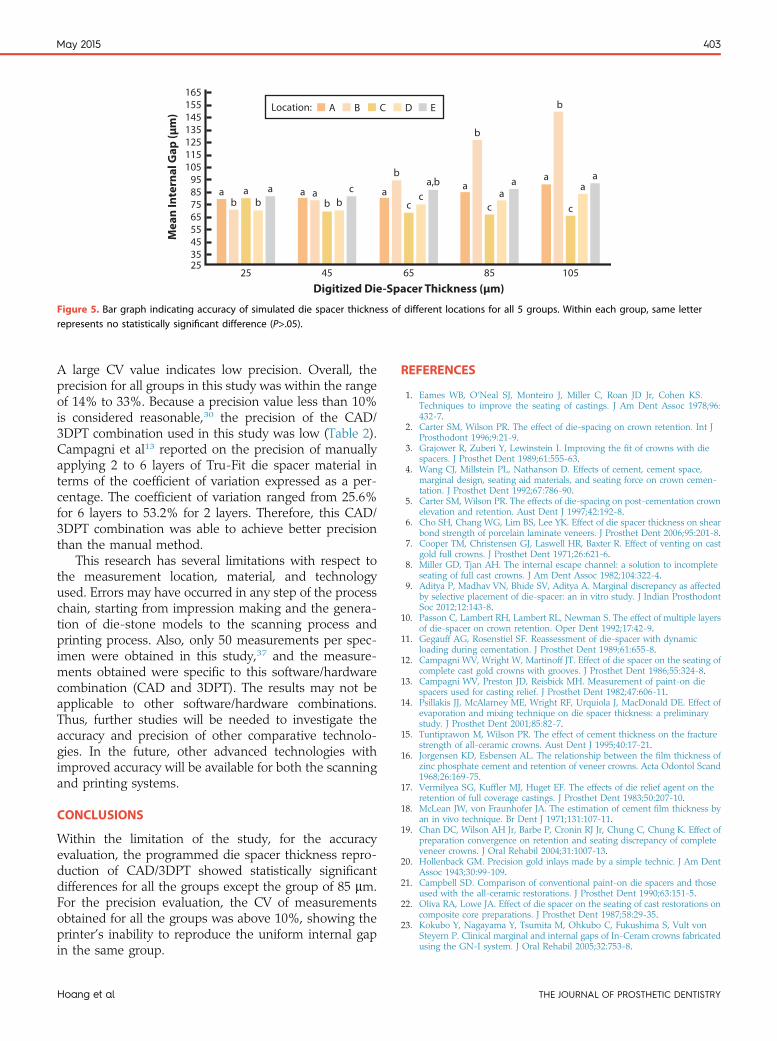
Figure 4. Scatter graph of measured means for each group and standarddeviations. Trend line equation and R2 value. Dotted line representstrend line of measured values. Solid line represents prescribed die spacerthickness.
402 Volume 113 Issue 5
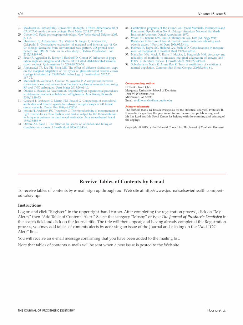
Table 2 summarizes the CV values for all 5 groups.The CV expressed in percentages ranged from 14% to33%. Figure 4 shows the overall mean internal gap(average of the 5 locations) versus the prescribed diespacer thickness, and Figure 5 displays the measuredinternal gaps for each prescribed die spacer thicknesssetting for the 5 locations. A marked deviation from thepredicted results (red line) is apparent.
DISCUSSION
The primary purpose of this study was to evaluate theaccuracy of the CAD/3DPT system. The overall statisticalanalyses rejected the accuracy null hypothesis of no dif-ference for the groups of 25 mm, 45 mm, 65 mm, and 105mm that were assessed (Table 1). This study indicated thatthe prescribed die spacer thickness values differed fromthe measured internal gaps of the resin copings manu-factured by 3DPT for all groups, except for the group of 85mm. Thus, accuracy was achieved for the combination ofCAD/3DPT at 85 mm. However, the CAD/3DPT systemwas unable to produce the same die spacer thickness forthe groups of 25 mm, 45 mm, 65 mm, and 105 mm.
The average internal gaps obtained from CAD/millingtechnology have been previously investigated. Kokuboet al23 investigated the internal gaps of 82 In-Ceramcrowns produced by CAD/milling technology where theprogrammed die spacer thickness was set at 50 mm. Theaverage measured internal gaps obtained ranged from165.9 to 200.3 mm, which were 3 to 4 times greater thanthe programmed die spacer thickness. Moldovan et al24
reported on the internal gap of zirconia copings pro-duced by CAD/Cercon and CAD/Cerec technologies. Theprogrammed die spacer thickness for CAD/Cercon was10 to 20 mm and CAD/Cerec was -100 mm; the averageinternal gaps obtained were 100 to 130 mm for CAD/Cercon and 60 to 70 mm for CAD/Cerec. In contrast,Bhaskaran et al26 reported on the marginal and internalgap of Co-Cr copings cast from 3D printed resin patternsto be 27.22 mm and 36.15 mm. In this study, the meanmeasured internal gaps of the group of 25 mm was 3times greater than the programmed die spacer thicknessvalue (25 mm). Clinically, the thicker and nonuniforminternal gaps will negatively affect the fracture strength ofthe ceramic crown. According to Tuntiprawon and
THE JOURNAL OF PROSTHETIC DENTISTRY
Wilson,15 ceramic crowns displayed a greater fracturestrength when the mean internal gap at the axial wallwas 73 mm.
From Figure 4, with the trend line equation at asetting where there is no die spacer thickness (0 mm) inthe CAD system, the graph indicates that a 65 mm in-ternal gap would still be expected. In addition, the trendline predicts that a measured die space of 25 mm wouldbe achieved with a programmed value of -147 mm. Thevalues of the internal gap observed for all the groupswere above the clinically acceptable maximum valuesuggested by a previous study of 73 mm for a ceramiccrown.15 The mean cement film thickness for RelyXUnicem was confirmed to be 15 mm, which is less thanthe smallest prescribed die spacer thickness. Because themean internal gap measured for all groups was greaterthan 70 mm, the influence of film thickness on incompleteseating for this experiment is unlikely.
The accuracy of the die spacer improved with largerprescribed die spacer thicknesses for locations A, D,and E. However, the mean internal gap at Location B(vertical location on the die) was consistently in therange 28 to 45 mm greater than the expected value(Fig. 5). This phenomenon might have been caused byan increased fit discrepancy between the coping andthe die at the vertical location. Because of the straightand vertical shape of Location B, coping deviationduring loading would be anticipated, resulting in anincrease in the internal gap. The opposite wasobserved for Location C, where the value of the in-ternal gap decreased as the programed die spacerthickness increased. A greater programed die spacerthickness might have prevented the seating interfer-ence at the occlusal-axial line angle, leading to a betterseated coping at the incisal portion, Location C.
Precision, expressed as the CV as a percentage, wasused to determine the reproducibility of the internal gaps.
Hoang et al
A B C D E
ab
cab
45
ac
a,bb
c
65
a
c
a
b
a
85
a
c
a
b
a
105
a a ab b
165155145135125115105
9585756555453525
Digitized Die-Spacer Thickness (µm)
Me
an
In
tern
al
Ga
p (
µm
)
25
Location:
Figure 5. Bar graph indicating accuracy of simulated die spacer thickness of different locations for all 5 groups. Within each group, same letterrepresents no statistically significant difference (P>.05).
May 2015 403
A large CV value indicates low precision. Overall, theprecision for all groups in this study was within the rangeof 14% to 33%. Because a precision value less than 10%is considered reasonable,30 the precision of the CAD/3DPT combination used in this study was low (Table 2).Campagni et al13 reported on the precision of manuallyapplying 2 to 6 layers of Tru-Fit die spacer material interms of the coefficient of variation expressed as a per-centage. The coefficient of variation ranged from 25.6%for 6 layers to 53.2% for 2 layers. Therefore, this CAD/3DPT combination was able to achieve better precisionthan the manual method.
This research has several limitations with respect tothe measurement location, material, and technologyused. Errors may have occurred in any step of the processchain, starting from impression making and the genera-tion of die-stone models to the scanning process andprinting process. Also, only 50 measurements per spec-imen were obtained in this study,37 and the measure-ments obtained were specific to this software/hardwarecombination (CAD and 3DPT). The results may not beapplicable to other software/hardware combinations.Thus, further studies will be needed to investigate theaccuracy and precision of other comparative technolo-gies. In the future, other advanced technologies withimproved accuracy will be available for both the scanningand printing systems.
CONCLUSIONS
Within the limitation of the study, for the accuracyevaluation, the programmed die spacer thickness repro-duction of CAD/3DPT showed statistically significantdifferences for all the groups except the group of 85 mm.For the precision evaluation, the CV of measurementsobtained for all the groups was above 10%, showing theprinter’s inability to reproduce the uniform internal gapin the same group.
Hoang et al
REFERENCES
1. Eames WB, O’Neal SJ, Monteiro J, Miller C, Roan JD Jr, Cohen KS.Techniques to improve the seating of castings. J Am Dent Assoc 1978;96:432-7.
2. Carter SM, Wilson PR. The effect of die-spacing on crown retention. Int JProsthodont 1996;9:21-9.
3. Grajower R, Zuberi Y, Lewinstein I. Improving the fit of crowns with diespacers. J Prosthet Dent 1989;61:555-63.
4. Wang CJ, Millstein PL, Nathanson D. Effects of cement, cement space,marginal design, seating aid materials, and seating force on crown cemen-tation. J Prosthet Dent 1992;67:786-90.
5. Carter SM, Wilson PR. The effects of die-spacing on post-cementation crownelevation and retention. Aust Dent J 1997;42:192-8.
6. Cho SH, Chang WG, Lim BS, Lee YK. Effect of die spacer thickness on shearbond strength of porcelain laminate veneers. J Prosthet Dent 2006;95:201-8.
7. Cooper TM, Christensen GJ, Laswell HR, Baxter R. Effect of venting on castgold full crowns. J Prosthet Dent 1971;26:621-6.
8. Miller GD, Tjan AH. The internal escape channel: a solution to incompleteseating of full cast crowns. J Am Dent Assoc 1982;104:322-4.
9. Aditya P, Madhav VN, Bhide SV, Aditya A. Marginal discrepancy as affectedby selective placement of die-spacer: an in vitro study. J Indian ProsthodontSoc 2012;12:143-8.
10. Passon C, Lambert RH, Lambert RL, Newman S. The effect of multiple layersof die-spacer on crown retention. Oper Dent 1992;17:42-9.
11. Gegauff AG, Rosenstiel SF. Reassessment of die-spacer with dynamicloading during cementation. J Prosthet Dent 1989;61:655-8.
12. Campagni WV, Wright W, Martinoff JT. Effect of die spacer on the seating ofcomplete cast gold crowns with grooves. J Prosthet Dent 1986;55:324-8.
13. Campagni WV, Preston JD, Reisbick MH. Measurement of paint-on diespacers used for casting relief. J Prosthet Dent 1982;47:606-11.
14. Psillakis JJ, McAlarney ME, Wright RF, Urquiola J, MacDonald DE. Effect ofevaporation and mixing technique on die spacer thickness: a preliminarystudy. J Prosthet Dent 2001;85:82-7.
15. Tuntiprawon M, Wilson PR. The effect of cement thickness on the fracturestrength of all-ceramic crowns. Aust Dent J 1995;40:17-21.
16. Jorgensen KD, Esbensen AL. The relationship between the film thickness ofzinc phosphate cement and retention of veneer crowns. Acta Odontol Scand1968;26:169-75.
17. Vermilyea SG, Kuffler MJ, Huget EF. The effects of die relief agent on theretention of full coverage castings. J Prosthet Dent 1983;50:207-10.
18. McLean JW, von Fraunhofer JA. The estimation of cement film thickness byan in vivo technique. Br Dent J 1971;131:107-11.
19. Chan DC, Wilson AH Jr, Barbe P, Cronin RJ Jr, Chung C, Chung K. Effect ofpreparation convergence on retention and seating discrepancy of completeveneer crowns. J Oral Rehabil 2004;31:1007-13.
20. Hollenback GM. Precision gold inlays made by a simple technic. J Am DentAssoc 1943;30:99-109.
21. Campbell SD. Comparison of conventional paint-on die spacers and thoseused with the all-ceramic restorations. J Prosthet Dent 1990;63:151-5.
22. Oliva RA, Lowe JA. Effect of die spacer on the seating of cast restorations oncomposite core preparations. J Prosthet Dent 1987;58:29-35.
23. Kokubo Y, Nagayama Y, Tsumita M, Ohkubo C, Fukushima S, Vult vonSteyern P. Clinical marginal and internal gaps of In-Ceram crowns fabricatedusing the GN-I system. J Oral Rehabil 2005;32:753-8.
THE JOURNAL OF PROSTHETIC DENTISTRY
404 Volume 113 Issue 5
24. Moldovan O, Luthardt RG, Corcodel N, Rudolph H. Three-dimensional fit ofCAD/CAM-made zirconia copings. Dent Mater 2011;27:1273-8.
25. Cooper KG. Rapid prototyping technology. New York: Marcel Dekker; 2005.p. 1-8.
26. Bhaskaran E, Azhagarasan NS, Miglani S, Ilango T, Krishna GP,Gajapathi B. Comparative evaluation of marginal and internal gap of Co-Cr copings fabricated from conventional wax pattern, 3D printed resinpattern and DMLS Tech: an in vitro study. J Indian Prosthodont Soc2013;13:189-95.
27. Beuer F, Aggstaller H, Richter J, Edelhoff D, Gernet W. Influence of prepa-ration angle on marginal and internal fit of CAD/CAM-fabricated zirconiacrown copings. Quintessence Int 2009;40:243-50.
28. Alghazzawi TF, Liu PR, Essig ME. The effect of different fabrication stepson the marginal adaptation of two types of glass-infiltrated ceramic crowncopings fabricated by CAD/CAM technology. J Prosthodont 2012;21:167-72.
29. Martorelli M, Gerbino S, Giudice M, Ausiello P. A comparison betweencustomized clear and removable orthodontic appliances manufactured usingRP and CNC techniques. Dent Mater 2013;29:e1-10.
30. Öhman C, Baleani M, Viceconti M. Repeatability of experimental proceduresto determine mechanical behaviour of ligaments. Acta Bioeng Biomech2009;11:19-23.
31. Gousard J, Lechevrel C, Martin PM, Rousel G. Comparison of monoclonalantibodies and tritated ligands for estrogen receptor asays in 241 breastcancer cytosols. Cancer Res 1986;46:4282-7.
32. Jensen PJ, Andersen PK, Thøgersen C. The reproducibility of measurement ofright ventricular ejection fraction and cardiac output by the thermodilutiontechnique in patients on mechanical ventilation. Acta Anaesthesiol Scand1994;38:486-9.
33. Olivera AB, Saito T. The effect of die spacer on retention and fitting ofcomplete cast crowns. J Prosthodont 2006;15:243-9.
Receive Tables of Co
Instructions
To receive tables of contents by e-mail, sign up through our Wodicals/ympr.
Log on and click “Register” in the upper right-hand corner. AAlerts,” then “Add Table of Contents Alert.” Select the categothe search field and click on the Journal title. The title will theprocess, you may add tables of contents alerts by accessing aAlert” link.
You will receive an e-mail message confirming that you have
Note that tables of contents e-mails will be sent when a new
THE JOURNAL OF PROSTHETIC DENTISTRY
34. Certification programs of the Council on Dental Materials, Instruments andEquipment. Specification No. 8. Chicago: American National StandardsInstitution/American Dental Association; 1977.
35. Wood KC, Berzins DW, Luo Q, Thompson GA, Toth JM, Nagy WW.Resistance to fracture of two all-ceramic crown materials following end-odontic access. J Prosthet Dent 2006;95:33-41.
36. Holmes JR, Bayne SC, Holland GA, Sulik WD. Considerations in measure-ment of marginal fit. J Prosthet Dent 1989;62:405-8.
37. Nawafleh NA, Mack F, Evans J, Mackay J, Hatamleh MM. Accuracy andreliability of methods to measure marginal adaptation of crowns andFDPs: a literature review. J Prosthodont 2013;22:419-28.
38. Subrahmanya Nairy K, Aruna Rao K. Tests of coefficients of variation ofnormal population. Commun Stat Simul Comput 2003;32:641-61.
Corresponding author:Dr Seok-Hwan ChoMarquette University School of Dentistry1801 W Wisconsin AveMilwaukee, WI 53233Email: [email protected]
AcknowledgmentsThe authors thank Dr Jessica Pruszynski for the statistical analyses, Professor R.Fournelle for granting the permission to use the microscope laboratory, andMr Lee Lord and Mr David Zanon for helping with the scanning and printing ofthe copings.
Copyright © 2015 by the Editorial Council for The Journal of Prosthetic Dentistry.
ntents by E-mail
eb site at http://www.journals.elsevierhealth.com/peri-
fter completing the registration process, click on “My ry “Mosby” or type The Journal of Prosthetic Dentistry in n appear, and having already completed the Registration
n issue of the Journal and clicking on the “Add TOC
been added to the mailing list.
issue is posted to the Web site.
Hoang et al