Embed Size (px)
Citation preview
Journ
alof
Cell
Scie
nce
Dictyostelium ACAP-A is an ArfGAP involved incytokinesis, cell migration and actincytoskeleton dynamics
Marco Dias1, Cedric Blanc1, Nelcy Thazar-Poulot2,3,4,5,6, Sabrina Ben Larbi3,4,7, Pierre Cosson1 andFrancois Letourneur3,4,7,*1Departement de Physiologie Cellulaire et Metabolisme, Centre Medical Universitaire, 1 rue Michel Servet, 1211 Geneva 4, Switzerland2CNRS, UMR5667, UMS3444, 15 parvis Rene Descartes BP 7000, Lyon, 69342, France3Universite de Lyon, Lyon, 69361, France4Universite Lyon 1, Villeurbanne, 69622, France5Ecole Normale Superieure de Lyon, Lyon, 69342, France6Institut National de la Recherche Agronomique, Lyon, 69364, France7CNRS, UMR5534, Centre de Genetique et de Physiologie Moleculaire et Cellulaire, 16 rue Raphael Dubois, Villeurbanne, 69622, France
*Author for correspondence ([email protected])
Accepted 19 November 2012Journal of Cell Science 126, 756–766� 2013. Published by The Company of Biologists Ltddoi: 10.1242/jcs.113951
SummaryACAPs and ASAPs are Arf-GTPase-activating proteins with BAR, PH, GAP and ankyrin repeat domains and are known to regulatevesicular traffic and actin cytoskeleton dynamics in mammalian cells. The amoeba Dictyostelium has only two proteins with this domain
organization, instead of the six in human, enabling a more precise functional analysis. Genetic invalidation of acapA resulted inmultinucleated cells with cytokinesis defects. Mutant acapA2 cells were hardly motile and their multicellular development wassignificantly delayed. In addition, formation of filopodial protrusions was deficient in these cells. Conversely, re-expression of ACAP-
A–GFP resulted in numerous and long filopodia-like protrusions. Mutagenesis studies showed that the ACAP-A actin remodelingfunction was dependent on its ability to activate its substrate, the small GTPase ArfA. Likewise, the expression of a constitutively activeArfANGTP mutant in wild-type cells led to a significant reduction in filopodia length. Together, our data support a role for ACAP-A in
the control of the actin cytoskeleton organization and dynamics through an ArfA-dependent mechanism.
Key words: ArfGAP, Cytokinesis, Migration, Actin cytoskeleton
IntroductionThe ADP-ribosylation factor (Arf) proteins are GTP-binding
proteins that control vesicular transport and remodeling of the
actin cytoskeleton (D’Souza-Schorey and Chavrier, 2006;
Gillingham and Munro, 2007; Donaldson and Jackson, 2011).
These proteins cycle between active GTP-bound and inactive
GDP-bound forms with distinct functions. The tight control of
this cycle relies on two key families of proteins. Arf-Guanine
nucleotide exchange factors (Arf-GEFs) catalyze the exchange of
GDP, whereas Arf-GTPase activating proteins (Arf-GAPs)
induce hydrolysis of GTP bound to Arf proteins. Besides their
catalytic functions, both GEFs and GAPs can serve as scaffold
for numerous effector proteins and can thus regulate cellular
physiology in an Arf-independent manner (Inoue and Randazzo,
2007; Randazzo et al., 2007).
In humans, 31 predicted ArfGAPs have been classified into ten
subfamilies (Kahn et al., 2008). In addition to a GAP domain,
four subfamilies (ASAP, ACAP, AGAP and ARAP), comprising
20 proteins have a pleckstrin homology (PH) and ankyrin (ANK)
repeat domains. ASAP and ACAP subfamilies each include three
members and exhibit at their N-terminus a BAR domain (Bin,
Amphiphysin and Rvs). ASAPs contain an additional SH3
domain at their C-termini. Both ASAPs and ACAPs mainly
function as regulator of the actin cytoskeleton dynamics (Inoue
and Randazzo, 2007; Randazzo et al., 2007). ASAPs associate
with three distinct cytoskeletal structures, focal adhesions,
circular dorsal rufles and invadopodia/podosomes but their
precise role in the formation and the dynamics of these
structures is still largely unknown. ACAPs regulate Arf6-
dependent actin remodeling (Inoue and Randazzo, 2007). For
instance both ACAP1 and ACAP2 associate with Arf6-induced
endocytic tubules and plasma membrane protrusions (Jackson
et al., 2000). In addition, ACAP1 function as part of an Arf6-
regulated clathrin vesicular coat involved in endocytic recycling
of integrin beta 1 that is critical for cell migration (Li et al., 2005;
Li et al., 2007). More recently, ACAP2 was shown to associate
with Rab35 (Kanno et al., 2010) and ACAP2 recruitment on
Arf6-positive endosomes through Rab35 regulates Arf6-
dependent neurite outgrowth of PC12 cells (Kobayashi and
Fukuda, 2012).
In the social amoeba Dictyostelium discoideum, there are only
12 genes with a predicted Arf-GAP domain (Chen et al., 2010).
ASAP, AGAP and ARAP proteins are missing but two proteins
(ACAP-A and -B) are highly homologous to human ACAPs
(Gillingham and Munro, 2007; Chen et al., 2010). These two
proteins are GAPs for ArfA, the only member of the Arf protein
756 Research Article

Journ
alof
Cell
Scie
nce
family identified in the Dictyostelium genome based on proteinsequence comparison studies (Weeks et al., 2005; Chen et al.,
2010). ACAP-A and ACAP-B have been proposed to shareredundant functions since the deletion of both genes is necessaryto induce relatively minor defects in spore biogenesis and in actindynamics during multicellular development (Chen et al., 2010).
These observations were quite surprising considering the criticalrole of mammalian ASAPs and ACAPs in actin cytoskeletonremodeling and membrane trafficking.
Filopodia are long (,10 mm) and thin (0.1–0.2 mm) finger-likemembrane protrusions involved in various cellular processes suchas cell adhesion, chemotaxis, and development (Wood and
Martin, 2002). Filopodia contain tight parallel bundles of actinfilaments with their barded ends pointing towards filopodial tips.In Dictyostelium, filopodia consist of a discontinuous actinfilaments network aligned in parallel or obliquely to the
filopodium shaft and the tip region comprises about ten shortactin filaments arranged to form a ‘terminal cone’ (Medalia et al.,2007). Filopodia formation has been shown to be driven by the
assembly of actin filaments. Thus actin nucleation appears as akey event in the initiation of filopodia, although the actualnucleation mechanism is still controversial (Faix and Rottner,
2006; Gupton and Gertler, 2007; Mattila and Lappalainen, 2008;Faix et al., 2009; Ahmed et al., 2010; Mellor, 2010; Yang andSvitkina, 2011). Two major mechanisms of initiation have beenproposed based on two distinct actin filament nucleators, the
Arp2/3 complex and formins. In the ‘convergent elongation’model, filopodia form from the elongation of pre-existinglamellipodial actin filaments induced by the activation of
nucleation-promoting factors such as WASP/WAVE-familyproteins that in turn activate the Arp2/3 complex. Conversely,in the ‘tip nucleation’ model, formins are proposed to ensure
actin nucleation and polymerisation at filopodial tips. Recentstudies suggest that both the Arp2/3 complex and formins couldcollaborate in filopodia initiation (Block et al., 2012;
Breitsprecher et al., 2012). In addition, several proteinsincluding IRSp53 (Ahmed et al., 2010) and Myosin X (Kerberand Cheney, 2011) have been shown to contribute to filopodiaformation suggesting that multiple mechanisms of filopodia
formation might exist in different cell types. In Dictyostelium,filopodia form mainly by actin tip nucleation since the Arp2/3complex is dispensable for their formation (Steffen et al., 2006)
and the Diaphanous related formin, dDia2 is required for theirextension (Schirenbeck et al., 2005). Although Rac1 interactswith dDia2 (Schirenbeck et al., 2005), the respective and
coordinated functions of Rho and Arf GTPases in filopodiaformation in Dictyostelium cells are mostly unknown.
In this study we explored the function of the Dictyostelium
ACAP-A protein. Dictyostelium is a genetically tractable
eukaryotic cell with remarkable membrane remodelingcapacities, as seen for instance during cell migration andphagocytosis. The presence of only two proteins exhibiting PH,
Arf-GAP and ANK domains makes this organism an attractivecellular model to study the function of Arf-GAP proteins withoutfunctional redundancy problems inherent to the presence of
numerous Arf-GAPs, as observed in human. Our results indicatethat ACAP-A, but not ACAP-B, is specifically involved incytokinesis, cell motility and actin cytoskeleton remodeling, in an
Arf-dependent manner. These new functions of ACAP-A werenot reported before, mainly because they were not assayed inprevious studies (Chen et al., 2010).
ResultsacapA2 cells exhibit impaired cytokinesis
To explore the function of ACAP-A, the corresponding gene wasdisrupted in Dictyostelium cells by targeted integration of theblasticidin selection marker. Several independent clones wereanalyzed and showed identical phenotypes, described hereafter.
Disruption was confirmed by genomic PCR and RT-PCR. Inaddition, immunoblotting of whole cell lysates with an ACAP-Aspecific anti-peptide confirmed that the ACAP-A protein was not
present in acapA2 cells, whereas stable transfection of GFP-tagged ACAP-A led to overexpression of ACAP–A-GFPcompared to the endogenous expression level in wild-type cells
(Fig. 1A). As observed in Fig. 1B, acapA2 cells showed a largerand more heterogenous size than wild-type (WT) cells. Todetermine if the large size of acapA2 cells was the consequence
Fig. 1. Morphological characterization and nuclei analysis of acapA2 cells.
(A) Whole cell lysates (106 cells/lane) were analyzed by electrophoresis (7%
polyacrylamide gel) and ACAP-A revealed with an anti-ACAP-A specific rabbit
anti-peptide. Identical amounts of protein were loaded in each lane as verified by
immunoblotting with an anti-p64 rabbit antibody. ACAP-A was not detected in
acapA2 cells, whereas acapA2 cells transfected with GFP-tagged ACAP-A
overexpressed the tagged protein. Calculated molecular weights for ACAP-A and
GFP-tagged ACAP-A were respectively 146 and 173 kDa. (B) Wild-type (WT),
acapA2 and mutant cells transfected with GFP-tagged ACAP-A (acapA2 +
ACAP-A-GFP) were observed by phase-contrast microscopy or (C) grown
overnight on glass coverslips, fixed and stained with DAPI. Scale bar: 10 mm.
(D) Histograms showing the distribution of nuclei/cell among wild-type (black),
acapA2 (white) and acapA2 + ACAP-A–GFP (grey) cells grown in plastic Petri-
dishes and in suspension for 4 days at 180 r.p.m. as indicated (n5500 cells).
Results are from one representative experiment repeated at least twice.
A Dictyostelium ArfGAP controling actin dynamics 757
Journ
alof
Cell
Scie
nce
of a failure in cytokinesis, the number of nuclei per cell wasanalyzed after DNA staining of fixed cells with 49,-6-diamidino-
2-phenylindole (DAPI) (Fig. 1C). Whereas 94% of WT cellsgrowing on a plastic substrate contained a single nucleus, 77% ofacapA2 showed two or more nuclei (Fig. 1D, left panel),indicating a defect in cytokinesis. Similarly, acapA2 cells
grown in suspension for four days also exhibited a cytokinesisdefect (51.6% of acapA2 cells with more than two nuclei versus13.8% for WT cells) (Fig. 1D, right panel). When acapA2 cells
were stably transfected with GFP-tagged ACAP-A, a normal cellsize and number of nuclei were restored (Fig. 1B–D). acapB2
cells did not show cytokinesis defects (supplementary material
Fig. S1A) pointing out that even if ACAP-A and ACAP-B bothhave ArfGAP activities (Chen et al., 2010), ACAP-A showsdistinct and specific functions.
To analyze the cytokinesis defect of acapA2 cells, time-lapse
video microscopy observations were performed on cells grownfor 24 hours on glass coverslips. Wild-type cells completedcytokinesis within 180 seconds after cell rounding up
(supplementary material Fig. S2A). Constriction of theequatorial cleavage furrow started after 90 seconds followed bythe formation of an intercellular bridge (120–150 seconds) and
abscission (180 seconds). One representative example ofdefective cytokinesis observed with acapA2 cells is shown insupplementary material Fig. S2B. Cell rounding up occurred, but
one of the presumptive daughter cells seemed to detach from thesubstrate and stayed above the other daughter cell for600 seconds. At the 660 seconds frame, both presumptivedaughter cells attached to the substrate and deep furrowing was
observed in the equatorial region. This equatorial furrowinglasted for 120 seconds before the furrow regressed and thedaughter cells eventually merged into a single cell containing
probably at least two nuclei. Aborted abscission of daughter cellswas also observed in large multinucleated acapA2 cells(supplementary material Fig. S2C). These time-lapse
observations confirm that cytokinesis is defective in acapA2
and suggest that ACAP-A might participate in constriction of theequatorial cleavage furrow and/or abscission mechanisms.Although cytokinesis defects were not previously reported in
acapA2/B2 double null cells (Chen et al., 2010), our results arein agreement with observations made in human cells in whichdepletion of ACAP1 similarly results in cytokinesis inhibition
(Rueckert and Haucke, 2012).
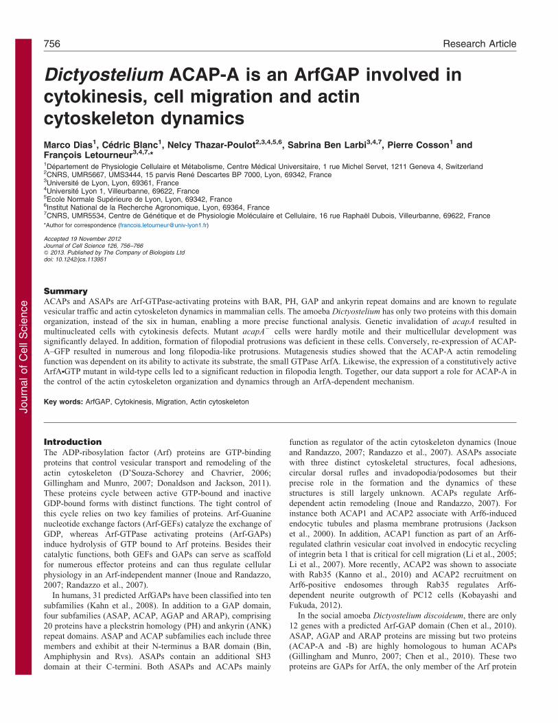
To determine whether ACAP-A localized to the cleavagefurrow, we made use of acapA2 cells expressing ACAP-A–GFP
since ACAP-A–GFP fully complemented the cytokinesis defectof acapA2 cells indicating that the fusion protein was functional.In interphase cells, ACAP-A–GFP was seen in the cytosol and
was enriched at the cell cortex (Fig. 2). In mitotic cells detectedby anti-tubulin staining, ACAP-A–GFP did not accumulate at thelevel of the cleavage furrow or the midbody. Instead, ACAP-A–GFP appeared still mainly in the cytosol and slightly enriched in
pericentrosomal regions in some cells. Like in mammalian cells,in Dictyostelium cytokinesis is dependent on the remodeling ofthe actin cytoskeleton, and many mutants with altered actin
cytoskeleton exhibit cytokinesis defects (Gebbie et al., 2004).Our observations suggest that, like ACAP proteins in mammaliancells, ACAP-A may be specifically required to regulate the
organization of the actin cytoskeleton.
In Dictyostelium, ArfA is the only ArfGTPase (Weeks et al.,2005; Chen et al., 2010). Protein sequence alignment (program
EMBOSS Needle, EMBL-EBI) with representative members of
the three classes of mammalian Arfs revealed that ArfA shared
83% identity and 88.5% similarity with human Arf1 (class I),
80.8% identity and 87.4% similarity with human Arf4 (class II),
and 64.3% identity and 78.6% similarity with human Arf6 (class
III). This high homology with class I/II human Arfs suggests that
ArfA might participate in the same cellular functions as these
human proteins. However, due to its significant similarity with
Arf6, ArfA might also play additional roles in actin cytoskeleton
dynamics and endocytic processes, as described for Arf6
(D’Souza-Schorey and Chavrier, 2006; Gillingham and Munro,
2007; Donaldson and Jackson, 2011). In mammalian cells, Arf6
is also required for cytokinesis and constitutively active Arf6
localizes to the plasma membrane at the site of cleavage furrow
ingression and midbody formation prior to the separation of the
daughter cells (Schweitzer and D’Souza-Schorey, 2002). Since
ArfA is a substrate for ACAP-A (Chen et al., 2010), we next
determined ArfA distribution during mitosis. To avoid massive
overexpression of ArfA, we transfected cells with plasmids that
allow inducible expression, upon doxycycline addition (Veltman
et al., 2009), of ArfA-Q71L or ArfA-T31N mutants fused to GFP
at their C-terminus. These mutations mimic respectively
constitutively activated (ArfNGTP) and dominant negative
(ArfNGDP) forms of human Arf1 that is the closest homolog of
ArfA (see above). After 48 hours of induction, ArfA mutants
were equally expressed in stably transfected WT cells (Fig. 3A)
and 17.9% of cells expressing ArfA-Q71L–GFP showed two
nuclei or more against 3.1% of ArfA-T31N–GFP expressing cells
(Fig. 3B). This cytokinesis defect induced by expression of
constitutively active ArfA is similar to that described in
mammalian cells upon overexpression of a GTPase-defective
Fig. 2. ACAP-A–GFP localization in mitotic cells. acapA2 + ACAP-A–
GFP cells were labeled with anti-tubulin antibody to detect mitotic cells and
analyzed by confocal microscopy. In interphase cells (upper row), ACAP-A–
GFP localized in the cytosol and at the cell surface. In mitotic cells, ACAP-
A–GFP did not accumulate on cleavage furrows (indicated by arrowheads)
but was slightly enriched in pericentrosomal regions (indicated by arrows) in
some cells. Scale bars: 5 mm.
Journal of Cell Science 126 (3)758
Journ
alof
Cell
Scie
nce
mutant of Arf6 (Arf6-Q67L, GTP-bound) (Schweitzer and
D’Souza-Schorey, 2002).
As previously described in cells constitutively expressing
GFP-tagged ArfA (Guetta et al., 2010), ArfA-Q71L–GFP and
ArfA-T31N–GFP mutants in interphase cells were mainly found
in the perinuclear region and showed a diffuse cytosolic
localization (Fig. 3D,E). In mitotic cells, neither ArfA-Q71L–
GFP nor ArfA-T31N–GFP was enriched at the cleavage furrow;
instead partial colocalization with tubulin was observed at the
spindle poles for both forms. Although a transient concentration
of ArfA at the cleavage furrow may have been overlooked, these
localization studies do not support a direct function of ArfA
during the abscission step. As described for Arf6 (D’Souza-
Schorey and Chavrier, 2006), ArfA might nevertheless play a role
in cytokinesis by regulating for instance membrane trafficking
processes.
acapA2 cells show reduced motility and streaming
When monitoring mitosis, we noticed that acapA2 cells were less
motile than wild-type cells. To further examine motility, cells
moving randomly on glass coverslips were imaged every
30 seconds for 30 minutes and 50 cells were analyzed. Mutant
acapA2 cells moved 5.462.9 mm from their initial positions
whereas WT and acapA2 + ACAP-A–GFP cells moved
36.7610.6 and 38.1612.9 mm, respectively (Fig. 4A). This was
accounted for by a marked difference in instantaneous speed:
acapA2 cells moved at 1.960.7 mm/minute while wild-type cells
moved at 5.160.6 mm/minute and complemented acapA2 cells at
4.760.6 mm/minute (data not shown). No migration defects were
observed in acapB2 cells (supplementary material Fig. S1B).
The motility defect of vegetative acapA2 cells prompted us to
examine whether multicellular development of starved cells was
altered in these cells. We first followed motility of individual
cells incubated in phosphate starvation buffer. Whereas motility
was still defective in acapA2 cell after one hour of starvation
(data not shown), WT and acapA2 cells moved respectively
61.9621.2 mm and 56.5617.2 mm from their initial positions
after 10 hours (Fig. 4B), thus indicating that motility was
restored in acapA2 cells during development.
We next observed by microscopy collective movement of cells
incubated into phosphate buffer in submerged conditions
(supplementary material Fig. S3A). After 10 hours, WT cells
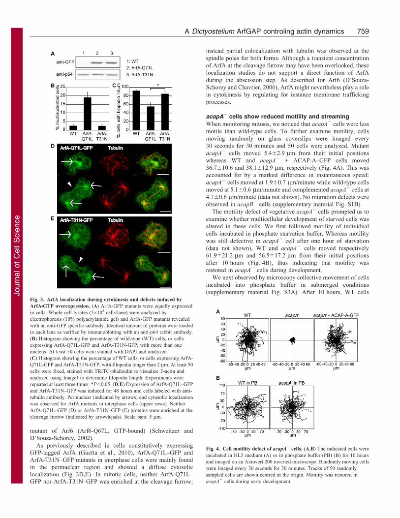
Fig. 4. Cell motility defect of acapA2 cells. (A,B) The indicated cells were
incubated in HL5 medium (A) or in phosphate buffer (PB) (B) for 10 hours
and imaged on an Axiovert 200 inverted microscope. Randomly moving cells
were imaged every 30 seconds for 30 minutes. Tracks of 50 randomly
sampled cells are shown centred at the origin. Motility was restored in
acapA2 cells during early development.
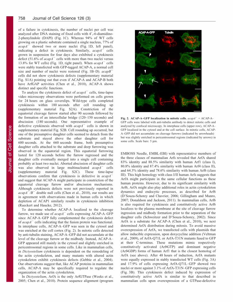
Fig. 3. ArfA localization during cytokinesis and defects induced by
ArfANGTP overexpression. (A) ArfA-GFP mutants were equally expressed
in cells. Whole cell lysates (56105 cells/lane) were analyzed by
electrophoresis (10% polyacrylamide gel) and ArfA-GFP mutants revealed
with an anti-GFP specific antibody. Identical amount of proteins were loaded
in each lane as verified by immunoblotting with an anti-p64 rabbit antibody.
(B) Histogram showing the percentage of wild-type (WT) cells, or cells
expressing ArfA-Q71L-GFP and ArfA-T31N-GFP, with more than one
nucleus. At least 50 cells were stained with DAPI and analyzed.
(C) Histogram showing the percentage of WT cells, or cells expressing ArfA-
Q71L-GFP and ArfA-T31N-GFP, with filopodia longer than 2 mm. At least 50
cells were fixed, stained with TRITC-phalloidin to visualize F-actin and
analyzed using ImageJ to determine filopodia length. Experiments were
repeated at least three times. *P,0.05. (D,E) Expression of ArfA-Q71L–GFP
and ArfA-T31N–GFP was induced for 48 hours and cells labeled with anti-
tubulin antibody. Perinuclear (indicated by arrows) and cytosolic localization
was observed for ArfA mutants in interphase cells (upper rows). Neither
ArfA-Q71L–GFP (D) or ArfA-T31N–GFP (E) proteins were enriched at the
cleavage furrow (indicated by arrowheads). Scale bars: 5 mm.
A Dictyostelium ArfGAP controling actin dynamics 759
Journ
alof
Cell
Scie
nce
gathered into streams moving towards aggregation centers.
Aggregates were then clearly individualized after 14 hours.Streaming and aggregation was also observed with acapA2 cells
but with a delay of 4 hours in comparison to wild-type cells. To
observe the late stage of development, cells were then plated on
solid starvation plates. Development of WT cells was complete
after 24 hours whereas acapA2 cells showed fruiting bodystructures with morphology comparable to wild-type cells only
after 36 hours (supplementary material Fig. S3C). In addition,
spore yield and viability were not affected in acapA2 cells (data
not shown). This last result suggests that double deletion of bothacapA and acapB genes might be required to observe spore
production defects previously reported in acapA2/B2 cells (Chen
et al., 2010).
Together our data suggest that ACAP-A does not affect
chemotaxis or development. During development, mutant cells
appear to migrate through a mechanism independent of ACAP-A.
The time delay of the development cycle observed in the absenceof ACAP-A might be due to defective migration until this ACAP-
A-independent migration mechanism proceeds and/or ACAP-Acontrol is released. These results are in agreement with theprevious report that acapA2, acapB2 and acapA2/B2 cells showa normal multicellular development (Chen et al., 2010).
Altered cell shape and organization of F-actin in acapA2
cells
The poor motility of acapA2 cells strongly suggested that theremodeling of the actin cytoskeleton might be defective in thesecells since actin-driven membrane protrusions are important for
cell migration. To determine the effect of ACAP-A on cell shapeand organization of F-actin, cells were labeled with TRITC-phalloidin to visualize F-actin (Fig. 5A). We also examined cell
morphology by scanning electron microscopy (Fig. 5B). Incontrast to WT and acapB 2 cells (supplementary material Fig.
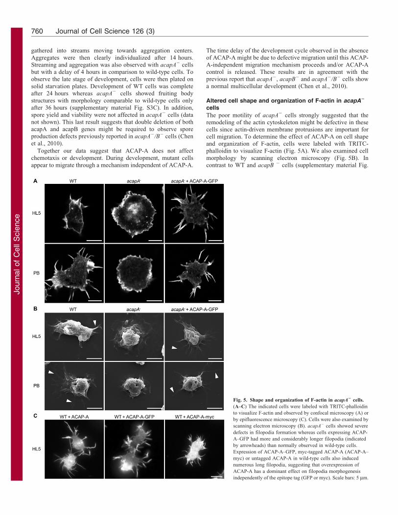
Fig. 5. Shape and organization of F-actin in acapA2 cells.
(A–C) The indicated cells were labeled with TRITC-phalloidin
to visualize F-actin and observed by confocal microscopy (A) or
by epifluorescence microscopy (C). Cells were also examined by
scanning electron microscopy (B). acapA2 cells showed severe
defects in filopodia formation whereas cells expressing ACAP-
A–GFP had more and considerably longer filopodia (indicated
by arrowheads) than normally observed in wild-type cells.
Expression of ACAP-A–GFP, myc-tagged ACAP-A (ACAP-A–
myc) or untagged ACAP-A in wild-type cells also induced
numerous long filopodia, suggesting that overexpression of
ACAP-A has a dominant effect on filopodia morphogenesis
independently of the epitope tag (GFP or myc). Scale bars: 5 mm.
Journal of Cell Science 126 (3)760
Journ
alof
Cell
Scie
nce
S1C,D), acapA2 cells showed severe defects in filopodia
morphology and number. Instead of filopodia, they mainly
displayed numerous membrane folds and lamellipodia
(Fig. 5A,B). In culture medium (HL5), 100% of acapA2 cells
had no filopodia at all or less than six filopodia in contrast to WT
cells which showed 75% of cells with less than six filopodia per
cell (Fig. 6B). Furthermore, in WT cells 14% of filopodia were
longer than 2 mm whereas filopodia this size were not seen in
acapA2 cells (Fig. 6C). Conversely, transfection of GFP-tagged
ACAP-A in acapA2 cells complemented this defect and
generated more numerous as well as considerably longer
filopodia than usually observed in WT cells (Fig. 5A). Thus,
90% of acapA2 + ACAP-A–GFP cells showed more than five
filopodia whereas no acapA2 cells and only 25% of WT cells did
(Fig. 6B). In acapA2 + ACAP-A–GFP cells, 71% of filopodia
were longer than 2 mm in contrast to 14% in WT cells (Fig. 6C).
Interestingly, expression of ACAP-A–GFP in WT cells also
induced numerous long filopodia (Fig. 5C), suggesting that
overexpression of ACAP-A–GFP has a dominant effect on
filopodia morphogenesis. Note that expression of untagged or
myc-tagged ACAP-A led to comparable results (Fig. 5C),
suggesting that the effect observed was caused by
overexpression of ACAP-A. Finally, expression of ACAP-B–
GFP in acapA2 cells did not restore filopodia biogenesis, further
underlining the distinct functions of ACAP-A and ACAP-B
(supplementary material Fig. S1E).
Incubation of Dictyostelium cells in phosphate buffer induces
numerous thin filopodial extensions, which are not observed in
some mutants defective in genes controlling actin cytoskeleton
remodeling (Gebbie et al., 2004). In this condition, acapA2 cells
formed filopodia that were short (87% filopodia #2 mm) in
comparison to those observed in WT cells (83% filopodia
.2 mm) (Fig. 5A,B and Fig. 6B,C). Note that the number and
length of filopodia induced by the expression of ACAP-A–GFP
were also influenced by phosphate buffer incubation (Fig. 6B,C).
Together these results indicate a specific requirement for ACAP-
A but not ACAP-B in the biogenesis of filopodia.
Next, to determine the dynamics of filopodia formed upon
expression of ACAP-A–GFP in acapA2 cells, we monitored
filopodia formation and retraction in live cells by time-lapse
video microscopy (Fig. 7). Filopodia extended with
instantaneous speed of 0.4960.06 mm/second (n510) in WT
cells (Fig. 7A) and 0.5560.11 mm/second (n510) in acapA2 +
ACAP-A–GFP cells (Fig. 7B,C). As previously reported
(Medalia et al., 2007), retraction was often associated with an
early filopodia bending step (Fig. 7D). These long filopodia did
not inhibit migration (Fig. 4A) and apparently extended soon
after pseudopodia protrusion (data not shown). We conclude that
ACAP-A–GFP overexpression do not interfere with normal
filopodia dynamics and cell migration.
ACAP-A-regulated actin remodeling depends on its GAP
activity
To test whether actin remodeling was dependent on the GAP
activity of ACAP-A, we mutated a conserved arginine (mutation
R633Q) known to be critical for ArfGAP activity (Mandiyan
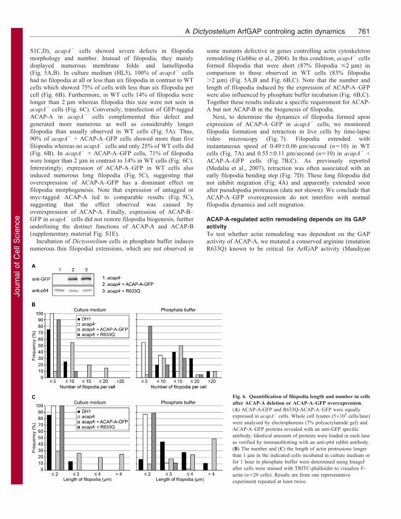
Fig. 6. Quantification of filopodia length and number in cells
after ACAP-A deletion or ACAP-A–GFP overexpression.
(A) ACAP-A-GFP and R633Q-ACAP-A–GFP were equally
expressed in acapA2 cells. Whole cell lysates (56105 cells/lane)
were analyzed by electrophoresis (7% polyacrylamide gel) and
ACAP-A–GFP proteins revealed with an anti-GFP specific
antibody. Identical amounts of proteins were loaded in each lane
as verified by immunoblotting with an anti-p64 rabbit antibody.
(B) The number and (C) the length of actin protrusions longer
than 1 mm in the indicated cells incubated in culture medium or
for 1 hour in phosphate buffer were determined using ImageJ
after cells were stained with TRITC-phalloidin to visualize F-
actin (n520 cells). Results are from one representative
experiment repeated at least twice.
A Dictyostelium ArfGAP controling actin dynamics 761
Journ
alof
Cell
Scie
nce
et al., 1999; Randazzo et al., 2000; Szafer et al., 2000; Jackson
et al., 2000). Protein expression level of mutant ACAP-A–GFP
stably expressed in acapA2 cells was comparable to that of
ACAP-A–GFP (Fig. 6A). However this mutant did not restore
filopodia formation (Fig. 6B,C) nor did it complement the
cytokinesis defect (data not shown) in acapA2 cells. This result
strongly suggests a role of ArfA in filopodia biogenesis. This
hypothesis was further comforted by the observation that
expression of a constitutively activated ArfANGTP mutant in
WT cells led to a significant reduction of filopodia length in
comparison to cells expressing a dominant negative ArfANGDP
mutant (Fig. 3C). Together these results suggest that the effect of
ACAP-A on the actin cytoskeleton is GAP-activity dependent
and likely associated to the control of ArfANGTP level in cells.
ACAP-A is not enriched in filopodia
In vertebrates, ASAPs and ACAPs are associated with
cytoskeleton structures (Inoue and Randazzo, 2007; Randazzo
et al., 2007). To test whether ACAP-A function in filopodia
biogenesis was linked to a hypothetic localization in membrane
protrusions, first acapA2 cells expressing ACAP–GFP were
observed by confocal microscopy of live cells. When a confocal
section was made within the cell body (CB, Fig. 8A,B), ACAP-
A–GFP was seen in the cytosol and accumulated at the plasma
membrane. In contrast, optical sections made at the level of the
cell contact with the substratum (CS, Fig. 8A,C) revealed that
ACAP-A–GFP was present but not concentrated in membrane
protrusions. We next labeled fixed cells with TRITC-phalloidin
to decorate F-actin. Colocalization of ACAP-A–GFP with
cortical F-actin was observed (CB, Fig. 8D) whereas ACAP-A–
GFP was not concentrated in filopodial F-actin-rich structures.
This suggests that ACAP-A might not be directly required for
the elongation of filopodial actin filaments or membrane
deformation. Note that a transient accumulation (undetected in
this study) of ACAP-A–GFP to filopodia cannot be formally
excluded.
Loss of ACAP-A induces similar phenotypes in AX2 and
DH1 strains
Our results indicating that ACAP-A plays a role in cytokinesis,
cell motility and actin cytoskeleton remodeling were in apparent
contradiction with a previous study that did not identify any such
role for ACAP-A (Chen et al., 2010). Since these differences may
be caused by the use of a different parental strain (AX2 versus
DH1), we also deleted ACAP-A in AX2, the parental strain used
in this previous work. Hereafter deleted strains are referred to as
AX2-acapA2 and DH1-acapA2 cells, respectively.
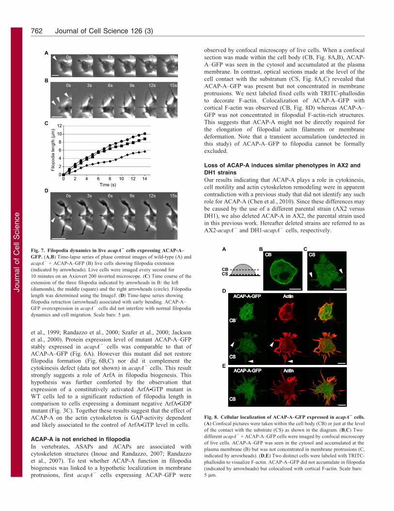
Fig. 7. Filopodia dynamics in live acapA2 cells expressing ACAP-A–
GFP. (A,B) Time-lapse series of phase contrast images of wild-type (A) and
acapA2 + ACAP-A–GFP (B) live cells showing filopodia extension
(indicated by arrowheads). Live cells were imaged every second for
10 minutes on an Axiovert 200 inverted microscope. (C) Time course of the
extension of the three filopodia indicated by arrowheads in B: the left
(diamonds), the middle (square) and the right arrowheads (circle). Filopodia
length was determined using the ImageJ. (D) Time-lapse series showing
filopodia retraction (arrowhead) associated with early bending. ACAP-A–
GFP overexpression in acapA2 cells did not interfere with normal filopodia
dynamics and cell migration. Scale bars: 5 mm.
Fig. 8. Cellular localization of ACAP-A–GFP expressed in acapA2 cells.
(A) Confocal pictures were taken within the cell body (CB) or just at the level
of the contact with the substrate (CS) as shown in the diagram. (B,C) Two
different acapA2 + ACAP-A–GFP cells were imaged by confocal microscopy
of live cells. ACAP-A–GFP was seen in the cytosol and accumulated at the
plasma membrane (B) but was not concentrated in membrane protrusions (C,
indicated by arrowheads). (D,E) Two distinct cells were labeled with TRITC-
phalloidin to visualize F-actin. ACAP-A–GFP did not accumulate in filopodia
(indicated by arrowheads) but colocalized with cortical F-actin. Scale bars:
5 mm.
Journal of Cell Science 126 (3)762
Journ
alof
Cell
Scie
nce
As expected, ACAP-A was not detected by western blotting inAX2-acapA2 cells (supplementary material Fig. S1F). DAPI
staining of fixed cells revealed that 45.8% of AX2-acapA2 cellsgrown on plastic dishes exhibited two or more nuclei, a featureobserved only in 2.4% of AX2 cells (supplementary material Fig.
S1G). Only 12.1% of AX2-acapA2 cells contained more thanthree nuclei against 52.8% of DH1-acapA2 cells (Fig. 1D). ThusACAP-A gene disruption in AX2 leads to a cytokinesis defectless pronounced than in DH1 cells, a phenotype that may be
missed if not specifically tested. AX2-acapA2 cells grown insuspension also exhibited a cytokinesis defect (supplementarymaterial Fig. S1G). Next, we observed that AX2-acapA2 cells
were hardly motile, with an instantaneous speed of 1.160.3 mm/minute while parental AX2 cells that moved at 3.960.6 mm/minute (supplementary material Fig. S1H). Motility of AX2-
acapA2 cells was restored during development since streamingand aggregation were observed with only minor time delayscompared to AX2 cells (supplementary material Fig. S3B,D).After 24 hours of development, both AX2 and AX2-acapA2 cells
showed fruiting bodies with comparable morphologies(supplementary material Fig. S3D). Finally, the organization ofF-actin was analyzed in cells labeled with TRITC-phalloidin
(supplementary material Fig. S1I–L). In culture medium (HL5),100% of AX2-acapA2 cells showed less than six filopodiaagainst 45% of AX2 cells (supplementary material Fig. S1J).
Whereas 47% of filopodia were longer than 2 mm in AX2 cells,such long filopods were not observed in AX2-acapA2 cells(supplementary material Fig. S1K). This defect in AX2-acapA2
cells was further amplified upon cell incubation in phosphatebuffer for one hour. AX2-acapA2 cells still formed shortfilopodia (90% filopodia #2 mm) while AX2 cells mainlyshowed long filopodia (86% filopodia .2 mm) (supplementary
material Fig. S1K). Remarkably, after 6 hours of incubation innutrient-depleted phosphate buffer, AX2-acapA2 cells showednumerous cortical spike-like protrusions (,1 mm) whereas AX2
cells still displayed long filopodia (supplementary material Fig.S1L). This result is in agreement with actin remodeling defectspreviously noticed in acapA2/B2 cells after 5–6 hours of
starvation (Chen et al., 2010).
We conclude that defects observed in acapA2 cells wereidentically observed in two parental Dictyostelium strains, further
reinforcing the role of ACAP-A in cytokinesis and actincytoskeleton dynamics. Partial discrepancies between ourresults and those of Chen and co-workers are discussed below.
DiscussionIn this study, we report the function of an Arf-GTPase activatingprotein, ACAP-A, in the model organism Dictyostelium
discoideum. We demonstrate for the first time that inactivationof the gene coding for ACAP-A results in strong phenotypes,
including cytokinesis defects, reduced motility and impairedactin cytoskeleton remodeling. We provide the first evidence thatthe GAP domain is essential for these functions suggesting that
ACAP-A regulates the actin skeleton organization and dynamicsin an ArfA-dependent manner.
ACAP-A and cytokinesis
In mammalian cells, Arf6 is required for cytokinesis (D’Souza-
Schorey and Chavrier, 2006). Hence overexpression of Arf6NGTP(Brown et al., 2001) and depletion of Arf6 using siRNA(Schweitzer and D’Souza-Schorey, 2002) cause cytokinesis
defects. In addition, depletion of ACAP1, a GAP for Arf6 that
is highly homologous to ACAP-A (Chen et al., 2010), impairs
cytokinesis and leads to the accumulation of binucleated cells
(Rueckert and Haucke, 2012). Here in Dictyostelium, cytokinesis
defects were comparably observed when manipulating ArfA
activity. This was first achieved by interfering with the GAP
activity of ACAP-A. Indeed, in cells deprived of ACAP-A
(acapA2 cells) inferred excess of ArfANGTP was associated with
concomitant cytokinesis defects. ArfA activity was also altered in
cells by overexpression of a constitutively active mutant of ArfA
(ArfANGTP) and this led to cells with multiple nuclei.
What is the function of ArfA and ACAP-A in cytokinesis? Our
microscopy studies did not allow to identify a particular
localization for ArfA and ACAP-A that would indicate an
obvious function in cytokinesis, for instance in cleavage furrow
ingression. In mammalian cells, Arf6NGTP localizes to the
cleavage furrow and the midbody in mitotic cells (Schweitzer
and D’Souza-Schorey, 2002). Numerous studies on Arf6 strongly
indicate that this GTPase regulates endosomal trafficking events
crucial for cytokinesis (Schweitzer et al., 2011; McKay and
Burgess, 2011; D’Souza-Schorey and Chavrier, 2006). In
Dictyostelium, ArfA is the only ArfGTPase (Weeks et al.,
2005; Chen et al., 2010) and thus, functions in the endocytic or
secretory pathways controlled by individual members of the three
different mammalian Arf classes might be supported by ArfA
alone in amoeba. To our knowledge, the role of ArfA in
endosomal trafficking has never been assessed, although
ArfANGDP was reported to associate with an arrestin related
protein localized to endocytic compartments (Guetta et al., 2010)
and ArfA was also identified as a phagosomal protein (Gotthardt
et al., 2006). ArfA is highly homologous to human Arf1 (Weeks
et. al., 2005; Chen et al., 2010) known to regulate membrane
trafficking in the secretory pathway. In mammalian cells,
secretory Golgi-derived vesicles traffic to the cleavage furrow
and the midbody region during cytokinesis (Goss and Toomre,
2008). Consistent with the localization of ArfA to the Golgi
apparatus (Guetta et al., 2010), the role of ArfA in cytokinesis
might be related to hypothetical functions in the secretory
pathway and Golgi structure. This hypothesis does not exclude
that ArfA (and ACAP-A) might also be required for the control of
actin remodeling during cytokinesis.
ACAP-A and cell migration
For the first time we report here that ACAP-A participates in
vegetative Dictyostelium cell migration. In mammalian cells,
downregulation as well as overexpression of the Arf6 GAP
ACAP1 has been shown to inhibit migration induced by
stimulation-dependent recycling of integrin b and controls of
Arf6NGTP levels (Li et al., 2005; Ma et al., 2007). The role of
Arf6 in cell migration is known to rely on its capacity to regulate
internalization and trafficking of integrins but also to remodel the
actin cytoskeleton notably via Rac1 trafficking to the plasma
membrane (Schweitzer et al., 2011; D’Souza-Schorey and
Chavrier, 2006). In Dictyostelium, Rac1 GTPases appear to
regulate cell motility (Dumontier et al., 2000; Filic et al., 2011)
and therefore ACAP-A might participate in Rac1 localization that
will regulate subsequently actin cytoskeleton dynamics.
Interestingly, during Dictyostelium development, ACAP-A does
not regulate cell motility since motility was restored in acapA2
cells upon starvation. This suggests that the migration machinery
A Dictyostelium ArfGAP controling actin dynamics 763
Journ
alof
Cell
Scie
nce
might be controlled by ArfA/ACAP-A independent signals
during development.
ACAP-A and actin cytoskeleton remodeling
Multi-domain ArfGAPs have been shown to regulate membrane
remodeling associated to actin polymerization (Randazzo and
Hirsch, 2004; Randazzo et al., 2007). Here we provide evidence
that ACAP-A regulates actin cytoskeleton remodeling in
Dictyostelium. First, deletion of ACAP-A impairs membrane
protrusions usually observed in vegetative Dictyostelium cells.
Second, overexpression of ACAP-A in acapA2 cells induces the
formation of numerous long filopodia. In addition, we show here
that actin remodeling is dependent on the GAP activity of ACAP-
A since a GAP defective ACAP-A mutant (ACAP-A R633Q)
expressed in acapA2 cells does not restore filopodia formation.
Therefore the effect of ACAP-A on actin remodeling appears as
an Arf-dependent activity and consequently might be associated
to well-known Arf functions such as the regulation of Rho family
GTPases that control actin dynamics (Schweitzer et al., 2011;
McKay and Burgess, 2011; D’Souza-Schorey and Chavrier,
2006). Hence, ArfA might control actin polymerization by
positive regulation of Rac1, which in turn would activate dDia2
responsible for filopodia elongation (Schirenbeck et al., 2005). In
agreement with the hypothetical Arf-dependent function of
ACAP-A, we observe that overexpression of a constitutively
activated ArfANGTP mutant leads to a significant reduction of
filopodia length. However the extent of this reduction is modest
in comparison to that observed in acapA2 cells in which
ArfANGTP is inferred to accumulate. Furthermore,
overexpression of a dominant negative ArfANGDP mutant, a
situation that should mimic ACAP-A overexpression, does not
enhance filopodia protrusions. Together these observations
suggest that ACAP-A could have additional Arf-independent
activities that affect actin cytoskeleton. ACAP-A might play for
instance a direct role in actin nucleation during filopodia
formation. However in contrast to dDia2, a known actin
nucleator localized at the tips of filopodia (Schirenbeck et al.,
2005), ACAP-A–GFP is apparently not enriched in these
domains, unless this localization is too transient to be detected
or is masked by the overexpression of the GFP-tagged protein.
Alternatively, ACAP-A might participate in membrane
deformation associated to filopodia extension. Such a role has
been described for proteins with an IMD domain (IRSp53 and
MIM homology Domain) similar to the BAR/I-BAR domain that
functions both as a sensor and inducer of membrane curvature
(Ahmed et al., 2010; Rao and Haucke, 2011). Indeed,
overexpression of the IMD domain of IRSp53 can induce
filopodia-like structures and other domains of IRSp53 interact
with several key players in actin dynamics (e.g. WAVE, N-
WASP, mDia1) suggesting that IRSp53 participates in the
mechanism of filopodia formation (Ahmed et al., 2010).
Interestingly, ACAP-A contains a BAR domain that might
share similar functions with the IMD domain of IRSp53.
However, the apparent cortical localization of ACAP-A–GFP
does not support this hypothesis. Alternatively, the BAR domain
(maybe interacting with the PH domain) could regulate the
association of ACAP-A with membranes enriched in ArfA or
might affect the GAP enzymatic activity as reported for the
ArfGAP ASAP1 (Jian et al., 2009). Additional experiments will
be further required to test these hypothesis and determine critical
domains of ACAP-A required for theses GAP-independentfunctions.
Comparison to previous studies on Dictyostelium ACAP-A
Finally, as mentioned above, there are apparent discrepancies
between our study and previously published results (Chen et al.,2010), since we demonstrate that the deletion of ACAP-A issufficient to induce dramatic defects in vegetative cells that were
unnoticed in this early work. However, upon close examination,the vast majority of the differences between our results and thisprevious study appear to reflect differences in the phenotypesanalyzed in both studies. First, the cytokinesis defect in acapA2
cells was not described in this previous study. Here we report thatAX2-acapA2 cells exhibit a cytokinesis defect, but lesspronounced than in DH1-acapA2 cells. Thus the cytokinesis
defect in AX2-acapA2 cells might be only revealed upon carefulquantitative analysis of DAPI stained nuclei, which is not aroutine procedure in all laboratories. Second, we noticed a strong
motility defect in acapA2 cells that had not been reported before.In our hands, this defect was observed both in AX2-acapA2 andDH1-acapA2 cells but time-lapse microscopy studies wererequired to uncover these cell migration defects. However, we
observed a nearly normal motility of acapA2 cells duringdevelopment, which is in agreement with previous resultsreporting normal chemotaxis, streaming and development in
acapA2/B2 cells (Chen et al., 2010). Third, the reduced numberand length of filopodia in vegetative acapA2 cells was notpreviously observed, whereas we demonstrate here that both
AX2-acapA2 and DH1-acapA2 cells have a similar defect inactin remodeling. However this anomaly is most striking whencells are incubated in phosphate buffer for one hour, a condition
that was not analyzed previously. Conversely, Chen and co-workers analyzed actin cytoskeleton of cells during development(5–6 hours of starvation) and reported that acapA2/B2 cellsdisplay a greater number of short actin protrusions in comparison
to wild-type cells (Chen et al., 2010), a phenotype also observedin this study. In summary, no significant contradiction isnoticeable between our results and previously published results.
It is likely that defects associated with inactivation of ACAP-Awere overlooked because they were not apparent in the assaysused previously (Chen et al., 2010).
Materials and MethodsCell culture, antibodies, gel electrophoresis and immunoblotting
D. discoideum strains DH1-10 (Cornillon et al., 2000) and AX2 were grown at22 C in HL5 medium and subcultured twice a week. Mouse monoclonal antibody(mAb) against p25 (H72) was described previously (Mercanti et al., 2006). mAb toGFP and myc (9E10) were purchased (Roche Diagnostics, Meylan, France).mAb to alpha tubulin (DM1A), 49,6-diamidino-2-phenylindole (DAPI) andtetramethylrhodamine B isothiocyanate (TRITC)-labeled phalloidin were fromSigma-Aldrich (St Quentin Fallavier, France). Polyclonal antibodies to ACAP-Aand p64 (DDB_G0282233) were raised in rabbits using KLH-coupled peptides(1063EKDKDYKNTRPKSK1076; 1311NKKPKKSKKSKPLE1324) and GST-p64(residues 427–522) recombinant protein, respectively (Covalab, Villeurbanne,France). The anti-ACAP-A antibody did not generate a signal inimmunofluorescence experiments (data not shown). SDS polyacrylamideelectrophoresis and immunoblotting were performed as previously described(Cornillon et al., 2000). Bands were detected by ECL (Thermo Scientific,Courtaboeuf, France) and a ChemiDoc MP imager (Bio-Rad, Marnes-la-Coquette,France).
Immunofluorescence microscopy
For immunofluorescence analysis, cells were applied on glass coverslips overnight,incubated or not in phosphate buffer (PB, 2 mM Na2HPO4, 14.7 mM KH2PO4,pH 6.5) then fixed with 4% paraformaldehyde for 30 minutes, washed andpermeabilized with methanol at 220 C for 2 minutes. Cells were incubated with
Journal of Cell Science 126 (3)764
Journ
alof
Cell
Scie
nce
the indicated antibodies for 1 hour, and then stained with correspondingfluorescent (Alexa Fluor 488/568) secondary antibodies (Molecular Probes/Invitrogen, Eugene, OR) for 30 minutes. The actin cytoskeleton was labeled byincubating paraformaldehyde-fixed cells for 1 hour in phosphate-buffered saline(PBS) containing 0.2% BSA and 1 mg/ml (TRITC)-labeled phalloidin. Cells wereobserved by laser scanning confocal microscopy (Zeiss LSM 510 Meta) orepifluorescence microscopy (Zeiss AxioImager Z1) when indicated. For nucleistaining, cells were treated as described above, incubated with DAPI for30 minutes and observed with a Zeiss AxioImager Z1 photomicroscope.
Scanning electron microscopy
For scanning electron microscopy, cells were incubated on glass coverslidesovernight in HL5. Cells were then incubated or not in PB for 1 hour before fixationusing 2% glutaraldehyde in HL5 for 30 minutes followed by 2% glutaraldehyde in100 mM PB (pH 7.14) for 30 minutes. Cells were rinsed and postfixed in 1%osmium tetroxide in 100 mM PB (pH 7.14) for 1 hour. The fixative was removed,and cells were progressively dehydrated through a 25–100% ethanol series. Afterair-drying, cells were sputter-coated in gold and viewed on a JEOL-JSM-7001 FAField Emission Scanning Electron Microscope.
Live cell imaging and analysis
Cells were incubated overnight in Lab-Tek (Nalgene) chambered coverglasses inHL5 medium and imaged on an Axiovert 200 inverted microscope with a motorizedstage. Randomly moving cells were imaged every 30 seconds for 30 minutes. Filmswere processed by manual centroid tracking of at least 50 cells with the ImageJanalysis software (NIH) and MTrackJ. For mitosis analysis, asynchronously growncells were imaged every 15 seconds for 1 hour. To document filopodia dynamics,cells were identically imaged every second for 10 minutes.
Plasmids and cell transfection
Full-length ACAP-A and mutants were produced by PCR using pairs ofoligonucleotides containing BamHI and XhoI sites in 59 and 39, respectively.PCR fragments were digested by BamHI and XhoI and cloned into BamHI/XhoIsites of pDXA-3C (Manstein et al., 1995) containing either GFP or a triple myc-tagfor C-terminal fusion. All constructs were sequenced (Genome express, Grenoble,France). Plasmids were linearized by ScaI and transfected in Dictyostelium byelectroporation as described (Cornillon et al., 2000). Clone selection was madewith 10 mg/ml G418. ArfA(Q71L) and ArfA(T31N) mutants (kindly provided byDr Laurence Aubry, CEA, Grenoble, France) were subcloned after PCRamplification into BglII/SpeI sites of the inducible expression vector pDM370(Veltman et al., 2009). After electroporation, transfectants were selected andmaintained in 25 mg/ml hygromycin. Expression was induced by adding 10 mg/mldoxycycline 2 or 3 days before analysis.
acapA knockout cells
To obtain the acapA knockout vector, the 59 fragment was amplified from genomicDNA with sens (ATGAGTGGGCAACAACCAACAACAGAA) and antisens(CATTTGATCATTAATACTATTAACAGA) oligonucleotides and cloned intopBlueScript vector (Stratagene, La Jolla, CA). The 39 fragment was obtained by PCRusing sense (GAATTTCATCCAGAAAATGCATTAAAT) and antisense(TGGATCAACCAATGTAATGTCAGCACC) oligonucleotides and cloned inpBlueScript containing the 59 fragment. After sequencing, the knockout vectorwas completed by inserting the blasticidin resistance cassette between the two 59 and39 fragments. The resulting plasmid was linearized by digestion with restrictionenzymes (KpnI and NotI) and electroporated into DH1-10 cells. Transformants wereselected in presence of 10 mg/ml blasticidin. Individual colonies were tested by PCRand the absence of expression ACAP-A mRNA was then verified by RT-PCR. TheACAP-A mutant retains residues 1–273 from the complete amino sequence, leavingthus only half of the BAR domain (102 out of 229 residues).
AcknowledgementsWe thank Laurence Aubry (U1038 Inserm/CEA/UJF CEA, Grenoble,France) for her generous gift of ArfA constructs and ChristopheAnjard (UMR5534, Lyon, France) for helpful suggestions.
FundingThis work was supported by grants from the Association pour laRecherche contre le Cancer (ARC) [grant number 1082 to F.L.]; andthe research program of the Region Rhone-Alpes (to F.L.). P.C.’slaboratory is funded by the Fonds National Suisse pour la RechercheScientifique.
Supplementary material available online at
http://jcs.biologists.org/lookup/suppl/doi:10.1242/jcs.113951/-/DC1
ReferencesAhmed, S., Goh, W. I. and Bu, W. (2010). I-BAR domains, IRSp53 and filopodium
formation. Semin. Cell Dev. Biol. 21, 350-356.
Block, J., Breitsprecher, D., Kuhn, S., Winterhoff, M., Kage, F., Geffers, R., Duwe,
P., Rohn, J. L., Baum, B., Brakebusch, C. et al. (2012). FMNL2 drives actin-based
protrusion and migration downstream of Cdc42. Curr. Biol. 22, 1005-1012.
Breitsprecher, D., Jaiswal, R., Bombardier, J. P., Gould, C. J., Gelles, J. and Goode,
B. L. (2012). Rocket launcher mechanism of collaborative actin assembly defined by
single-molecule imaging. Science 336, 1164-1168.
Brown, F. D., Rozelle, A. L., Yin, H. L., Balla, T. and Donaldson, J. G. (2001).
Phosphatidylinositol 4,5-bisphosphate and Arf6-regulated membrane traffic. J. Cell
Biol. 154, 1007-1018.
Chen, P. W., Randazzo, P. A. and Parent, C. A. (2010). ACAP-A/B are ArfGAP
homologs in dictyostelium involved in sporulation but not in chemotaxis. PLoS ONE
5, e8624.
Cornillon, S., Pech, E., Benghezal, M., Ravanel, K., Gaynor, E., Letourneur, F.,
Bruckert, F. and Cosson, P. (2000). Phg1p is a nine-transmembrane protein
superfamily member involved in dictyostelium adhesion and phagocytosis. J. Biol.
Chem. 275, 34287-34292.
D’Souza-Schorey, C. and Chavrier, P. (2006). ARF proteins: roles in membrane traffic
and beyond. Nat. Rev. Mol. Cell Biol. 7, 347-358.
Donaldson, J. G. and Jackson, C. L. (2011). ARF family G proteins and their
regulators: roles in membrane transport, development and disease. Nat. Rev. Mol. Cell
Biol. 12, 362-375.
Dumontier, M., Hocht, P., Mintert, U. and Faix, J. (2000). Rac1 GTPases control
filopodia formation, cell motility, endocytosis, cytokinesis and development in
Dictyostelium. J. Cell Sci. 113, 2253-2265.
Faix, J. and Rottner, K. (2006). The making of filopodia. Curr. Opin. Cell Biol. 18, 18-
25.
Faix, J., Breitsprecher, D., Stradal, T. E. and Rottner, K. (2009). Filopodia: Complex
models for simple rods. Int. J. Biochem. Cell Biol. 41, 1656-1664.
Filic, V., Marinovic, M., Faix, J. and Weber, I. (2012). A dual role for Rac1 GTPases
in the regulation of cell motility. J. Cell Sci. 125, 387-398.
Gebbie, L., Benghezal, M., Cornillon, S., Froquet, R., Cherix, N., Malbouyres, M.,
Lefkir, Y., Grangeasse, C., Fache, S., Dalous, J. et al. (2004). Phg2, a kinase
involved in adhesion and focal site modeling in Dictyostelium. Mol. Biol. Cell 15,
3915-3925.
Gillingham, A. K. and Munro, S. (2007). The small G proteins of the Arf family and
their regulators. Annu. Rev. Cell Dev. Biol. 23, 579-611.
Goss, J. W. and Toomre, D. K. (2008). Both daughter cells traffic and exocytose
membrane at the cleavage furrow during mammalian cytokinesis. J. Cell Biol. 181,
1047-1054.
Gotthardt, D., Blancheteau, V., Bosserhoff, A., Ruppert, T., Delorenzi, M. and
Soldati, T. (2006). Proteomics fingerprinting of phagosome maturation and evidence
for the role of a Galpha during uptake. Mol. Cell. Proteomics 5, 2228-2243.
Guetta, D., Langou, K., Grunwald, D., Klein, G. and Aubry, L. (2010). FYVE-
dependent endosomal targeting of an arrestin-related protein in amoeba. PLoS ONE 5,
e15249.
Gupton, S. L. and Gertler, F. B. (2007). Filopodia: the fingers that do the walking. Sci.
STKE 2007, re5.
Inoue, H. and Randazzo, P. A. (2007). Arf GAPs and their interacting proteins. Traffic
8, 1465-1475.
Jackson, T. R., Brown, F. D., Nie, Z., Miura, K., Foroni, L., Sun, J., Hsu, V. W.,
Donaldson, J. G. and Randazzo, P. A. (2000). ACAPs are arf6 GTPase-activating
proteins that function in the cell periphery. J. Cell Biol. 151, 627-638.
Jian, X., Brown, P., Schuck, P., Gruschus, J. M., Balbo, A., Hinshaw, J. E. and
Randazzo, P. A. (2009). Autoinhibition of Arf GTPase-activating protein activity by
the BAR domain in ASAP1. J. Biol. Chem. 284, 1652-1663.
Kahn, R. A., Bruford, E., Inoue, H., Logsdon, J. M., Jr, Nie, Z., Premont, R. T.,
Randazzo, P. A., Satake, M., Theibert, A. B., Zapp, M. L. et al. (2008). Consensus
nomenclature for the human ArfGAP domain-containing proteins. J. Cell Biol. 182,
1039-1044.
Kanno, E., Ishibashi, K., Kobayashi, H., Matsui, T., Ohbayashi, N. and Fukuda, M.
(2010). Comprehensive screening for novel rab-binding proteins by GST pull-down
assay using 60 different mammalian Rabs. Traffic 11, 491-507.
Kerber, M. L. and Cheney, R. E. (2011). Myosin-X: a MyTH-FERM myosin at the tips
of filopodia. J. Cell Sci. 124, 3733-3741.
Kobayashi, H. and Fukuda, M. (2012). Rab35 regulates Arf6 activity through
centaurin-b2 (ACAP2) during neurite outgrowth. J. Cell Sci. 125, 2235-2243.
Li, J., Ballif, B. A., Powelka, A. M., Dai, J., Gygi, S. P. and Hsu, V. W. (2005).
Phosphorylation of ACAP1 by Akt regulates the stimulation-dependent recycling of
integrin beta1 to control cell migration. Dev. Cell 9, 663-673.
Li, J., Peters, P. J., Bai, M., Dai, J., Bos, E., Kirchhausen, T., Kandror, K. V. and
Hsu, V. W. (2007). An ACAP1-containing clathrin coat complex for endocytic
recycling. J. Cell Biol. 178, 453-464.
Ma, Z., Nie, Z., Luo, R., Casanova, J. E. and Ravichandran, K. S. (2007). Regulation
of Arf6 and ACAP1 signaling by the PTB-domain-containing adaptor protein GULP.
Curr. Biol. 17, 722-727.
Mandiyan, V., Andreev, J., Schlessinger, J. and Hubbard, S. R. (1999). Crystal
structure of the ARF-GAP domain and ankyrin repeats of PYK2-associated protein
beta. EMBO J. 18, 6890-6898.
A Dictyostelium ArfGAP controling actin dynamics 765
Journ
alof
Cell
Scie
nce
Manstein, D. J., Schuster, H. P., Morandini, P. and Hunt, D. M. (1995). Cloningvectors for the production of proteins in Dictyostelium discoideum. Gene 162, 129-134.
Mattila, P. K. and Lappalainen, P. (2008). Filopodia: molecular architecture andcellular functions. Nat. Rev. Mol. Cell Biol. 9, 446-454.
McKay, H. F. and Burgess, D. R. (2011). ‘Life is a highway’: membrane traffickingduring cytokinesis. Traffic 12, 247-251.
Medalia, O., Beck, M., Ecke, M., Weber, I., Neujahr, R., Baumeister, W. and Gerisch,G. (2007). Organization of actin networks in intact filopodia. Curr. Biol. 17, 79-84.
Mellor, H. (2010). The role of formins in filopodia formation. Biochim. Biophys. Acta
1803, 191-200.Mercanti, V., Charette, S. J., Bennett, N., Ryckewaert, J. J., Letourneur, F. and
Cosson, P. (2006). Selective membrane exclusion in phagocytic and macropinocyticcups. J. Cell Sci. 119, 4079-4087.
Randazzo, P. A. and Hirsch, D. S. (2004). Arf GAPs: multifunctional proteins thatregulate membrane traffic and actin remodelling. Cell. Signal. 16, 401-413.
Randazzo, P. A., Andrade, J., Miura, K., Brown, M. T., Long, Y. Q., Stauffer, S.,
Roller, P. and Cooper, J. A. (2000). The Arf GTPase-activating protein ASAP1regulates the actin cytoskeleton. Proc. Natl. Acad. Sci. USA 97, 4011-4016.
Randazzo, P. A., Inoue, H. and Bharti, S. (2007). Arf GAPs as regulators of the actincytoskeleton. Biol. Cell 99, 583-600.
Rao, Y. and Haucke, V. (2011). Membrane shaping by the Bin/amphiphysin/Rvs(BAR) domain protein superfamily. Cell. Mol. Life Sci. 68, 3983-3993.
Rueckert, C. and Haucke, V. (2012). The oncogenic TBC domain protein USP6/TRE17 regulates cell migration and cytokinesis. Biol. Cell 104, 22-33.
Schirenbeck, A., Bretschneider, T., Arasada, R., Schleicher, M. and Faix, J. (2005).
The Diaphanous-related formin dDia2 is required for the formation and maintenance
of filopodia. Nat. Cell Biol. 7, 619-625.
Schweitzer, J. K. and D’Souza-Schorey, C. (2002). Localization and activation of the
ARF6 GTPase during cleavage furrow ingression and cytokinesis. J. Biol. Chem. 277,
27210-27216.
Schweitzer, J. K., Sedgwick, A. E. and D’Souza-Schorey, C. (2011). ARF6-mediated
endocytic recycling impacts cell movement, cell division and lipid homeostasis.
Semin. Cell Dev. Biol. 22, 39-47.
Steffen, A., Faix, J., Resch, G. P., Linkner, J., Wehland, J., Small, J. V., Rottner, K.
and Stradal, T. E. (2006). Filopodia formation in the absence of functional WAVE-
and Arp2/3-complexes. Mol. Biol. Cell 17, 2581-2591.
Szafer, E., Pick, E., Rotman, M., Zuck, S., Huber, I. and Cassel, D. (2000). Role of
coatomer and phospholipids in GTPase-activating protein-dependent hydrolysis of
GTP by ADP-ribosylation factor-1. J. Biol. Chem. 275, 23615-23619.
Veltman, D. M., Keizer-Gunnink, I. and Haastert, P. J. (2009). An extrachromosomal,
inducible expression system for Dictyostelium discoideum. Plasmid 61, 119-125.
Weeks, G., Gaudet, P. and Insall, R. H. (2005). The small GTPase superfamily. In
Dictyostelium Genomics (ed. W. F. Loomis and A. Kuspa), pp. 173-201. Norfolk, UK:
Horizon Bioscience.
Wood, W. and Martin, P. (2002). Structures in focus – filopodia. Int. J. Biochem. Cell
Biol. 34, 726-730.
Yang, C. and Svitkina, T. (2011). Filopodia initiation: focus on the Arp2/3 complex and
formins. Cell Adhes. Migr. 5, 402-408.
Journal of Cell Science 126 (3)766