Embed Size (px)
Citation preview

Submitted 24 December 2017, Accepted 8 June 2018, Published 29 June 2018
Corresponding Author: Rekhani Hansika Perera – e-mail – [email protected] 141
Diaporthe collariana sp. nov., with prominent collarettes associated
with Magnolia champaca fruits in Thailand
Perera RH1, 2, Hyde KD2, 3, 4, Dissanayake AJ2, 5, Jones EBG6, Liu JK1, Wei D3, 7
and Liu ZY1*
1 Guizhou Key Laboratory of Agricultural Biotechnology, Guizhou Academy of Agricultural Sciences, Guiyang,
Guizhou Province 550006, P.R. China. 2 Center of Excellence in Fungal Research, Mae Fah Luang University, Chiang Rai 57100, Thailand. 3 Key Laboratory for Plant Biodiversity and Biogeography of East Asia (KLPB), Kunming Institute of Botany, Chinese
Academy of Science, Kunming 650201, Yunnan, China. 4 World Agroforestry Centre, East and Central Asia, 132 Lanhei Road, Kunming 650201, P.R. China. 5 Beijing Key Laboratory of Environmental Friendly Management on Fruit diseases and Pests in North China, Institute
of Plant and Environment Protection, Beijing Academy of Agriculture and Forestry Sciences, Beijing 100097, People’s
Republic of China. 6 Department of Botany and Microbiology, College of Science, King Saud University, P.O. Box: 2455, Riyadh, 1145,
Saudi Arabia. 7 Department of Entomology and Plant Pathology, Faculty of Agriculture, Chiang Mai University, Chiang Mai, 50200,
Thailand.
Perera RH, Hyde KD, Dissanayake AJ, Jones EB, Liu JK, Wei D, Liu ZY 2018 – Diaporthe
collariana sp. nov., with prominent collarettes associated with Magnolia champaca fruits in
Thailand. Studies in Fungi 3(1), 141–151, Doi 10.5943/sif/3/1/16
Abstract
We are studying seed and fruit inhabiting fungi in Thailand and this paper introduces a new
species, Diaporthe collariana, from Magnolia champaca fruits, collected in Chiang Rai Province.
Molecular analysis of a combined ITS, TEF1, TUB and CAL sequence DNA and morphological
data provide evidence to justify the new species. Diaporthe collariana is characterized by
producing alpha and beta conidia, and conidiogenous cells with prominent, flared collarettes. The
new species is compared with closely related species in the genus.
Key words – Diaporthaceae – morphology – new species – phylogeny – seed/fruit fungi
Introduction
Diaporthe species are plant pathogens, endophytes or saprobes, found on a wide range of
hosts (Gomes et al. 2013, Gao et al. 2014, Dissanayake et al. 2017a, b, c). Previously species of this
genus were considered as host-specific. However, as the same species can be found on more than
one host, this is no longer valid (Rehner & Uecker 1994, Gomes et al. 2013, Dissanayake et al.
2017b). Currently, 171 species of Diaporthe, have been described from various plant hosts
worldwide and species rank supported with molecular data (Gomes et al. 2013, Dissanayake et al.
2017a, b, c, Gao et al. 2017). However, most old epithets of Diaporthe lack molecular data and
some morphological descriptions lack informative data (Dayarathne et al. 2016, Gao et al. 2017,
Index Fungorum 2017). Taxonomy of the genus relies largely on molecular phylogenies (Udayanga
et al. 2012, Gomes et al. 2013), as few morphological characters can be used in species delimitation
Studies in Fungi 3(1): 141–151 (2018) www.studiesinfungi.org ISSN 2465-4973
Article
Doi 10.5943/sif/3/1/16 Copyright © Institute of Animal Science, Chinese Academy of Agricultural Sciences
142
(Sutton 1980, Rehner & Uecker 1994, Chi et al. 2007, Hyde et al. 2011, Dissanayake et al. 2017b,
Gao et al. 2017). Currently, the pairwise dissimilarities of the internal transcribed spacer (ITS),
translation elongation factor 1-α (TEF1), partial beta tubulin (TUB), histone H3 (HIS) and
calmodulin (CAL) loci are useful when defining a new species (Udayanga et al. 2012, Gomes et al.
2013, Jeewon & Hyde 2016, Dissanayake et al. 2017b, Gao 2017, Santos et al. 2017).
The leaf spot causing pathogenic species of Diaporthe (as Phomopsis micheliae Sankaran et
al.) was identified from leaves of Magnolia champaca (=Michelia champaca) in India (Sankaran et
al. 1987). It is characterized by simple septate conidiophores (9–36 × 1–1.5 µm), fusiform to
ellipsoid alpha conidia, and filiform, hamate beta conidia (16–34 × 1.5 µm) (Sankaran et al. 1987).
A homonym, P. micheliae C.Q. Chang et al., which was collected from living branches of Michelia
alba in China, was introduced by Chang et al. (2005). However, this was not considered as a
validly published species, since the name was already published by Sankaran et al. (1987)
(Hawksworth & David 1989 – Art. 53.1). Gao et al. (2017) treated Phomopsis micheliae as a
synonymy of Diaporthe michelina (C.Q. Chang et al.) Y.H. Gao & L. Cai.
In the current study, an undescribed species of Diaporthe is recognized by DNA sequence
analysis, together with morphological characterization of asexual morphic structures.
Materials & methods
Sample collection, morphological examination and isolation
Specimens were collected from Chiang Rai, Thailand during August 2017, and macroscopic
and microscopic characters were observed in the laboratory. Fungal structures were observed using
a Motic dissecting microscope (SMZ 168) and a Nikon ECLIPSE 80i compound microscope. Free
hand sections of conidiomata were taken and mounted in water for microscopic study.
Conidiophores, conidiogenous cells and conidia were mounted in Congo red for detailed
observations. Photomicrography was carried out using a Canon 450D digital camera fitted to the
microscope. Measurements were taken with the Tarosoft (R) Image Frame Work software. The
images used for illustrating the fungi were processed with Adobe Photoshop CS5 v. 12.0 software
(Adobe Systems, USA). Single conidial colonies were established as described in Chomnunti et al.
(2014). Pure cultures were obtained on Potato Dextrose Agar (PDA) and incubated at room
temperature of 28°C. To induce sporulation, cultures were incubated at 28 °C, in the dark.
Conidiomata produced on PDA, were also illustrated following the above procedure.
Herbarium specimens were deposited in the Mae Fah Luang University (MFLU) herbarium,
Chiang Rai, Thailand. Living cultures were deposited in the Culture Collection at Mae Fah Luang
University (MFLUCC). Facesoffungi and Index Fungorum numbers were registered as explained in
Jayasiri et al. (2015) and Index Fungorum (2017). Species are delineated based on DNA sequence
data as in Jeewon & Hyde (2016).
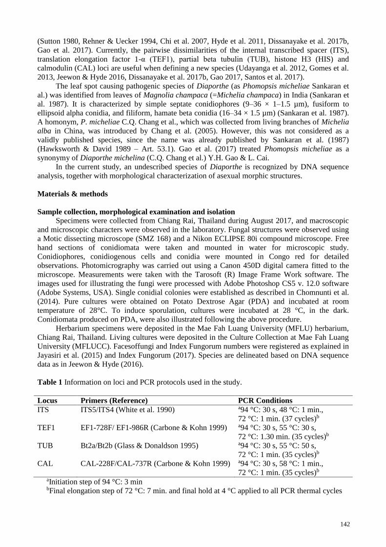
Table 1 Information on loci and PCR protocols used in the study.
Locus Primers (Reference) PCR Conditions
ITS ITS5/ITS4 (White et al. 1990) a94 °C: 30 s, 48 °C: 1 min.,
72 °C: 1 min. (37 cycles)b
TEF1 EF1-728F/ EF1-986R (Carbone & Kohn 1999) a94 °C: 30 s, 55 °C: 30 s,
72 °C: 1.30 min. (35 cycles)b
TUB Bt2a/Bt2b (Glass & Donaldson 1995) a94 °C: 30 s, 55 °C: 50 s,
72 °C: 1 min. (35 cycles)b
CAL CAL-228F/CAL-737R (Carbone & Kohn 1999) a94 °C: 30 s, 58 °C: 1 min.,
72 °C: 1 min. (35 cycles)b
aInitiation step of 94 °C: 3 min bFinal elongation step of 72 °C: 7 min. and final hold at 4 °C applied to all PCR thermal cycles
143
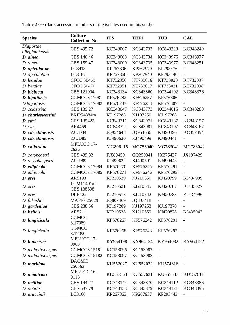
Table 2 GenBank accession numbers of the isolates used in this study
Species Culture
Collection No. ITS TEF1 TUB CAL
Diaporthe
alleghaniensis CBS 495.72 KC343007 KC343733 KC843228 KC343249
D. alnea CBS 146.46 KC343008 KC343734 KC343976 KC343977
D. alnea CBS 159.47 KC343009 KC343735 KC343977 KC343251
D. apiculatum LC3418 KP267896 KP267970 KP293476 -
D. apiculatum LC3187 KP267866 KP267940 KP293446 -
D. betulae CFCC 50469 KT732950 KT733016 KT733020 KT732997
D. betulae CFCC 50470 KT732951 KT733017 KT733021 KT732998
D. bicincta CBS 121004 KC343134 KC343860 KC344102 KC343376
D. biguttusis CGMCC3.17081 KF576282 KF576257 KF576306 -
D.biguttusis CGMCC3.17082 KF576283 KF576258 KF576307 -
D. celastrina CBS 139.27 KC343047 KC343773 KC344015 KC343289
D. charlesworthii BRIP54884m KJ197288 KJ197250 KJ197268 -
D. citri CBS 135422 KC843311 KC843071 KC843187 KC843157
D. citri AR4469 KC843321 KC843081 KC843197 KC843167
D. citrichinensis ZJUD34 JQ954648 JQ954666 KJ490396 KC357494
D. citrichinensis ZJUD85 KJ490620 KJ490499 KJ490441 -
D. collariana MFLUCC 17-
2636 MG806115 MG783040 MG783041 MG783042
D. cotoneastri CBS 439.82 FJ889450 GQ250341 JX275437 JX197429
D. discoidispora ZJUD89 KJ490622 KJ490501 KJ490443 -
D. ellipicola CGMCC3.17084 KF576270 KF576245 KF576291 -
D. ellipicola CGMCC3.17085 KF576271 KF576246 KF576295 -
D. eres AR5193 KJ210529 KJ210550 KJ420799 KJ434999
D. eres LCM11401a =
CBS 138598 KJ210521 KJ210545 KJ420787 KJ435027
D. eres DLR12a KJ210518 KJ210542 KJ420783 KJ434996
D. fukushii MAFF 625029 JQ807469 JQ807418 - -
D. gardeniae CBS 288.56 KJ197289 KJ197252 KJ197270 -
D. helicis AR5211 KJ210538 KJ210559 KJ420828 KJ435043
D. longicicola CGMCC
3.17089 KF576267 KF576242 KF576291 -
D. longicicola CGMCC
3.17090 KF576268 KF576243 KF576292 -
D. lonicerae MFLUCC 17-
0963 KY964198 KY964154 KY964082 KY964122
D. mahothocarpus CGMCC3 15181 KC153096 KC153087 - -
D. mahothocarpus CGMCC3 15182 KC153097 KC153088 - -
D. maritima DAOMC
250563 KU552027 KU552022 KU574616 -
D. momicola MFLUCC 16-
0113 KU557563 KU557631 KU557587 KU557611
D. neilliae CBS 144.27 KC343144 KC343870 KC344112 KC343386
D. nobilis CBS 587.79 KC343153 KC343879 KC344121 KC343395
D. oraccinii LC3166 KP267863 KP267937 KP293443 -
144
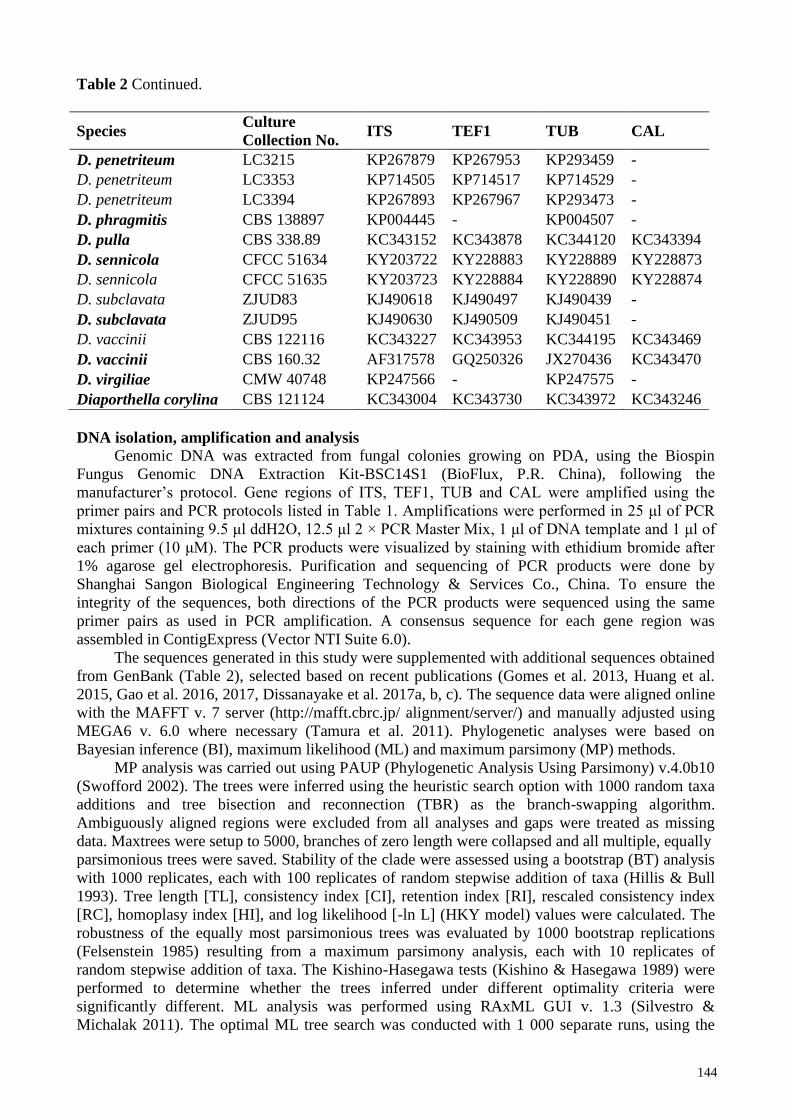
Table 2 Continued.
Species Culture
Collection No. ITS TEF1 TUB CAL
D. penetriteum LC3215 KP267879 KP267953 KP293459 -
D. penetriteum LC3353 KP714505 KP714517 KP714529 -
D. penetriteum LC3394 KP267893 KP267967 KP293473 -
D. phragmitis CBS 138897 KP004445 - KP004507 -
D. pulla CBS 338.89 KC343152 KC343878 KC344120 KC343394
D. sennicola CFCC 51634 KY203722 KY228883 KY228889 KY228873
D. sennicola CFCC 51635 KY203723 KY228884 KY228890 KY228874
D. subclavata ZJUD83 KJ490618 KJ490497 KJ490439 -
D. subclavata ZJUD95 KJ490630 KJ490509 KJ490451 -
D. vaccinii CBS 122116 KC343227 KC343953 KC344195 KC343469
D. vaccinii CBS 160.32 AF317578 GQ250326 JX270436 KC343470
D. virgiliae CMW 40748 KP247566 - KP247575 -
Diaporthella corylina CBS 121124 KC343004 KC343730 KC343972 KC343246
DNA isolation, amplification and analysis
Genomic DNA was extracted from fungal colonies growing on PDA, using the Biospin
Fungus Genomic DNA Extraction Kit-BSC14S1 (BioFlux, P.R. China), following the
manufacturer’s protocol. Gene regions of ITS, TEF1, TUB and CAL were amplified using the
primer pairs and PCR protocols listed in Table 1. Amplifications were performed in 25 μl of PCR
mixtures containing 9.5 μl ddH2O, 12.5 μl 2 × PCR Master Mix, 1 μl of DNA template and 1 μl of
each primer (10 μM). The PCR products were visualized by staining with ethidium bromide after
1% agarose gel electrophoresis. Purification and sequencing of PCR products were done by
Shanghai Sangon Biological Engineering Technology & Services Co., China. To ensure the
integrity of the sequences, both directions of the PCR products were sequenced using the same
primer pairs as used in PCR amplification. A consensus sequence for each gene region was
assembled in ContigExpress (Vector NTI Suite 6.0).
The sequences generated in this study were supplemented with additional sequences obtained
from GenBank (Table 2), selected based on recent publications (Gomes et al. 2013, Huang et al.
2015, Gao et al. 2016, 2017, Dissanayake et al. 2017a, b, c). The sequence data were aligned online
with the MAFFT v. 7 server (http://mafft.cbrc.jp/ alignment/server/) and manually adjusted using
MEGA6 v. 6.0 where necessary (Tamura et al. 2011). Phylogenetic analyses were based on
Bayesian inference (BI), maximum likelihood (ML) and maximum parsimony (MP) methods.
MP analysis was carried out using PAUP (Phylogenetic Analysis Using Parsimony) v.4.0b10
(Swofford 2002). The trees were inferred using the heuristic search option with 1000 random taxa
additions and tree bisection and reconnection (TBR) as the branch-swapping algorithm.
Ambiguously aligned regions were excluded from all analyses and gaps were treated as missing
data. Maxtrees were setup to 5000, branches of zero length were collapsed and all multiple, equally
parsimonious trees were saved. Stability of the clade were assessed using a bootstrap (BT) analysis
with 1000 replicates, each with 100 replicates of random stepwise addition of taxa (Hillis & Bull
1993). Tree length [TL], consistency index [CI], retention index [RI], rescaled consistency index
[RC], homoplasy index [HI], and log likelihood [-ln L] (HKY model) values were calculated. The
robustness of the equally most parsimonious trees was evaluated by 1000 bootstrap replications
(Felsenstein 1985) resulting from a maximum parsimony analysis, each with 10 replicates of
random stepwise addition of taxa. The Kishino-Hasegawa tests (Kishino & Hasegawa 1989) were
performed to determine whether the trees inferred under different optimality criteria were
significantly different. ML analysis was performed using RAxML GUI v. 1.3 (Silvestro &
Michalak 2011). The optimal ML tree search was conducted with 1 000 separate runs, using the
145
default algorithm of the program from a random starting tree for each run. The final tree was
selected among suboptimal trees from each run by comparing likelihood scores with the
GTRGAMMA nucleotide substitution model. MrBayes v. v. 3.2.0 was used to generate the
phylogenetic trees under optimal criteria per data partition (Ronquist & Huelsenbeck 2003).
Bayesian analysis was performed using MrBayes v. 3.2.0. The best-fit evolutionary models for
phylogenetic analyses were selected separately for ITS, TEF1, TUB and CAL gene regions using
MrModeltest v. 2.2 (Nylander 2004). The GTR+I+G model was selected for ITS and TUB, while
GTR+G was selected for TEF1 and CAL, separately, and incorporated into the analysis. Two
parallel analyses of each consisting of six Markov Chain Monte Carlo (MCMC) chains, run from
random trees for 6 000 000 generations were sampled every 100 generations resulting in 20 000
total trees. The first 10 000 trees, representing the burn in phase of the analyses were discarded
from each run. The remaining trees were used to calculate posterior probabilities (PP) in the
majority rule consensus tree. Trees were viewed by FigTree v1.4
(http://tree.bio.ed.ac.uk/software/figtree/) and edited using Microsoft PowerPoint 2010.
Results
Phylogenetic analyses
Single gene analyses of ITS, TEF1 and TUB were carried out for all the available sequences
of Diaporthe species to compare the topology of the trees and clade stability (data not shown).
Based on those analyses and blast results, 48 isolates were selected (including the outgroup taxon)
for the combined gene analysis of ITS, TEF1, TUB and CAL (Table 2). The aligned dataset
comprised 1766 characters including gaps (ITS: 1–468, TEF1: 469–922, TUB: 923–1283 and CAL:
1284–1766), of which 970 were constant, 393 parsimony-informative and 403 parsimony-
uninformative. The parsimony analysis resulted in 8 equally most parsimonious trees (TL = 1628
steps, CI = 0.641, RI = 0.731, RC = 0.469, HI = 0.359). Bayesian inference and maximum
parsimony analyses of the combined data set yielded trees with similar topologies to maximum
likelihood tree. The best scoring RAxML tree with a final likelihood value of -10272.773331 is
presented (Fig. 1). The matrix comprised 732 distinct alignment patterns, with 19.13% of
undetermined characters or gaps. Estimated base frequencies were as follows; A = 0.220005, C =
0.316011, G = 0.233364, T = 0.230620; substitution rates AC = 1.337142, AG = 3.640863, AT =
1.113669, CG = 1.137669, CT = 5.044583, GT = 1.000000; gamma distribution shape parameter α
= 0.501532.
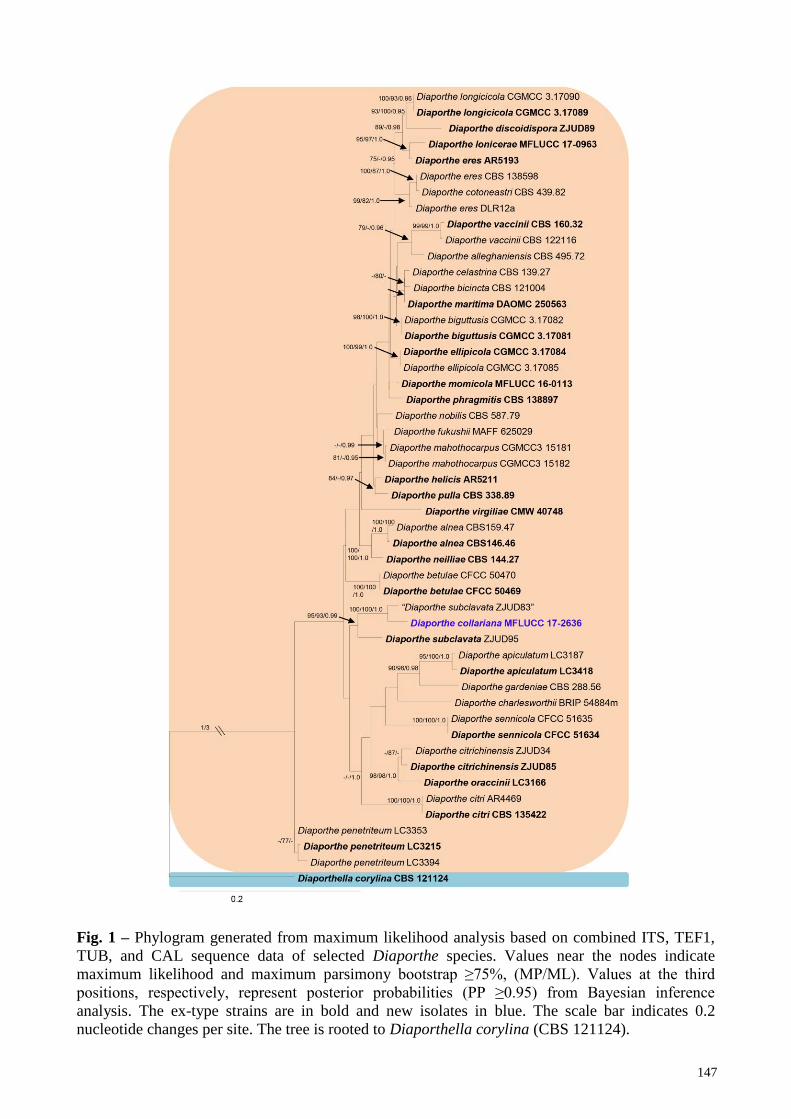
In the phylogenetic analysis, our new isolate Diaporthe collariana clustered with D.
subclavata isolate ZJUD83, with a high statistical support (100% MLBT, 100% MPBT, 1.00 PP).
The ex-type strain of D. subclavata also shows a close relationship to D. collariana.
Taxonomy
Diaporthe collariana R. H. Perera & K. D. Hyde sp. nov. Figs 2, 3
Indexfungorum: IF554061; Facesoffungi: FoF03909
Etymology – Named after its prominently flared collarettes.
Saprobic on Magnolia champaca. Asexual morph from the natural substrate – Conidiomata 190–
325 μm wide, 310–550 μm high, pycnidial, eustromatic, subepidermal, semi immersed, scattered,
globose to ampullifom or irregular, black, outer surface smooth, convoluted to unilocular, singly
ostiolate, with prominent necks 150–290 μm long. Peridium 18–25 μm thick, 5–9 cells thick,
consisting brown to hyaline cells of textura angularis. Conidial mass globose or sometimes
exuding in cirrhi, white to pale-yellow. Alpha conidiophores 12.1–20.6 × 2.4–3.2 μm ( = 16.6 ×
2.8 μm), densely aggregated, ampulliform to subcylindrical, rarely septate and branched, hyaline.
Alpha conidiogenous cells 10–17 × 1.3–2.4 μm ( = 13.7 × 1.8 μm) subcylindrical, tapering
towards the apex, hyaline, with visible periclinal thickening, collarette prominent, up to 6 μm long,
5.7 μm wide. Alpha conidia 4.2–6.2 × 1.5–2 μm ( = 5.2 × 1.7 μm), less common than beta
146
conidia, oblong to ellipsoidal, apex bluntly rounded, base obtuse to subtruncate, aseptate, straight,
guttulate, hyaline, smooth-walled. Beta conidiophores 10.3–19 × 1.4–3.5 μm ( = 14.6 × 2.6 μm),
densely aggregated, subcylindrical, filiform or obconical, branched and septate, hyaline. Beta
conidiogenous cells 3.8–14 × 1.4–2.2 μm ( = 7.9 × 1.8 μm) subcylindrical, tapering towards the
apex, hyaline, with visible periclinal thickening, collarette prominent, up to 6.6 μm long, 5.7 μm
wide. Beta conidia 22–31.3 × 0.8–1.6 μm ( = 27.7–1.2 μm), commonly found, straight, curved or
hamate, hyaline, smooth-walled. Gamma conidia not observed. Asexual morph on PDA –
Conidiomata 600–636 μm wide, 1045–1170 μm high, pycnidial, aggregated in small groups,
globose to ampullifom, unilocular, black, with a prominent neck. Peridium consisting brown cells
of textura angularis. Conidial mass globose or sometimes exuding in cirrhi, white to pale-yellow.
Alpha conidiophores 12–20 × 2.4–3.2 μm ( = 17.2 × 2.8 μm), densely aggregated, ampulliform
to subcylindrical, rarely septate and branched, hyaline. Alpha conidiogenous cells 11.1–17 × 1.3–
2.4 μm ( = 14.4 × 1.8 μm) subcylindrical, tapering towards the apex, hyaline, with visible
periclinal thickening, collarette prominent, up to 3.5 μm long, 3.2 μm wide. Alpha conidia 4.7–5.6
× 1.7–2.2 μm ( = 5.2 × 1.9 μm), less common than beta conidia, oblong to ellipsoidal, apex
bluntly rounded, base obtuse to subtruncate, aseptate, straight, bi-guttulate, hyaline, smooth-walled.
Beta conidiophores 13.2–20.8 × 1.3–4.1 μm ( = 17.4 × 3.6 μm), densely aggregated,
subcylindrical, filiform or obconical, branched and septate, hyaline. Beta conidiogenous cells 8.8–
13.4 × 1.7–2.3 μm ( = 10.8 × 2.1 μm) subcylindrical, tapering towards the apex, hyaline, with
visible periclinal thickening, collarette prominent, up to 3.5 μm long, 3.2 μm wide. Beta conidia
22–31.7 × 1.1–1.6 μm ( = 28.8–1.3 μm), commonly found, straight, curved or hamate, hyaline,
smooth-walled. Gamma conidia not observed. Sexual morph – Undetermined.
Culture characters – Conidia germinating on WA (Water Agar) within 12 h and germ tubes
produced from one end. Colonies growing on PDA, reaching 6 cm in 7 days at 25°C, flat, initially
white, aerial mycelium forming concentric rings with cottony texture, white to olivaceous, reverse
zonate with white and ash-brown rings. Sporulate on PDA after 2 months incubation period in dark,
at 25°C.
Material examined – THAILAND, Chiang Rai, Mae Fah Luang University premises, on dried
fruits and pedicels of Magnolia champaca (L.) Baill. ex Pierre (Magnoliaceae), 17 August 2017, S.
Boonmee, Fruit 3 (MFLU 17-2770, holotype), MFLU 17-2845 dried culture on PDA, ex-type
living culture, MFLUCC 17-2636. (GenBank: LSU: MG806114)
Notes – Our new fungus Diaporthe collariana nested in between two D. subclavata strains
and was more related to strain ZJUD83, which was collected from a fruit of Citrus maxima cv.
Shatianyou in China, with very good support (Fig. 1). Nucleotide comparison reveals 5 (1.3%)
differences in the ITS region, 10 (2.1%) in the TEF1 region, 11 (1.4%) in the TUB region. The ex-
type strain of D. subclavata (ZJUD95) is the next phylogenetically closest isolate to Diaporthe
collariana (Fig. 1). Nucleotide comparison reveals 15 (3.8%) were distinct in the ITS region, 46
(9.7%) in the TEF1 region, 10 (1.2%) in the TUB region. CAL region is not available for D.
subclavata strains in GenBank (Huang et al. 2015). Diaporthe collariana differs from D.
subclavata in the presence of beta conidia. Furthermore, D. collariana produces prominent
collarettes while collarets are absent in D. subclavata. On PDA, D. collariana produces smaller
alpha conidia, which are oblong to ellipsoidal (4.7–5.6 × 1.7–2.2 μm), while D. subclavata
produces fusiform to clavate conidia (5.5–7.2 × 2.2–2.9 μm) (Huang et al. 2015). The placement of
D. collariana in between two isolates of D. subclavata is rather intriguing. However, by comparing
available gene sequences of D. subclavata strains, we confirm that ZJUD83 is different from its ex-
type ZJUD95. This is further discussed below.
Diaporthe micheliae, is another species which lacks molecular data in the GenBank, and was
also isolated from the same host as D. collariana (Sankaran et al. 1987). However, D. collariana
can be distinguished from D. micheliae by having prominent collarettes which are absent in D.
micheliae, branched conidiophores (vs. simple conidiophores), and smaller alpha conidia (4.7–5.6 ×
1.7–2.2 vs. 4.6–8.2(-11.5) × 2–2.8 µm) (Sankaran et al. 1987).
147
Fig. 1 – Phylogram generated from maximum likelihood analysis based on combined ITS, TEF1,
TUB, and CAL sequence data of selected Diaporthe species. Values near the nodes indicate
maximum likelihood and maximum parsimony bootstrap ≥75%, (MP/ML). Values at the third
positions, respectively, represent posterior probabilities (PP ≥0.95) from Bayesian inference
analysis. The ex-type strains are in bold and new isolates in blue. The scale bar indicates 0.2
nucleotide changes per site. The tree is rooted to Diaporthella corylina (CBS 121124).

148
Fig. 2 – Diaporthe collariana (MFLU 17-2770, holotype). a Herbarium material. b Conidioma on
host substrate. c, d Section through conidiomata. e Peridium. f Alpha and beta conidia inside the
same conidiomata. g, h Conidiophores with alpha conidia (in Congo red). i, j Conidiophores with
beta conidia (in Congo red). k, l Beta conidia. m Alpha conidia. n Germinating conidium.
o, p Colony on PDA. Scale bars: b, c = 200 µm, d = 50 µm, e–g = 50 µm, h–l = 20 µm, m, n = 10
µm.
149
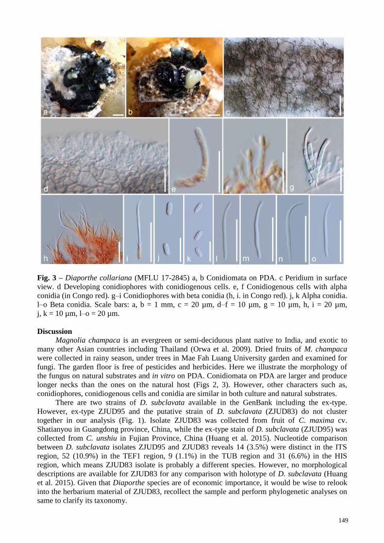
Fig. 3 – Diaporthe collariana (MFLU 17-2845) a, b Conidiomata on PDA. c Peridium in surface
view. d Developing conidiophores with conidiogenous cells. e, f Conidiogenous cells with alpha
conidia (in Congo red). g–i Conidiophores with beta conidia (h, i. in Congo red). j, k Alpha conidia.
l–o Beta conidia. Scale bars: a, b = 1 mm, c = 20 µm, d–f = 10 µm, g = 10 µm, h, i = 20 µm,
j, k = 10 µm, l–o = 20 µm.
Discussion
Magnolia champaca is an evergreen or semi-deciduous plant native to India, and exotic to
many other Asian countries including Thailand (Orwa et al. 2009). Dried fruits of M. champaca
were collected in rainy season, under trees in Mae Fah Luang University garden and examined for
fungi. The garden floor is free of pesticides and herbicides. Here we illustrate the morphology of
the fungus on natural substrates and in vitro on PDA. Conidiomata on PDA are larger and produce
longer necks than the ones on the natural host (Figs 2, 3). However, other characters such as,
conidiophores, conidiogenous cells and conidia are similar in both culture and natural substrates.
There are two strains of D. subclavata available in the GenBank including the ex-type.
However, ex-type ZJUD95 and the putative strain of D. subclavata (ZJUD83) do not cluster
together in our analysis (Fig. 1). Isolate ZJUD83 was collected from fruit of C. maxima cv.
Shatianyou in Guangdong province, China, while the ex-type stain of D. subclavata (ZJUD95) was
collected from C. unshiu in Fujian Province, China (Huang et al. 2015). Nucleotide comparison
between D. subclavata isolates ZJUD95 and ZJUD83 reveals 14 (3.5%) were distinct in the ITS
region, 52 (10.9%) in the TEF1 region, 9 (1.1%) in the TUB region and 31 (6.6%) in the HIS
region, which means ZJUD83 isolate is probably a different species. However, no morphological
descriptions are available for ZJUD83 for any comparison with holotype of D. subclavata (Huang
et al. 2015). Given that Diaporthe species are of economic importance, it would be wise to relook
into the herbarium material of ZJUD83, recollect the sample and perform phylogenetic analyses on
same to clarify its taxonomy.
150
Acknowledgements The Research of Featured Microbial Resources and Diversity Investigation in Southwest
Karst area (Project No. 2014FY120100) is thanked for financial support. Kevin D. Hyde thanks the
Chinese Academy of Sciences, project number 2013T2S0030, for the award of a Visiting
Professorship for Senior International Scientists at Kunming Institute of Botany. The authors
extend their appreciation to the International Scientific Partnership Program ISPP at King Saud
University for funding this research work through ISPP#0089.
References
Carbone I, Kohn LM. 1999 – A method for designing primer sets for speciation studies in
filamentous ascomycetes. Mycologia 91, 553–556.
Chang CQ, Cheng YH, Xiang MM, Jiang ZD, Chi PK. 2005 – New species of Phomopsis on
woody plants in Fujian province. Mycosystema 24, 6–11.
Chomnunti P, Hongsanan S, Aguirre-Hudson B, Tian Q et al. 2014 – The sooty moulds. Fungal
Diversity 66, 1–36.
Chi P, Jiang Z, Xiang M. 2007 – Flora Fungorum Sinicorum 34: Phomopsis. Science, Beijing.
Dayarathne MC, Boonmee S, Braun U, Crous PW et al. 2016 – Taxonomic utility of old names in
current fungal classification and nomenclature: Conflicts, confusion and clarifications.
Mycosphere 7, 1622–1648.
Dissanayake AJ, Camporesi E, Hyde KD, Zhang Wei, Yan JY, Li XH. 2017a – Molecular
phylogenetic analysis reveals seven new Diaporthe species from Italy. Mycosphere 8, 853–
877.
Dissanayake AJ, Phillips AJL, Hyde KD, Yan JY, Li XH. 2017b – The current status of species in
Diaporthe. Mycosphere 8, 1106–1156.
Dissanayake AJ, Zhang W, Liu M, Hyde KD et al. 2017c – Diaporthe species associated with
peach tree dieback in Hubei, China. Mycosphere 8, 533–549.
Felsenstein J. 1985 – Confidence limits on phylogenies: an approach using the bootstrap. Evolution
39, 783–791.
Gao Y, Liu F, Cai L. 2016 – Unravelling Diaporthe species associated with Camellia. Systematics
and Biodiversity 14, 102–117.
Gao Y, Liu F, Duan W, Crous PW, Cai L. 2017 – Diaporthe is paraphyletic. IMA Fungus 8, 153–
187.
Gao YH, Su YY, Sun W, Cai L. 2014 – Diaporthe species occurring on Lithocarpus glabra in
China, with descriptions of five new species. Fungal Biology 119, 295–309.
Glass NL, Donaldson GC. 1995 – Development of primer sets designed for use with the PCR to
amplify conserved genes from filamentous ascomycetes. Applied and Environmental
Microbiology 61, 1323–1330.
Gomes RR, Glienke C, Videira SI, Lombard L, Groenewald JZ, Crous PW. 2013 – Diaporthe: a
genus of endophytic, saprobic and plant pathogenic fungi. Persoonia: Molecular Phylogeny
and Evolution of Fungi. 31, 1–41.
Hawksworth DL, David JC. 1989 – Family names. Index of Fungi Supplement – C.A.B.
International. Wallingford, U.K.
Hillis DM, Bull JJ. 1993 – An empirical test of bootstrapping as a method for assessing confidence
in phylogenetic analysis. Systematic Biology 42, 182–192.
Huang F, Udayanga D, Wang X, Hou X et al. 2015 – Endophytic Diaporthe associated with Citrus:
A phylogenetic reassessment with seven new species from China. Fungal Biology 119, 331–
347.
Ronquist F, Huelsenbeck JP. 2003 – MrBayes 3: Bayesian phylogenetic inference under mixed
models Bioinformatics 19, 1572–1574.
Hyde KD, McKenzie EHC, KoKo TW. 2011 – Towards incorporating anamorphic fungi in a
natural classification–checklist and notes for 2010. Mycosphere 2, 1–88.
151
Index Fungorum. 2017 – www.indexfungorum.org.
Jayasiri SC, Hyde KD, Ariyawansa HA, Bhat J et al. 2015 – The Faces of Fungi database: fungal
names linked with morphology, phylogeny and human impacts. Fungal Diversity 74, 3–18.
Jeewon R, Hyde KD. 2016 – Establishing species boundaries and new taxa among fungi:
recommendations to resolve taxonomic ambiguities. Mycosphere 7, 1669–1677.
Kishino H, Hasegawa M. 1989 – Evaluation of the maximum likelihood estimate of the
evolutionary tree topologies from DNA sequence data. Journal of Molecular Evolution 29,
170–179.
Nylander JAA. 2004 – MrModeltest v2. Program distributed by the author Evolutionary Biology
Centre, Uppsala University, Sweden.
Orwa C, Mutua A, Kindt R, Jamnadass R, Anthony S. 2009 – Agroforestree Database:a tree
reference and selection guide version 4.0
(http://www.worldagroforestry.org/treedb/AFTPDFS/Michelia_champaca.PDF)
Rehner SA, Uecker FA. 1994 – Nuclear ribosomal internal transcribed spacer phylogeny and host
diversity in the coelomycete Phomopsis. Canadian Journal of Botany 72, 1666–1674.
Sankaran KV, Florence EJM, Sharma JK. 1987 – Two new species of Phomopsis from India.
Transactions of the British Mycological Society 89, 404–407.
Santos L, Alves A, Alves R. 2017 – Evaluating multi-locus phylogenies for species boundaries
determination in the genus Diaporthe. PeerJ 5, e3120.
Silvestro D, Michalak I. 2011 – raxmlGUI: a graphical front-end for RAxML. Organisms.
Diversity and Evolution 12, 335–337.
Swofford DL. 2002 – PAUP*: phylogenetic analysis using parsimony, version 4.0 b10. Sinauer
Associates, Sunderland.
Sutton BC. 1980 – The coelomycetes. Fungi imperfecti with pycnidia, acervuli and stromata.
Commonwealth Mycological Institute, Kew, Surrey, England.
Tamura K, Peterson D, Peterson N, Stecher G, Nei M, Kumar S. 2011 – MEGA5: molecular
evolutionary genetics analysis using maximum likelihood, evolutionary distance, and
maximum parsimony methods. Molecular Biology and Evolution 28, 2731–2739.
Udayanga D, Liu XZ, Crous PW, McKenzie EH, Chukeatirote E, Hyde KD. 2012 – A multi-locus
phylogenetic evaluation of Diaporthe (Phomopsis). Fungal Diversity 56, 157–171.
White TJ, Bruns T, Lee S, Taylor J. 1990 – Amplification and direct sequencing of fungal
ribosomal RNA genes for phylogenetics. In: Innis MA, Gelfand DH, Sninsky JJ, White TJ
(eds), PCR Protocols: a guide to methods and applications. Academic Press, New York, pp.
315–322.