Embed Size (px)
Citation preview
Kidney International, Vol. 6 (1974), p. 177—183
Dialysis of methylguanidine
SERGIO GIOVANNETTI and GIuLIAN0 BARSOTTI
Cattedra Semelotica Medica, Università di Pisa, Pisa, Italy
Dialysis of methylguanidine. The dialysis clearance of methyl-guanidine (MG) was measured in Vitro and in ViVo; the fall in itsplasma concentration was measured after peritoneal dialysisand hemodialysis in uremic patients and on plasma and muscletissue of uremic dogs; the postdialysis plasma rebound wasevaluated after hemodialysis and peritoneal dialysis; and,finally, MG binding to plasma proteins was measured in vitro atdifferent pH values. The dialysis clearance of MG was lower invivo than in vitro and it decreased during the course of hemo-dialysis. Its protein binding, rising as the plasma pH rose duringhemodialysis, accounted for its dialytic behavior. The slowtransfer of MG from tissue, occurring during dialysis as shownby the experiments on uremic dogs, accounted for its highrebound in plasma after hemodialysis. Rebound after peritonealdialysis was lower so that the MG plasma concentrations, 12 hrafter the end of two dialytic procedures, were not different. It isconcluded that MG is the only known "uremic toxin" thus farstudied whose dialytic removal is equally obtained with bothperitoneal dialysis and hemodialysis schedules which insure thatthe patient is maintained in good clinical condition.
La dialyse de Ia mdthylguanidine. La clearance de Ia methyl-guanidine (MG) au cours de la dialyse a été mesurée in vivo et invitro. La diminution de la concentration plasmatique aprCsdialyse péritonéale et hémodialyse a été mesurée chez lesmalades urémiques et dans le plasma et le tissu musculaire dechiens urémiques. Le rebond post dialytique a etC CvaluC aprèshemodialyse et dialyse péritonéale. Enfin la liaison de MG auxprotéines a Cté mesurée in vitro pour différentes valeurs du pH.La clearance de MG au cours de Ia dialyse est inférieure in vivoa ce qu'elle est in vitro et diminue au cours de l'hCmodialyse. Laliaison aux protéines, qui augmente en même temps au le pHs'élève au cours de l'hémodialyse, rend compte de ce comporte-ment dialytique. Le transfert lent de MG des tissus qui survientpendant l'hémodialyse, ainsi que le montrent les experienceschez le chien, rend compte du rebond post dialytique élevé. Lesrebonds aprés dialyse péritonéaie sont plus faibles si bien que laconcentration plasmatique de MG est semblable 12 heuresaprés la fin de chacun des deux modes de dialyse. Ii est concluque la MG est la seule "toxine urémique" connue, et étudiéejusque là, dont Ia soustraction par dialyse Soit obtenue dans desconditions égales aussi bien avec la dialyse péritonéale quel'hémodialyse dans des conditions qui assurent un état cliniquesatisfaisant.
Methylguanidine (MG) administered repeatedly tonormal dogs in large doses has been found to induce
Received for publication October 9, 1973;and in revised form March 14, 1974.© 1974, by the International Society of Nephrology.
177
symptoms resembling those of uremia, such as anemia,anorexia, vomiting, gastric ulcers, peripheral neuro-pathy, decreased intestinal absorption of calcium andhypertriglyceridemia [1, 2]. When smaller doses wereused, so as to maintain MG plasma concentrations atvalues resembling those of chronically ill nondialyzeduremic patients (300 to 500 g/lO0 ml), similar butmilder symptoms were induced [3].
These findings suggest that the retention of MG inrenal failure may provide an important source of"uremic toxin", but this possibility stands in sharpcontrast with "the middle molecule hypothesis"which states that the toxic symptoms of uremiamay be caused by substance(s) with a mol wtbetween 300 and 1000 [4]. The latter hypothesis ismainly supported by the observation that uremicpatients on maintenance peritoneal dialysis [5] orhemodialysis at a low dialysate flow rate [6, 7] respondas well to treatment as do those on a standard hemo-dialysis program, despite the fact that the plasmaconcentrations of the low mol wt metabolites such asurea (U; mol wt, 60) and creatinine (CR; mol wt, 113)are higher in the former patients.
A logical explanation for this clinical finding mightbe provided by the existence of toxic molecules ofintermediate mol wt which would pass more readilythrough the peritoneal membrane than throughcellophane, and whose removal would not be de-creased during hemodialysis at low rates of dialysateflow [4].
One would expect that MG, having a mol wt (73)which is intermediate between that of U and CR,would accumulate in the body fluids of patients onperitoneal dialysis or hemodialysis at low dialysateflow rate and that, if so, such an event would provide astrong argument against its proposed importance as a"uremic toxin".
In the present study this problem has been examinedby measuring the dialyzer clearance rates of MG in
178 Giovannetti/Barsotti
vitro and in uremic patients; by measuring its dialyticremoval from 40 liters of "artificial uremic blood"and its decrease in concentration in the plasma ofuremic patients subjected to peritoneal dialysis orhemodialysis from plasma and muscle tissue of uremicdogs; by measuring the rebound of its plasma con-centration 12 hr after the termination of hemodialysisand peritoneal dialysis; and, finally, by evaluating itsbinding to plasma proteins at different pH values.
Methods
The uremic patients we examined had receivedmaintenance dialysis for at least three months prior tothis study; all were anuric or extremely oliguric (lessthan 100 ml of urine/24 hr) with an endogenouscreatine clearance less than 2 ml/min. In addition,studies were also carried out on six mongrel dogs inwhich uremia was induced via bilateral ureteralligation.
The "artificial uremic blood" employed in the invitro experiments was a water-based solution of U(about 250 mg/100 ml), CR (about 15 mg/lO0 ml) andMG (about 250 g/l00 ml). For measurements ofdialyzer clearance rates in vitro (six experiments), thissolution was recirculated at 200 mI/mm for 12 hr,through a two-layer Kiil dialyzer assembled with a"150 P" Cuprophane membrane. Dialysate, con-sisting of tap water, was circulated in "single pass"and the temperature of both the "artificial uremicblood" and dialysate was maintained at 37°C. Thevarious flow rates of dialysate (Fig. 1) were measuredon the outflow line with a graduated cyclinder andchronometer. The same artificial kidney and type ofmembrane were employed for measurements of thedialyzer clearance of MG on uremic patients (nineexperiments). Both in vitro and in vivo, these mea-surements were made 20 mm after the beginning ofdialysis and at the 4th, 8th and 12th hr. Two rates ofdialysate flow (QD) were examined in vivo: 200 and500 ml/min, while the blood flow rate (QB) wasmaintained as constant as possible at 200 ml/min bymeans of a blood pump. The "air bubble" methodwas also employed as an additional determinant ofthe rate of blood flow when clearance rates weremeasured.
For both in vitro and in vivo measurements ofdialyzer clearances, as well as for those duringperitoneal dialysis, the following formula [8] wasemployed:
= CD.QD,
where CD and CD are the concentrations of the
examined metabolite in dialysate and arterial blood,respectively.
The predialysis and postdialysis concentrations ofU, CR and MG were measured in the following con-ditions: 1) on 40 liters of "artificial uremic blood"circulating for eight hours through a multipointdialyzer (assembled with "150 P" Cuprophane mem-brane) at QB 250 and QD750 ml/min (five experiments);2) on the plasma of 28 uremic patients on an inter-mittent program consisting of two dialyses per weekcarried out with a multipoint dialyzer under the sameconditions as the in vitro experiments; 3) on theplasma of eight uremic patients on a program of twodialyses per week of 12 hrs' duration carried out witha standard two-layer Kiil dialyzer at QB 200 and QD500 ml/min; 4) on the plasma samples of 11 uremicpatients on twice-weekly peritoneal dialysis of 18 to20 hrs' duration each, with a total dialysate exchange of80 to 100 liters; and 5) on the plasma and muscle tissueof six dogs on the third day of anuria following liga-ture of the ureters. These animals were dialyzed forsix hours with a standard two-layer Kiil kidney utiliz-ing a femoral artery and vein for access to the systemiccirculation. These experiments were carried out whilethe dogs were under general anesthesia, induced andmaintained with pentobarbital administered i.v. in therequired amounts. The measurements of U, CR andMG in muscle tissue were performed on symmetrical,fresh wet samples of the same muscles (femoralquadriceps), using the procedure previously de-scribed [3].
In 8 patients on maintenance peritoneal dialysis andin 12 on the hemodialysis program (two dialyses perweek of 8 hrs' duration utilizing a multipoint artificialkidney), the concentrations of U, CR and MG weremeasured immediately before dialysis, immediatelyafter dialysis and 12 hr later, for evaluation of thephenomenon of plasma rebound.
The binding of MG to plasma proteins was evalu-ated on eight samples of normal human plasma towhich MG had been added at concentrations rangingfrom 100 to 200 g/ml. The pH of an aliquot of thisplasma was adjusted to about 7.3 and that of anotheraliquot, to about 7.5, by adding a concentrated solutionof NaCO; each aliquot was then placed in a Cupro-phane bag (obtained from coil dialyzer tubing) andeach bag was placed in the upper part of a glass tubehaving a porous septum across its middle segment.These were then placed in tubes that were centrifugedfor six hours at 6000 rpm at room temperature. TheMG concentrations were then measured in the plasmacontained in the Cuprophane bags and in the protein-free ultrafiltrate which collected in the bottom of thecentrifuge tubes. Reproducibility of this procedure was
Dialysis of methylguanidine 179
determined in six experiments by using the sameplasma samples with added MG, and it was found tobe satisfactory.
Determinations of U, and CR, in plasma samples,'artificial uremic blood", dialysate (for the experi-ments reported in Fig. 2) and the aqueous extracts ofmuscle tissue of dogs, were performed with an Auto-analyzer. For measurements of MG, blood sampleswere centrifuged immediately after they were drawn soas to minimize the equilibration of MG betweenred blood cells and plasma [8]; plasma was thenfrozen until the time of measurement according to themethod previously described [9]. This method is basedon the absorption of MG by a strong cation exchangeresin, from which CR and arginine are removedcompletely with a NaOH solution, and MG is theneluted with HC1. The eluate is evaporated and theSakaguchi reaction is performed on the redissolveddry residue. Activated charcoal is not employed.
Results
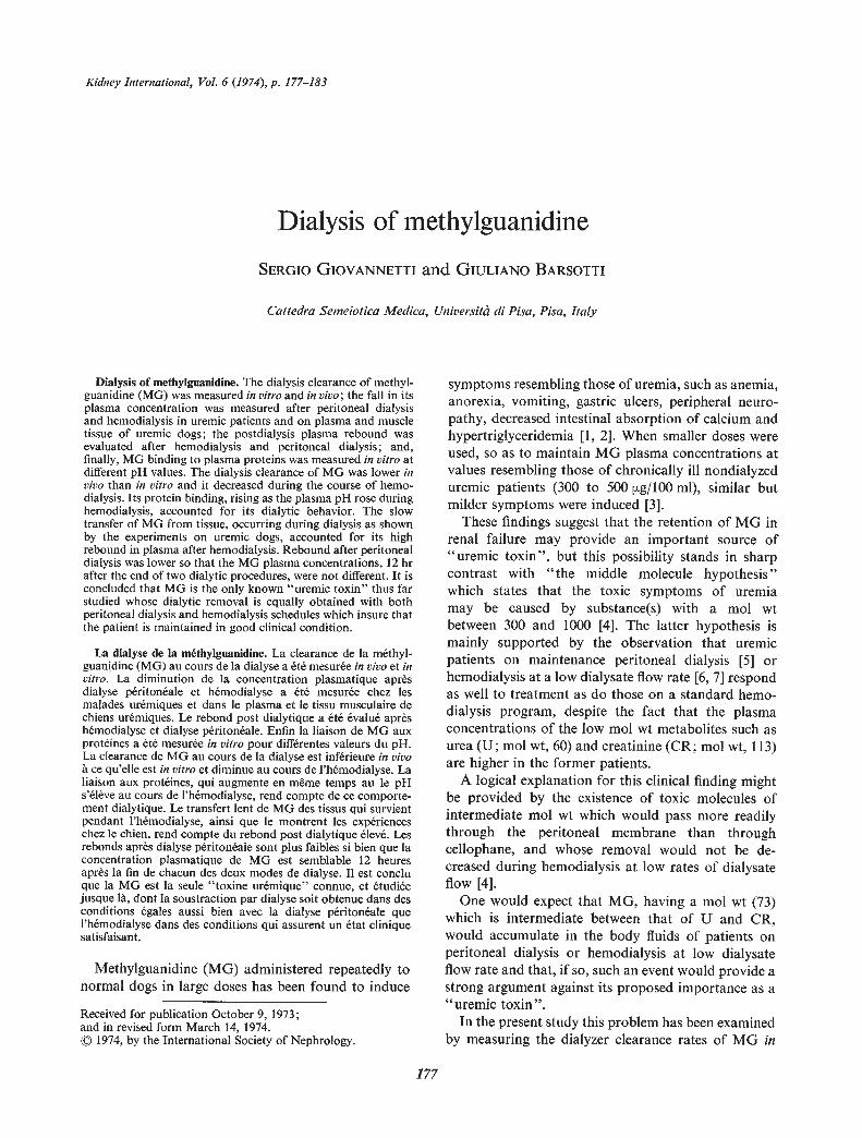
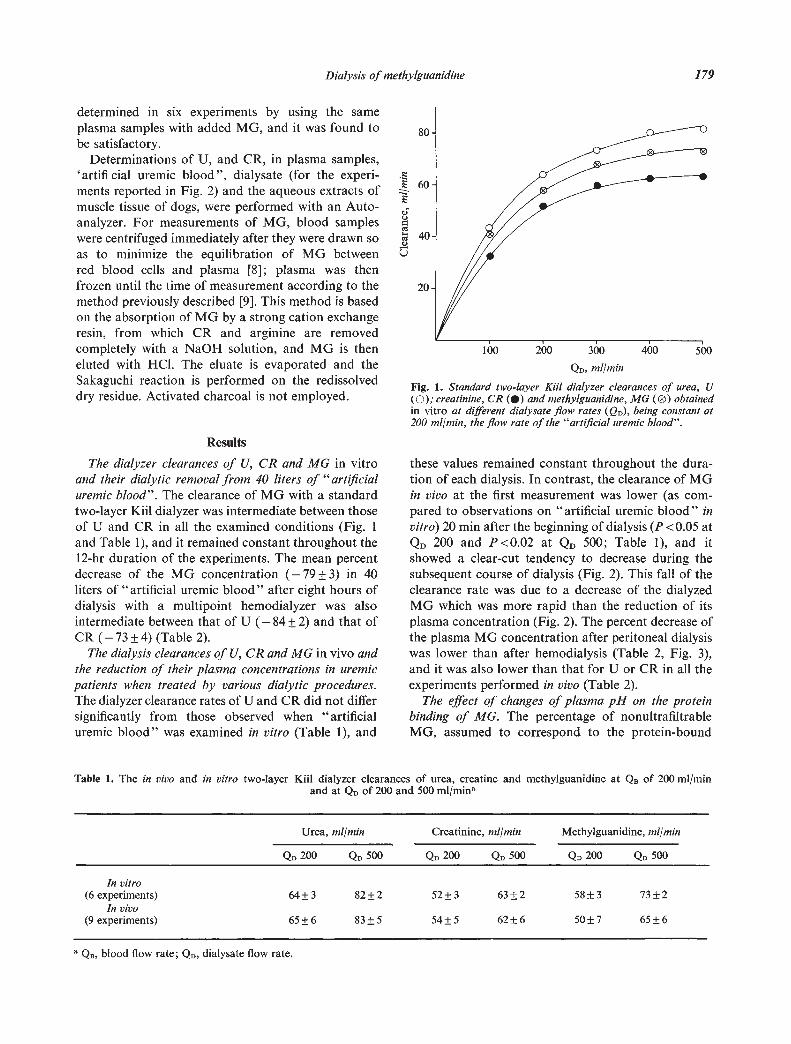
The dialyzer clearances of U, CR and MG in vitroand their dialytic removal from 40 liters of "artificialuremic blood". The clearance of MG with a standardtwo-layer Kiil dialyzer was intermediate between thoseof U and CR in all the examined conditions (Fig. 1and Table I), and it remained constant throughout the12-hr duration of the experiments. The mean percentdecrease of the MG concentration (—79±3) in 40liters of "artificial uremic blood" after eight hours ofdialysis with a multipoint hemodialyzer was alsointermediate between that of U (—84±2) and that ofCR (—73 (Table 2).
The dialysis clearances of U, CR and MG in vivo andthe reduction of their plasma concentrations in uremicpatients when treated by various dialytic procedures.The dialyzer clearance rates of U and CR did not differsignificantly from those observed when "artificialuremic blood" was examined in vitro (Table 1), and
300
QD, mi/mm
Fig. 1. Standard two-layer Ku! dialyzer clearances of urea, U(0), creatinine, CR () and methylguanidine, MG (0) obtainedin vitro at different dialysate flow rates (QD), being constant at200 mi/mm, the flow rate of the "artificial uremic blood".
these values remained constant throughout the dura-tion of each dialysis. In contrast, the clearance of MGin vivo at the first measurement was lower (as com-pared to observations on "artificial uremic blood" invitro) 20 mm after the beginning of dialysis (P <0.05 atQD 200 and P <0.02 at QD 500; Table 1), and itshowed a clear-cut tendency to decrease during thesubsequent course of dialysis (Fig. 2). This fall of theclearance rate was due to a decrease of the dialyzedMG which was more rapid than the reduction of itsplasma concentration (Fig. 2). The percent decrease ofthe plasma MG concentration after peritoneal dialysiswas lower than after hemodialysis (Table 2, Fig. 3),and it was also lower than that for U or CR in all theexperiments performed in vivo (Table 2).
The effect of changes of plasma pH on the proteinbinding of MG. The percentage of nonultrafiltrableMG, assumed to correspond to the protein-bound
Table 1. The in vivo and in vitro two-layer Kill dialyzer clearances of urea, creatine and methylguanidine at QB of 200 mI/mmand at QD of 200 and 500 ml/min
Urea, ml/min Creatinine, mi/mm Methylguanidine, ml/min
QD 200 Q1, 500 200 QD 500 QD 200 QD 500
In vitro(6 experiments) 64 3 82 2 52 3 63 2 58 3 73 2
In vivo(9 experiments) 65±6 83±5 54±5 62±6 50±7 65±6
QB, blood flow rate; QD, dialysate flow rate.
80
60E
C)C.)aceI-Ce0)
40
20.
180 Giovannetti/Barsotti
Table 2. Percent decreases of urea, creatinine and methylguanidine concentrations obtained on 40 liters of "artificial uremic blood" (AUB)and on the plasma of uremic patients, with different hemodialytic procedures and with peritoneal dialysis
Experiments(N)
Urea Creatinine Methylguanidine
Before After Decrease Before After Decrease Before After Decreasedialysis dialysis % dialysis dialysis % dialysis dialysis %
mg/l00 ml mg/l00 ml mg/l00 ml mg/100 ml ig/l00 ml g/100 ml
8-hr multipoint dialysis,40litersofAUB 258±58 40±12 —84±2 15±4 4±0.7 —73±4 261±37 54±6 —79±3
(5)8-hr multipoint dialysis,
uremic patients 240±41 87±27 —64±8 14±3 6±2 —52±6 132±66 68±30 —45±9twice a week
(28)I 2-hr, standard two-layer
Kiil, uremic patients 181 41 62±21 —65 5 11 5 5 3 —61 8 128 29 70±14 —44 2twice a week
(8)18- to 20-hr peritoneal
dialysis 254±127 158±47 —34±14 15±3 10±3 —29±7 123±60 88±38 27±8twice a week
(11)
moiety, was significantly higher (P <0.001) at pH7.51 0.04 (29.6±5.9%) than at pH 7.31 0.03 (15.2
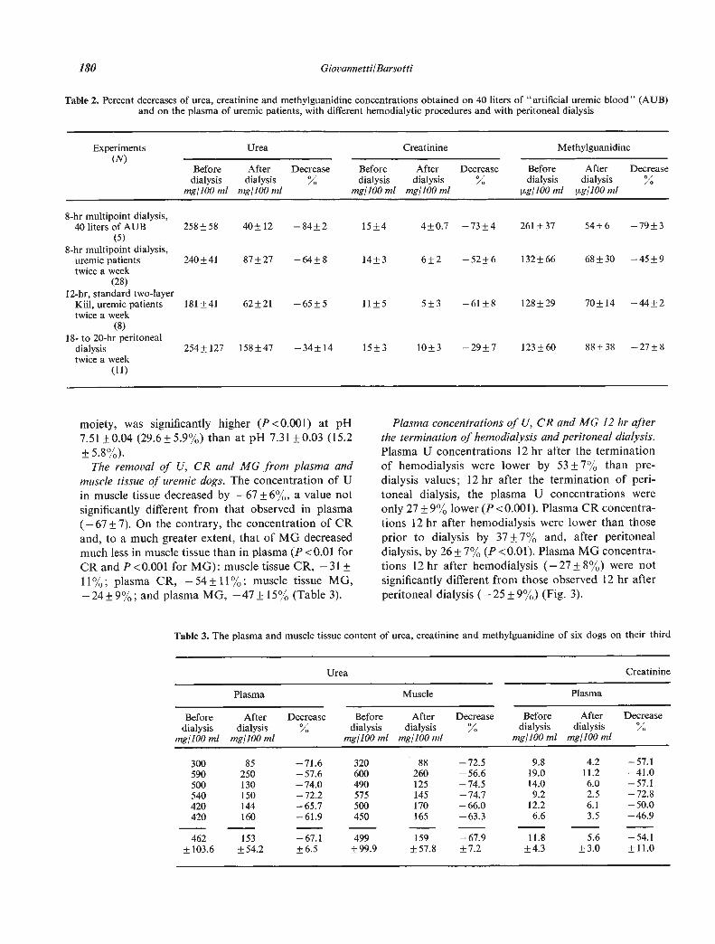
The removal of U, CR and MG from plasma andmuscle tissue of uremic dogs. The concentration of Uin muscle tissue decreased by —67±6%, a value notsignificantly different from that observed in plasma(—67 7). On the contrary, the concentration of CRand, to a much greater extent, that of MG decreasedmuch less in muscle tissue than in plasma (P <0.01 forCR and P<0.00l for MG): muscle tissue CR, —3111%; plasma CR, —54± 11%; muscle tissue MG,—24±9%; and plasma MG, —47±15% (Table 3).
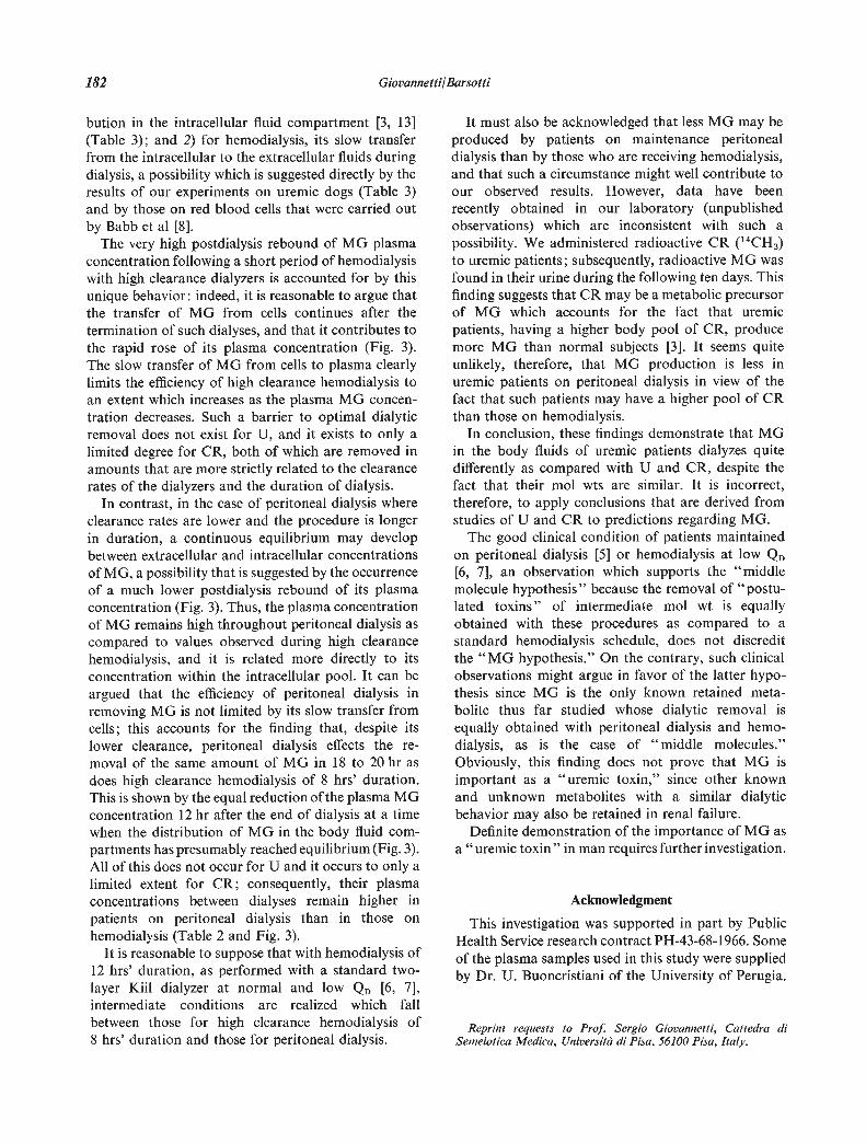
Plasma concentrations of U, CR and MG 12 hr afterthe termination of hemodialysis and peritoneal dialysis.Plasma U concentrations 12 hr after the terminationof hemodialysis were lower by 53 7% than pre-dialysis values; 12 hr after the termination of pen-toneal dialysis, the plasma U concentrations wereonly 27 9% lower (P <0.001). Plasma CR concentra-tions 12 hr after hemodialysis were lower than thoseprior to dialysis by 37 7% and, after peritonealdialysis, by 26±7% (P<0,0l). Plasma MG concentra-tions 12 hr after hemodialysis (—27±8%) were notsignificantly different from those observed 12 hr afterperitoneal dialysis (—25±9%) (Fig. 3).
Table 3. The plasma and muscle tissue content of urea, creatinine and methylguanidine of six dogs on their third
Urea Creatinine
Plasma Muscle Plasma
Beforedialysis
mg/l0O ml
Afterdialysis
mg/lOO ml
Decrease%
Beforedialysis
mg/lOO ml
Afterdialysis
mg/loom!
Decrease%
Beforedialysis
mg/lOU ml
Afterdialysis
mg/lOU ml
Decrease%
300590500540420420
85250130150144160
—71.6—57.6—74.0—72.2—65.7—61.9
320600490575500450
88260125145170165
—72.5—56.6—74.5—74.7—66.0—63.3
9.819.014.09.2
12.26.6
4.211.26.02.56.13.5
—57.1—41.0—57.1—72.8—50.0—46.9
462 153 —67.1 499 159 —67.9 11.8 5.6 —54.1±11.0
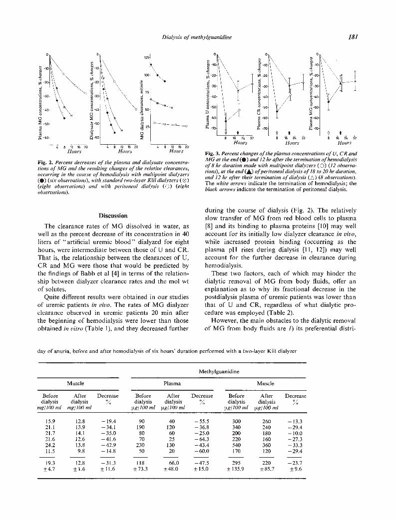
Fig. 2. Percent decreases of the plasma and dialysate concentra-tions of MG and the resulting changes of the relative clearances,occurring in the coarse of hemodialysis with multipoint dialyzers() (six observations), with standard two-layer Ku! dialyzers (®)(eight observations) and with peritoneal dialysis (0) (eightobservations).
Discussion
The clearance rates of MG dissolved in water, aswell as the percent decrease of its concentration in 40liters of "artificial uremic blood" dialyzed for eighthours, were intermediate between those of U and CR.That is, the relationship between the clearances of U,CR and MG were those that would be predicted bythe findings of Babb et al [4] in terms of the relation-ship between dialyzer clearance rates and the mol wtof solutes.
Quite different results were obtained in our studiesof uremic patients in vivo. The rates of MG dialyzerclearance observed in uremic patients 20 mm afterthe beginning of hemodialysis were lower than thoseobtained in vitro (Table 1), and they decreased further
during the course of dialysis (Fig. 2). The relativelyslow transfer of MG from red blood cells to plasma[8] and its binding to plasma proteins [10] may wellaccount for its initially low dialyzer clearance in vivo,while increased protein binding (occurring as theplasma pH rises during dialysis [11, 12]) may wellaccount for the further decrease in clearance duringhemodialysis.
These two factors, each of which may hinder thedialytic removal of MG from body fluids, offer anexplanation as to why its fractional decrease in thepostdialysis plasma of uremic patients was lower thanthat of U and CR, regardless of what dialytic pro-cedure was employed (Table 2).
However, the main obstacles to the dialytic removalof MG from body fluids are 1) its preferential distri-
day of anuria, before and after hemodialysis of six hours' duration performed with a two-layer Kiil dialyzer
Methylguanidine
Muscle Plasma Muscle
Beforedialysis
mg/lOU ml
Afterdialysis
mg/IOU ml
Decrease Beforedialysis
sg/lO0 ml
Afterdialysis
pg/100 ml
Decrease%
Beforedialysis
sg/1OO ml
Afterdialysis
p.g/IOO ml
Decrease%
15.921.121.721.624.211.5
12.813.914.112.613.89,8
—19.4—34.1—35.0—41.6—42.9—14.8
901908070
23050
401206025
13020
—55.5—36.8—25.0—64.3—43.4—60.0
300340200220540170
260240180160360120
—13.3—29.4—10.0—27.3—33.3—29.4
19.3 12.8 —31.3 118 66.0 —47.5 295 220 —23.7
Dialysis of methylguanidine 181
t
C0
C
C0
S
0
12 16 20Hours Hours Hours Fig. 3. Percent changes of the plasma concentrations of U, CR and
MG at the end () and 12 hr after the termination of hemodialysisof 8 hr duration made with multipoint dialyzers (0) (12 observa-tions), at the end (A) ofperitoneal dialysis of 18 to 20 hr duration,and 12 hr after their termination of dialysis (A) (8 observations).The white arrows indicate the termination of hemodialysis; theblack arrows indicate the termination of peritoneal dialysis.
182 Giovannetti/Barsotti
bution in the intracellular fluid compartment [3, 13](Table 3); and 2) for hemodialysis, its slow transferfrom the intracellular to the extracellular fluids duringdialysis, a possibility which is suggested directly by theresults of our experiments on uremic dogs (Table 3)and by those on red blood cells that were carried outby Babb et al [8].
The very high postdialysis rebound of MG plasmaconcentration following a short period of hemodialysiswith high clearance dialyzers is accounted for by thisunique behavior: indeed, it is reasonable to argue thatthe transfer of MG from cells continues after thetermination of such dialyses, and that it contributes tothe rapid rose of its plasma concentration (Fig. 3).The slow transfer of MG from cells to plasma clearlylimits the efficiency of high clearance hemodialysis toan extent which increases as the plasma MG concen-tration decreases. Such a barrier to optimal dialyticremoval does not exist for U, and it exists to only alimited degree for CR, both of which are removed inamounts that are more strictly related to the clearancerates of the dialyzers and the duration of dialysis.
In contrast, in the case of peritoneal dialysis whereclearance rates are lower and the procedure is longerin duration, a continuous equilibrium may developbetween extracellular and intracellular concentrationsof MG, a possibility that is suggested by the occurrenceof a much lower postdialysis rebound of its plasmaconcentration (Fig. 3). Thus, the plasma concentrationof MG remains high throughout peritoneal dialysis ascompared to values observed during high clearancehemodialysis, and it is related more directly to itsconcentration within the intracellular pool. It can beargued that the efficiency of peritoneal dialysis inremoving MG is not limited by its slow transfer fromcells; this accounts for the finding that, despite itslower clearance, peritoneal dialysis effects the re-moval of the same amount of MG in 18 to 20 hr asdoes high clearance hemodialysis of 8 hrs' duration.This is shown by the equal reduction of the plasma MGconcentration 12 hr after the end of dialysis at a timewhen the distribution of MG in the body fluid com-partments has presumably reached equilibrium (Fig. 3).All of this does not occur for U and it occurs to only alimited extent for CR; consequently, their plasmaconcentrations between dialyses remain higher inpatients on peritoneal dialysis than in those onhemodialysis (Table 2 and Fig. 3).
It is reasonable to suppose that with hemodialysis of12 hrs' duration, as performed with a standard two-layer Kiil dialyzer at normal and low Q [6, 7],intermediate conditions are realized which fallbetween those for high clearance hemodialysis of8 hrs' duration and those for peritoneal dialysis.
It must also be acknowledged that less MG may beproduced by patients on maintenance peritonealdialysis than by those who are receiving hemodialysis,and that such a circumstance might well contribute toour observed results. However, data have beenrecently obtained in our laboratory (unpublishedobservations) which are inconsistent with such apossibility. We administered radioactive CR (14CH3)to uremic patients; subsequently, radioactive MG wasfound in their urine during the following ten days. Thisfinding suggests that CR may be a metabolic precursorof MG which accounts for the fact that uremicpatients, having a higher body pool of CR, producemore MG than normal subjects [3]. It seems quiteunlikely, therefore, that MG production is less inuremic patients on peritoneal dialysis in view of thefact that such patients may have a higher poo1 of CRthan those on hemodialysis.
In conclusion, these findings demonstrate that MGin the body fluids of uremic patients dialyzes quitedifferently as compared with U and CR, despite thefact that their mol wts are similar. It is incorrect,therefore, to apply conclusions that are derived fromstudies of U and CR to predictions regarding MG.
The good clinical condition of patients maintainedon peritoneal dialysis [5] or hemodialysis at low QD[6, 7], an observation which supports the "middlemolecule hypothesis" because the removal of "postu-lated toxins" of intermediate mol wt is equallyobtained with these procedures as compared to astandard hemodialysis schedule, does not discreditthe "MG hypothesis." On the contrary, such clinicalobservations might argue in favor of the latter hypo-thesis since MG is the only known retained meta-bolite thus far studied whose dialytic removal isequally obtained with peritoneal dialysis and hemo-dialysis, as is the case of "middle molecules."Obviously, this finding does not prove that MG isimportant as a "uremic toxin," since other knownand unknown metabolites with a similar dialyticbehavior may also be retained in renal failure.
Definite demonstration of the importance of MG asa "uremic toxin" in man requires furtherinvestigation.
Acknowledgment
This investigation was supported in part by PublicHealth Service research contract PH-43-68-l 966. Someof the plasma samples used in this study were suppliedby Dr. U. Buoneristiani of the University of Perugia.
Reprint requests to Prof Sergio Giovannetti, Cattedra diSemetotica Medica, Università di Pisa, 56100 Pisa, Italy.
Dialysis of methylguanidine 183
References
1. GIOVANNETTI S, BiceNi M, BALESTRI PL, NAVALESI R,GIAGNONI P, DE MATTEIS A, FERRO-MILONE P, PERFErII C:Uraemia-like syndrome in dogs chronically intoxicated withmethylguanidine and creatinine. C/in Sci 36:445—452, 1969
2. BALESTRI PL, BIAGINI M, RINDI P, GIOVANNETTI S: Uremictoxins. Arch intern Med 126:843—845, 1970
3. GIOVANNETTI S, BALESTRI PL, BARsorrI G: Methylguani-dine in uremia. Arch intern Med 131:709—713, 1973
4. BABB AL, POPOVICH RP, CHRISTOPHER TG, SCRIBNER BH:The genesis of the square meter-hour hypothesis. Trans AmSoc Art/f intern Organs 17:81—91, 1971
5. TENCKHOFF H, CURTIS FK: Experience with maintenanceperitoneal dialysis in the home. Trans Am Soc Artif InternOrgans 16:90—95, 1970
6. CHRiSTOPHER TG, CAME! V, HARKER LA, HURST PE,PoPovIcH RP, BABB AL, SCRIBNER BH: A study of hemo-dialysis with lowered dialysate flow rate. Trans Am SocArt/f intern Organs 17:92—95, 1971
7. CAMBI V, DALL'AGLIO P, SAVAZZI G, ARI5I L, ROSSI E,MIGONE L: Clinical assessment of haemodialysis patients
with reduced small molecules removal. Proc EDTA 9:67—73,1973
8. BABE AL, POPOVICH RP, FARRELL PC, BLAG CR: The effectof erythrocyte mass transfer rates on solute clearancemeasurements during hemodialysis. Proc EDTA 9:303—319, 1972
9. MENICHINI GC, GONELLA M, BARSOTFI G, GIOVANNETTI S:Determination of methylguanidine in serum and urine fromnormal and uremic subjects. Experientia 27: 1157, 1971
10. FARRELL PC, BRIB NL, FRY DELOSS L, PoPovIcH RP,BROVIAC JW, BABB AL: Comparison of "in vitro" and "invivo" solute-protein binding interaction in normal anduremic subjects. Trans Am Soc Art/f Intern Organs 18 :268—276, 1972
11. TESSA-MORRELL E, PARSONS FM: Regulation of acid-basecomposition by dialysis. Proc EDTA 4:421—424, 1967
12. ROSENBAUM BJ, COBURN JW, SHINABERGER JH, MASSRY SG:Acid-base status during the interdialitic period in patientsmaintained with chronic hemodialysis. Ann intern Med 71:1105—1111, 1969
13. MENICHINI GC, GIOVANNETTI S: A new method for mea-suring guanidine in uremia. Experientia 29:506—507, 1973