Embed Size (px)
Citation preview

Page 1 of 18
Diagnostic yield of Digital Breast Tomosynthesis (DBT) vsDigital Mammography (DM) in assessing breast cancer: astudy on surgical specimens
Poster No.: C-1390
Congress: ECR 2012
Type: Scientific Paper
Authors: R. Girometti1, C. Zuiani1, A. Taibi2, S. Vecchio3, R. Fazzin1, M.
Bazzocchi1; 1Udine/IT, 2Ferrara/IT, 3Pontecchio Marconi/IT
Keywords: Breast, Mammography, Experimental, Comparative studies,Experimental investigations, Technical aspects
DOI: 10.1594/ecr2012/C-1390
Any information contained in this pdf file is automatically generated from digital materialsubmitted to EPOS by third parties in the form of scientific presentations. Referencesto any names, marks, products, or services of third parties or hypertext links to third-party sites or information are provided solely as a convenience to you and do not inany way constitute or imply ECR's endorsement, sponsorship or recommendation of thethird party, information, product or service. ECR is not responsible for the content ofthese pages and does not make any representations regarding the content or accuracyof material in this file.As per copyright regulations, any unauthorised use of the material or parts thereof aswell as commercial reproduction or multiple distribution by any traditional or electronicallybased reproduction/publication method ist strictly prohibited.You agree to defend, indemnify, and hold ECR harmless from and against any and allclaims, damages, costs, and expenses, including attorneys' fees, arising from or relatedto your use of these pages.Please note: Links to movies, ppt slideshows and any other multimedia files are notavailable in the pdf version of presentations.www.myESR.org

Page 2 of 18
Purpose
Background
• State-of-the-art mammography, including digital mammography (DM), is ofpivotal importance to achieve early diagnosis of breast cancer, and the onlyimaging tool proved to reduce mortality [1]. However, the overlap of fibro-glandular tissue in dense breasts may affect sensitivity and specificity of DMby masking or mimicking a cancer [2].
• Digital Breast Tomosynthesis (DBT) is a novel imaging tool aimed toovercome tissue overlap, since it is based on a moving x-ray source anda digital detector used to obtain a three-dimensional (3D) volume of thinsection data. Images are then reconstructed with proper algorithms to obtaina set of thin image sections parallel to the breast platform [3].
• DBT is still under investigation. This technique is expected to helpradiologists in improving: (i) lesions detectability; (ii) the characterization ofmasses and architectural distortions (especially in dense breasts); (iii) thedefinition of lesions in terms of dimensions and margins [2-3].
Purpose
The aim of our study was to compare DBT and DM in terms of: (i) diagnostic yield (DY)for cancer (primary endpoint); (ii) the agreement in assessing lesion size (secondaryendpoint).
Methods and Materials
Patients
• Over a 1-month period, we prospectively obtained 22 consecutive surgicalspecimens (13 whole-breasts and 9 quadrants) from 22 patients (age range41-82 years, mean 62.0 years). Patients were referred to surgery for biopsy-proven cancers in 21 cases and for one axillary nodal metastasis in 1 casewith a CUP (Cancer of Unknown Primary) syndrome.
• Pathologic examination of the specimen was used as the standard ofreference.
Imaging protocol
• We sequentially performed DM and DBT by using the same digital system(Giotto TOMO, IMS, Bologna, Italy), which implements: (i) a W-target x-raysource, combined with Rh-filter or Ag-filter depending on breast thickness;(ii) an a-Se digital detector (ANRAD LMAM), with a sensitive area

Page 3 of 18
of 24×30 cm2 and a squared pixel pitch of 0.085 mm. Surgical specimenswere positioned in the system according to criteria showed in Fig. 1 on page4 .
• We performed DM by acquiring one reference "cranio-caudal" projectionfollowed by one orthogonal projection obtained by rotating the specimen of90° (Fig. 1 on page 4 ).
• The DM position corresponding to the largest thickness of the specimen wasused to performed one single DBT view. For DBT, the movable x-ray sourcespanned an overall angular range of ±20°, acquiring 13 projections at therequested position.Exposure parameters were determined by Automatic Exposure Control(AEC). AEC for DBT in one view was defined so as to deliver a radiationdoseapproximately 1.4 times that for digital mammography in one view.Image reconstruction was based on an iterative method that used TotalVariation (TV) regularisation, and by default reconstructed voxel sizeof 0.085 mm×0.085 mm×1.0 mm.
Image analysis
• Images were reviewed in consensus on a dedicated workstation (Raffaello,IMS, Bologna, Italy) by two radiologists with more than 10 years ofexperience in breast imaging. Both readers were blinded to lesionsdimension and pre- and post-surgical histopatological results. DM and DBTimages of same patients were presented in independent reading sessionsseparated by a 4 week interval in order to avoid recall bias. In each readingsession, DM and DBT cases were showed randomly.
• For each specimen, readers were asked to record - both on DM or DBTimages - the number of lesions they detected, together with the lesion typeaccording to the following categories [4]: mass, microcalcifications cluster,and mixed lesions (in the case of a combination of the two former types).Readers measured the diameter of each lesion by using the electroniccalliper. Measurements were performed on the DM projection or DTB sliceshowing the largest lesion diameter.
• Additionally, readers were asked to express, on a subjective basis: (i) thedetectability of lesions, defined as lesions visibility against the backgroundof fibro-glandular tissue (detectability score); (ii) the visibility of lesionsmargins (margins score) . Detectability was expressed with a 1-5 score asfollows: 1=very low; 2=low; 3=mild; 4=good; 5=very good. For the purposeof analysis, readers assessed the background density of each specimen byusing the Breast Imaging reporting and Data System (BIRADS) criteria [4].Margins visibility was expressed as a dichotomic value as follows: 1 when#50% of lesion margins were visible and 0 when <50% of margins werevisible.
• Finally, readers expressed the subjective impression of DM or DBT imagequality by using the above 1-5 score (image quality score).

Page 4 of 18
Data Analysis
• Analysis was performed on a per-lesion basis.• First, we estimated the diagnostic yield (DY) of DM and DBT as the ratio
between the number of correctly detected lesions over the total number oflesions found at histopathological examination.
• Second, we estimated the agreement between DBT and DM in measuringlesions size by using the Bland-Altman analysis. The intraclass correlationcoefficient (ICC) was calculated accordingly.
• Third, we compared the detectability and image quality scores of the twotechniques by using the Wilcoxon signed-rank test, and estimated theagreement in attributing the margins score with the Cohen's k statistic. Kwas interpreted as follows: 0.0-0.20 = poor; 0.21-0.40 = fair; 0.41-0.60 =moderate; 0.61-0.80 = good; 0.81-1.0 = very good.
• Statistical significance was assumed for an alfa value less than 0.5. Binomialexact 95% C.I. were calculated for proportions.
Images for this section:
Page 5 of 18
Fig. 1: The surgical specimen was positioned on the sistem plate (red line) by usingthree surgical wires of different lenght as reference (A). A DM "cranio-caudal" referenceprojection was obtained accordingly. The second DM orthogonal projection was obtainedafter rotating the specimen of 90° (clockwise direction for specimens from the right breastand counter-clockwise direction for specimens from the left breast) (B). A single DBTview was obtained in A or B position corresponding to largest specimen thickness. Ort.= orthogonal projection.

Page 6 of 18
Results
Final diagnosis
• A total of twenty-seven lesions on 21 specimens were confirmed atpathologic examination. Four specimens showed 2 lesions each, whereas 1specimen showed 3 lesions. The remaining specimen from the patient withCUP syndrome was excluded from analysis, since it showed no lesions atpathological examination.
• An overview of lesions types and appearance on DM and/or DBT is shownin Table 1 on page 7.
Diagnostic yield and agreement in measuring lesions size
• DBT detected 27/27 lesions, corresponding to a DY of 100% (95% C.I.87.2-100), whereas DM detected 24/27 lesions, corresponding to a DYof 88.8% (95% C.I. 70.8-97.6). Lesions missed by DM were: 3 IDC withintraductal component appearing as a mass (Fig. 2 on page 8 ), a mixedlesion and a cluster of microcalcifications (Fig. 3 on page 9 ).
• Largest lesions diameters as measured by DBT or DM on 24/27 lesionsvisible with both techniques are reported in Table 2 on page 10.According to the Bland and Altman analysis, average difference betweenDBT and DM measurements was 0.4 mm, with 95% limits of agreement of-4.9 to +5.8 mm (). ICC was 0.92. Lesions missed by DM ranged 3-9 mm indiameter.
Comparison of subjective scores
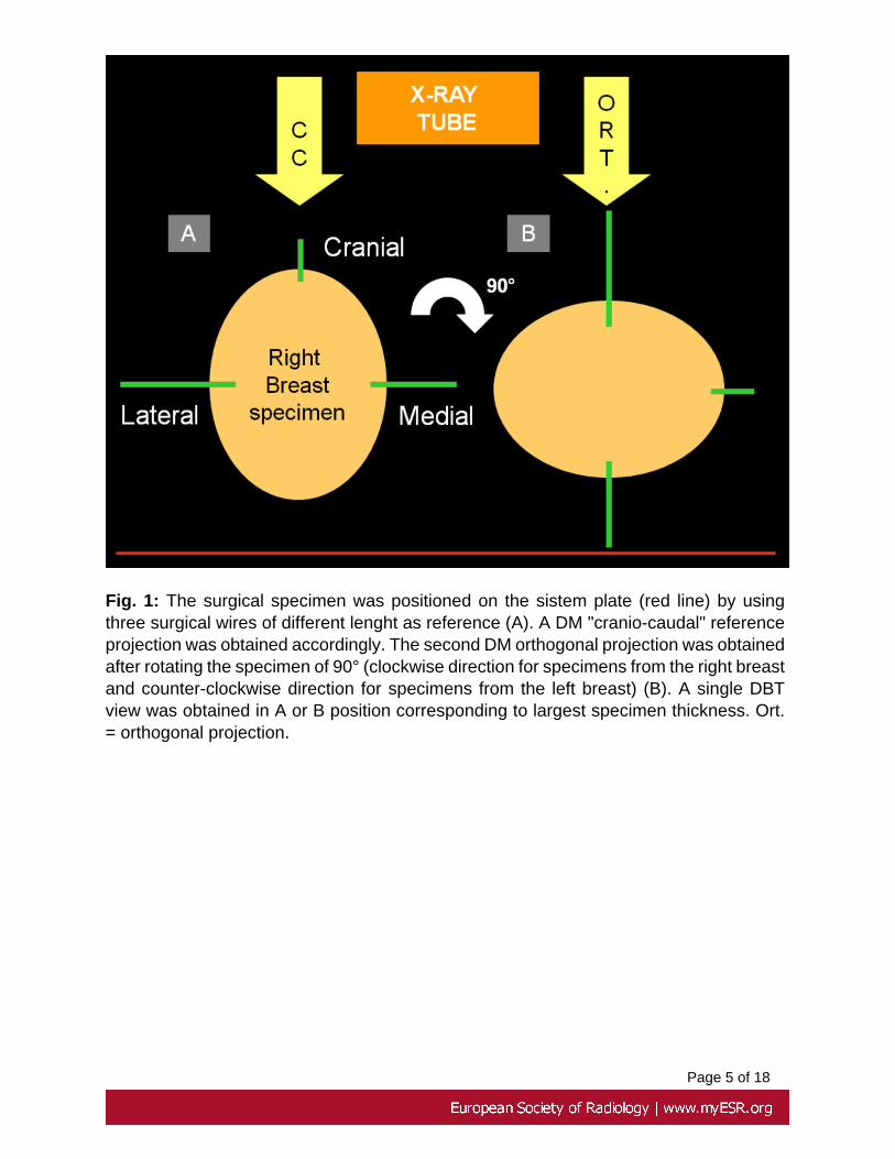
• No significant difference (p>0.5) was found in detectability and image qualityscores attributed by readers to DBT and DM (Table 3 on page 12) (Fig. 5on page 13 - Fig. 6 on page 14 ). Specimen density was assessed asfollows: 8 cases BI-RADS 1-2 and 14 cases BI-RADS 3-4. Lesions missedby DM occurred in specimens with a BI-RADS density of 2 in one case (ICDwith i.s. component appearing as a mass showed in Fig. 2 on page 8)and 3-4 in two cases (remaining lesions cited above, including that showedin Fig. 3 on page 9).
• There was no agreement (k=0.043) in the margins visibility score asattributed by using DBT and DM (24/27 comparable lesions). This becausereaders detected more than 50% of lesions margins in 23/24 lesions (95,8%;95% C.I. 78.8-99.9) with DBT and in 8/24 patients only with DM (33.4%;95% C.I. 15.6-55.3) (Fig. 7 on page 15 ). Of note, the margins score forDM cases was assumed to be the highest one of two projections.

Page 7 of 18
Images for this section:
Fig. 1: The surgical specimen was positioned on the sistem plate (red line) by usingthree surgical wires of different lenght as reference (A). A DM "cranio-caudal" referenceprojection was obtained accordingly. The second DM orthogonal projection was obtainedafter rotating the specimen of 90° (clockwise direction for specimens from the right breastand counter-clockwise direction for specimens from the left breast) (B). A single DBTview was obtained in A or B position corresponding to largest specimen thickness. Ort.= orthogonal projection.

Page 8 of 18
Table 1: Distribution of breast lesions on 21 surgical specimens according to theappearance on DM and/or DBT. * 1/10 specimen containing 3 lesions and 2/10specimens containing 2 lesions each. **1/8 specimen containing 2 lesions. ***1/4specimen containing 2 lesions.

Page 9 of 18
Fig. 2: 82 years-old patient operated for an IDC with i.s. component of the right breast(confirmed at histopathological examination of the specimen). Specimen density wasassessed as 2 according to BI-RADS criteria. Readers missed the lesion both on DMcranio-caudal (A) and orthogonal (B) projections. However, the lesion was correctlydetected as a mass in the 1-mm DBT thin slice (arrow in C). Lesion is magnified in D.

Page 10 of 18
Fig. 3: 40 years-old patient who underwent left skin-sparing mastectomy because of twoIDC with i.s. component as confirmed by histopathological examination of the specimen.BI-RADS density of the specimen was assessed as 3. By using DM (A), readers were ableto detect the largest mixed lesion only (arrow). DBT led to correctly detect one additionallesion appearing as a 3 mm large cluster of microcalcifications (circle in B). The regioncontaining the lesions is magnified in C.

Page 11 of 18
Table 2: Lesions size as assessed by DM and DBT.

Page 12 of 18
Fig. 4: Results of Bland and Altman analysis for DBT and DM agreement in terms oflesions size measurement.

Page 13 of 18
Table 3: Average subjective scores attibuted by readers to lesions detectability andimage quality for both imaging techniques. N.S. = not significant.
Page 14 of 18
Fig. 5: 55 years-old patient who underwent surgery for IDC. Readers attributed the samedetectability score value of 4 to the lesion identified both on DM (A) and DBT 1-mm thinslice (B). BI-RADS of the specimen was assessed as 1.

Page 15 of 18
Fig. 6: 44 years-old patient operated for IDC with i.s. component. DM (A) and DBT (B)had the same image quality score of 5.

Page 16 of 18
Fig. 7: 56 years-old patient who underwent surgery for IDC with i.s. component. DMcranio-caudal projection only is shown (A). As evident by the comparison with DBTimage (B), lesions margins are better identified all around the lesion contour with the 3Dtechnique. Lesion is magnified in C.

Page 17 of 18
Conclusion
Limitations
• Main study limitation is the small sample size. However, the collection ofnew cases is a work-in-progress in our Institution. Additional results seem toconfirm the trend we showed in this poster.
Conclusions
• Regardless of lesion type (mass and/or microcalcifications), DBT showedhigher DY than DM by retrieving small lesions, prevalently in densespecimens. This is in accordance with previous results showing that DBTincreases the sensitivity for cancer by reducing breast tissues overlapping[5-6].
• DBT showed a reasonable agreement with DM in assessing lesions size(measurements not exceeding 6 mm). DBT and DM were equivalent alsoin terms of lesions visibility and image quality, suggesting that DBT has thepotential to replace DM in the evaluation of surgical specimens. This is inaccordance with the better margins definition we observed by using DBT.Further studies are required to assess whether this assumption is valid forthe clinical scenario.
References
1. Kolb TM, Lichy J, Newhouse JH. Comparison of the performance ofscreening mammography, physical examination, and breast USandevaluation of factors that influence them: an analysis of 27,825 patientevaluations. Radiology 2002; 225(1): 165-75
2. Park JM, Franken EA, Garg M, et al. Breast Tomosynthesis: presentconsiderations and future applications. Radiographics 2007; 27:S231-S240
3. Lewin JM, Niklason L. Advanced applications of Digital Mammography:Tomosynthesis and contrast-enhanced Digital Mammography. SeminRoentgenol 2007; 42(4):243-252
4. D'Orsi CJ, Mendelson, EB, Ikeda DM, et al: Breast Imaging Reporting andData System: ACR BI-RADS - Breast Imaging Atlas, Reston, VA, AmericanCollege of Radiology, 2003
5. Baker JA, Lo JY. Breast tomosynthesis: state of the art and review of theliterature. Academic Radiology 2011; 18: 1298-1310
6. Gennaro G, Toledano A, Di Maggio C, et al Digital Breast Tomosynthesisversus Digital Mammography: a clinical performance study. Eur Radiol 2010;20: 1545-1553

Page 18 of 18
Personal Information
Thank you for the interest in this poster.
For any questions, do not hesitate to contact me.
R. Girometti
Aknowledgments
The Authors want to thank Dr. Cristina Molinari for the revision of the manuscript.
![arXiv:1912.11027v2 [eess.IV] 27 Dec 2019 · Robust breast cancer detection in mammography and digital breast tomosynthesis using annotation-efficient deep learning approach William](https://img.pdfslide.us/doc/110x75/5f02ff077e708231d4070694/arxiv191211027v2-eessiv-27-dec-2019-robust-breast-cancer-detection-in-mammography.jpg)