Embed Size (px)
Citation preview

22
HematologyJJAAIIMMEE SSAAMMOOUURR,, MMVVZZ,, PP hhDD,, DD iippll EECCAAMMSS
CHAPTER
Diagnostic Value of
Hematology is the discipline of medical science thatstudies the blood and blood-forming tissues, and is cur-rently considered an integral part of clinical laboratorydiagnostics in avian medicine. Hematology assays sel-dom provide an etiological diagnosis, but they remain,nevertheless, indispensable diagnostic tools to evaluatehealth and disease in individuals, to monitor the prog-ress of diseases, to evaluate the response to therapy andto offer a prognosis.
The routine collection and processing of blood samplesallows the evaluation of the hematologic response todisease. In addition, the creation of hematology data-bases is important in establishing reference values forvarious avian species.
In the past 15 years, significant advances have beenmade in the use of hematology assays in the differentialdiagnosis of pathologic conditions in avian species. Thisappears to have developed parallel to other areas suchas nutrition, wellness examinations, anesthesia, surgeryand therapeutics.
The processing of hematology samples also has beenenhanced in recent years. In the past, automatic analysisof avian blood samples was basically limited to total redcell counts using the cell countersa that were availableand making manual adjustments of the thresholds andcurrent aperture settings. More recently, the analysis ofavian blood samples has received a significant boostwith the advent of more comprehensive and accurateautomatic analytical systems based on laser flow cytome-try.b This methodology is based on the measurement ofscattered laser light, which fluctuates with the size of the
22_Hematology.qxd 8/23/2005 1:05 PM Page 587
Cl inica l Avian Medic ine - Volume I I588
cell, the complexity of the cell (eg, overall shape,nucleus-to-cytoplasm ratio, granulation), and the sizeand shape of the nucleus after blood cells are exposedto a laser beam. The laser flow cytometry unit producesa graphic display containing a total optical white cellcount, white cell differential count expressed in percent-age, absolute values and total red cell count, hemoglo-bin measurement by the cyanmethemoglobin method,thrombocyte count, and white cell count by cell-lysingimpedance measurement of cell nuclei.14 However, theuse of laser flow cytometric technology in avian speciesis not free from deficiencies.
There are certain pathological conditions in which thepresence of enlarged thrombocytes (commonly referredto as megathrombocytes) in the blood film appear to bea characteristic hemoresponse. For instance, in thehoubara bustard (Chlamydotis undulata macqueenii),the mean thrombocyte measurements in birds undergo-ing chronic inflammation (severe shoulder injury as aresult of repeated crashing against the enclosure wall)were 9.22 ± 0.21 µm length and 8.10 ± 0.19 µm widthcompared with 5.47 ± 0.12 µm length and 4.96 ± 0.10µm width in clinically normal birds.9 The mean diameterof lymphocytes in clinically normal houbara bustards is7.7 µm;51 however, there are other species, such as thekori bustard (Ardeotis kori), in which the presence oflarge and small thrombocytes in the same blood filmappears to be normal.51 It is, therefore, probable that asample containing megathrombocytes would yield ahigh lymphocyte count under an automatic analyticalsystem, as it would be impossible for even a sophisti-cated unit to differentiate between lymphocytes andmegathrombocytes. When dealing with such species, thesoftware would require some adjustments in order toproperly differentiate these cells. This would obviouslyimply the need to carry out extensive calibration basedon repeated manual assessments on a significant num-ber of samples.
Furthermore, in certain species it is relatively commonto find large and small lymphocytes in the same bloodfilm.4,12,26,31,35 This phenomenon has been observed inmany psittacine species. Clinically normal kori bustards,for instance, demonstrated a mean diameter for smalllymphocytes of 7.2 ± 0.12 µm, whereas the mean diam-eter of large lymphocytes was 10.7 ± 0.16 µm.31 There-
fore, total white blood cell counts and differential whiteblood cell counts cannot be accepted as reliable in everyclinical case and in every species if the values were esti-mated by laser flow cytometry. It is, therefore, highly rec-ommended to re-evaluate these samples using manualmethods. Clearly, the clinician must be fully familiar withthe materials and methods of hematology analysis inorder to assess and understand the results.
BBlloooodd SSaammppllee CCoolllleeccttiioonnIt is essential that blood samples be obtained from avianspecies by or under the supervision of a veterinarianwho is experienced with avian venipuncture. The assis-tant, if one is used, also should be comfortable withrestraint and handling techniques. The techniques usedvary according to personal preferences and the speciesbeing handled. Materials needed for blood sample col-lection, ie, syringes, slides, tubes, should be labeled inadvance and readily accessible (Table 22.1).
MMEETTHHOODDThe total blood volume in clinically normal birds is inthe range of 6 to 11 ml per 100 g of body weight.54
Thus, a bird weighing 250 g would have approximately15 to 27.5 ml of blood, of which, in a clinically normalindividual, up to 10% (1.5-2.7 ml) can be safely with-drawn without having any detrimental effect on thepatient. However, 0.2 to 0.3 ml of blood is generally suf-ficient to carry out a comprehensive hematology exami-nation in a bird.
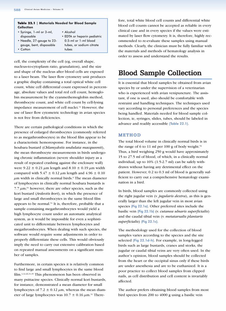
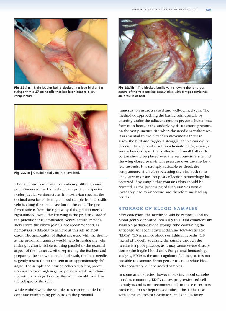
In birds, blood samples are commonly collected usingthe right jugular vein (v. jugularis dextra), as this is gen-erally larger than the left jugular vein in most avianspecies (Fig 22.1a). Other preferred sites include thebasilic vein (Fig 22.1b) (v. cutanea ulnaris superficialis)and the caudal tibial vein (v. metatarsalis plantarissuperficialis) (Fig 22.1c).
The methodology used for the collection of blood samples varies according to the species and the siteselected (Fig 22.1d-h). For example, in long-leggedbirds such as large bustards, cranes and storks, thejugular or caudal tibial veins are very often used. In theauthor’s opinion, blood samples should be collectedfrom the heart or the occipital sinus only if these birdsare under anesthesia and are to be euthanized. It is apoor practice to collect blood samples from clippednails, as cell distribution and cell content is invariablyaffected.
The author prefers obtaining blood samples from mostbird species from 200 to 4000 g using a basilic vein
Table 22.1 | Materials Needed for Blood SampleCollection
• Syringe, 1-ml or 3-ml,disposable
• Needle, 27-gauge to 23-gauge, bent, disposable
• Cotton
• Alcohol• EDTA or heparin pediatric
0.5-ml or 1-ml bloodtubes, or sodium citratetubes
22_Hematology.qxd 8/23/2005 1:05 PM Page 588
Chapter 22 | D I A G N O S T I C V A L U E O F H E M A T O L O G Y 589
while the bird is in dorsal recumbency, although mostpractitioners in the US dealing with psittacine speciesprefer jugular venipuncture. In most avian species, theoptimal area for collecting a blood sample from a basilicvein is along the medial section of the vein. The pre-ferred side is from the right wing if the practitioner isright-handed, while the left wing is the preferred side ifthe practitioner is left-handed. Venipuncture immedi-ately above the elbow joint is not recommended, ashemostasis is difficult to achieve at this site in mostcases. The application of digital pressure with the thumbat the proximal humerus would help in raising the vein,making it clearly visible running parallel to the externalaspect of the humerus. After separating the feathers andpreparing the site with an alcohol swab, the bent needleis gently inserted into the vein at an approximately 45°angle. The sample can now be collected, taking precau-tion not to exert high negative pressure while withdraw-ing with the syringe because this will invariably result inthe collapse of the vein.
While withdrawing the sample, it is recommended tocontinue maintaining pressure on the proximal
humerus to ensure a raised and well-defined vein. Themethod of approaching the basilic vein dorsally byentering under the adjacent tendon prevents hematomaformation because the underlying tissue exerts pressureon the venipuncture site when the needle is withdrawn.It is essential to avoid sudden movements that canalarm the bird and trigger a struggle, as this can easilylacerate the vein and result in a hematoma or, worse, asevere hemorrhage. After collection, a small ball of drycotton should be placed over the venipuncture site andthe wing closed to maintain pressure over the site for afew seconds. It is strongly advisable to check thevenipuncture site before releasing the bird back to itsenclosure to ensure no post-collection hemorrhage hasoccurred. Any sample that contains clots should berejected, as the processing of such samples wouldinvariably lead to imprecise and therefore misleadingresults.
SSTTOORRAAGGEE OOFF BBLLOOOODD SSAAMMPPLLEESSAfter collection, the needle should be removed and theblood gently deposited into a 0.5 to 1.0 ml commerciallyavailable pediatric blood storage tube containing theanticoagulant agent ethylenediamine tetra-acetic acid(EDTA) (1.5 mg/ml of blood) or lithium heparin (1.8mg/ml of blood). Squirting the sample through the needle is a poor practice, as it may cause severe disrup-tion to the fragile blood cells. For general hematologyanalysis, EDTA is the anticoagulant of choice, as it is notpossible to estimate fibrinogen or to count white bloodcells accurately in heparinized samples.
In some avian species, however, storing blood samplesin tubes containing EDTA causes progressive red cellhemolysis and is not recommended; in these cases, it ispreferable to use heparinized tubes. This is the casewith some species of Corvidae such as the jackdaw
Fig 22.1a | Right jugular being blocked in a love bird and asyringe with a 27 ga needle that has been bent to allowvenipuncture.
Fig 22.1b | The blocked basilic vein showing the torturousnature of the vein making cannulation with a hypodermic nee-dle difficult at best.
Fig 22.1c | Caudal tibial vein in a love bird.
22_Hematology.qxd 8/23/2005 1:05 PM Page 589

Cl inica l Avian Medic ine - Volume I I590
Fig 22.1d | The ventral surface of the wing of a cadaver withthe covert feathers removed to show the location for superficialulnaris vein lancing (arrow). Lancing the basilic vein in birdsunder 100 gms avoids subcutaneous hematomas and the possi-bility of death due to exsanguination. The site has a series ofnatural depressions over the vein that serve to allow the blood topool. The depressions are formed by the insertion of the second-ary feathers intermittently elevating and depressing the wing der-mis just caudal to the border of the flexor carpi ulnaris muscle.
Fig 22.1e | Lancet used in a finger stick technique for bloodsampling for glucose analysis in humans. The lancet still has themetal tip in place in the plastic cap.
Fig 22.1f | Lancet with the cap removed and the 1 mm tipexposed.
Fig 22.1g | The lancet readied at the site to lance thesuperficial ulnaris vein.
Fig 22.1h | The superficial ulnaris vein has been lanced andblood is being drawn into a micro capillary tube. A cotton ball isapplied to the site until hemostasis is achieved.
Figs 22.1d-h | Small Birds - Basilic Vein
Fig 22.2a | The improved Neubauer counting chamber andthe method for counting red blood cells. The total red blood cellcount is performed by counting the number of cells contained inthe 25 groups of 16 small squares (shaded) at the 4 cornersquares and center square in the central area of the chamber.Closely ruled triple lines (illustrated in the drawing as thick lines)separate these squares.
22_Hematology.qxd 8/23/2005 1:06 PM Page 590
Chapter 22 | D I A G N O S T I C V A L U E O F H E M A T O L O G Y 591
(Corvus monedula) and raven (Corvus corax); Gruidaesuch as the black-necked crowned crane (Balearicapavonina) and gray-necked crowned crane (Balearicaregulorum); Cracidae such as the black curassow (Craxalector); Phasianidae such as the brush turkey (Alecturalathami); Bucerotidae such as the crowned hornbill(Tockus alboterminatus); and the ostrich (Struthiocamelus).3,25 Storing blood samples in sodium citrate
tubes is recommended when sending samples to a com-
mercial laboratory for processing using laser flow
cytometry.14,19
Commercially available collection tubes usually have
printed labels. A pencil or ballpoint pen is used to enter
the date and identification of the bird, preferably prior
to filling the tube with the collected blood sample.
Always remember the rule of thumb in clinical pathol-
ogy: label tubes, not lids.
TTRRAANNSSPPOORRTTAATTIIOONN OOFF BBLLOOOODDSSAAMMPPLLEESS
In avian practice, hematology samples are commonly
sent to commercial laboratories for processing (Table
22.2). Therefore, it is essential to be familiar with and to
submit samples in full compliance with current local
mail and courier regulations.
HHeemmaattoollooggyy LLaabboorraattoorryy AAnnaallyyssiissAlthough the hematology laboratory analysis described
in this chapter were developed primarily for testing
human blood and are in full compliance with the recom-
mendations of the International Committee for
Standardization in Hematology,34 these have been
adapted and used successfully in avian hematology.
Ideally, laboratory analysis should be carried out within
3 to 4 hours after collection. Many laboratories in the
USA request that a smear be made immediately and sent
along with the EDTA tube. If this is not possible, sam-
ples should be refrigerated at 8 to 12° C or within a suit-
able container for processing within 24 to 48 hours.
Refrigerated samples are not ideal for hematology test-
ing, as the cells invariably suffer some changes. Only an
experienced hematologist would be able to differentiate
these changes from true hemoresponses to particular
medical disorders. Samples should not be exposed to
extreme environmental conditions or excessive shaking,
as this will affect the quality of the sample. Any form of
mouth pipetting with a Thoma pipette or any other
pipette with or without tubing is not acceptable within
clinical laboratory practices.
The amount of blood available for testing from small
birds (eg, <80 g) is very often limited, making it impos-
sible to carry out a full range of analyses. The clinician
should bear this in mind and request the analysis in
order of priority (Table 22.3).
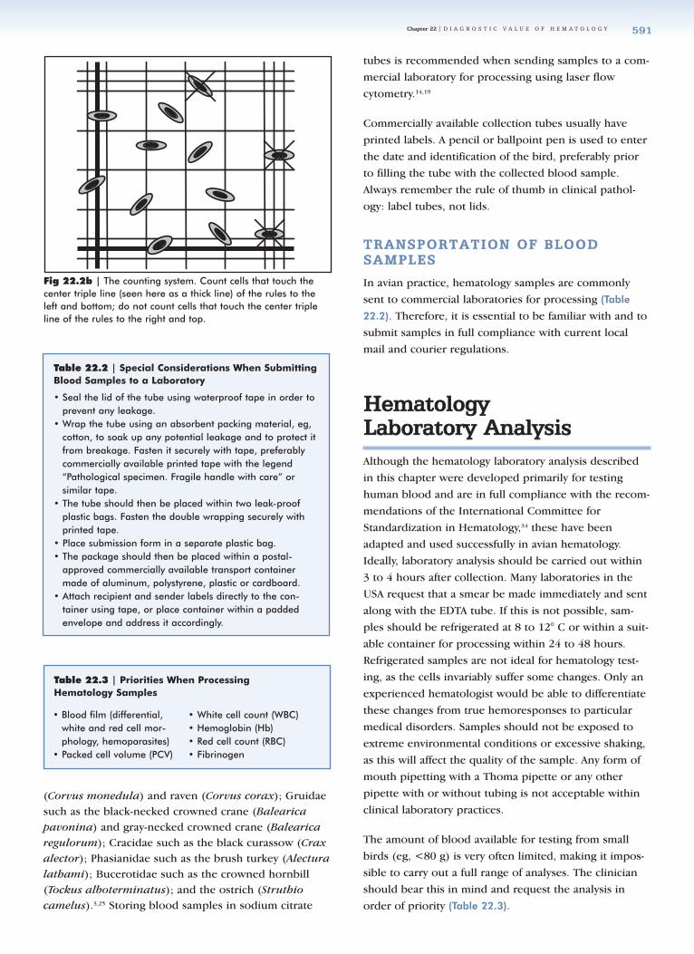
Fig 22.2b | The counting system. Count cells that touch thecenter triple line (seen here as a thick line) of the rules to theleft and bottom; do not count cells that touch the center tripleline of the rules to the right and top.
Table 22.2 | Special Considerations When SubmittingBlood Samples to a Laboratory
• Seal the lid of the tube using waterproof tape in order toprevent any leakage.
• Wrap the tube using an absorbent packing material, eg,cotton, to soak up any potential leakage and to protect itfrom breakage. Fasten it securely with tape, preferablycommercially available printed tape with the legend“Pathological specimen. Fragile handle with care” or similar tape.
• The tube should then be placed within two leak-proofplastic bags. Fasten the double wrapping securely withprinted tape.
• Place submission form in a separate plastic bag.• The package should then be placed within a postal-
approved commercially available transport containermade of aluminum, polystyrene, plastic or cardboard.
• Attach recipient and sender labels directly to the con-tainer using tape, or place container within a paddedenvelope and address it accordingly.
Table 22.3 | Priorities When Processing Hematology Samples
• Blood film (differential,white and red cell mor-phology, hemoparasites)
• Packed cell volume (PCV)
• White cell count (WBC)• Hemoglobin (Hb)• Red cell count (RBC)• Fibrinogen
22_Hematology.qxd 8/23/2005 1:06 PM Page 591
Cl inica l Avian Medic ine - Volume I I592
TTHHEE TTOOTTAALL RREEDD BBLLOOOODD CCEELLLLCCOOUUNNTT ((RRBBCC XX 1100 1122//LL))
The total red blood cell count is in itself an important
hematology assay, but it also is essential for the estima-
tion of mean corpuscular volume (MCV) and mean cor-
puscular hemoglobin (MCH). Many laboratories prefer
to estimate RBC using an automatic system, as this is
more precise than manual methods. The materials, solu-
tions and method described in Table 22.4, together with
Fig 22.2a,b, apply to a manual technique.
HHEEMMOOGGLLOOBBIINN EESSTTIIMMAATTIIOONN ((HHbb gg//ddll ))
In avian species, estimation of hemoglobin is hampered
by the presence of nuclei in the erythrocytes. Hemo-
globin estimation relies on the colorimetric measurement
of hemoglobin released after the lysing of the erythro-
cytes. Hemoglobin can be estimated using automatic
methods or manual methods (Table 22.5). Commercial
laboratories that estimate hemoglobin using an automatic
hematology analyzer have to take into consideration the
photometric interference of the free nuclei after lysing of
the erythrocyte. In the manual method, it is essential to
remove the nuclei from the preparation because its pres-
ence could yield unreliable results. The nuclei can be
deposited by low-speed centrifugation, but because some
hemoglobin remains attached to the nuclei, colorimetric
readings are commonly low. This can be overcome by
estimating hemoglobin as cyanmethemoglobin using
alkaline Drabkin’s cyanide-ferricyanide solution or as oxy-
hemoglobin using ammonia solution. In both cases, the
estimation is carried out using a spectrophotometer at
the absorbance reading of 540 nm. A calibration graph
should be made using commercially available hemoglo-
bin standards to express hemoglobin as oxyhemoglobin.
Conversely, hemoglobin can be estimated directly as oxy-
hemoglobin using a commercially available hemoglobi-
nometer. This is the preferred method used and recom-
mended by the author.
PPAACCKKEEDD CCEELLLL VVOOLLUUMMEE EESSTTIIMMAATTIIOONNPPCCVV %% ((HHEEMMAATTOOCCRRIITT HHcctt LL//LL))
Packed cell volume (PCV) is an important hematologic
assay because it provides an easy and objective way of
estimating the number of erythrocytes in the sample. It
also is essential for the calculation of the mean corpus-
cular volume (MCV) and mean corpuscular hemoglobin
concentration (MCHC). In avian species, PCV is best esti-
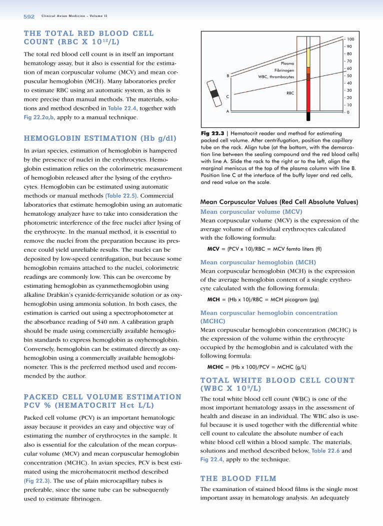
mated using the microhematocrit method described
(Fig 22.3). The use of plain microcapillary tubes is
preferable, since the same tube can be subsequently
used to estimate fibrinogen.
Mean Corpuscular Values (Red Cell Absolute Values)
Mean corpuscular volume (MCV)Mean corpuscular volume (MCV) is the expression of theaverage volume of individual erythrocytes calculatedwith the following formula:
MCV = (PCV x 10)/RBC = MCV femto liters (fl)
Mean corpuscular hemoglobin (MCH)Mean corpuscular hemoglobin (MCH) is the expressionof the average hemoglobin content of a single erythro-cyte calculated with the following formula:
MCH = (Hb x 10)/RBC = MCH picogram (pg)
Mean corpuscular hemoglobin concentration(MCHC) Mean corpuscular hemoglobin concentration (MCHC) isthe expression of the volume within the erythrocyteoccupied by the hemoglobin and is calculated with thefollowing formula:
MCHC = (Hb x 100)/PCV = MCHC (g/L)
TTOOTTAALL WWHHIITTEE BBLLOOOODD CCEELLLL CCOOUUNNTT((WWBBCC XX 1100 99//LL))The total white blood cell count (WBC) is one of themost important hematology assays in the assessment ofhealth and disease in an individual. The WBC also is use-ful because it is used together with the differential whitecell count to calculate the absolute number of eachwhite blood cell within a blood sample. The materials,solutions and method described below, Table 22.6 andFig 22.4, apply to the technique.
TTHHEE BBLLOOOODD FFIILLMMThe examination of stained blood films is the single mostimportant assay in hematology analysis. An adequately
Fig 22.3 | Hematocrit reader and method for estimatingpacked cell volume. After centrifugation, position the capillarytube on the rack. Align tube (at the bottom, with the demarca-tion line between the sealing compound and the red blood cells)with line A. Slide the rack to the right or to the left, align themarginal meniscus at the top of the plasma column with line B.Position line C at the interface of the buffy layer and red cells,and read value on the scale.
Plasma
Fibrinogen
WBC, thrombocytes
RBC
B
A
C
- 100
- 90
- 80
- 70
- 60
- 50
- 40
- 30
- 20
- 10
- 0
22_Hematology.qxd 8/23/2005 1:06 PM Page 592

Chapter 22 | D I A G N O S T I C V A L U E O F H E M A T O L O G Y 593
Methodc
• Label sample tubes using a permanent marker. • Use an automatic dispenser to transfer 4 ml of either
formol citrate solution or Natt and Herrick’s solution intosample tube.
• Wait for 5 minutes to allow working solution to reachroom temperature.
• Aspirate 20 µl of whole blood from storage tube usingmicropipette, wipe side of pipette tip carefully using tis-sue and dispense on the side of sample tube to make adilution of 1:200.
• Avoid touching the distal opening of the pipette tip withthe tissue, as this will cause capillary shift of blood intothe tissue.
• Avoid immersing the pipette tip into the diluting fluid.This is a poor laboratory practice.
• Place sample tube in roller mixer and wait for 3 minutes.• Clean Neubauer hemocytometer and coverslip using a
dry, lint-free cloth or laboratory lens tissue.• Place coverslip onto hemocytometer and slide gently over
it, making sure Newton’s rings (colored interference pat-tern) appear on both sides of the contact surfaces.
• Withdraw a small aliquot of the diluted sample using aplain capillary tube.
• Fill up one side of the hemocytometer by touching gentlythe intersection between coverslip and hemocytometerwith the loaded capillary tube. Avoid air bubbles andunderfilling or overfilling.
• Place filter paper at the bottom of the Petri dish. Positiontwo toothpicks on either side of the dish. Wet filter paperlightly with distilled water. Rest hemocytometer on tooth-picks. Cover Petri dish. Leave for 5 minutes for the cellsto settle down.
• The hemocytometer is now ready for use.• Count cells contained in the four corner and central
squares in the mid section of the hemocytometer.Following the “L” rule: count cells that touch the centertriple line of the ruling on the left and the bottom sides;do not count cells that touch the center triple line of theruling on the right and the top sides (see Figs 22.2a,b).
• Calculate red blood cell count using: N/100 = RBC x 1012/L
Note: N = number of cells counted in 160 small squares.
Materials and Equipment
• Automatic dispenser, 0-50 ml• Disposable sample tube with lid, 5 ml• Micropipette, 20 µl and corresponding tip• Roller mixer• Plain capillary tubes• Improved Neubauer hemocytometer and coverslip• Laboratory lens tissue• Petri dish 8.5 cm diameter• Filter paper 8.5 cm diameter• Toothpick• Distilled water• Microscope, preferably with phase contrast capability
Test Systems
The Unopette 365851 systemc
is probably the most popularmethod used for manual red blood cell count in avianspecies. It uses 10 µl of whole blood in 1.9 ml of 0.85%saline, resulting in a 1:200 dilution. The two other com-monly used systems are based on using either formol citrate solution (Dacie’s fluid) or Natt and Herrick’s solu-tion, depending on whether the examination is carried outwith or without phase contrast microscopy. Dacie’s formolcitrate solution is the least known diluting fluid, but oneused and recommended by the author.
Working Solutions
1. BD Unopette 365851c red blood count manual hematology test
2. Natt and Herrick’s solution (for use without phase contrast microscopy)
Note: Allow solution to stand overnight. Filter before use.
3. Formol citrate solution or Dacie’s fluid (for use with phase contrast microscopy)
Note: Refrigerate at 8 to 12° C.
NaCl 3.88 g
Na2SO4 2.5 g
Na2HPO4 12 H2O 2.91 g
KH2PO4 0.25 g
Formaldehyde 40% 7.5 ml
Methyl violet 2B 0.1 g
Distilled water to 1000 ml
Formaldehyde 10% 10 ml
Trisodium citrate 31.3 g
Distilled water 100 ml
Table 22.4 | Manual Total Red Blood Cell (RBC) Count Hematology Test
22_Hematology.qxd 8/23/2005 1:06 PM Page 593
prepared blood film provides the differential white bloodcell count and absolute white blood cell count, thethrombocyte count and the hemoparasite examination.
Preparation of the Blood Film
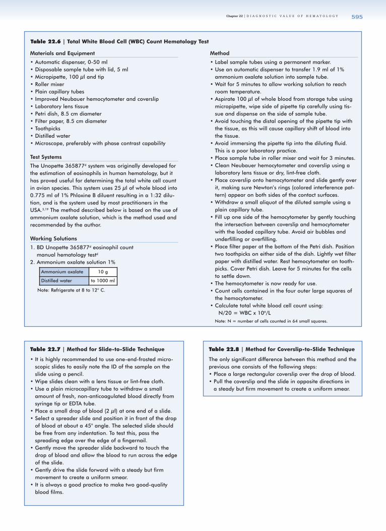
Blood films can be made from a drop of fresh, non-anti-coagulated blood directly from the tip of the syringe.Conversely, films can be made from blood stored inEDTA within 2 to 3 hours after collection. There are twogenerally accepted methods for the preparation of bloodfilms in hematology: the slide-to-slide technique (Fig
22.5, Table 22.7) and the coverslip-to-slide technique(Fig 22.6, Table 22.8). A two cover-slip technique is notdescribed here. The most popular method among avian
clinicians is the coverslip-to-slide technique, as smudg-
ing of blood red cells is generally minimized.
Fixation and Staining of the Blood Film
It is commonly accepted that blood films can be pre-
pared and be fixed and stained at a later date. This is
incorrect; blood films should at least be fixed immedi-
ately after preparation, particularly if made in a hot and
humid environment or under cold and freezing condi-
tions. Blood films should not be exposed to direct sun-
light, moisture of any kind or vapor from chemicals
(formaldehyde in particular), as this would invariably
affect cell morphology.
Cl inica l Avian Medic ine - Volume I I594
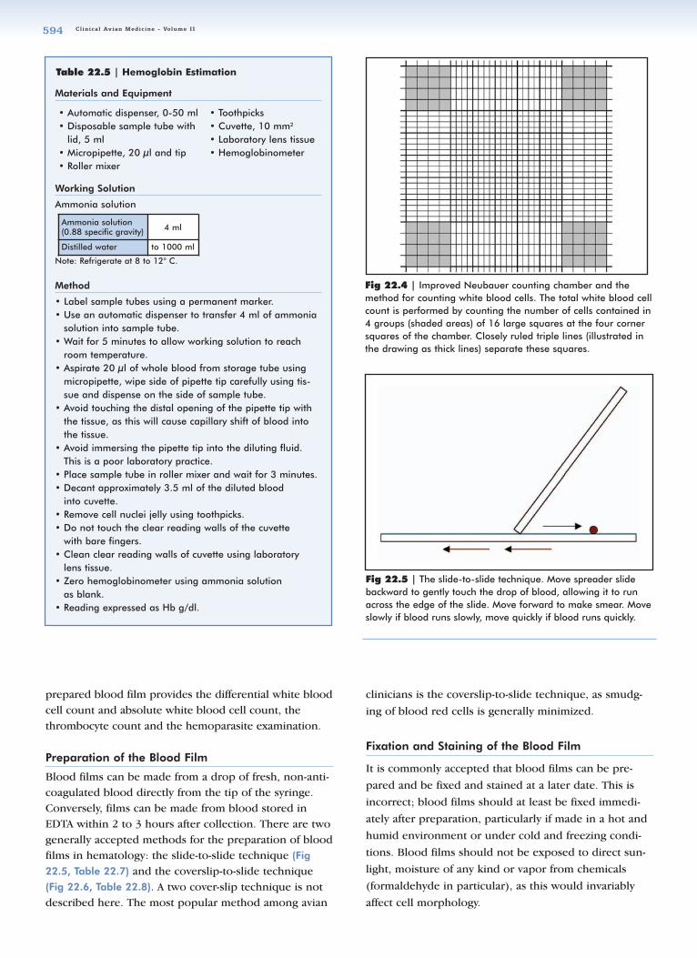
Fig 22.4 | Improved Neubauer counting chamber and themethod for counting white blood cells. The total white blood cellcount is performed by counting the number of cells contained in4 groups (shaded areas) of 16 large squares at the four cornersquares of the chamber. Closely ruled triple lines (illustrated inthe drawing as thick lines) separate these squares.
Materials and Equipment
Working Solution
Ammonia solution
Note: Refrigerate at 8 to 12° C.
Method
• Label sample tubes using a permanent marker. • Use an automatic dispenser to transfer 4 ml of ammonia
solution into sample tube.• Wait for 5 minutes to allow working solution to reach
room temperature.• Aspirate 20 µl of whole blood from storage tube using
micropipette, wipe side of pipette tip carefully using tis-sue and dispense on the side of sample tube.
• Avoid touching the distal opening of the pipette tip withthe tissue, as this will cause capillary shift of blood intothe tissue.
• Avoid immersing the pipette tip into the diluting fluid.This is a poor laboratory practice.
• Place sample tube in roller mixer and wait for 3 minutes.• Decant approximately 3.5 ml of the diluted blood
into cuvette.• Remove cell nuclei jelly using toothpicks.• Do not touch the clear reading walls of the cuvette
with bare fingers.• Clean clear reading walls of cuvette using laboratory
lens tissue.• Zero hemoglobinometer using ammonia solution
as blank.• Reading expressed as Hb g/dl.
Ammonia solution(0.88 specific gravity) 4 ml
Distilled water to 1000 ml
Table 22.5 | Hemoglobin Estimation
• Automatic dispenser, 0-50 ml• Disposable sample tube with
lid, 5 ml• Micropipette, 20 µl and tip• Roller mixer
• Toothpicks• Cuvette, 10 mm2
• Laboratory lens tissue• Hemoglobinometer
Fig 22.5 | The slide-to-slide technique. Move spreader slidebackward to gently touch the drop of blood, allowing it to runacross the edge of the slide. Move forward to make smear. Moveslowly if blood runs slowly, move quickly if blood runs quickly.
22_Hematology.qxd 8/23/2005 1:08 PM Page 594
Chapter 22 | D I A G N O S T I C V A L U E O F H E M A T O L O G Y 595
Materials and Equipment
• Automatic dispenser, 0-50 ml• Disposable sample tube with lid, 5 ml• Micropipette, 100 µl and tip• Roller mixer• Plain capillary tubes• Improved Neubauer hemocytometer and coverslip• Laboratory lens tissue• Petri dish, 8.5 cm diameter • Filter paper, 8.5 cm diameter• Toothpicks• Distilled water• Microscope, preferably with phase contrast capability
Test Systems
The Unopette 365877d system was originally developed forthe estimation of eosinophils in human hematology, but ithas proved useful for determining the total white cell countin avian species. This system uses 25 µl of whole blood into0.775 ml of 1% Phloxine B diluent resulting in a 1:32 dilu-tion, and is the system used by most practitioners in theUSA.3,19 The method described below is based on the use ofammonium oxalate solution, which is the method used andrecommended by the author.
Working Solutions
1. BD Unopette 365877d eosinophil count manual hematology testd
2. Ammonium oxalate solution 1%
Note: Refrigerate at 8 to 12° C.
Method
• Label sample tubes using a permanent marker.• Use an automatic dispenser to transfer 1.9 ml of 1%
ammonium oxalate solution into sample tube.• Wait for 5 minutes to allow working solution to reach
room temperature.• Aspirate 100 µl of whole blood from storage tube using
micropipette, wipe side of pipette tip carefully using tis-sue and dispense on the side of sample tube.
• Avoid touching the distal opening of the pipette tip withthe tissue, as this will cause capillary shift of blood intothe tissue.
• Avoid immersing the pipette tip into the diluting fluid.This is a poor laboratory practice.
• Place sample tube in roller mixer and wait for 3 minutes.• Clean Neubauer hemocytometer and coverslip using a
laboratory lens tissue or dry, lint-free cloth.• Place coverslip onto hemocytometer and slide gently over
it, making sure Newton’s rings (colored interference pat-tern) appear on both sides of the contact surfaces.
• Withdraw a small aliquot of the diluted sample using aplain capillary tube.
• Fill up one side of the hemocytometer by gently touchingthe intersection between coverslip and hemocytometerwith the loaded capillary tube. Avoid air bubbles andunderfilling or overfilling.
• Place filter paper at the bottom of the Petri dish. Positiontwo toothpicks on either side of the dish. Lightly wet filterpaper with distilled water. Rest hemocytometer on tooth-picks. Cover Petri dish. Leave for 5 minutes for the cellsto settle down.
• The hemocytometer is now ready for use.• Count cells contained in the four outer large squares of
the hemocytometer. • Calculate total white blood cell count using:
N/20 = WBC x 109/L Note: N = number of cells counted in 64 small squares.
Ammonium oxalate 10 g
Distilled water to 1000 ml
Table 22.6 | Total White Blood Cell (WBC) Count Hematology Test
Table 22.7 | Method for Slide-to-Slide Technique
• It is highly recommended to use one-end-frosted micro-scopic slides to easily note the ID of the sample on theslide using a pencil.
• Wipe slides clean with a lens tissue or lint-free cloth.• Use a plain microcapillary tube to withdraw a small
amount of fresh, non-anticoagulated blood directly fromsyringe tip or EDTA tube.
• Place a small drop of blood (2 µl) at one end of a slide.• Select a spreader slide and position it in front of the drop
of blood at about a 45° angle. The selected slide shouldbe free from any indentation. To test this, pass thespreading edge over the edge of a fingernail.
• Gently move the spreader slide backward to touch thedrop of blood and allow the blood to run across the edgeof the slide.
• Gently drive the slide forward with a steady but firmmovement to create a uniform smear.
• It is always a good practice to make two good-qualityblood films.
Table 22.8 | Method for Coverslip-to-Slide Technique
The only significant difference between this method and theprevious one consists of the following steps:• Place a large rectangular coverslip over the drop of blood.• Pull the coverslip and the slide in opposite directions in
a steady but firm movement to create a uniform smear.
22_Hematology.qxd 8/23/2005 1:08 PM Page 595

Cl inica l Avian Medic ine - Volume I I596
Fixation
In general, freshly prepared blood films should beimmersed in absolute methanol within a Coplin jar for 5to 10 minutes immediately after preparation. Fixedblood films can then be stored within commerciallyavailable slide storage boxes (eg, under field conditions)and be stained at a later date. Blood films also can bestained immediately after fixation.
The importance of adequate fixation of blood films fromavian species cannot be overemphasized. The intracyto-plasmic granules of the heterophils and basophils arewater soluble; therefore, blood films should be ade-quately fixed before staining in order to preserve theintegrity of these structures. A significant problem inavian hematology is the presence of smudged red cellnuclei as a consequence of hemolysis in poorly fixedblood films. This is one of the main reasons why clini-cians and commercial laboratories are now inclined touse stains that are prepared in absolute methanol (eg,Wright-Giemsa stain, Leishman stain) and are used at fullstrength so films are fixed and stained at the same time.If absolute methanol within a Coplin jar is used for fixa-tion in your laboratory, it must be replaced as soon as itbegins showing chemical fatigue. This would depend onthe number of slides fixed and the environmental condi-tions within the laboratory.
Staining
Most Romanovsky stains used for staining human andmammalian blood films are suitable for staining avian
blood films. However, the results obtained with variousstains may be slightly different, and the selection ofstains is generally accepted as a matter of personal pref-erence. Commonly used stains include Wright stain,Giemsa stain, Wright-Giemsa stain, Leishman stain,Wright-Leishman stain, May-Grünwald stain and May-Grünwald-Giemsa stain. In the author’s opinion, rapidstains on their own, eg, Diff Quick and Rapid Diff, donot produce adequate quality for the differentiation ofsubtle blood cell structures and those of hematozoa.This is particularly important with respect to the mor-phological characteristics of the granulocytes.
Automatic slide stainers facilitate staining a relativelylarge number of blood films at the same time, producingconsistent results and eliminating variations that mayoccur with manual techniques. However, this type ofequipment is relatively expensive to purchase and main-tain and is more appropriate for high-volume commer-cial laboratories.
It is important that clinicians or laboratory techniciansrecall the basic principles of hematology when stainingblood films. The pH of the stains should be checkedeach time new stock is prepared. Some stains, particu-larly those prepared from powder, should be adequatelyfiltered. Glassware should be properly washed, rinsedwith distilled water and dried thoroughly before use.Many of the common artifacts on blood films are due tocareless preparation and improper methodology.
The staining method currently used and recommended
Fig 22.7 | A microcapillary tube after incubation and centrifu-gation, and the measurements necessary for the estimation offibrinogen.
Fig 22.6 | The coverslip-to-slide technique. Place coversliponto drop of blood. Apply gentle pressure downward. Moveslide and coverslip in opposite directions to make smear.
22_Hematology.qxd 8/23/2005 1:09 PM Page 596

Chapter 22 | D I A G N O S T I C V A L U E O F H E M A T O L O G Y 597
by the author is a slightly modified technique4 describedin Table 22.9.
The placement of a coverslip using a commercially avail-able mounting medium over the blood smear isoptional. Additionally, the mounting of blood filmsoffers several advantages such as preventing scratchingduring transport, protection against damage duringexcessive manipulation (eg, teaching material) andenhancing visualization for optimal examination andphotography.
Morphologic and Staining Characteristics of RedBlood Cells, White Blood Cells and Thrombocytes
Normal red blood cells appear elliptical and have ellipti-cal nuclei; the cytoplasm stains uniformly eosinophilic,and the nuclei is dark purple in color (modified Wright-Giemsa stain).
In general, the widely known “Romanovsky stains” con-tain blue azure that reacts with acid groups, includingthose of nucleic acids and proteins of the nucleus andcytoplasm and eosin Y, which has a particular affinity forbasic groups of hemoglobin. When used in differentavian species, the slight variations observed may be theresult of true species diversity or simply variations in thematerials and methods used from individual to individ-ual or from laboratory to laboratory.
Adequate knowledge of the morphology and stainingcharacteristics of the different blood cells is of the utmostimportance for the differentiation and classification ofthose blood cells (Table 22.10 and Figs 22.8-22.43).
Differential White Blood Cell and Absolute White Blood Cell Count
For the differential white blood cell count and absolutewhite blood cell count, the film should be examinedthoroughly under high-power magnification, under oil(1000x). The recommended topographic site is on theshoulder of the blood film. The shoulder is the edge ofthe oval-shaped end of a smear. This is the area wherethe blood cells are in one layer and are slightly segre-gated, thus facilitating examination.
In general terms, 100 white blood cells should becounted and classified according to the morphologicand staining characteristics. Counting is usually carriedout using a commercially available manual or electronicdifferential cell counter. The differential white blood cellcount is expressed as a percentage of the individual cellgroup. The percentage of each cell group is then con-verted into absolute numbers by reference to the totalWBC using the following formula:
Table 22.9 | Wright-Giemsa Staining Procedure
Working Stain
• 3 g Wright stain powder• 0.3 g Giemsa stain powder• 5 ml glycerol• To 1000 ml absolute methanol (acetone free)• Filter and store
Method
• Prepare thin blood smears.• Place on staining rack.• Flood smear with Wright-Giemsa stain, allow to stand for
3 minutes.• Add equal amount of Sørensen’s pH 6.5-6.8 buffer,
depending on batch stain.• Mix gently by blowing using a pipette until metallic green
sheen forms on the surface, allow to stand for 6 minutes.• Rinse with buffer, allowing to stand for 1 minute for
differentiation.• Wash copiously with buffer.• Wipe the back of smear with tissue to remove excess stain.• Prop in rack until dry.
Table 22.10 | Morphologic and Staining Characteristics ofDifferent Blood Cells
Blood Cell MorphologicCharacteristics
StainingCharacteristics
Erythrocyte Mature cells
Medium size, oval elon-gated shape, central ovalelongated nucleus
Cytoplasm: uniform paleorange to red-pink;Nucleus: purple-red, condensed, clumped chromatin
Immature cells
Smaller than mature cell,round to semi-oval, relatively larger nucleus
Polychromatic, cytoplasmpale to dark blue
Heterophil Medium size, round shape,bilobed nucleus
Colorless cytoplasm, rod-to cigar-shaped brick redto pale blue granules
Eosinophil Medium size, round shape,bilobed nucleus
Pale blue cytoplasm, roundto oval brick red to paleblue granules
Basophil Small size, round shape,unlobed nucleus
Pale blue cytoplasm, vari-able number of small,medium and large darkred-purple granules
Lymphocyte Small to medium size, typi-cally round to triangularshape, centrally positionedlarge round nucleus; ingeneral, 25 cytoplasm:75nucleus; ratio, coarsely condensed to highly condensed chromatin
Pale blue cytoplasm
Monocyte Large size, typically roundshape, eccentrically posi-tioned kidney-shapednucleus; in general 75cytoplasm:25 nucleus ratio,cytoplasm lace-likeappearance, often mediumsize vacuoles, coarselycondensed chromatin
Cytoplasm pale blue topale gray
Thrombocyte Small, oval to rectangularshape, nucleus oval to rectangular
Cytoplasm colorless to paleblue, large vacuoles,nucleus highly condenseddark purple-red chromatin
22_Hematology.qxd 8/23/2005 1:09 PM Page 597
Cl inica l Avian Medic ine - Volume I I598
(Percentage of white blood cell counted x total WBC)/100 =absolute No. x 109/L
Thrombocyte CountThrombocytes are usually counted while performing thedifferential white blood cell count. Valid and reliableresults cannot be obtained if there is evidence of throm-bocyte clumping.
The absolute number of thrombocytes is estimated byusing the following formula:
(No. of thrombocytes counted/100) x WBC = thrombocytes x 109/L
Figs 22.44-22.48 and Tables 22.12-22.18 are offered asreferences for interpreting hemotological findings.
FFIIBBRRIINNOOGGEENN EESSTTIIMMAATTIIOONN (( gg//LL))Fibrinogen is a plasma protein essential for normalblood coagulation, but also is one of the acute reactiveproteins that are detected in increased levels in associa-tion with medical disorders involving infection andinflammation (see Tables 22.11, 22.12 and 22.18).
Hemoparasite Examination
Hemoparasite examination is carried out on thin, good-quality blood films. Prior to a differential white cellcount using high-power magnification (1000x), theblood film should be examined under low-power magni-fication (eg, 200x or 400x) in order to detect large extracellular hemoparasites (eg, microfilariae), which couldbe missed if the film is examined only under high-powermagnification. The examination under low-power magni-fication should concentrate on areas not commonlyexamined under high-power magnification, eg, head andtail of the blood smear. The blood film should be exam-ined in full in a systematic way and following a consis-tent pathway.
A blood parasite quantitative assessment should be car-ried out, in certain cases and under certain circum-stances, by examining 1000 red cells (in the case ofintracytoplasmic parasitic forms) and determining the
number of red cells containing hemoparasites (eg,Haemoproteus spp., Babesia spp.). The number is thenexpressed in percentage and this usually constitutes thedegree of parasitemia.
If hemoparasites are observed during routine examina-tion of the blood film under low- or high-power magnifi-cation, it is imperative to immediately prepare additionalblood films. Ideally, a fresh blood sample should beobtained to prepare these new blood films from non-anticoagulated blood. This is of the utmost importance ifa rare parasite is observed in the film. Blood filmsshould be fixed but unstained when sending them toparasitologists, who have their own preferences forstains and staining procedures.
Table 22.11 | Fibrinogen Estimation
Materials and Equipment Needed for Fibrinogen Estimation
• Microcapillary tube rack• Microhematocrit centrifuge• Microhematocrit reader• Water bath 56° C ± 1° C• Microcapillary tube holder• Microscope with measuring eyepiece
and stage Vernier scale• Timer
Method (following estimation of packed cell volume)
• Place microcapillary tube in tube rack.• Place loaded rack in water bath at 56° C for 3 minutes
(make sure the entire plasma column is immersed).• Centrifuge microcapillary tubes again at 10,000 to 12,000
“g force” for 5 minutes.• Place microcapillary tubes in tube holder and, using the
microscope measuring eyepiece and the stage Vernier ofthe microscope, take reading at the upper and lower limitsof the protein layer and at the upper limit of the plasmacolumn (see Fig 22.7).
• Estimate fibrinogen with the following formula: (B - A)/(C- A) x 100 = fibrinogen g/L
Note: It is essential to perform this analysis on blood stored in EDTAbecause the analysis would be invalidated if performed on samplesstored in heparin or on samples containing clots.
22_Hematology.qxd 8/23/2005 1:09 PM Page 598
Chapter 22 | D I A G N O S T I C V A L U E O F H E M A T O L O G Y 599
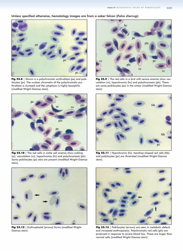
Unless specified otherwise, hematology images are from a saker falcon (Falco cherrug):
Fig 22.9 | The red cells in a bird with severe anemia show vac-uolation (vc), hypochromia (hc) and polychromasia (plc). Thereare some poikilocytes (pc) in the smear (modified Wright-Giemsastain).
Fig 22.8 | Shown is a polychromatic erythroblast (pe) and poik-ilocytes (pc). The nuclear chromatin of the polychromatic ery-throblast is clumped and the cytoplasm is highly basophilic(modified Wright-Giemsa stain).
Fig 22.10 | The red cells in sickle cell anemia show sickling(sc), vacuolation (vc), hypochromia (hc) and polychromasia (plc).Some poikilocytes (pc) also are present (modified Wright-Giemsastain).
Fig 22.11 | Hypochromic (hc), teardrop-shaped red cells (tds)and poikilocytes (pc) are illustrated (modified Wright-Giemsastain).
Fig 22.12 | Erythroplastid (arrows) forms (modified Wright-Giemsa stain).
Fig 22.13 | Poikilocytes (arrows) are seen in metabolic defectsand increased erythropoiesis. Polychromatic red cells (plc) areproduced in response to severe blood loss. These are larger thannormal cells (modified Wright-Giemsa stain).
22_Hematology.qxd 8/23/2005 1:09 PM Page 599

Cl inica l Avian Medic ine - Volume I I600
Fig 22.14 | Shown are teardrop-shaped red cells (tds) andpolychromasia (plc). Teardrop-shaped cells are indications of tox-icosis (May-Grünwald Giemsa stain).
Fig 22.15 | Two normal heterophils (arrows). Heterophils arecharacterized by brick red, elongated intracytoplasmic granulesand bilobed nuclei (modified Wright-Giemsa stain).
Fig 22.16 | In this eosinophil (arrow), note the numerous smalland medium-sized, dark purple-colored granules located mainlyin the periphery of the cytoplasm (modified Wright-Giemsa stain).
Fig 22.17 | An eosinophil (arrow) from an eclectus parrot(Eclectus roratus). Note the numerous small intracytoplasmicgranules widespread across the cytoplasm. The granules staindark purple in color (modified Wright-Giemsa stain).
Fig 22.18 | An eosinophil (arrow) from a kori bustard (Ardeotiskori). Note the large, round, orange-colored granules character-istic of this species (May-Grünwald Giemsa stain).
Fig 22.19 | A slightly disrupted eosinophil (arrow) from a lessersulphur-crested cockatoo (Cacatua sulphurea). The medium-sized,round granules are blue in color (May-Grünwald Giemsa stain).
22_Hematology.qxd 8/23/2005 1:09 PM Page 600

Chapter 22 | D I A G N O S T I C V A L U E O F H E M A T O L O G Y 601
Fig 22.20 | In this eosinophil (arrow) from a saker falcon (Falcocherrug), the granules are not stained, giving the impression ofnumerous irregular vacuoles within the cytoplasm (May-GrünwaldGiemsa stain).
Fig 22.21 | In this eosinophil (arrow) is seen a similar-stainingartifactual difference, as in the previous figure. The granules arenot stained, giving the impression of numerous vacuoles withinthe cytoplasm (Diff Quik stain).
Fig 22.22 | These eosinophil (arrow) granules are well stained,irregular in shape and size, stained purple or dark purple (modi-fied Wright-Giemsa stain). The author highly recommends theuse of this stain for routine hematology.
Fig 22.23 | A basophil (arrow) is characterized by the presenceof large, round, dark purple granules widespread across the cyto-plasm and an unlobed nucleus (modified Wright-Giemsa stain).
Fig 22.24 | A normal monocyte (arrow) is a relatively large cellwith a kidney-shaped nucleus and abundant, slightly opaque,blue-gray, “lace-like” cytoplasm (modified Wright-Giemsa stain).
Fig 22.25 | A normal lymphocyte (ly) and a normal thrombo-cyte (th). Lymphocytes are regular round cells with a central orslightly eccentric nuclei, and with a varying amount of pale bluecytoplasm (modified Wright-Giemsa stain).
22_Hematology.qxd 8/23/2005 1:10 PM Page 601

Cl inica l Avian Medic ine - Volume I I602
Fig 22.26 | Two normal thrombocytes (arrows) from a kori bus-tard (Ardeotis kori). Thrombocytes are round or irregular cellswith completely dark purple and dense round or oval nuclei, andclear blue-gray cytoplasm. In some species, a few cytoplasmicprojections can be observed. Sometimes it can be very difficult todifferentiate between thrombocytes and small lymphocytes (May-Grünwald Giemsa stain).
Fig 22.27 | Shown are a normal thrombocyte (th), normal lym-phocyte (ly) and a normal monocyte (mo) for comparison ofthree different mononuclear cells. Thrombocytes and small lym-phocytes can be very similar. In order to differentiate betweenthem, the appearance of the nuclear chromatin has to be closelyexamined (modified Wright-Giemsa stain).
Fig 22.28 | Two toxic heterophils (th) and a megathrombocyte(mth). One of the heterophils shows a lack of lobulation of thenucleus (left shift); both show loss of granulation and the cyto-plasm is stained basophilic. The megathrombocyte is significantlylarger than a normal thrombocyte. The cytoplasm is basophilic,the nucleus cytoplasm ratio is increased and it has scallopedcytoplasmic margins (modified Wright-Giemsa stain).
Fig 22.30 | A toxic heterophil (arrow) with a lack of nuclearlobulation (left shift) and loss of cytoplasmic granulation. Only afew large, round, dark purple granules are present and the cyto-plasm is basophilic (modified Wright-Giemsa stain).
Fig 22.31 | A toxic heterophil (arrow). The heterophil showsloss of nuclear lobulation (left shift) and loss of cytoplasmic gran-ulation. There are very few large, dark purple granules and thecytoplasm is basophilic (modified Wright-Giemsa stain).
Fig 22.29 | A toxic heterophil (arrow) showing loss of nuclearlobulation (left shift) and loss of cytoplasmic granulation. Thegranules are round, large and stained dark purple, and the cyto-plasm is basophilic (modified Wright-Giemsa stain).
22_Hematology.qxd 8/23/2005 1:10 PM Page 602
Chapter 22 | D I A G N O S T I C V A L U E O F H E M A T O L O G Y 603
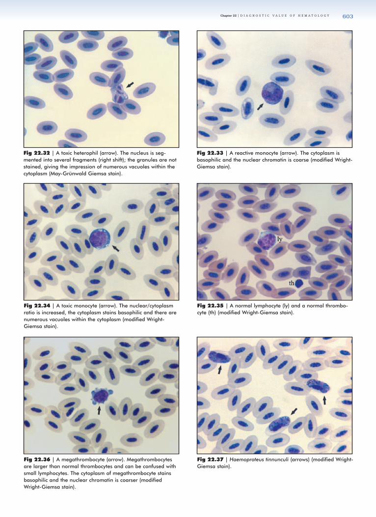
Fig 22.32 | A toxic heterophil (arrow). The nucleus is seg-mented into several fragments (right shift); the granules are notstained, giving the impression of numerous vacuoles within thecytoplasm (May-Grünwald Giemsa stain).
Fig 22.33 | A reactive monocyte (arrow). The cytoplasm isbasophilic and the nuclear chromatin is coarse (modified Wright-Giemsa stain).
Fig 22.34 | A toxic monocyte (arrow). The nuclear/cytoplasmratio is increased, the cytoplasm stains basophilic and there arenumerous vacuoles within the cytoplasm (modified Wright-Giemsa stain).
Fig 22.35 | A normal lymphocyte (ly) and a normal thrombo-cyte (th) (modified Wright-Giemsa stain).
Fig 22.36 | A megathrombocyte (arrow). Megathrombocytesare larger than normal thrombocytes and can be confused withsmall lymphocytes. The cytoplasm of megathrombocyte stainsbasophilic and the nuclear chromatin is coarser (modifiedWright-Giemsa stain).
Fig 22.37 | Haemoproteus tinnunculi (arrows) (modified Wright-Giemsa stain).
22_Hematology.qxd 8/23/2005 1:10 PM Page 603

Cl inica l Avian Medic ine - Volume I I604
Fig 22.38 | Haemoproteus psittaci (arrows) from a green-winged macaw (Ara chloroptera).
Fig 22.40 | Microfilaria sp. (modified Wright-Giemsa stain). Fig 22.41 | Leucocytozoon toddi (lct), Haemoproteus tinnunculi(hpt) and normal heterophil (ht) (modified Wright-Giemsa stain).
Fig 22.42 | Leucocytozoon simondi from a Canada goose(Branta canadensis).
Fig 22.43 | Plasmodium vaughani schizont from a robin (Turdusmigratorius).
Fig 22.39 | Babesia shortti (arrows) (modified Wright-Giemsastain).
Ken
dall
Har
r
M.
Gri
ener
22_Hematology.qxd 8/23/2005 1:11 PM Page 604

Chapter 22 | D I A G N O S T I C V A L U E O F H E M A T O L O G Y 605
Fig 22.46 | The Hct value continued to increase steadily from0.23 ± 0.7 L/L at 1 month of age to 0.399 ± 0.9 L/L at 5months of age and remained fairly constant until the age of 12to 15 months, when the value increased to 0.47 ± 0.9 L/L.
The following is a collection of hematology values and an interpretation guide for the avian veterinarian:
Fig 22.44 | The RBC increased steadily for the first 4 monthsfrom 1.28 ± 0.06 x 10¹²/L at 1 month of age, increasing gradu-ally up to the age of 4 months to 2.06 ± 0.08 x 10¹²/L. Afterthis time, the RBC remained fairly constant. The RBC value atthe age of 12 to 15 months was 2.08 ± 0.06 x 10¹²/L.
Fig 22.45 | The HB value followed a similar pattern as forRBC, with a value of 7.5 ± 0.2 g/dl at the age of 1 month,increasing to 12.1 ± 0.3 g/dl at 4 months of age. This valueremained fairly constant until the age of 12 months, when itincreased to 14.2 ± 0.4 g/dl.
Fig 22.47 | The WBC count at 1 month of age was 8.78 ±0.45 x 10⁹/L, increasing to 15.6 ± 0.7 x 10⁹/L at 7 months, thendecreasing slightly to 14.5 ± 0.5 x 10⁹/L at 9 months of age.
Fig 22.48 | The fibrinogen value was 1.76 ± 0.18 g/L at 1month of age, increasing steadily to 3.0 ± 0.2 g/L at the age of7 months.
AAggee--rreellaatteedd HHeemmaattoollooggiicc CChhaannggeessAge-related hematologic findings in kori bustard (Ardeotis kori) chicks during their growth and development are presented(Figs 22.44-22.48). Blood samples were collected from 16 clinically normal chicks at 1-month intervals. The tenth samplingwas obtained at 15 months of age.
22_Hematology.qxd 8/23/2005 1:11 PM Page 605

Cl inica l Avian Medic ine - Volume I I606
Table 22.12 | Evaluating the RBC, HB, PCV and Red Cell Indices
Hematologic Findings Possible Causes
Polycythemia: Increased packed cell volume(PCV) or hematocrit (Hct) and red blood cellcount (RBC)
Absolute: Primary polycythemiaPolycythemia veraSecondary polycythemia, reaction to hypoxia Physiological: Adaptation to high altitudes Pathological: Chronic circulatory or respiratory disease (ie, COPD or asthma of macaws), iron storage disease, rickets
Hypoxic increase in erythropoietin productionNon-hypoxic, autonomous increase in erythropoietin production
Relative: Dehydration, different etiologies
PCV >56% or Hct >0.56 L/L Dehydration in most birds, relatively normal in small (<100 g) psittacine and passerine birds, especially cockatiels
Anemia: Decreased PCV or Hct and RBC Absolute: Hemorrhage (trauma, coagulation disorders, ectoparasitism, endoparasitism); increased redcell destruction (hemoparasites, some bacterial infections, autoimmune hemolytic anemia); decreased redcell production (nutritional deficiencies, chronic infection, chronic renal disease, avian leukoses, toxicosis)
Relative: Overhydration
Low hemoglobin (Hb) value:(eg, <11.0 g/dl)
Anemia in adult birds
Low mean corpuscular hemoglobin con-centration (MCHC) value: (eg, <29.0 g/dl)
Possible iron and other element deficiency
Table 22.15 | Evaluating Leukocytosis/Heterophilia/Normal Heterophils
Monocyte Count Possible Causes
Normal InfectiousAcute: Gram-negative septicemias. Tuberculosis
(granulomas in Falconiformes andGalliformes, but not in Psittaciformes, inthese species exclusive accumulation ofepithelioid cells [Gerlach, personal communication, 2002]), coligranulomatosis,salmonellosis, yersiniosis and pasteurellosis
Monocytosis Fungal: Aspergillosis, severe candidiasisParasitic: Trichomonaisis, capillariaiasis, maggot
infestationsMiscellaneous: Foreign body inhalation pneumonia,
focal peritonitis, chronic ulcerativelesions, old open wounds
Acute and chronic: Pox and herpesvirus infections,chlamydophilosis
Chronic: Granulomatous or purulent infections/infestations
Monocytopenia Non-infectiousMaggot infestation, burns, lead/smoke intoxication,egg yolk peritonitis
MonocyteCount
Serial Sampling
Possible Causes ofHemogram Changes
Monocytosis WBC reduced Healing of soft tissue damageor bone fractures without thecomplications of severe infec-tion or necrosis, relative cellnumbers depend on thedegree of chronicity
Normal monocyte count
WBC stays elevated Acute or chronic inflammationwithout severe tissue necrosis
Return to normal within24-72 hours in theabsence of stressor
Stress
Note: Monocytes are slow-reacting cells of the immune system, stillchanging when other values are already close to normal levels.
Table 22.13 | Evaluating the Leukocytosis/ToxicHeterophilia with Left Shift
Table 22.16 | Evaluating Lymphocytosis with Reactive Lymphocytes (seen rarely in species with a strong heterophilic leukogram)
Hematologic Findings Possible Causes
Premature lymphoidcells, mitotic figures,anemia
Lymphoid leukosis; marked lymphocytosiswith or without immature cells indicates lym-phocytic leukemia, while marked lymphocy-tosis with predominantly mature, small lym-phocytes with scalloped cell margins indi-cates lymphoid neoplasia
Blood parasites with orwithout lymphocytosisand/or anemia
Haemoproteus and Leucocytozoon spp. usually without manifesting clinical signswith the exception of young birds; Babesia,Plasmodium spp. may cause life-threateningcondition with severe anemia and in somecases lymphocytosis
Monocytosis Strong chronic stimulation of the immunesystem, eg, chronic inflammation, chronicviremias (leukopenia, lymphocytosis inchronic viral antigen exposure), chronicaspergillosis, immune-mediated diseases
Table 22.14 | Evaluating the Leukocytosis/Toxic Heterophilia with Left Shift (cont)
WBC and Differential
Monocyte Count Humoral CellularResponse toContinuedPresence ofPathogen
Prognosis
Normal WBC/toxicheterophilsand/or reactivelymphocytes
Normal monocytecount - acute
Additional abnormal-ities-immature cells(left shift), anemia,bone marrow damage, excessivedemand
Poor
Monocytosis -chronic anemiadue to bone marrow damage =depression/aplasticanemia
No additional abnormalities
Excellent
Note: Causes for bone marrow suppression and anemia include infec-tions such as viral, bacterial endotoxins in gram-negative septicemia,neoplastic, toxic such as lead toxicosis, metabolic such as high estrogenlevels, emaciation.
HHeemmoorreessppoonnsseess
WWBBCC,, DDIIFFFFEERREENNTTIIAALL WWHHIITTEE BBLLOOOODD CCEELLLL CCOOUUNNTT
22_Hematology.qxd 8/23/2005 1:11 PM Page 606

Chapter 22 | D I A G N O S T I C V A L U E O F H E M A T O L O G Y 607
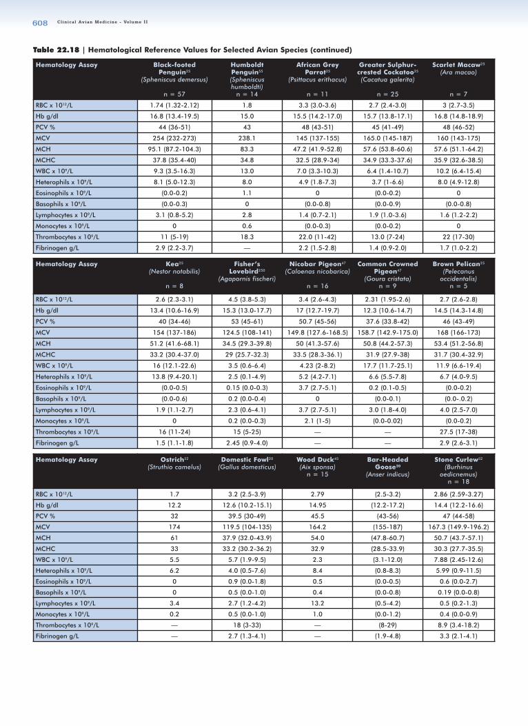
Table 22.18 | Hematological Reference Values for Selected Avian Species
Hematology Assay Egyptian Vulture25
(Neophron perc-nopterus)
n = 4
Common Buzzard25
(Buteo buteo)
n = 6
Golden Eagle25
(Aquila chrysaetos)
n = 4
Saker Falcon50
(Falco cherrug)
n = 25
Barn Owl25
(Tyto alba)
n = 10
RBC x 1012/L 2.3 (1.9-2.6) 2.4 (2.2-2.7) 2.4 (1.9-2.7) 2.65 (2.0-3.9) 2.7 (2.2-3.0)
Hb g/dl 14.8 (13.3-16.5) 12.9 (11.6-14.6) 13.8 (12.1-15.2) 15.3 (13.3-21.2) 14.2 (12.7-16.4)
PCV % 43 (37-46) 38 (34-42) 41 (35-47) 47 (42-53) 46 (42-51)
MCV 190 (183-206) 159 (151-171) 174 (160-184) 183.1 (135.8-219.5) 176 (145-216)
MCH 67.7 (65.2-72.9) 53.8 (48.8-57.5) 58.9 (56.3-62.7) 60.7 (50.6-78.9) 51.1 (44.9-60.7)
MCHC 35.2 (35.0-35.5) 33.9 (31.4-36.0) 34.0 (32.3-35.9) 60.7 (50.6-78.9) 31.8 (28.9-34.9)
WBC x 109/L 7.6 (4.7-10.6) 9.1 (4.6-13.9) 13.1 (11.7-14.7) 33.2 (28.3-40) 16.6 (11.5-22.3)
Heterophils x 109/L 4.0 (1.2-5.5) 5.5 (2.3-8.8) 10.4 (9.5-12.7) 4.1 (2.1-5.9) 8.9 (5.2-12.5)
Eosinophils x 109/L 0.3-1.4 0.1-3.1 0.2-0.6 0 0
Basophils x 109/L 0 0.0-0.6 0.0-0.2 0 0
Lymphocytes x 109/L 2.5 (1.5-3.4) 1.7 (1.1-2.4) 2.2 (1.6-3.2) 1.3 (0.5-2.2) 5.0 (2.5-7.5)
Monocytes x 109/L 0.0-0.4 0 0 0.2 (0-0.6) 0
Thrombocytes x 109/L 13 (6-15) 27 (18-36) 14 (4-21) 0.41 (0.17-0.76) 33 (14-58)
Fibrinogen g/L 1.6 (1.0-1.9) 2.3 (1.3-3.3) 2.9 (2.0-4.1) 2.8 (1.7-4.7) 2.7 (1.9-3.3)
Hematology Assay Crowned Crane25
(Balearica regulorum)
n = 33
Greater Flamingo25
(Phoenicopterusruber)n = 9
Rosy Flamingo43
(Phoenicopterus ruber ruber)
n = 25
White Stork25
(Ciconia ciconia)
n = 16
Kori Bustard31
(Ardeotis kori)
n = 28
RBC x 1012/L 2.8 (2.4-3.1) 2.6 (2.3-2.8) 1.4 (1.1-1.8) 2.4 (2.1-2.7) 2.3 (1.74-2.95)
Hb g/dl 15.6 (11.9-18.8) 17.3 (15.9-19.6) 13.4 (9.2-17.6) 15.8 (14.4-17.7) 14.1 (11.9-15.9)
PCV % 47 (44-52) 50 (47-57) 47.8 (37.9-57.8) 45 (41-48) 47 (39.5-52.5)
MCV 171 (156-182) 193 (170-207) 326.6 (234.3-419.0) 189 (172-195) 208.5 (161.9-275.4)
MCH 64.3 (59.8-70.2) 66.2 (57.6-70.0) 91.5 (57.8-125.3) 67.2 (60.2-69.9) 62.4 (48-84.6)
MCHC 36.2 (34.5-39.2) 34.4 (33.5-35.2) 28.1 (20.4-35.8) 35.3 (31-36.9) 30.0 (29.7-34.9)
WBC x 109/L 11.1 (6.3-15.6) 2.4 (0.9-3.4) 8.7 (1.5-15.8) 10.8 (7-14.3) 7.3 (3.0-12.8)
Heterophils x 109/L 8.2 (4.1-13.3) 1.2 (0.2-3.0) 3.9 (1.0-11.4) 9.2 (5.1-14.9) 3.9 (0.9-9.25)
Eosinophils x 109/L (0.0-1.3) (0.0-0.4) (0.0-0.3) (0.0-0.7) 0.3 (0.0-1.1)
Basophils x 109/L (0.1-0.8) (0.0-0.4) (0.0-0.8) (0.0-0.5) 0.2 (0.0-0.8)
Lymphocytes x 109/L 1.6 (0.6-2.7) 0.9 (0.4-1.6) 5.2 (0.8-9.6) 0.8 (0.2-1.6) 2.2 (0.41-5.4)
Monocytes x 109/L (0.0-0.3) (0.0-0.2) 0.5 (0-1.8) (0.0-0.3) 0.6 (0.0-1.5)
Thrombocytes x 109/L 3.6 (5-18) 4 (2-7) — 19 (8-32) 5.5 (1.49-18.0)
Fibrinogen g/L — — — 2.3 (1.7-3.2) 2.42 (1.42-4.5)
Table 22.17 | Evaluating Leukopenia
Hematologic Findings Possible Causes
Toxic heterophils, immature cells,anemia, monocytosis, reactivelymphocytes
Final stage of immune response, severe bone marrow dam-age, gram-negative septicemia, infected wounds, circovirusin psittacines, marked loss of skin, eg, burns, marked tissuenecrosis, smoke intoxication. Grave prognosis.
Normal morphology,relative heterophilia, absolutelymphopenia
Initial stage of stress
Reactive lymphocytes, relativelymphocytosis, progressive orintermittent leukopenia
Acute viral infection (toxic heterophils in pox and her-pesvirus infections)
22_Hematology.qxd 8/23/2005 1:11 PM Page 607
Cl inica l Avian Medic ine - Volume I I608
Hematology Assay Ostrich52
(Struthio camelus)Domestic Fowl20
(Gallus domesticus)Wood Duck45
(Aix sponsa)n = 15
Bar-HeadedGoose20
(Anser indicus)
Stone Curlew52
(Burhinus oedicnemus)
n = 18
RBC x 1012/L 1.7 3.2 (2.5-3.9) 2.79 (2.5-3.2) 2.86 (2.59-3.27)
Hb g/dl 12.2 12.6 (10.2-15.1) 14.95 (12.2-17.2) 14.4 (12.2-16.6)
PCV % 32 39.5 (30-49) 45.5 (43-56) 47 (44-58)
MCV 174 119.5 (104-135) 164.2 (155-187) 167.3 (149.9-196.2)
MCH 61 37.9 (32.0-43.9) 54.0 (47.8-60.7) 50.7 (43.7-57.1)
MCHC 33 33.2 (30.2-36.2) 32.9 (28.5-33.9) 30.3 (27.7-35.5)
WBC x 109/L 5.5 5.7 (1.9-9.5) 2.3 (3.1-12.0) 7.88 (2.45-12.6)
Heterophils x 109/L 6.2 4.0 (0.5-7.6) 8.4 (0.8-8.3) 5.99 (0.9-11.5)
Eosinophils x 109/L 0 0.9 (0.0-1.8) 0.5 (0.0-0.5) 0.6 (0.0-2.7)
Basophils x 109/L 0 0.5 (0.0-1.0) 0.4 (0.0-0.8) 0.19 (0.0-0.8)
Lymphocytes x 109/L 3.4 2.7 (1.2-4.2) 13.2 (0.5-4.2) 0.5 (0.2-1.3)
Monocytes x 109/L 0.2 0.5 (0.0-1.0) 1.0 (0.0-1.2) 0.4 (0.0-0.9)
Thrombocytes x 109/L — 18 (3-33) — (8-29) 8.9 (3.4-18.2)
Fibrinogen g/L — 2.7 (1.3-4.1) — (1.9-4.8) 3.3 (2.1-4.1)
Hematology Assay Kea25
(Nestor notabilis)
n = 8
Fisher’sLovebird250
(Agapornis fischeri)
Nicobar Pigeon47
(Caloenas nicobarica)
n = 16
Common CrownedPigeon47
(Goura cristata)n = 9
Brown Pelican25
(Pelecanusoccidentalis)
n = 5
RBC x 1012/L 2.6 (2.3-3.1) 4.5 (3.8-5.3) 3.4 (2.6-4.3) 2.31 (1.95-2.6) 2.7 (2.6-2.8)
Hb g/dl 13.4 (10.6-16.9) 15.3 (13.0-17.7) 17 (12.7-19.7) 12.3 (10.6-14.7) 14.5 (14.3-14.8)
PCV % 40 (34-46) 53 (45-61) 50.7 (45-56) 37.6 (33.8-42) 46 (43-49)
MCV 154 (137-186) 124.5 (108-141) 149.8 (127.6-168.5) 158.7 (142.9-175.0) 168 (166-173)
MCH 51.2 (41.6-68.1) 34.5 (29.3-39.8) 50 (41.3-57.6) 50.8 (44.2-57.3) 53.4 (51.2-56.8)
MCHC 33.2 (30.4-37.0) 29 (25.7-32.3) 33.5 (28.3-36.1) 31.9 (27.9-38) 31.7 (30.4-32.9)
WBC x 109/L 16 (12.1-22.6) 3.5 (0.6-6.4) 4.23 (2-8.2) 17.7 (11.7-25.1) 11.9 (6.6-19.4)
Heterophils x 109/L 13.8 (9.4-20.1) 2.5 (0.1-4.9) 5.2 (4.2-7.1) 6.6 (5.5-7.8) 6.7 (4.0-9.5)
Eosinophils x 109/L (0.0-0.5) 0.15 (0.0-0.3) 3.7 (2.7-5.1) 0.2 (0.1-0.5) (0.0-0.2)
Basophils x 109/L (0.0-0.6) 0.2 (0.0-0.4) 0 (0.0-0.1) (0.0-.0.2)
Lymphocytes x 109/L 1.9 (1.1-2.7) 2.3 (0.6-4.1) 3.7 (2.7-5.1) 3.0 (1.8-4.0) 4.0 (2.5-7.0)
Monocytes x 109/L 0 0.2 (0.0-0.3) 2.1 (1-5) (0.0-0.02) (0.0-0.2)
Thrombocytes x 109/L 16 (11-24) 15 (5-25) — — 27.5 (17-38)
Fibrinogen g/L 1.5 (1.1-1.8) 2.45 (0.9-4.0) — — 2.9 (2.6-3.1)
Table 22.18 | Hematological Reference Values for Selected Avian Species (continued)
Hematology Assay Black-footedPenguin25
(Spheniscus demersus)
n = 57
HumboldtPenguin55
(Spheniscus humboldti)
n = 14
African GreyParrot25
(Psittacus erithacus)
n = 11
Greater Sulphur-crested Cockatoo25
(Cacatua galerita)
n = 25
Scarlet Macaw25
(Ara macao)
n = 7
RBC x 1012/L 1.74 (1.32-2.12) 1.8 3.3 (3.0-3.6) 2.7 (2.4-3.0) 3 (2.7-3.5)
Hb g/dl 16.8 (13.4-19.5) 15.0 15.5 (14.2-17.0) 15.7 (13.8-17.1) 16.8 (14.8-18.9)
PCV % 44 (36-51) 43 48 (43-51) 45 (41-49) 48 (46-52)
MCV 254 (232-273) 238.1 145 (137-155) 165.0 (145-187) 160 (143-175)
MCH 95.1 (87.2-104.3) 83.3 47.2 (41.9-52.8) 57.6 (53.8-60.6) 57.6 (51.1-64.2)
MCHC 37.8 (35.4-40) 34.8 32.5 (28.9-34) 34.9 (33.3-37.6) 35.9 (32.6-38.5)
WBC x 109/L 9.3 (3.5-16.3) 13.0 7.0 (3.3-10.3) 6.4 (1.4-10.7) 10.2 (6.4-15.4)
Heterophils x 109/L 8.1 (5.0-12.3) 8.0 4.9 (1.8-7.3) 3.7 (1-6.6) 8.0 (4.9-12.8)
Eosinophils x 109/L (0.0-0.2) 1.1 0 (0.0-0.2) 0
Basophils x 109/L (0.0-0.3) 0 (0.0-0.8) (0.0-0.9) (0.0-0.8)
Lymphocytes x 109/L 3.1 (0.8-5.2) 2.8 1.4 (0.7-2.1) 1.9 (1.0-3.6) 1.6 (1.2-2.2)
Monocytes x 109/L 0 0.6 (0.0-0.3) (0.0-0.2) 0
Thrombocytes x 109/L 11 (5-19) 18.3 22.0 (11-42) 13.0 (7-24) 22 (17-30)
Fibrinogen g/L 2.9 (2.2-3.7) — 2.2 (1.5-2.8) 1.4 (0.9-2.0) 1.7 (1.0-2.2)
22_Hematology.qxd 8/23/2005 1:11 PM Page 608
Chapter 22 | D I A G N O S T I C V A L U E O F H E M A T O L O G Y 609
References andSuggested Reading
1. Alonso JA, et al: Hematology and blood chemistry of free-livingyoung great bustards (Otistarda). Comp Biochem Physiol97A:611-614, 1990.
2. Averbeck C: Hematology and blood chemistry of healthy andclinically abnormal great black-backed gulls (Larus marinus)and herring gulls (Larus argenta-tus). Avian Pathol 21:215-223,1992.
3. Campbell TW: Hematology. InRitchie BW, Harrison GJ, HarrisonLR (eds): Avian Medicine: Princi-ples and Application. Brentwood,TN, HBD Int’l, 1994, pp 176-199.
4. Campbell TW: Avian Hematology and Cytology. Ames, Iowa StateUniversity Press, 1995, pp 3-19.
5. Clubb SL, et al: Hematologic andserum biochemical referenceintervals in juvenile eclectus par-rots. J Assoc Avian Vet 4:218-225,1990.
6. Clubb SL, et al: Hematologic andserum biochemical referenceintervals in juvenile cockatoos. JAssoc Avian Vet 5:16-21, 1991.
7. Clubb SL, et al: Hematologic andserum biochemical referenceintervals in juvenile macaws (Arasp). J Assoc Avian Vet 5:154-162,1991.
8. Dacie JV, Lewis SM: PracticalHematology 8th ed. Edinburgh,Churchill Livingstone, 1995.
9. D’Aloia M-A, et al: Hemopatho-logical responses to chronicinflammation in the houbara bus-tard (Chlamydotis undulatamacqueenii). Com Haem Int4:203-206, 1994.
10. D’Aloia M-A, et al: Normal hema-tology and age-related findings inrufous-crested bustards (Eupodotis ruficrista). Com Haem Int 5:10-12,1995.
11. D’Aloia M-A, et al: Normal hema-tology of the white bellied(Eupodotis senegalensis), littleblack (Eupodotis afra) andHeuglin’s (Neotis heuglinii) bus-tards. Com Haem Int 6:46-49,1996.
12. Dein FJ: Hematology. In Harrison GJ, Harrison LR (eds): ClinicalAvian Medicine and Surgery.Philadelphia, WB Saunders Co,1986, pp 174-191.
13. Dorrestein GM: Cytology and haemocytology. In Beynon PH,Forbes NA, Lawton MPC (eds):Manual of Psittacine Birds.Cheltenham, Glos, Br Small Anim
Vet Assoc, 1996, pp 38-48. 14. Fudge AM: Clinical application of
laser flow cytometry to avianhematology analysis, Proc AssocAvian Vet, 1995, pp 17-18.
15. Fudge AM: Clinical hematology and chemistry of ratites. In TullyTN, Shane SM (eds): Ratites:Management, Medicine, andSurgery. Malabar, FL, KriegerPublishing, 1996, pp 105-114.
16. Fudge AM: Avian clinical patholo-gy: Hematology and chemistry. InAltman RB, et al (eds): AvianMedicine and Surgery.Philadelphia, WB Saunders Co,1997, pp 142-157.
17. Fudge AM: Problem-orientedapproach to blood panel inter-pretation. Proc Assoc Avian Vet,1998, pp 285-299.
18. Fudge AM: Avian cytology andhematology. Proc Assoc Avian Vet,1998, pp 357-369.
19. Fudge AM (ed): LaboratoryMedicine: Avian and Exotic Pets.Philadelphia, WB Saunders Co,2000, pp 1-8.
20. Gulland FMD, Hawkey CM: Avianhematology. Vet Annual 30:126-136, 1990.
21. Harris DJ: Clinical tests. In TullyTN, Lawton MPC, Dorrestein GM(eds): Avian Medicine. Oxford,Butterworth Heinemann, 2000,pp 43-51.
22. Hauska H, Gerlach H: The devel-opment of the red blood cell pat-tern of growing parrot nestlings.Proc Assoc Avian Vet, 1995, pp178-182.
23. Hauska H, Gerlach H: The devel-opment of the white blood cellpattern of growing parrotnestlings. Proc Assoc Avian Vet,1995, pp 183-186.
24. Hauska H, Redig PT: Morphologi-cal changes in the white hemo-gram of raptors. Proc Assoc AvianVet, 1997, pp 205-208.
25. Hawkey CM, Samour JH: Thevalue of clinical hematology inexotic birds. In Jacobson ER,Kollias GV Jr (eds): ContemporaryIssues in Small Animal Practice.London, Churchill Livingstone,1988, pp 109-142.
26. Hawkey C, et al: Normal and clin-ical hematology of captive cranes(Gruiformes). Avian Pathol 12:73-84, 1983.
27. Hawkey C, et al: Haematologicalchanges in domestic fowl (Gallusgallus) and cranes (Gruiformes)with Mycobacterium avium infec-tion. Avian Pathol 19:223-234,1990.
28. Hernandez M: Raptor clinical
hematology. Proc Assoc Avian Vet,1991, pp 420-433.
29. Hernandez M, Martin S, Fores P:Clinical hematology and bloodchemistry values for the commonbuzzard (Buteo buteo). J Rapt Res24(4):113-119, 1990.
30. Howlett JC: Clinical and diagnos-tic procedures. In Samour JH(ed): Avian Medicine. London,Harcourt Publishers Ltd, 2000, pp28-50.
31. Howlett JC, et al: Normal hema-tology of captive adult kori bus-tards (Ardeotis kori). Com HaemInt 5:102-105, 1995.
32. Howlett JC, et al: Haemoproteusin the houbara bustard(Chlamydotis undulata mac-queenii) and the rufous-crestedbustard (Eupodotis ruficrista) inthe United Arab Emirates. AvianPathol 25:4-55, 1996.
33. Howlett JC, et al: Age-relatedhematology changes in captive-reared kori bustards (Ardeotiskori). Com Haem Int 8:26-30,1998.
34. International Council forStandardization in Hematology:Guidelines for the evaluation ofblood cell analyzers includingthose used for differential leuco-cyte and reticulocyte countingand cell marker applications. Clinand Lab Haem 16:157, 1994.
35. Jain CJ: Essentials of VeterinaryHematology. Philadelphia, Leaand Febiger, 1993, pp 19-53.
36. Jennings IB: Hematology. InBeynon PH, Forbes NA, Harcourt-Brown NH (eds): Manual ofRaptors, Pigeons and Waterfowl.Cheltenham, Glos, Brit SmallAnim Vet Assoc, 1996, pp 68-78.
37. Jimenez A, et al: Clinical hematol-ogy of the great bustard (Otistarda). Avian Pathol 20:675-680,1991.
38. Lane RA: Avian hematology. InRosskopf W, Woerpel R (eds):Diseases of Cage and Aviary Birds3rd ed. Baltimore, Lea and Febiger,1996, pp 739-772.
39. Lind PJ, et al: Morphology of theavian eosinophil in raptors. JAssoc Avian Vet 4:33-38, 1990.
40. Maxwell MH: Avian blood leuco-cyte responses to stress. WorldPoult Sci J 49:34-43, 1993.
41. Maxwell MH, Robertson GW: Theavian basophilic leukocyte: Areview. World Poult Sci J 51:307-325, 1995.
42. Maxwell MH, Hocking PM,Robertson GW: Differential leuco-cyte response to various degreesof food restriction in broilers,
turkeys and ducks. Br Poult Sci33:177-187, 1992.
43. Merritt EL, Fritz CL, Ramsay EC:Hematologic and serum biochem-ical values in captive Americanflamingos (Phoenicopterus ruberruber). J Avian Med Surg10(3):163-167, 1996.
44. Mikaelian I: Variations circan-nuales des parametres hema-tologiques de l’outarde houbara(Chlamydotis undulata).Professional thesis (in French).Ecole Nationale Veterinaire deLyon, Université Claude Bernard,Lyon France, 1993.
45. Mulley RC: Hematology of thewood duck (Chenonetta jubata).J Wildl Dis 16(2):271-273, 1980.
46. Palomeque J, Pinto D, Viscor G:Hematologic and blood chemistryvalues of the Masai ostrich(Struthio camelus). J Wildl Dis27(1):34-40, 1991.
47. Peinado VI, et al: Hematology andplasma chemistry in endangeredpigeons. J Zoo Wildl Med23(1):65-71, 1992.
48. Pendl H: Avian hematology forpractitioners. Proc Assoc AvianVet, 2001, pp 387-400.
49. Samour JH, Peirce M: Babesiashortti infection in a saker falcon(Falco cherrug altaicus). Vet Rec139:167-168, 1996.
50. Samour JH, D’Aloia M-A, HowlettJC: Normal hematology of thesaker falcon (Falco cherrug).Com Haem Int 6:50-52, 1996.
51. Samour JH, et al: Normal hema-tology of the houbara bustard(Chlamydotis undulata mac-queenii). Com Haem Int 4:198-202, 1994.
52. Samour JH, et al: Normal hema-tology and blood chemistry ofcaptive adult stone curlews(Burhinus oedicnemus). ComHaem Int 8:219-224, 1998.
53. Stewart JS: Husbandry, medicaland surgical management ofratites. Proc Assoc Avian Vet,1989, pp 119-122.
54. Sturkie PD: Avian Physiology 2nded. London, Bailliére, Tindall andCassell, 1965.
55. VanderHeyden N: Evaluation andinterpretation of the avianhemogram. Sem Avian Exotic PetMed 3(1):5-13, 1994.
56. Villouta G, Hargreaves R, RiverosV: Haematological and clinicalbiochemistry findings in captiveHumboldt penguins (Spheniscushumboldti). Avian Pathol 26:851-858, 1997.
Products Mentioned in the Texta. Coulter Counter ZF, Beckman Coulter Inc, Fullerton,
CA, USA www.beckmancoulter.comb. Cell Dyn 3500, Abbott Laboratories, Abbott Park, IL,
USA www.abbottdiagnostics.comc. BD Unopette 365851 red blood count manual hema-
tology test, Becton Dickinson Co, Franklin Lakes, NJ, USA www.bd.comd. BD Unopette 365877 eosinophil count manual hema-
tology test, Becton Dickinson Co, Franklin Lakes, NJ, USA
Dedication
This chapter is dedicated to Dr. Christine M. Hawkey.
Acknowledgments
The author thanks HRH Prince Fahad bin Sultan binAbdulaziz Al Saud for his support to the advancement offalcon medicine, and to Mr. Basil Al Abbasi, DirectorGeneral, for his continuing interest in the clinical andresearch program of the Falcon Specialist Hospital andResearch Institute; to Shinto K. John; GenerosoQuiambao; Dr. Jesus Naldo.
22_Hematology.qxd 8/23/2005 1:11 PM Page 609

Cl inica l Avian Medic ine - Volume I I610
22_Hematology.qxd 8/23/2005 1:11 PM Page 610