Embed Size (px)
Citation preview
Diagnostic Tests in Renal Disease
Susan Hou M.D.
Loyola University Medical Center
Case One
• A 27 year old woman joins a group of friends to climb Mt. Washington 3 days after finishing her internship. On reaching the top she notes severe muscle pain and dark brown urine.
• The muscle pain grows worse and the brown urine continues. Three days after the climb, she goes to the emergency room. Urinalysis is dipstick positive for blood. There are no red cells on microscopic exam.
• What’s going on here?
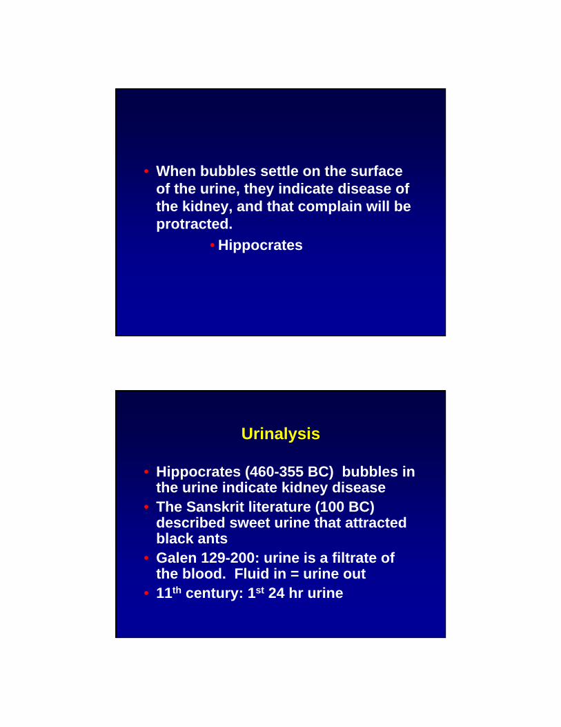
• When bubbles settle on the surface of the urine, they indicate disease of the kidney, and that complain will be protracted.
• Hippocrates
Urinalysis
• Hippocrates (460-355 BC) bubbles in the urine indicate kidney disease
• The Sanskrit literature (100 BC) described sweet urine that attracted black ants
• Galen 129-200: urine is a filtrate of the blood. Fluid in = urine out
• 11th century: 1st 24 hr urine
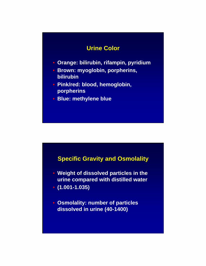
Urine Color
• Orange: bilirubin, rifampin, pyridium• Brown: myoglobin, porpherins,
bilirubin• Pink/red: blood, hemoglobin,
porpherins• Blue: methylene blue
Specific Gravity and Osmolality
• Weight of dissolved particles in the urine compared with distilled water
• (1.001-1.035)
• Osmolality: number of particles dissolved in urine (40-1400)
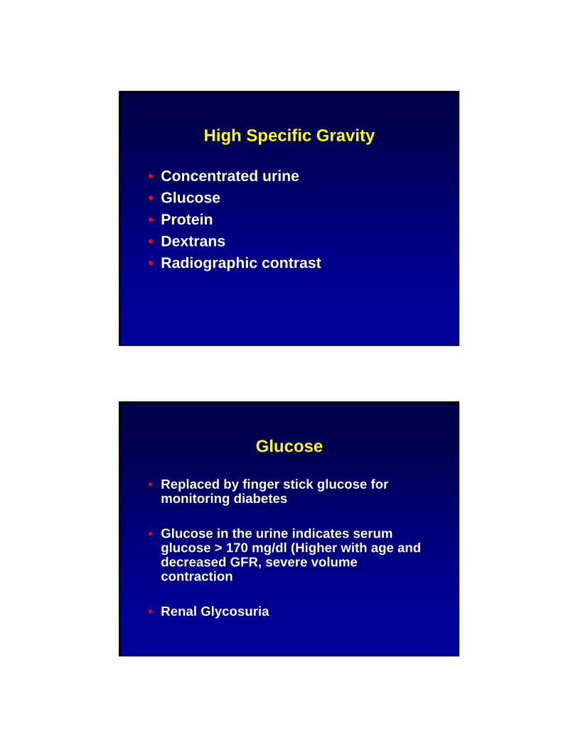
High Specific Gravity
• Concentrated urine• Glucose• Protein• Dextrans• Radiographic contrast
Glucose
• Replaced by finger stick glucose for monitoring diabetes
• Glucose in the urine indicates serum glucose > 170 mg/dl (Higher with age and decreased GFR, severe volume contraction
• Renal Glycosuria
Ketones
• Fasting (starvation ketosis)
• Ketoacidosis
Occult Blood
• Red cells
• If dipstick is positive for blood and microscopic shows no RBCs• Hemoglobin• Myoglobin• Very dilute urine
Case 1
• The woman knew her diagnosis when she first saw brown urine.
• Brown urine after extreme exercise particularly in some one who is deconditioned is classic presentation of rhabdomyolysis.
• ER visit was delayed because doctors make bad decisions about their own care.
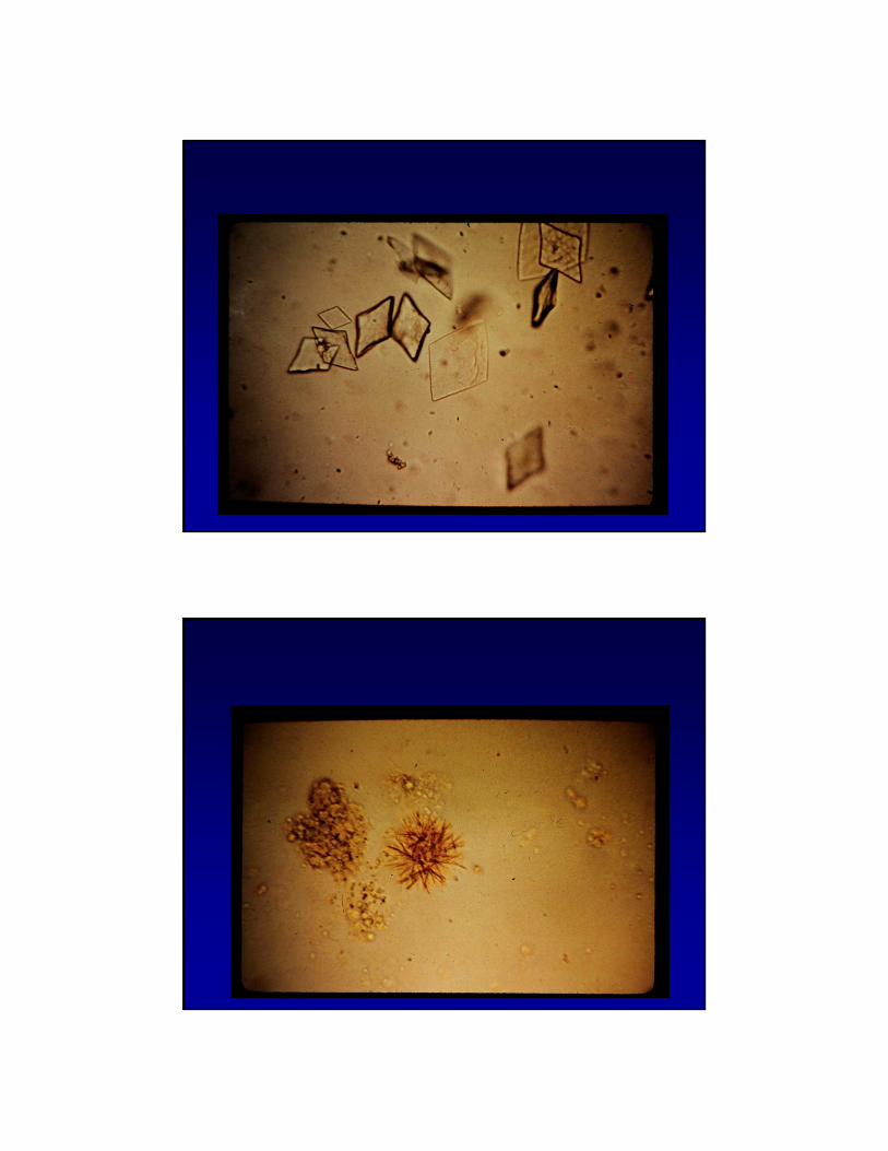
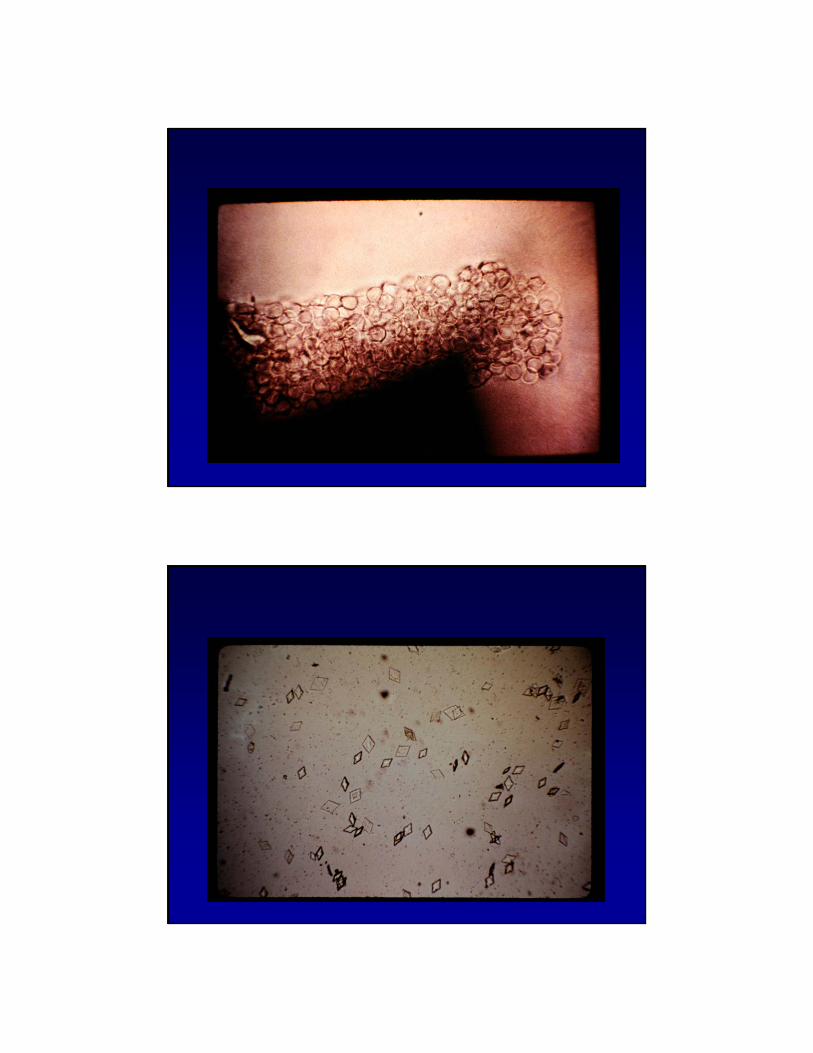
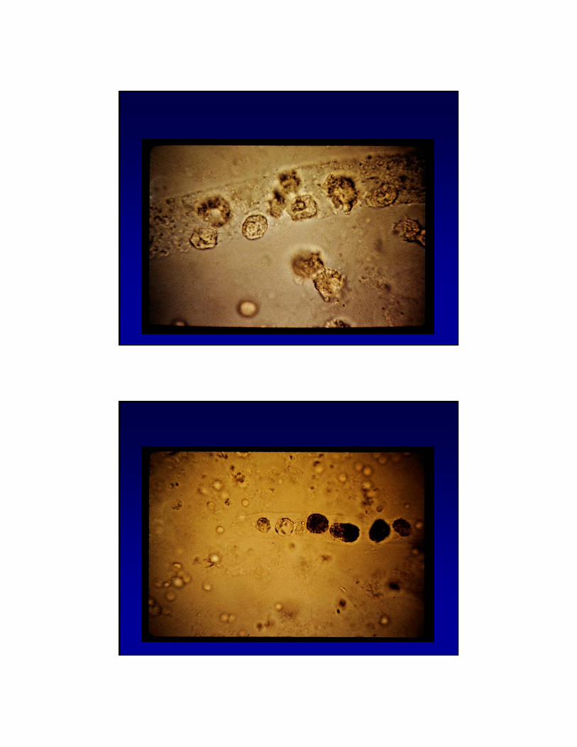
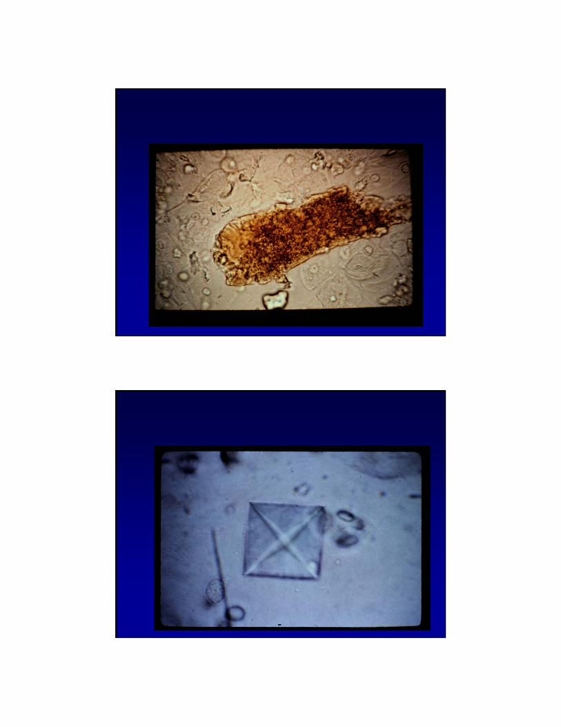
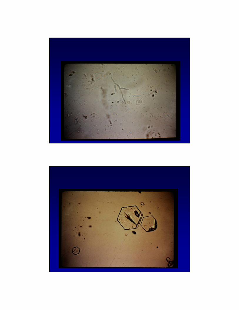
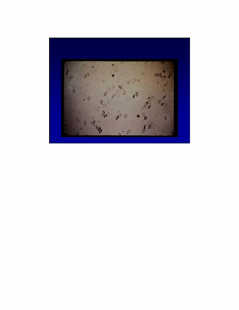
Microscopic Elements
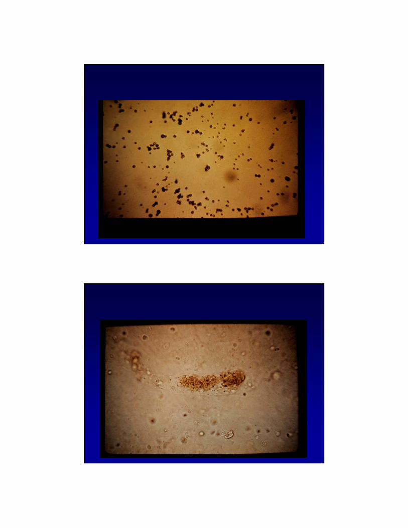
• Cells• Casts• Crystals• Bacteria• Yeast
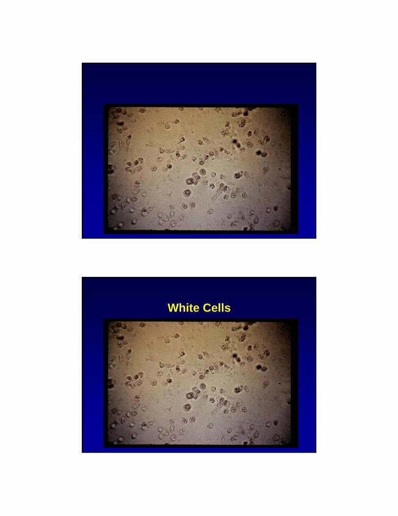
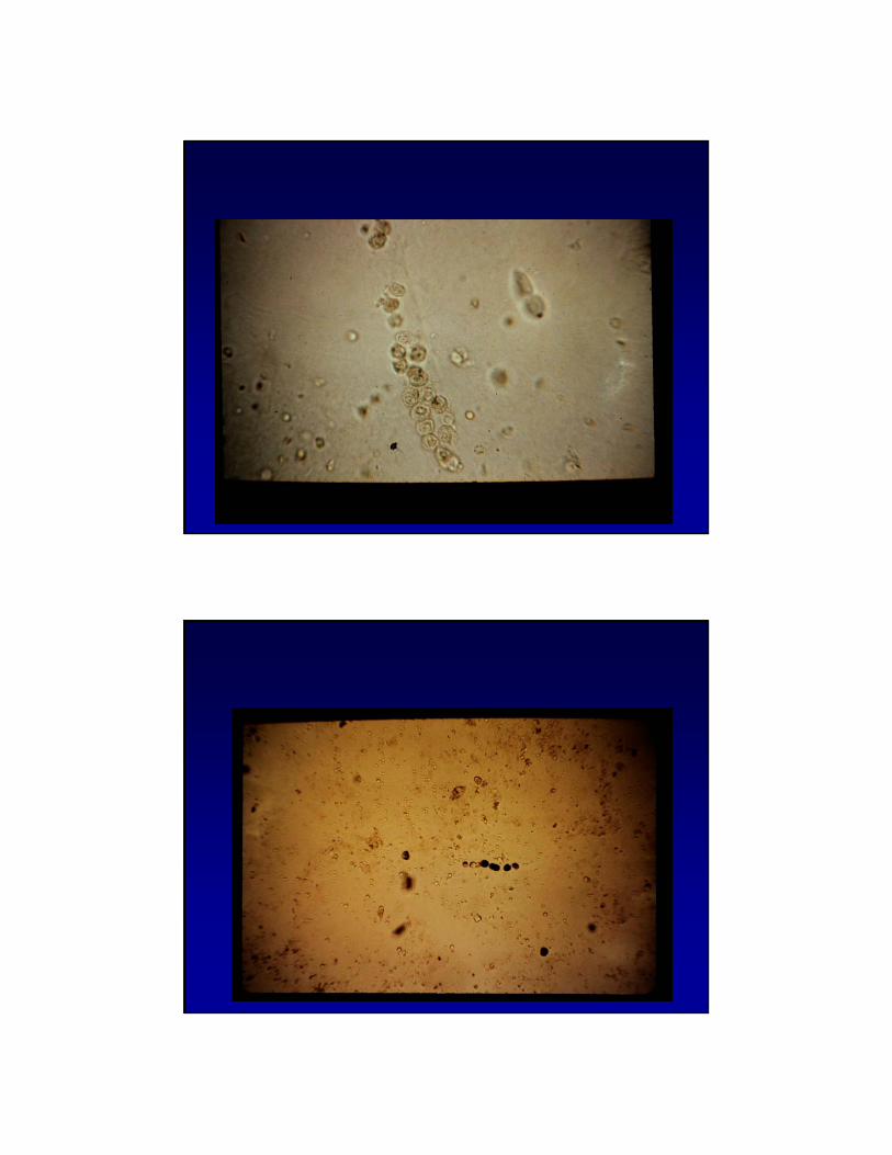
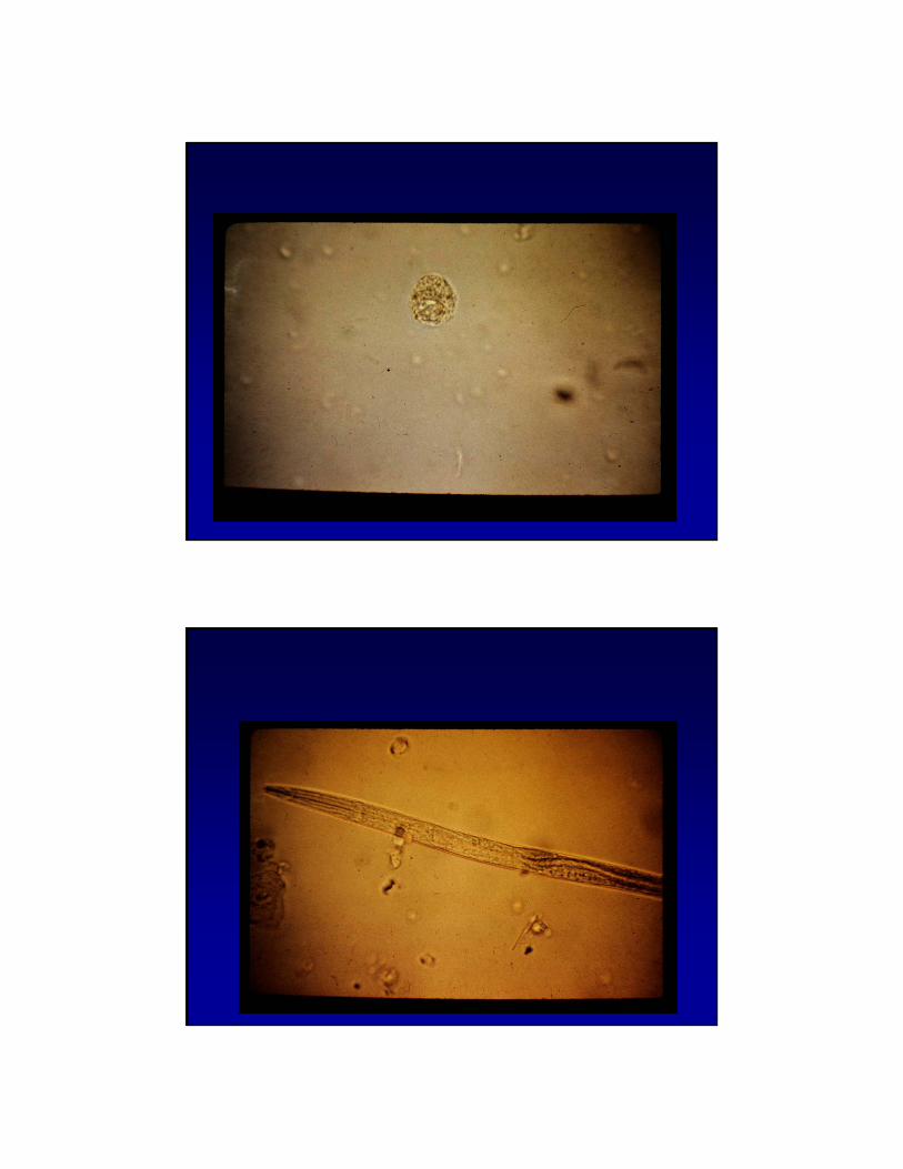
White Cells
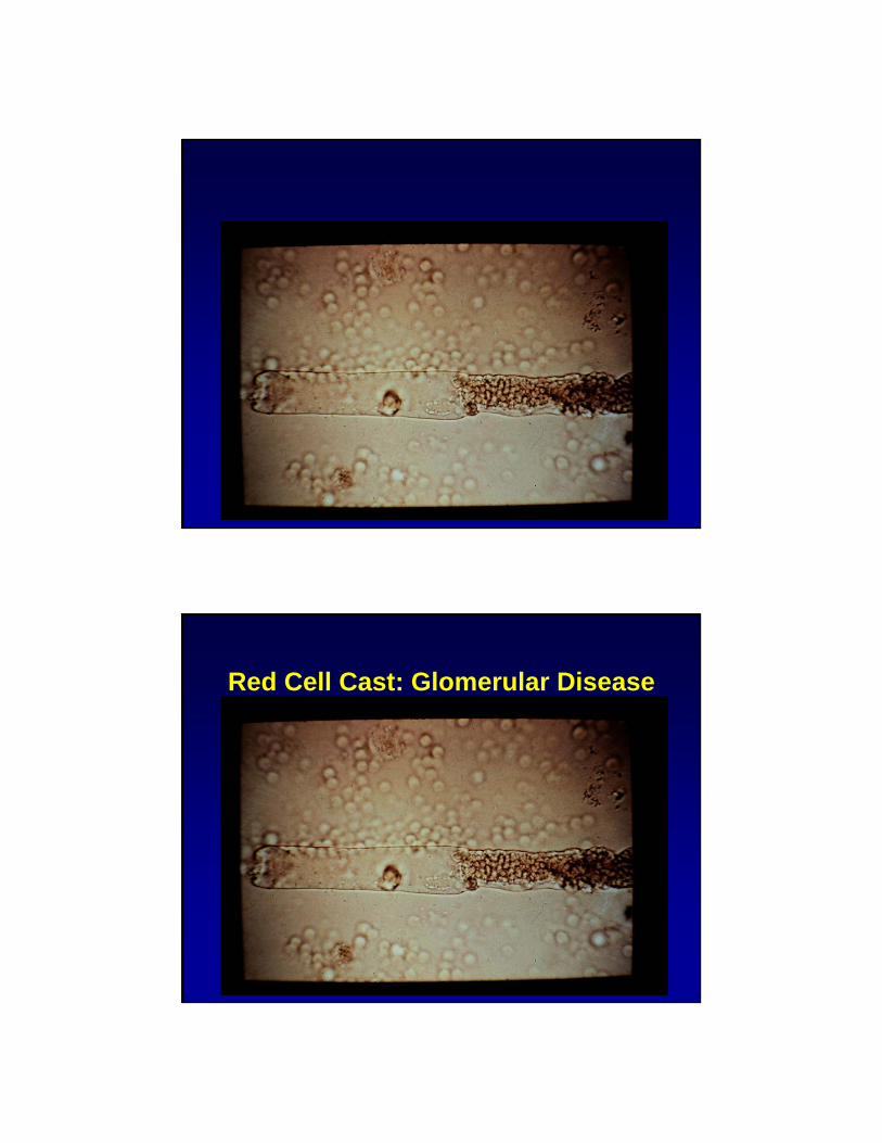
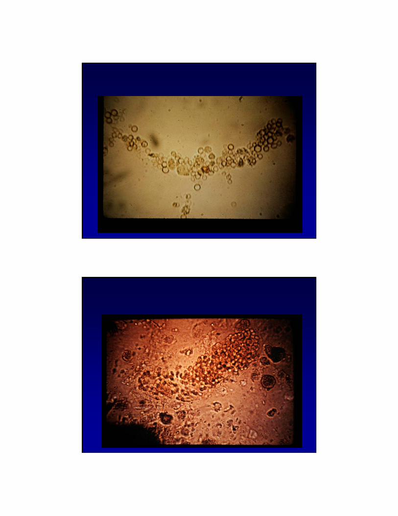
Red Cell Cast: Glomerular Disease
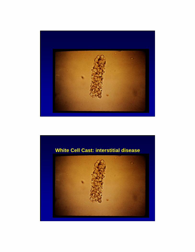
White Cell Cast: interstitial disease
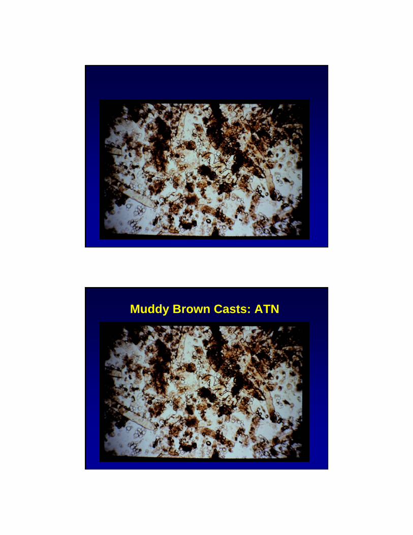
Muddy Brown Casts: ATN
Urine Protein:Categories of persistent proteinuria
• Overflow: Capacity to reabsorb normally filtered protein in proximal tubules over whelmed due to overproduction:e.g.lightchains,hemoglobinuria and myoglobinuria
• Tubular proteinuria: Decreased reabsorptionof filtered proteins by tubules due to tubulointerstitial damage ; usually <2 gm
• Glomerular proteinuria: Microalbuminuria to overt proteinuria usually>3.5 gm
Proteinuria
• >150-300 mg/24 hrs indicates renal disease. Increase to 300 mg/dl in pregnancy
• > 3.5 grams: glomerular disease• Tubular proteinuria• Microalbuminuria: >30 mg of albumin
in 24 hrs. Screening for diabetic nephropathy
Dipstick for Protein
• Proteinuria without renal disease: exercise, fever, high venous pressure
• Depends on Urine concentration• Does not detect Bence-Jones
proteins
Protein: Creatinine Ratio
• Protein : Creatinine ratio estimates 24 hr urine protein
• Useful for children• Hard to collect 24 h urine even in adults• Protein: creatinine ratio should be <0.15• Microalbumin in mg : creatinine in grams
ratio should be less than 30
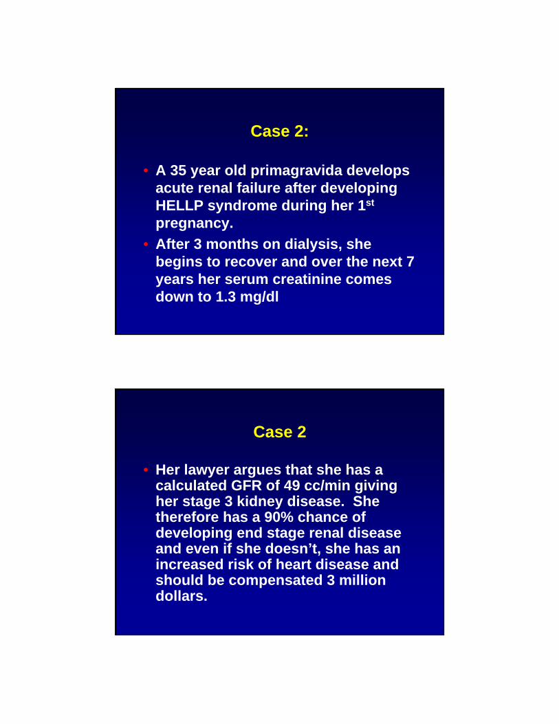
Case 2:
• A 35 year old primagravida develops acute renal failure after developing HELLP syndrome during her 1st
pregnancy.• After 3 months on dialysis, she
begins to recover and over the next 7 years her serum creatinine comes down to 1.3 mg/dl
Case 2
• Her lawyer argues that she has a calculated GFR of 49 cc/min giving her stage 3 kidney disease. She therefore has a 90% chance of developing end stage renal disease and even if she doesn’t, she has an increased risk of heart disease and should be compensated 3 million dollars.
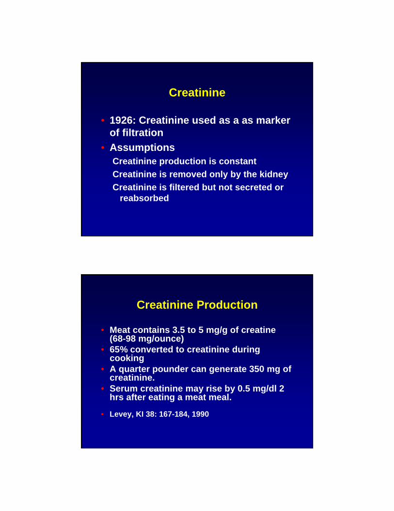
Creatinine
• 1926: Creatinine used as a as marker of filtration
• AssumptionsCreatinine production is constantCreatinine is removed only by the kidneyCreatinine is filtered but not secreted or
reabsorbed
Creatinine Production
• Meat contains 3.5 to 5 mg/g of creatine(68-98 mg/ounce)
• 65% converted to creatinine during cooking
• A quarter pounder can generate 350 mg of creatinine.
• Serum creatinine may rise by 0.5 mg/dl 2 hrs after eating a meat meal.
• Levey, KI 38: 167-184, 1990
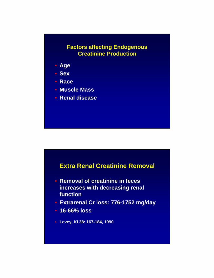
Factors affecting Endogenous Creatinine Production
• Age• Sex• Race• Muscle Mass• Renal disease
Extra Renal Creatinine Removal
• Removal of creatinine in feces increases with decreasing renal function
• Extrarenal Cr loss: 776-1752 mg/day• 16-66% loss
• Levey, KI 38: 167-184, 1990
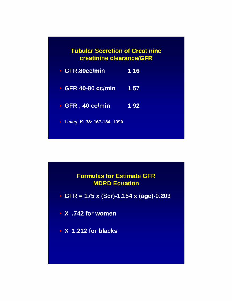
Tubular Secretion of Creatininecreatinine clearance/GFR
• GFR.80cc/min 1.16
• GFR 40-80 cc/min 1.57
• GFR , 40 cc/min 1.92
• Levey, KI 38: 167-184, 1990
Formulas for Estimate GFRMDRD Equation
• GFR = 175 x (Scr)-1.154 x (age)-0.203
• X .742 for women
• X 1.212 for blacks
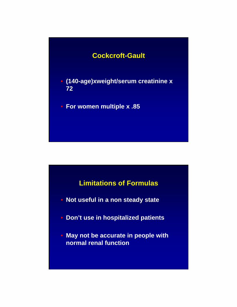
Cockcroft-Gault
• (140-age)xweight/serum creatinine x 72
• For women multiple x .85
Limitations of Formulas
• Not useful in a non steady state
• Don’t use in hospitalized patients
• May not be accurate in people with normal renal function
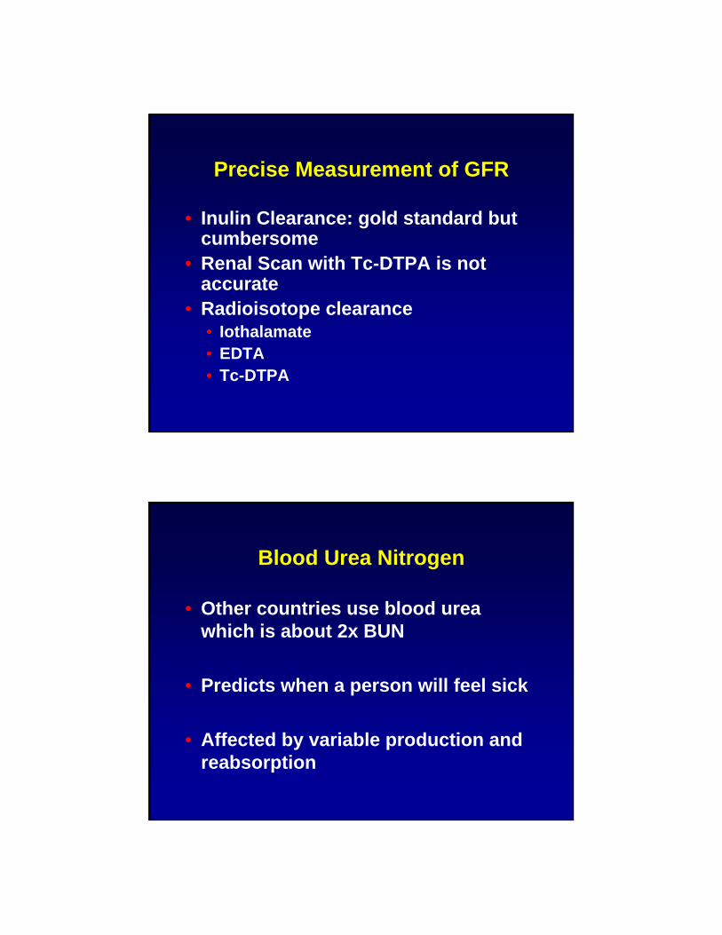
Precise Measurement of GFR
• Inulin Clearance: gold standard but cumbersome
• Renal Scan with Tc-DTPA is not accurate
• Radioisotope clearance• Iothalamate• EDTA• Tc-DTPA
Blood Urea Nitrogen
• Other countries use blood urea which is about 2x BUN
• Predicts when a person will feel sick
• Affected by variable production and reabsorption
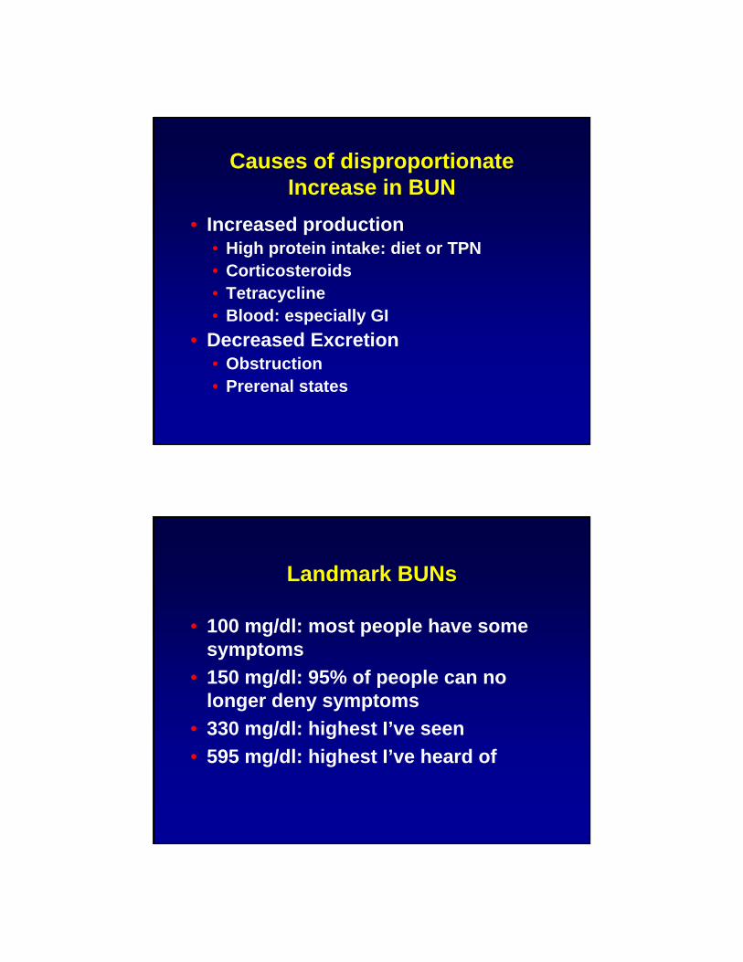
Causes of disproportionate Increase in BUN
• Increased production• High protein intake: diet or TPN• Corticosteroids• Tetracycline• Blood: especially GI
• Decreased Excretion• Obstruction• Prerenal states
Landmark BUNs
• 100 mg/dl: most people have some symptoms
• 150 mg/dl: 95% of people can no longer deny symptoms
• 330 mg/dl: highest I’ve seen• 595 mg/dl: highest I’ve heard of
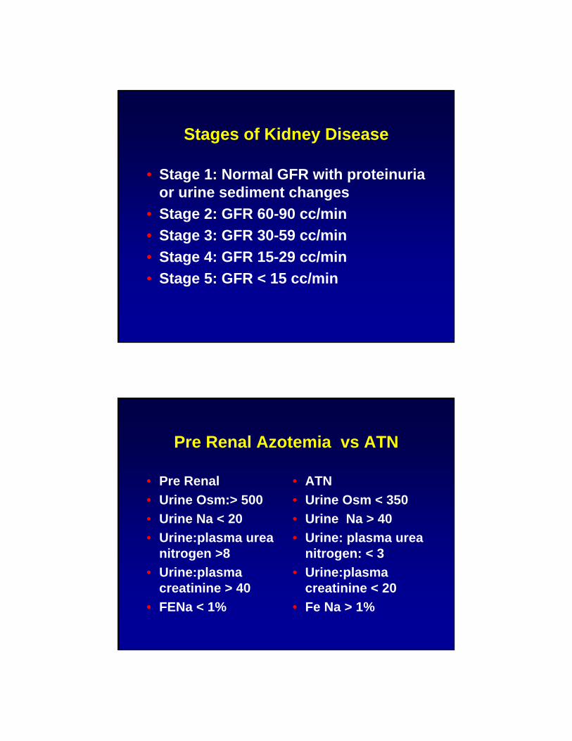
Stages of Kidney Disease
• Stage 1: Normal GFR with proteinuriaor urine sediment changes
• Stage 2: GFR 60-90 cc/min• Stage 3: GFR 30-59 cc/min• Stage 4: GFR 15-29 cc/min• Stage 5: GFR < 15 cc/min
Pre Renal Azotemia vs ATN
• Pre Renal• Urine Osm:> 500• Urine Na < 20• Urine:plasma urea
nitrogen >8• Urine:plasma
creatinine > 40• FENa < 1%
• ATN• Urine Osm < 350• Urine Na > 40• Urine: plasma urea
nitrogen: < 3• Urine:plasma
creatinine < 20• Fe Na > 1%

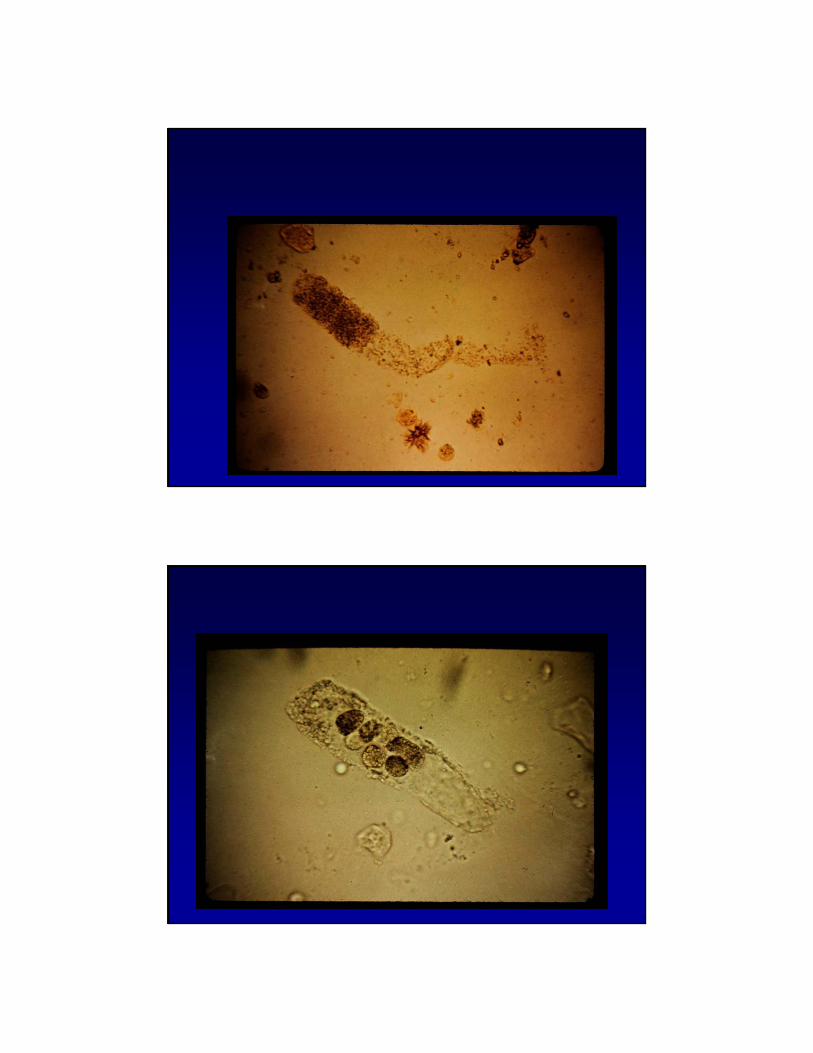
Muddy Brown Casts
Fractional Excretion of Sodium
• The fraction of filtered sodium which ends up in the final urine
Urine [Na] x Plasma [Creatinine] x 100Plasma [Na] x Urine [Creatinine]
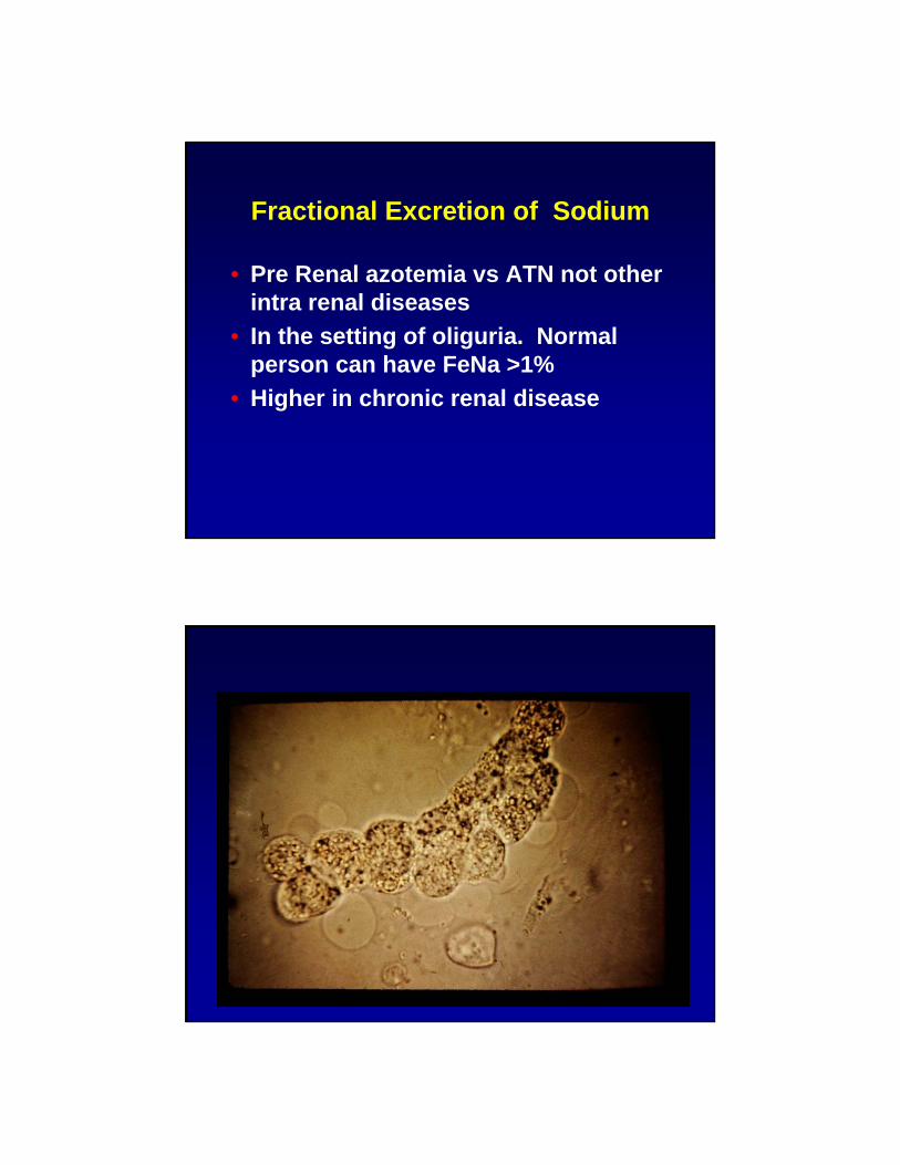
Fractional Excretion of Sodium
• Pre Renal azotemia vs ATN not other intra renal diseases
• In the setting of oliguria. Normal person can have FeNa >1%
• Higher in chronic renal disease
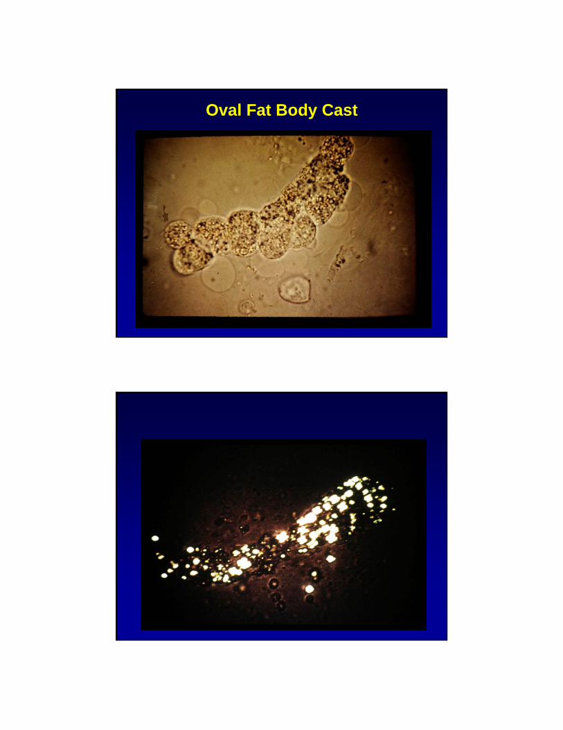
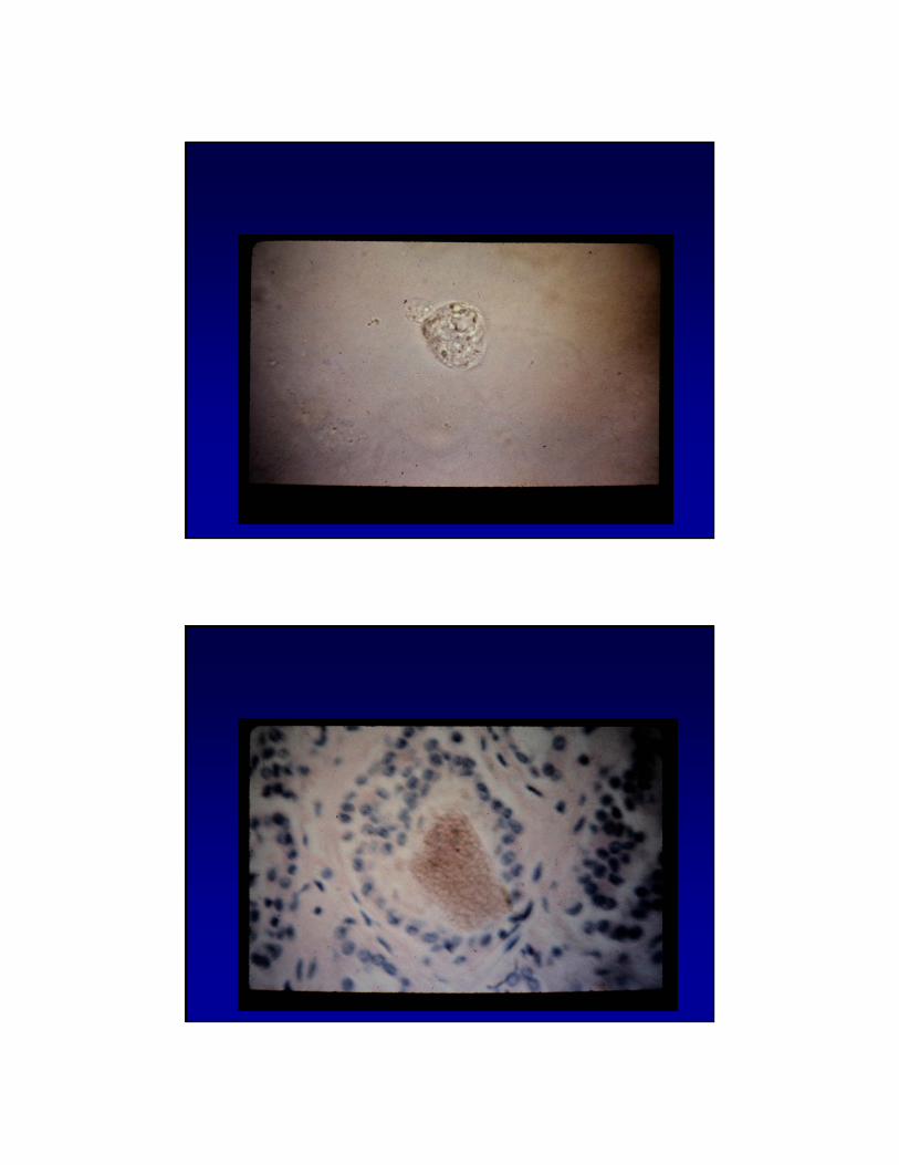
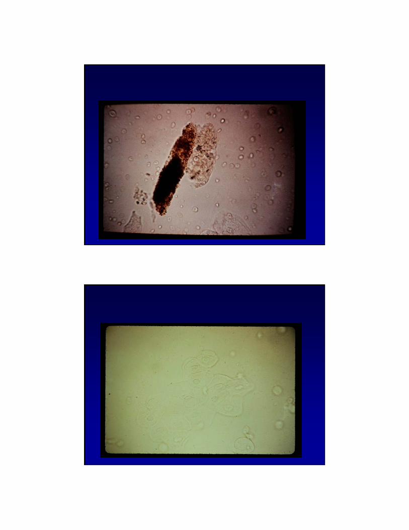
Oval Fat Body Cast
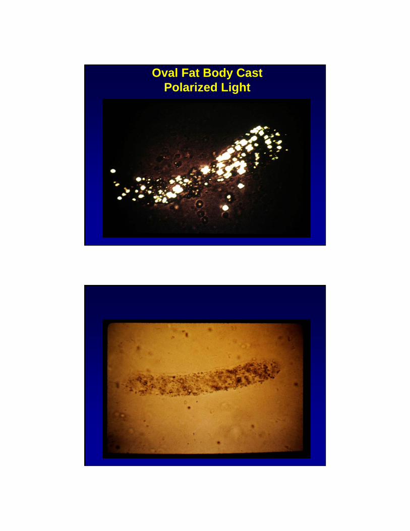
Oval Fat Body CastPolarized Light
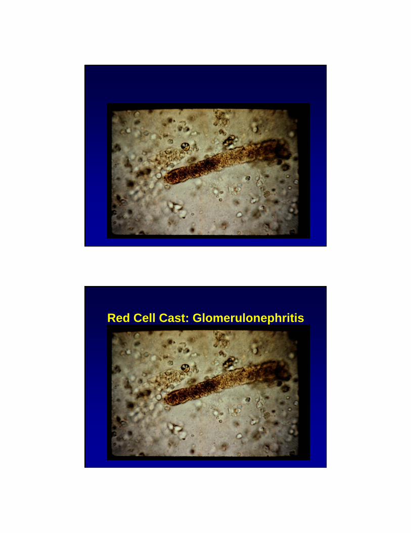
Red Cell Cast: Glomerulonephritis
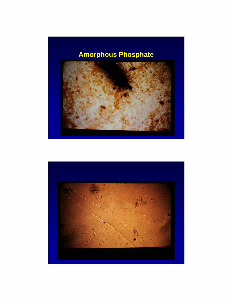
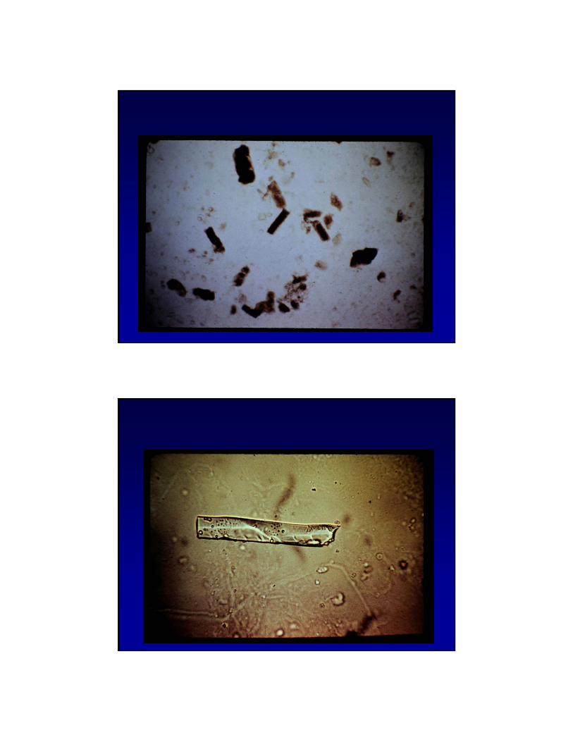
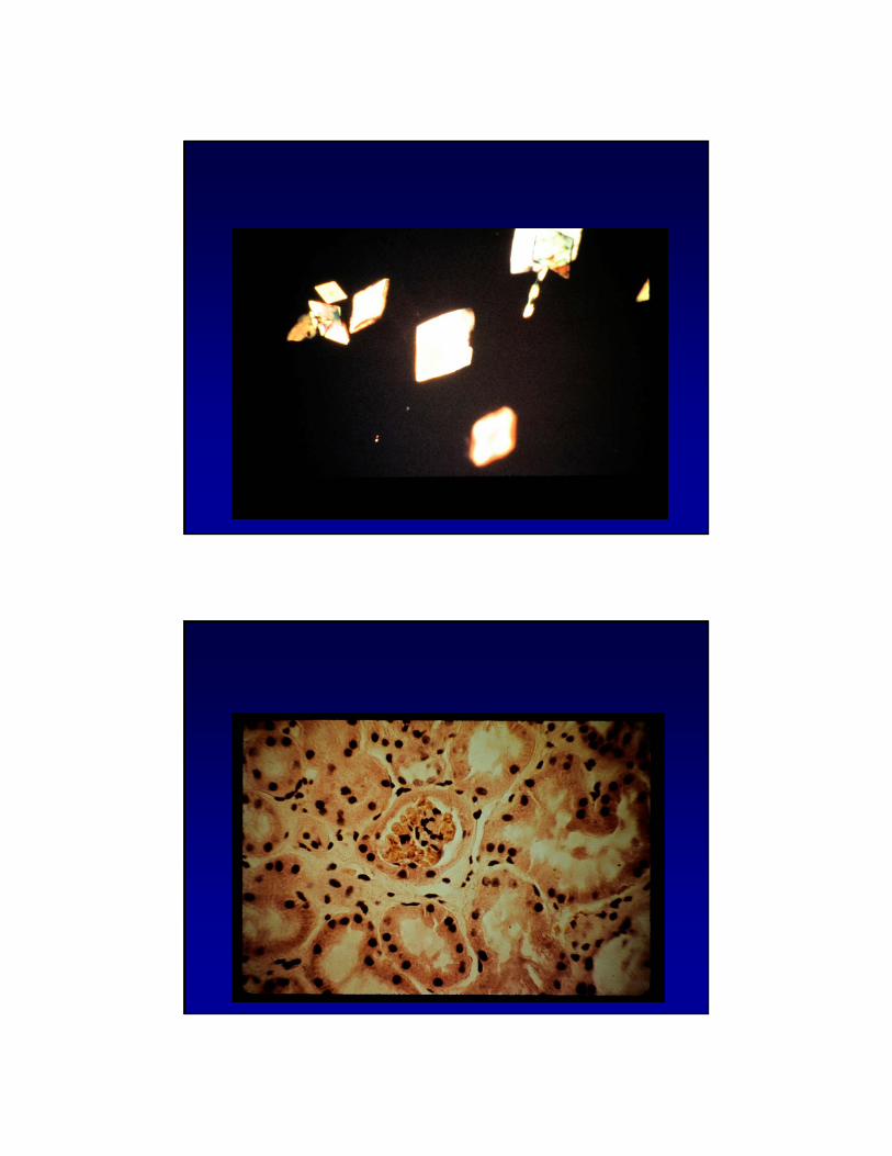
Amorphous Phosphate