Embed Size (px)
Citation preview
Diagnostic Protein Electrophoresis &
Immunofixation
Dr. John J. Haddad, Ph.D., PD.R.F.
St. Elias Medical Laboratories
Proteins and Electrophoresis: An Overview
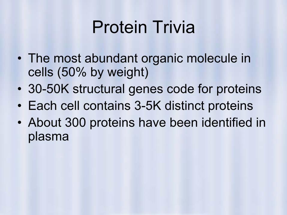
Protein Trivia
• The most abundant organic molecule in cells (50% by weight)
• 30-50K structural genes code for proteins• Each cell contains 3-5K distinct proteins• About 300 proteins have been identified in
plasma
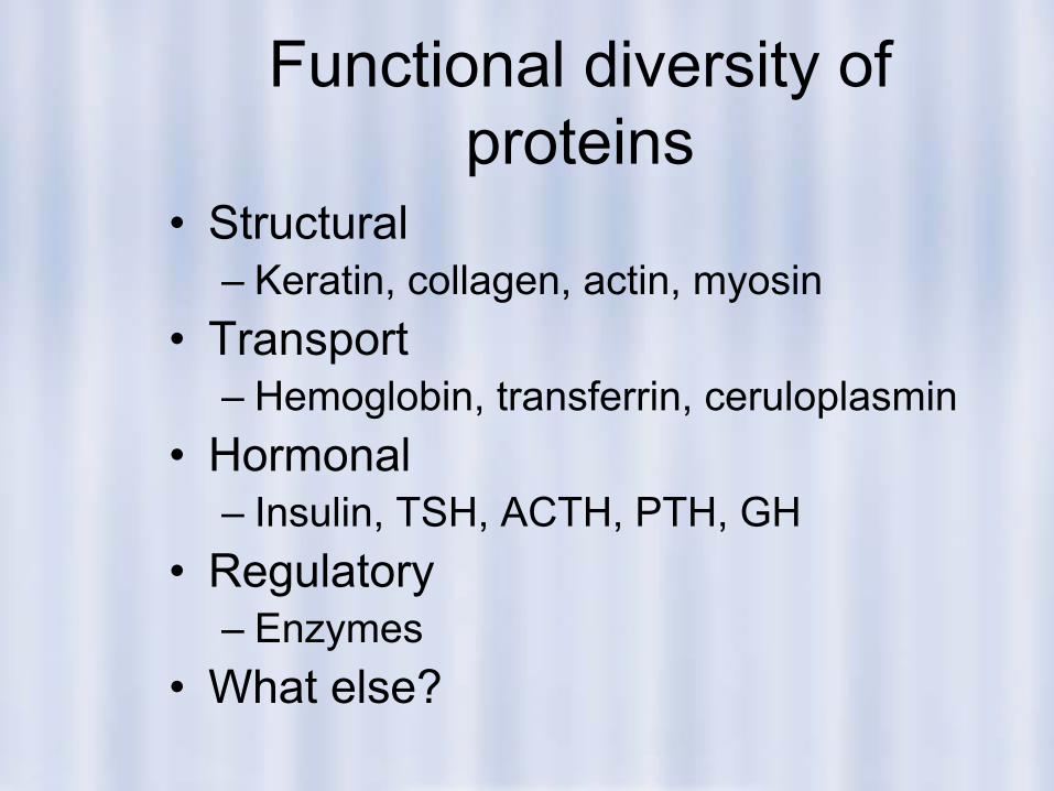
Functional diversity of proteins
• Structural– Keratin, collagen, actin, myosin
• Transport– Hemoglobin, transferrin, ceruloplasmin
• Hormonal– Insulin, TSH, ACTH, PTH, GH
• Regulatory– Enzymes
• What else?
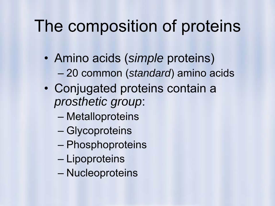
The composition of proteins
• Amino acids (simple proteins)– 20 common (standard) amino acids
• Conjugated proteins contain a prosthetic group:– Metalloproteins– Glycoproteins– Phosphoproteins– Lipoproteins– Nucleoproteins
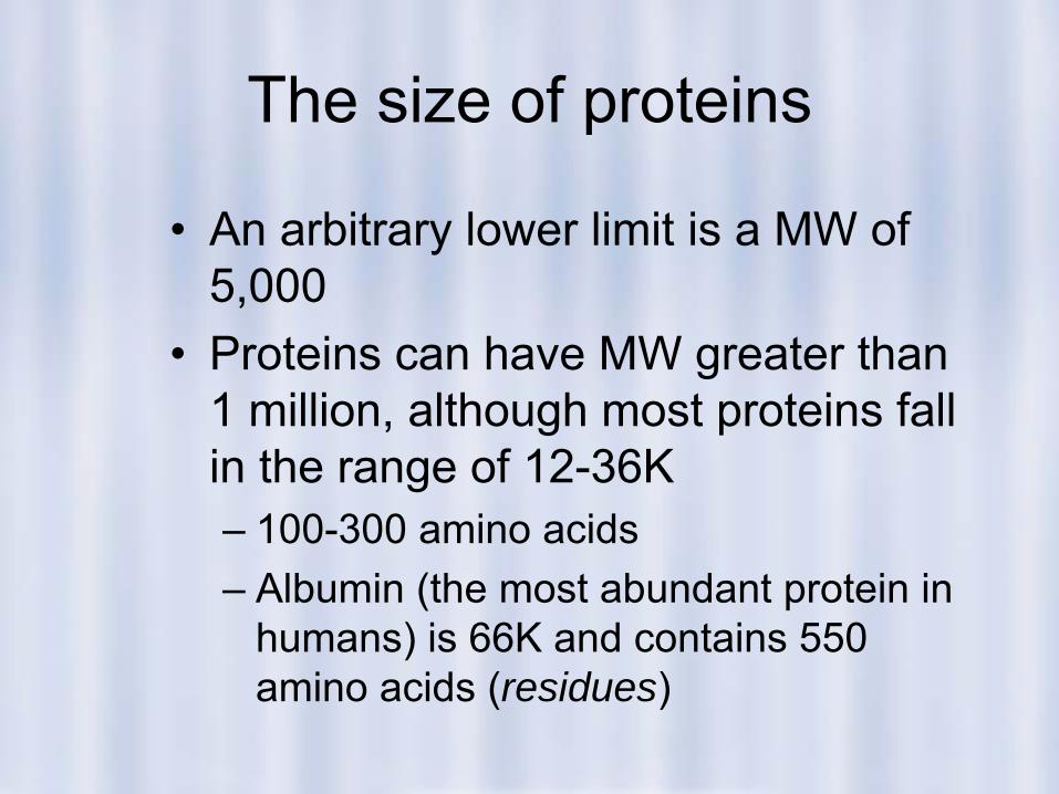
The size of proteins
• An arbitrary lower limit is a MW of 5,000
• Proteins can have MW greater than 1 million, although most proteins fall in the range of 12-36K– 100-300 amino acids– Albumin (the most abundant protein in
humans) is 66K and contains 550 amino acids (residues)
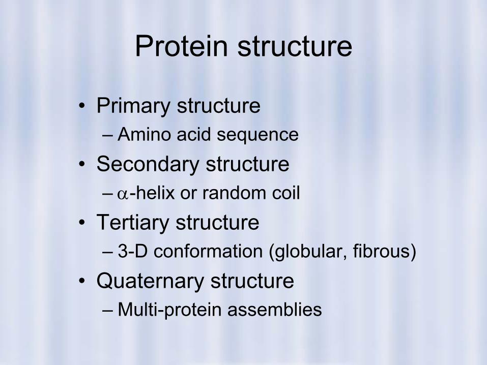
Protein structure
• Primary structure– Amino acid sequence
• Secondary structure– α-helix or random coil
• Tertiary structure– 3-D conformation (globular, fibrous)
• Quaternary structure– Multi-protein assemblies
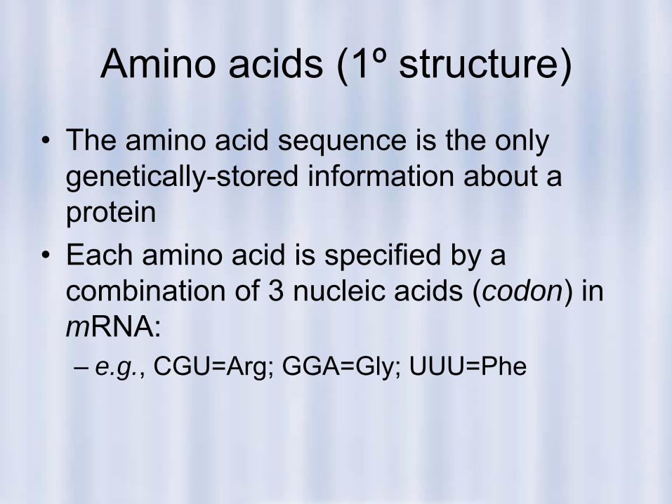
Amino acids (1º structure)
• The amino acid sequence is the only genetically-stored information about a protein
• Each amino acid is specified by a combination of 3 nucleic acids (codon) in mRNA:– e.g., CGU=Arg; GGA=Gly; UUU=Phe
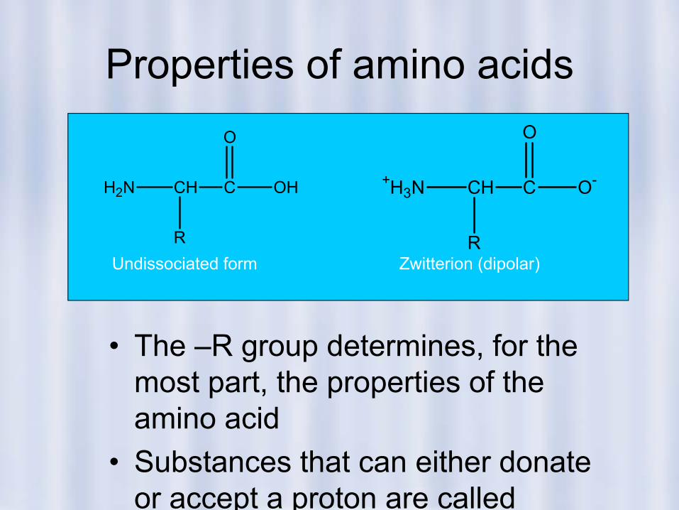
Properties of amino acids
• The –R group determines, for the most part, the properties of the amino acid
• Substances that can either donate or accept a proton are called
+H3N CH C
R
O-
O
H2N CH C
R
OH
O
Undissociated form Zwitterion (dipolar)
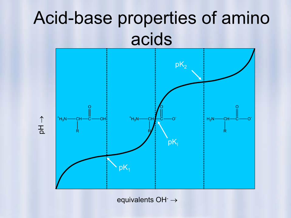
Acid-base properties of amino acids
pH →
equivalents OH- →
pK1
pKI
pK2
H2N CH C
R
O-
O
+H3N CH C
R
OH
O
+H3N CH C
R
O-
O

Acidic and basic amino acids
• Acidic– Asp R=CH2COO-
– Glu R=(CH2)2COO-
• Basic– Lys R=(CH2)4NH3
+
– Arg R= (CH2)3NHC(NH2)2+
– His R: CH2
N
NH2+
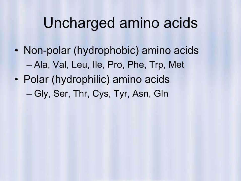
Uncharged amino acids
• Non-polar (hydrophobic) amino acids– Ala, Val, Leu, Ile, Pro, Phe, Trp, Met
• Polar (hydrophilic) amino acids– Gly, Ser, Thr, Cys, Tyr, Asn, Gln
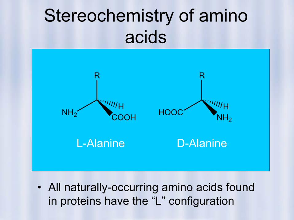
Stereochemistry of amino acids
• All naturally-occurring amino acids found in proteins have the “L” configuration
R
NH2 COOHH
R
HOOC NH2
H
L-Alanine D-Alanine
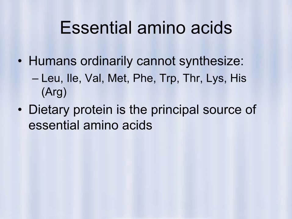
Essential amino acids
• Humans ordinarily cannot synthesize:– Leu, Ile, Val, Met, Phe, Trp, Thr, Lys, His
(Arg)• Dietary protein is the principal source of
essential amino acids
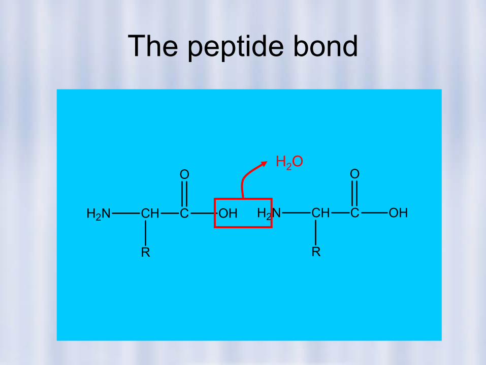
The peptide bond
H2N CH C
R
OH
O
H2N CH C
R
OH
OH2O
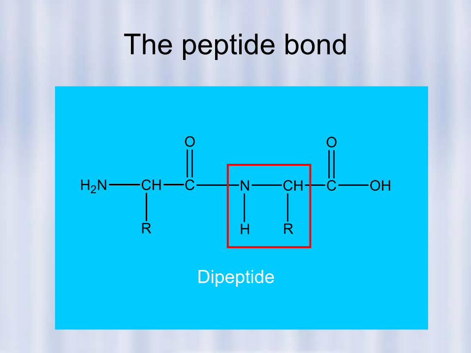
The peptide bond
H2N CH C
R
O
N CH
R
C
O
OH
H
Dipeptide
Amino acid composition and protein properties
• The –R groups determine, for the most part, the properties of the protein
• Proteins rich in Asp, Glu are acidic (albumin is an example)
• Post-translational modifications of proteins have significant effects on their properties, as well.
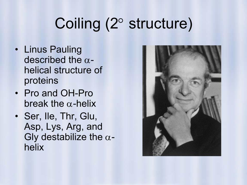
Coiling (2° structure)• Linus Pauling
described the α-helical structure of proteins
• Pro and OH-Pro break the α-helix
• Ser, Ile, Thr, Glu, Asp, Lys, Arg, and Gly destabilize the α-helix
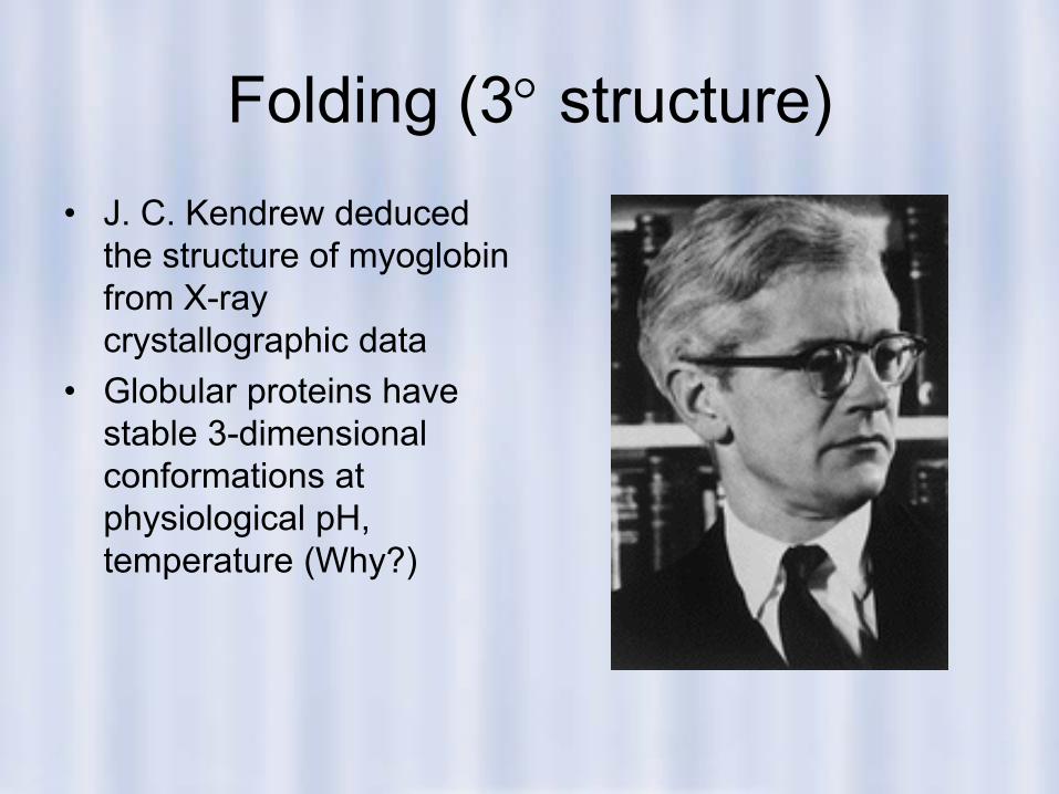
Folding (3° structure)• J. C. Kendrew deduced
the structure of myoglobin from X-ray crystallographic data
• Globular proteins have stable 3-dimensional conformations at physiological pH, temperature (Why?)

Myoglobin
• Protein 3°structure is influenced by αand β regions
• Proteins fold in order to expose hydrophilic regions, and sequester hydrophobic regions
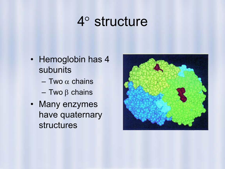
4° structure
• Hemoglobin has 4 subunits– Two α chains– Two β chains
• Many enzymes have quaternary structures

Technical Electrophoresis
The Separation
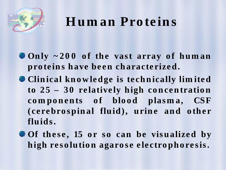
Human Proteins
Only ~200 of the vast array of humanproteins have been characterized.Clinical knowledge is technically limitedto 25 – 30 relatively high concentrationcomponents of blood plasma, CSF(cerebrospinal fluid), urine and otherfluids.Of these, 15 or so can be visualized byhigh resolution agarose electrophoresis.
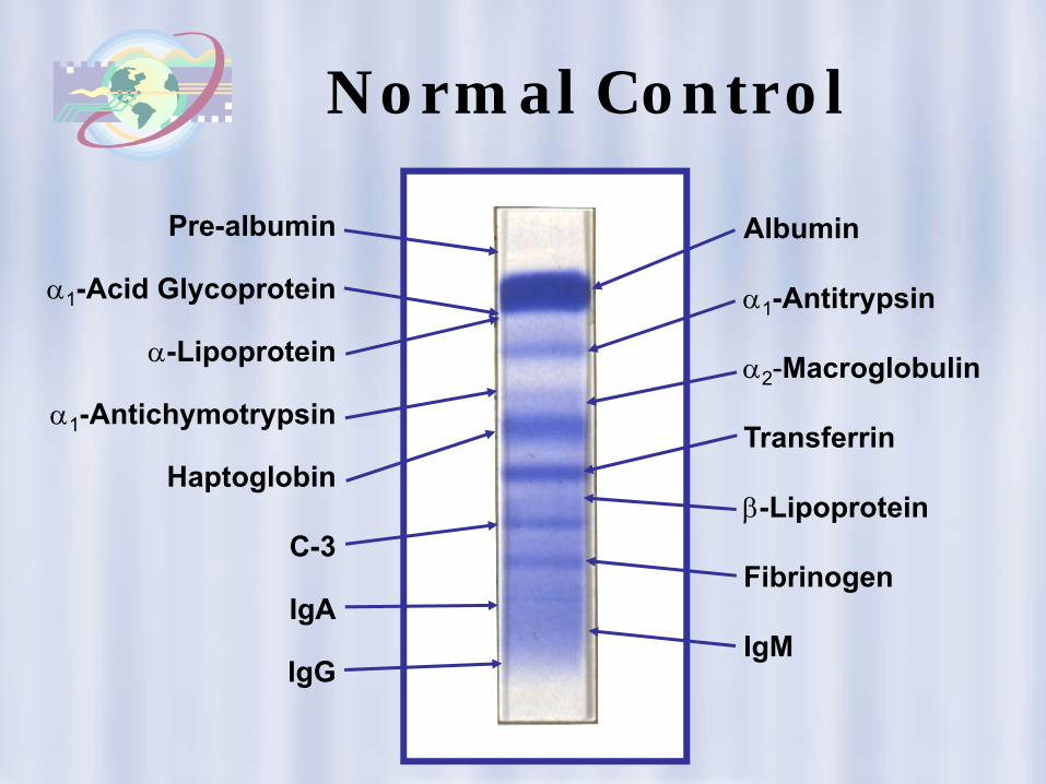
Normal Control
Pre-albumin
α1-Acid Glycoprotein
α-Lipoprotein
α1-Antichymotrypsin
Haptoglobin
C-3
IgA
IgG
Albumin
α1-Antitrypsin
α2-Macroglobulin
Transferrin
β-Lipoprotein
Fibrinogen
IgM
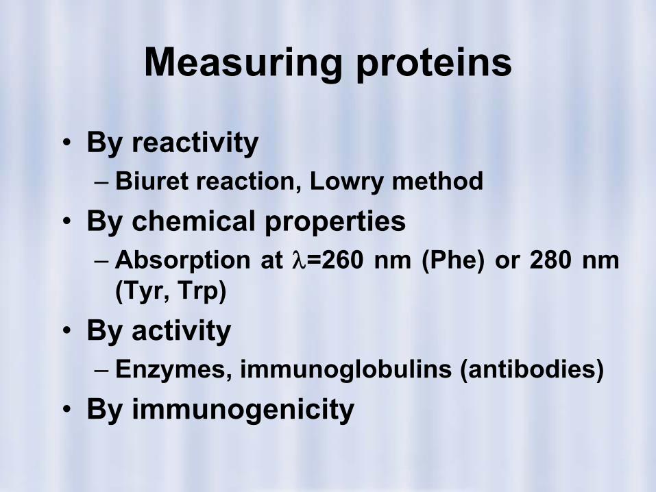
Measuring proteins
• By reactivity– Biuret reaction, Lowry method
• By chemical properties– Absorption at λ=260 nm (Phe) or 280 nm
(Tyr, Trp)• By activity
– Enzymes, immunoglobulins (antibodies)• By immunogenicity
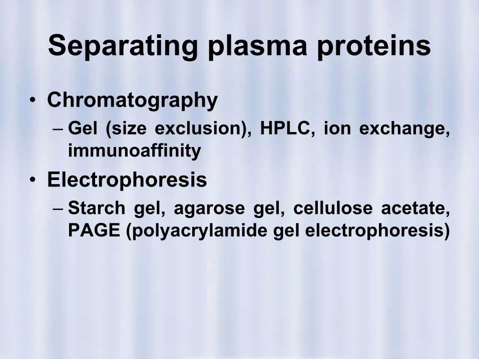
Separating plasma proteins
• Chromatography– Gel (size exclusion), HPLC, ion exchange,
immunoaffinity• Electrophoresis
– Starch gel, agarose gel, cellulose acetate,PAGE (polyacrylamide gel electrophoresis)
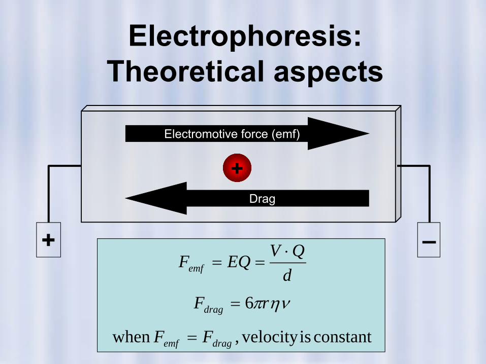
Electrophoresis: Theoretical aspects
+
+ –
Electromotive force (emf)
Drag
constantisvelocity,when
6
dragemf
drag
emf
FF
rFdQVEQF
=
=
⋅==
ηνπ
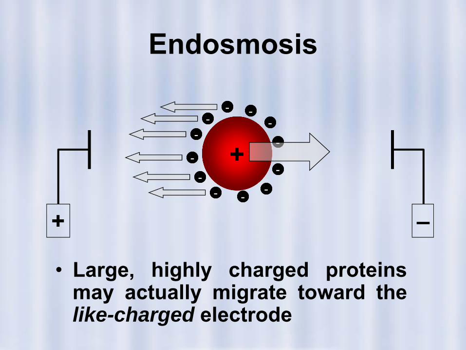
Endosmosis
• Large, highly charged proteinsmay actually migrate toward thelike-charged electrode
+ –
+-
- --
-
-
-
-
--
- -
Optimizing electrophoresis
• Optimal electrophoretic separations mustbalance speed and resolution
– Higher voltage increases speed, but heatcauses evaporation of the buffer and maydenature proteins or even cause gel molting
– Higher ionic strength (buffer) increasesconductivity, but enhances endosmoticeffects
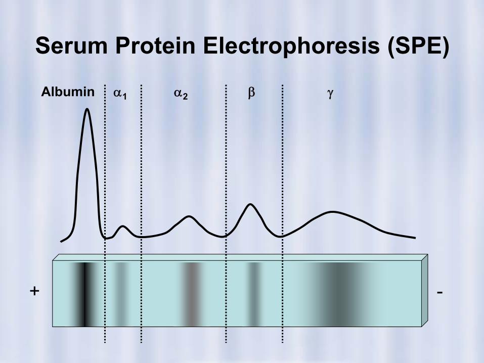
Serum Protein Electrophoresis (SPE)
+ -
Albumin α1 α2 β γ
Albumin• Most abundant protein in plasma
(approximately half of totalprotein)– Synthesized in liver– t½=15-19 days
• Principal functions– Maintaining fluid balance– Carrier– Anti-oxidant activity– Buffer
Clinical significance of albumin
• Hyperalbuminemia is rare and ofno clinical significance
• Hypoalbuminemia– Increased loss (nephrotic
syndrome)– Decreased production (nutritional
deficit, liver failure)• Analbuminemia• Bisalbuminemia, dimeric albumin
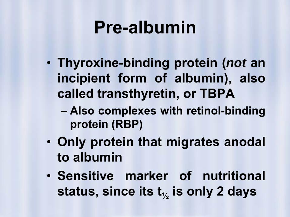
Pre-albumin
• Thyroxine-binding protein (not anincipient form of albumin), alsocalled transthyretin, or TBPA– Also complexes with retinol-binding
protein (RBP)• Only protein that migrates anodal
to albumin• Sensitive marker of nutritional
status, since its t½ is only 2 days
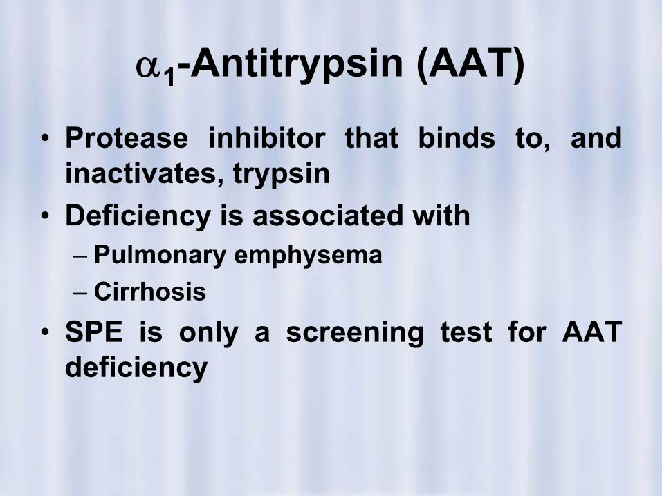
α1-Antitrypsin (AAT)
• Protease inhibitor that binds to, andinactivates, trypsin
• Deficiency is associated with– Pulmonary emphysema– Cirrhosis
• SPE is only a screening test for AATdeficiency
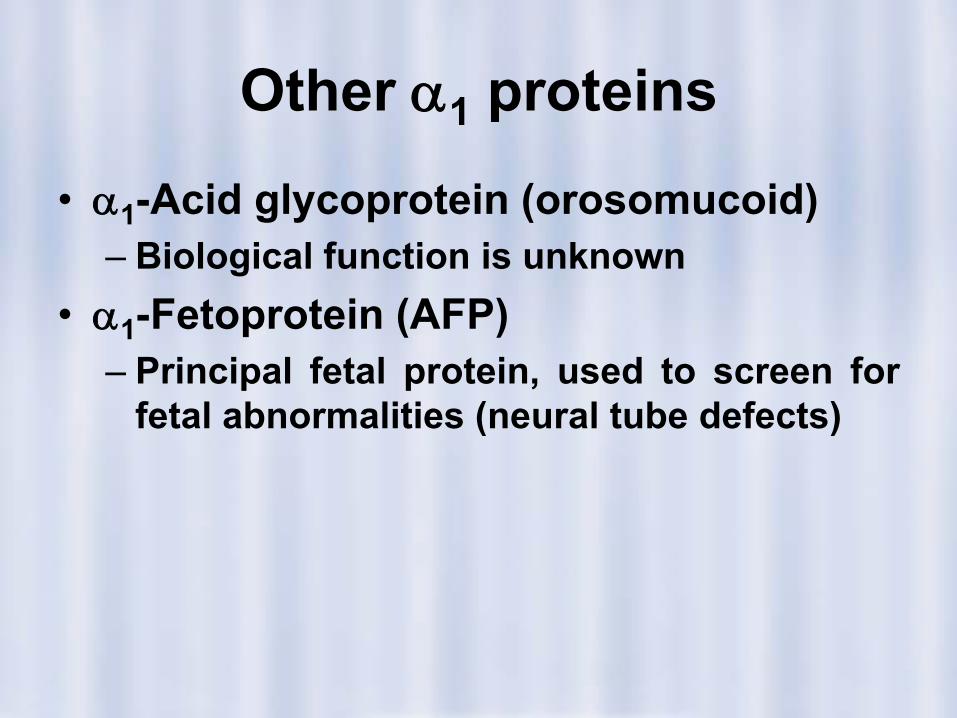
Other α1 proteins
• α1-Acid glycoprotein (orosomucoid)– Biological function is unknown
• α1-Fetoprotein (AFP)– Principal fetal protein, used to screen for
fetal abnormalities (neural tube defects)

α2-Macroglobulin
• Largest non-immunoglobulin inplasma
• Protease inhibitor• Increased in nephrotic syndrome
(size)• Complete genetic deficiency is
unknown
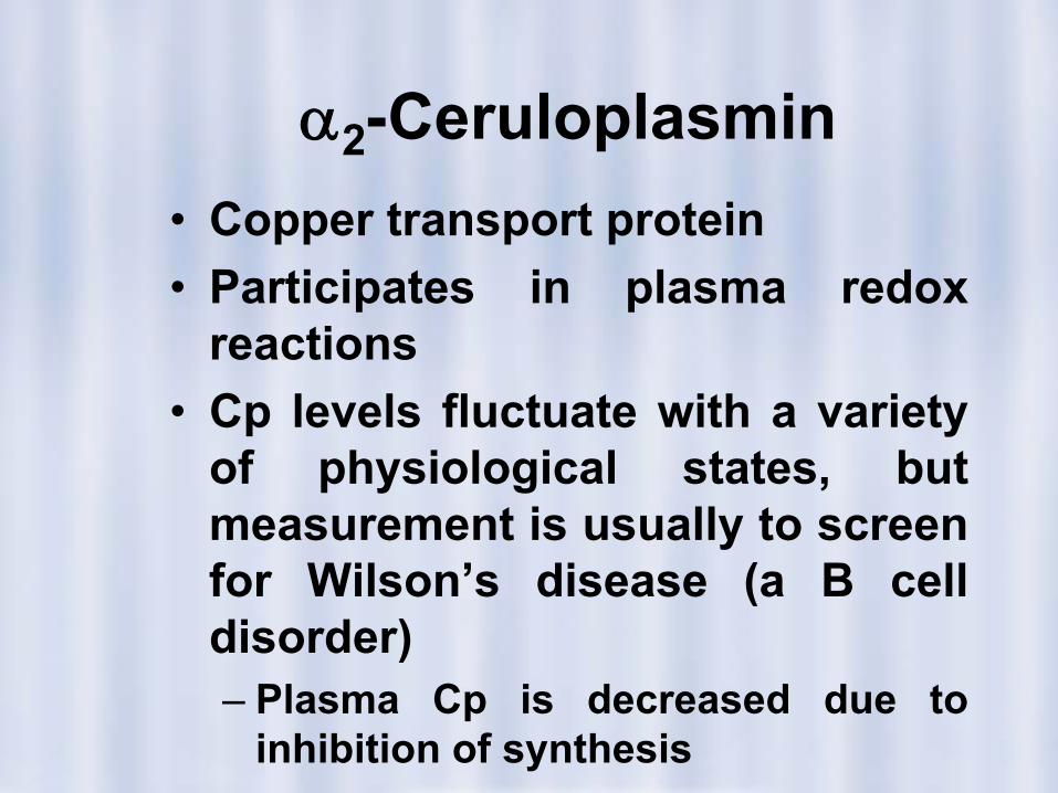
α2-Ceruloplasmin• Copper transport protein• Participates in plasma redox
reactions• Cp levels fluctuate with a variety
of physiological states, butmeasurement is usually to screenfor Wilson’s disease (a B celldisorder)– Plasma Cp is decreased due to
inhibition of synthesis
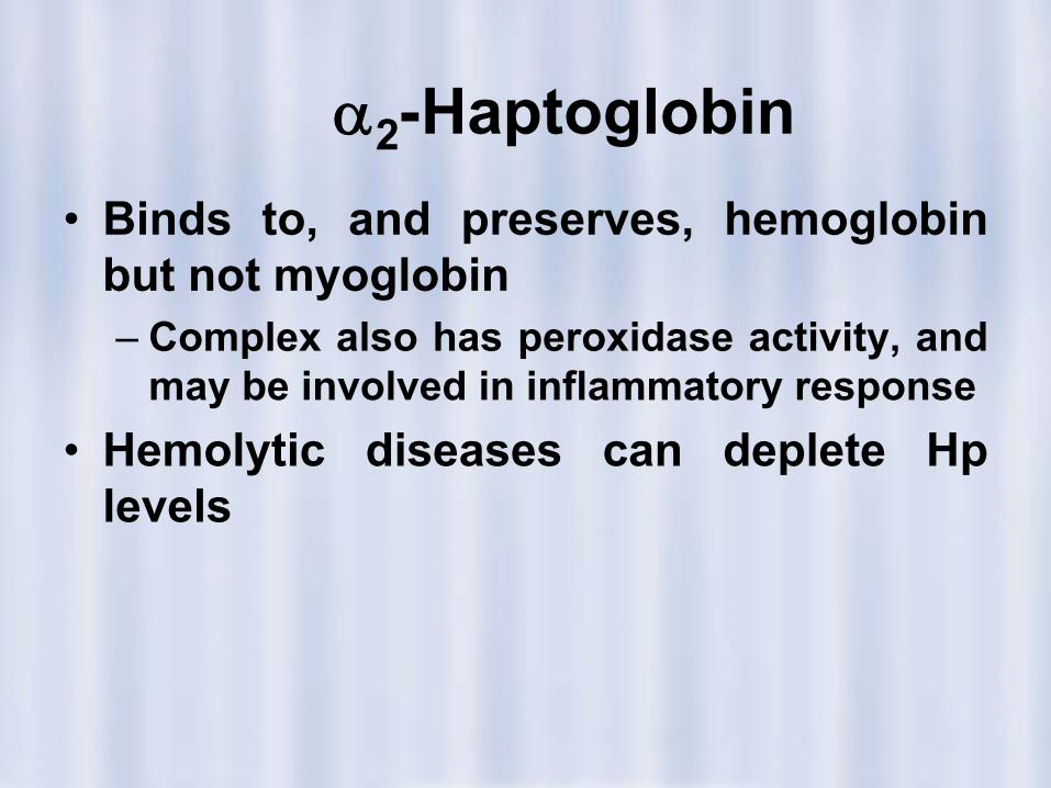
α2-Haptoglobin• Binds to, and preserves, hemoglobin
but not myoglobin– Complex also has peroxidase activity, and
may be involved in inflammatory response• Hemolytic diseases can deplete Hp
levels
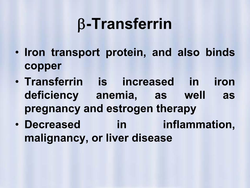
β-Transferrin
• Iron transport protein, and also bindscopper
• Transferrin is increased in irondeficiency anemia, as well aspregnancy and estrogen therapy
• Decreased in inflammation,malignancy, or liver disease
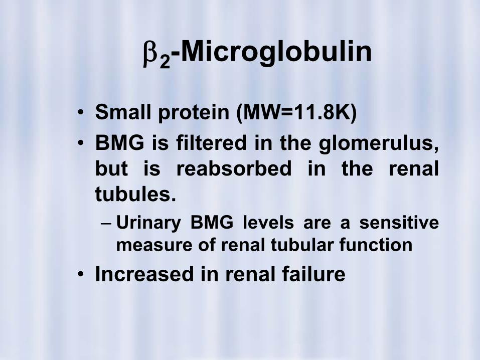
β2-Microglobulin
• Small protein (MW=11.8K)• BMG is filtered in the glomerulus,
but is reabsorbed in the renaltubules.– Urinary BMG levels are a sensitive
measure of renal tubular function• Increased in renal failure
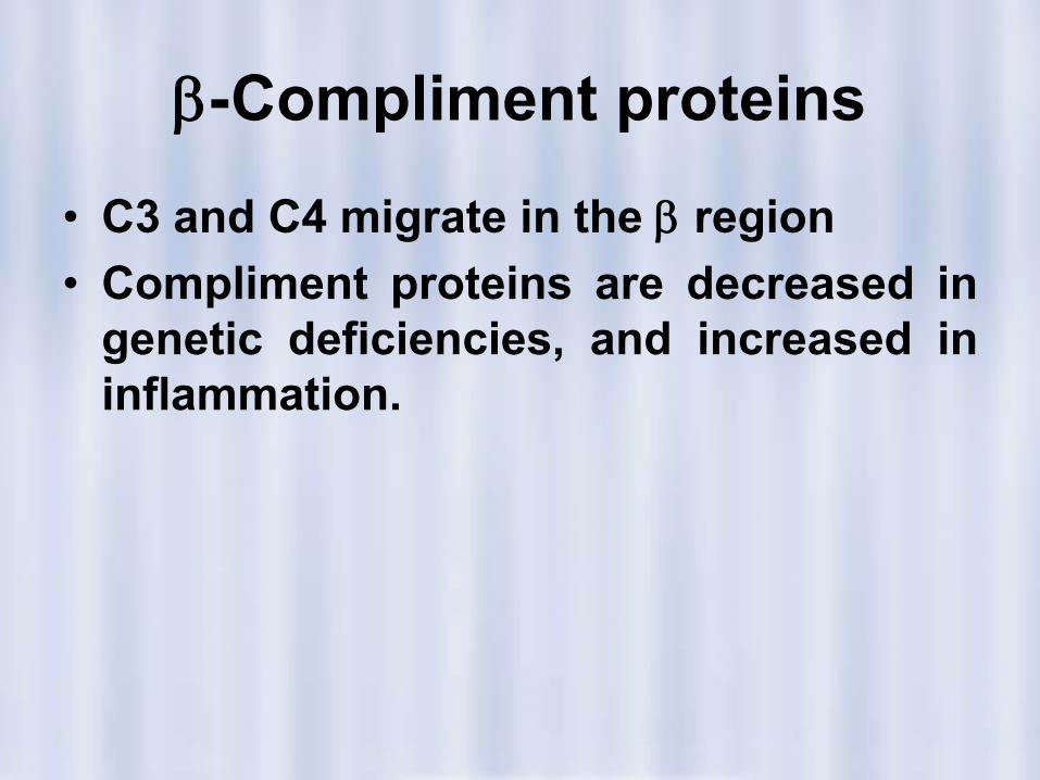
β-Compliment proteins
• C3 and C4 migrate in the β region• Compliment proteins are decreased in
genetic deficiencies, and increased ininflammation.
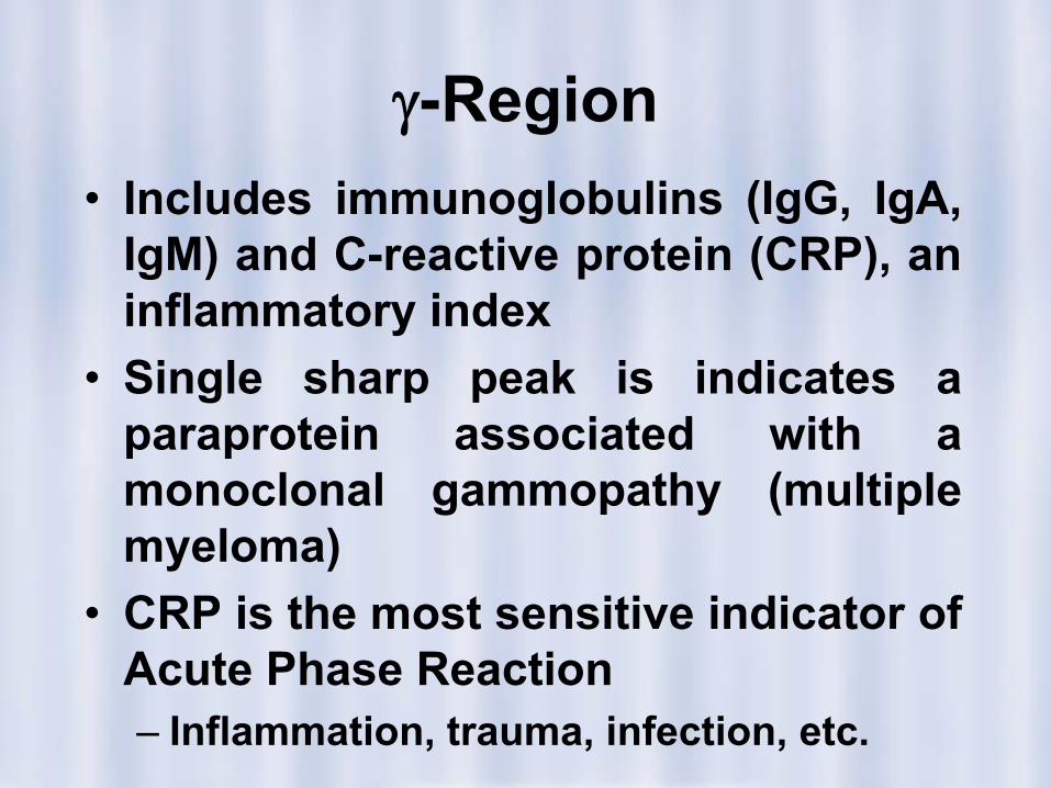
γ-Region• Includes immunoglobulins (IgG, IgA,
IgM) and C-reactive protein (CRP), aninflammatory index
• Single sharp peak is indicates aparaprotein associated with amonoclonal gammopathy (multiplemyeloma)
• CRP is the most sensitive indicator ofAcute Phase Reaction– Inflammation, trauma, infection, etc.
Protein and Immunofixation Electrophoresis
The Test
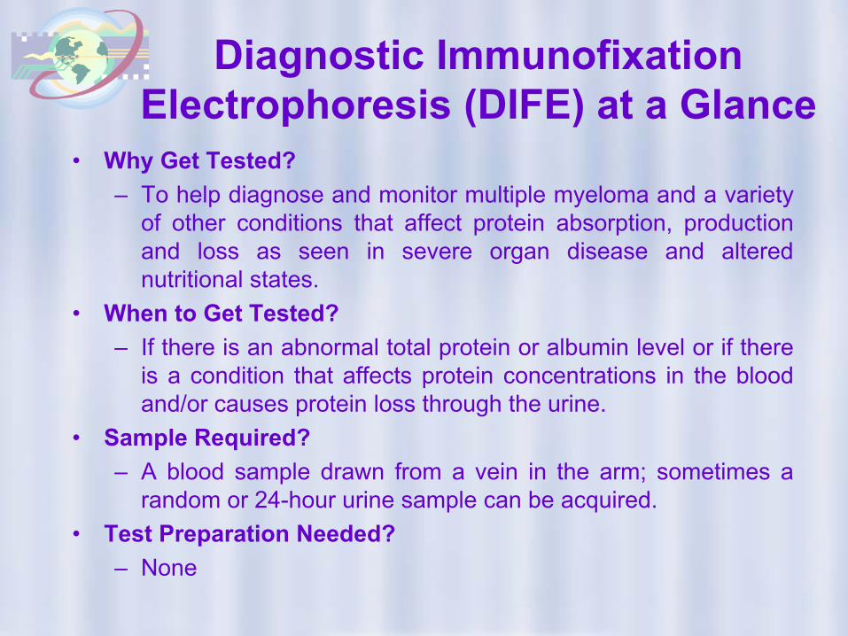
Diagnostic Immunofixation Electrophoresis (DIFE) at a Glance
• Why Get Tested?– To help diagnose and monitor multiple myeloma and a variety
of other conditions that affect protein absorption, productionand loss as seen in severe organ disease and alterednutritional states.
• When to Get Tested?– If there is an abnormal total protein or albumin level or if there
is a condition that affects protein concentrations in the bloodand/or causes protein loss through the urine.
• Sample Required?– A blood sample drawn from a vein in the arm; sometimes a
random or 24-hour urine sample can be acquired.• Test Preparation Needed?
– None
How is the DIFE test used?• Electrophoresis is normally used to identify the presence
or absence of abnormal proteins, and to identify whendifferent groups of proteins are increased or decreased inserum or urine.
• It is frequently used to detect and identify monoclonalproteins (an excessive production of one specificimmunoglobulin).
• Protein and immunofixation electrophoresis are used tohelp detect, diagnose and monitor the course andtreatment of conditions associated with these abnormalproteins, including multiple myeloma and a few relateddiseases.
How is DIFE used? Cont’d
• Protein is usually normally excreted in the urine in verysmall amounts. When it is present in moderate to largeamounts, it may indicate a problem with the kidneys(glomerulonephritis) or multiple myeloma.
• The primary reason protein and immunofixationelectrophoresis are requested on urine is to look formonoclonal protein production.
• This protein may show up in both the serum and urine,or it may only be seen in the urine.
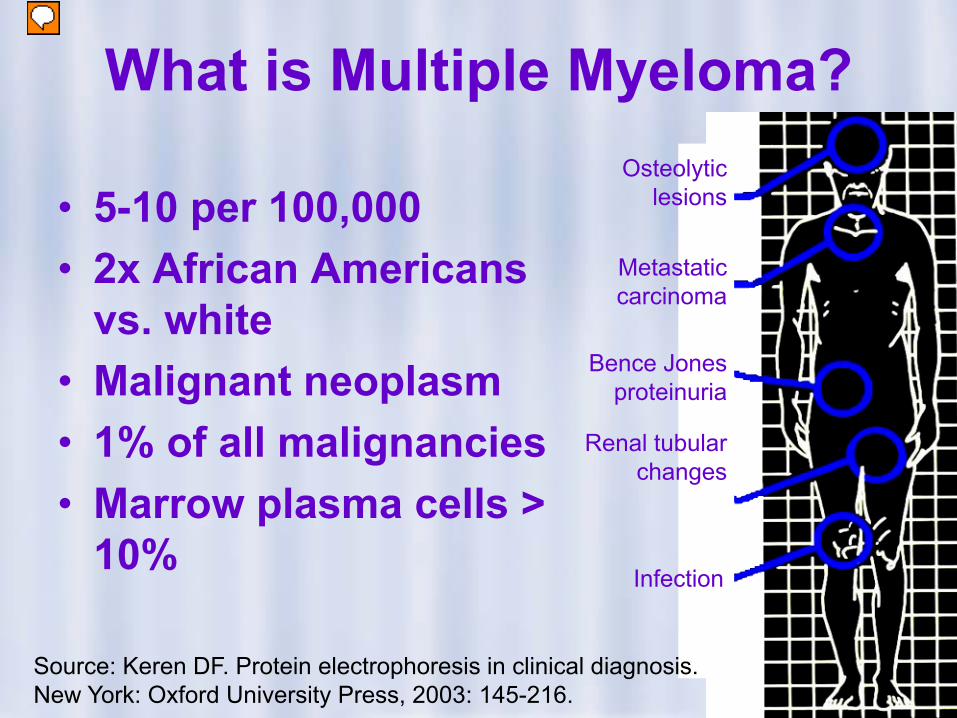
What is Multiple Myeloma?
• 5-10 per 100,000• 2x African Americans
vs. white• Malignant neoplasm• 1% of all malignancies• Marrow plasma cells >
10%
Osteolytic lesions
Metastatic carcinoma
Bence Jones proteinuria
Infection
Renal tubular changes
Source: Keren DF. Protein electrophoresis in clinical diagnosis. New York: Oxford University Press, 2003: 145-216.
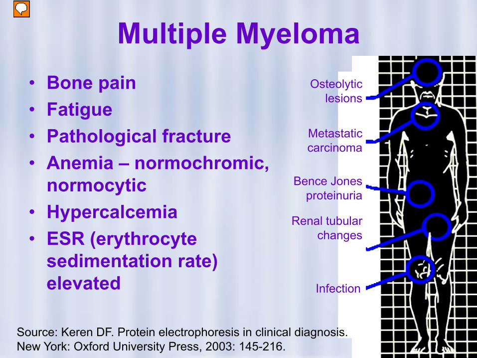
Multiple Myeloma• Bone pain• Fatigue• Pathological fracture• Anemia – normochromic,
normocytic• Hypercalcemia• ESR (erythrocyte
sedimentation rate) elevated
Osteolytic lesions
Metastatic carcinoma
Bence Jones proteinuria
Infection
Renal tubular changes
Source: Keren DF. Protein electrophoresis in clinical diagnosis. New York: Oxford University Press, 2003: 145-216.
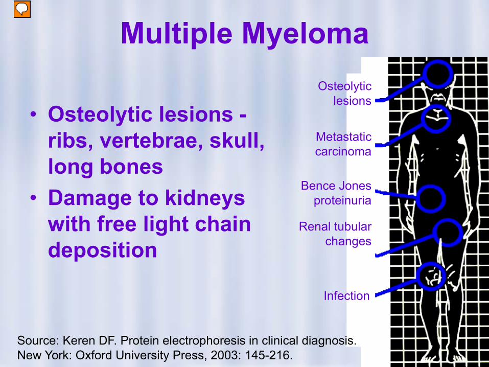
Multiple Myeloma
• Osteolytic lesions -ribs, vertebrae, skull, long bones
• Damage to kidneys with free light chain deposition
Osteolytic lesions
Metastatic carcinoma
Bence Jones proteinuria
Infection
Renal tubular changes
Source: Keren DF. Protein electrophoresis in clinical diagnosis. New York: Oxford University Press, 2003: 145-216.
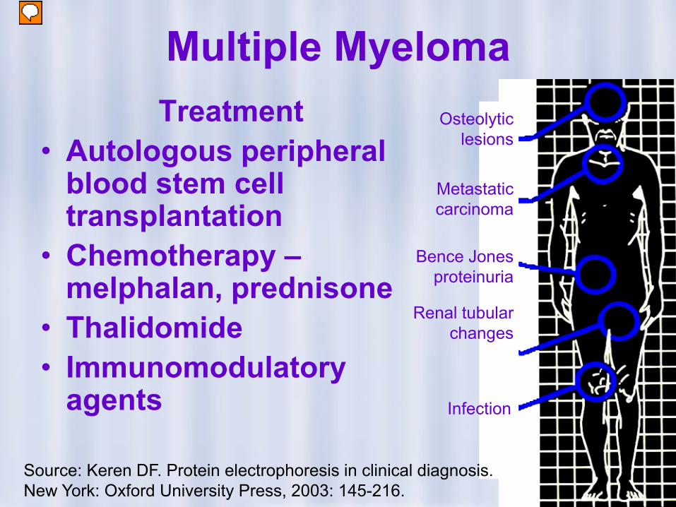
Multiple MyelomaTreatment
• Autologous peripheral blood stem cell transplantation
• Chemotherapy –melphalan, prednisone
• Thalidomide• Immunomodulatory
agents
Osteolytic lesions
Metastatic carcinoma
Bence Jones proteinuria
Infection
Renal tubular changes
Source: Keren DF. Protein electrophoresis in clinical diagnosis. New York: Oxford University Press, 2003: 145-216.
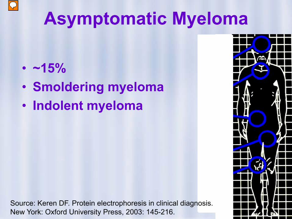
Asymptomatic Myeloma
• ~15% • Smoldering myeloma• Indolent myeloma
Source: Keren DF. Protein electrophoresis in clinical diagnosis. New York: Oxford University Press, 2003: 145-216.
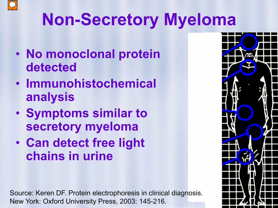
Non-Secretory Myeloma
• No monoclonal protein detected
• Immunohistochemical analysis
• Symptoms similar to secretory myeloma
• Can detect free light chains in urine
Source: Keren DF. Protein electrophoresis in clinical diagnosis. New York: Oxford University Press, 2003: 145-216.
How is DIFE used? Cont’d
• An example is Bence-Jones protein, which is the freelight chain component of antibodies (normally,antibodies are composed of four parts, two identicalheavy (H) chains and two identical light (L) chains.)
• Sometimes, in multiple myeloma, only one or the otheris produced, or it may be produced in excess.
• The small size of Bence-Jones protein allows it to passthrough the kidneys by filtration and enter the urine.
How is DIFE used? Cont’d
• Urine protein electrophoresis may also be used to helpdiagnose the cause and estimate the severity ofprotein excretion due to kidney damage or disease.
• This damage or disease may be due to diabetes,chronic inflammation, an autoimmune condition, or amalignancy (cancerous).
• Electrophoresis is not usually necessary to assess theloss of small to moderate amounts of protein due totemporary conditions, such as a urinary tract infectionor an acute inflammation.
When is DIFE requested?
• Protein electrophoresis may be requested when adoctor or laboratory technologist is investigatingsymptoms that suggest multiple myeloma, symptomssuch as bone pain, anemia, tiredness, unexplainedfractures and recurrent infections.
• It may also be used as a follow-up to other laboratorytests, such as an i) abnormal total protein and/oralbumin level, ii) elevated urine protein levels, iii)elevated calcium levels, and iv) low white or red bloodcell counts (CBC).
When is DIFE requested? Cont’d
• Immunofixation electrophoresis is usually orderedwhen the protein electrophoresis test shows thepresence of an abnormal protein band that may be animmunoglobulin.
When is DIFE requested? Cont’d
• Electrophoresis tests are most frequently requestedwhen there is a disease or condition that causes amonoclonal protein to be produced.
• Once a disease or condition has been diagnosed,electrophoresis may be used at regular intervals tomonitor the course of the disease and theeffectiveness of treatment (if any).
• As disease progresses, the amount of protein goes up;with treatment, it usually goes down.
When is DIFE requested? Cont’d
• Monoclonal protein production may be due to amalignant disease, such as multiple myeloma, but itmay also be due to a monoclonal gammopathy ofundetermined significance (MGUS).
• Most patients with MGUS have no symptoms but theymust continue to be monitored regularly as some maydevelop or progress into multiple myeloma after anumber of years. The reasons for this relapse areobscure.
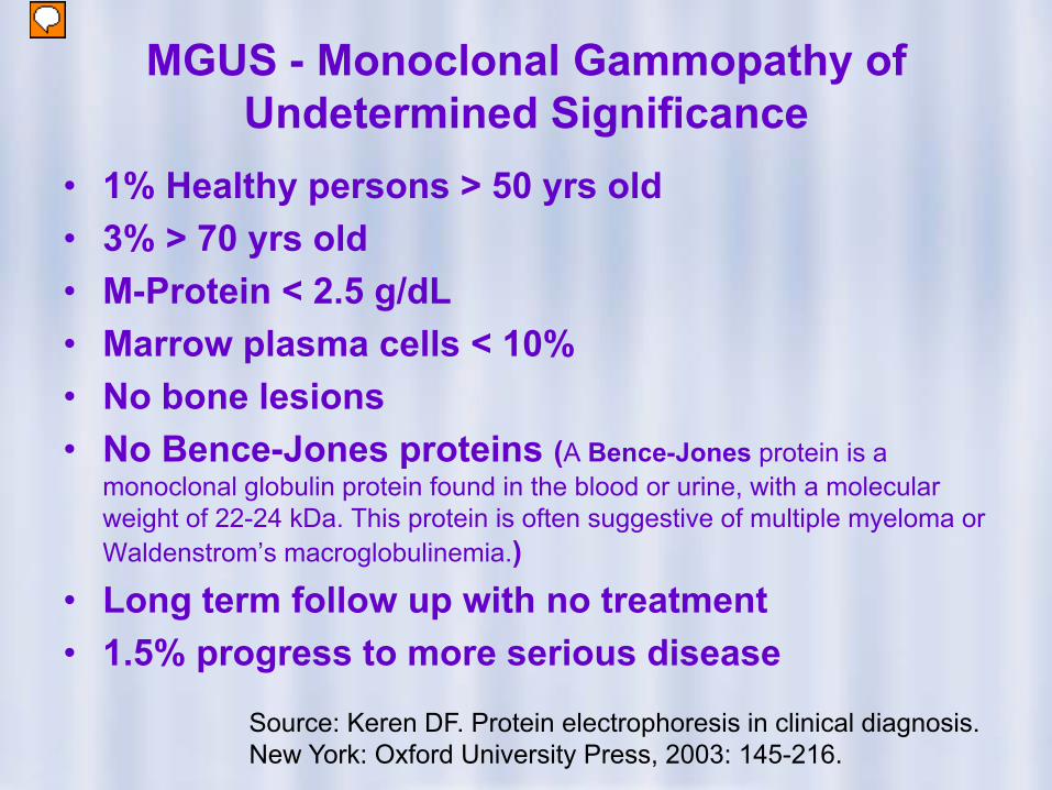
MGUS - Monoclonal Gammopathy of Undetermined Significance
• 1% Healthy persons > 50 yrs old• 3% > 70 yrs old• M-Protein < 2.5 g/dL• Marrow plasma cells < 10%• No bone lesions• No Bence-Jones proteins (A Bence-Jones protein is a
monoclonal globulin protein found in the blood or urine, with a molecular weight of 22-24 kDa. This protein is often suggestive of multiple myeloma or Waldenstrom’s macroglobulinemia.)
• Long term follow up with no treatment• 1.5% progress to more serious disease
Source: Keren DF. Protein electrophoresis in clinical diagnosis. New York: Oxford University Press, 2003: 145-216.
When is DIFE requested? Cont’d
• Serum protein electrophoresis may also be used whensymptoms suggest an inflammatory condition, anautoimmune disease, an acute or chronic infection, akidney or liver disorder, or a protein-losing condition,even if the total protein and/or albumin concentrationsare apparently normal.
• Urine protein electrophoresis may be used when thereis protein detected in the urine or when the doctorsuspects a monoclonal protein may be present.
What does the DIFE test result mean?
• Protein and immunofixation electrophoresis tests givea rough estimate of how much of each protein ispresent.
• The value of protein electrophoresis lies in theproportions of proteins and in the patterns they createon the electrophoresis graph (gel).
• The value of immunofixation electrophoresis is in theidentification of the presence of a particular type ofimmunoglobulin or fragment of an immunoglobulin.
What does the DIFE test result mean?
• For example, diagnostically certain conditions ordiseases may be associated with decreases orincreases in various serum proteins.
• This is reflected according to the following illustrations:
DIFE Clinical Significance
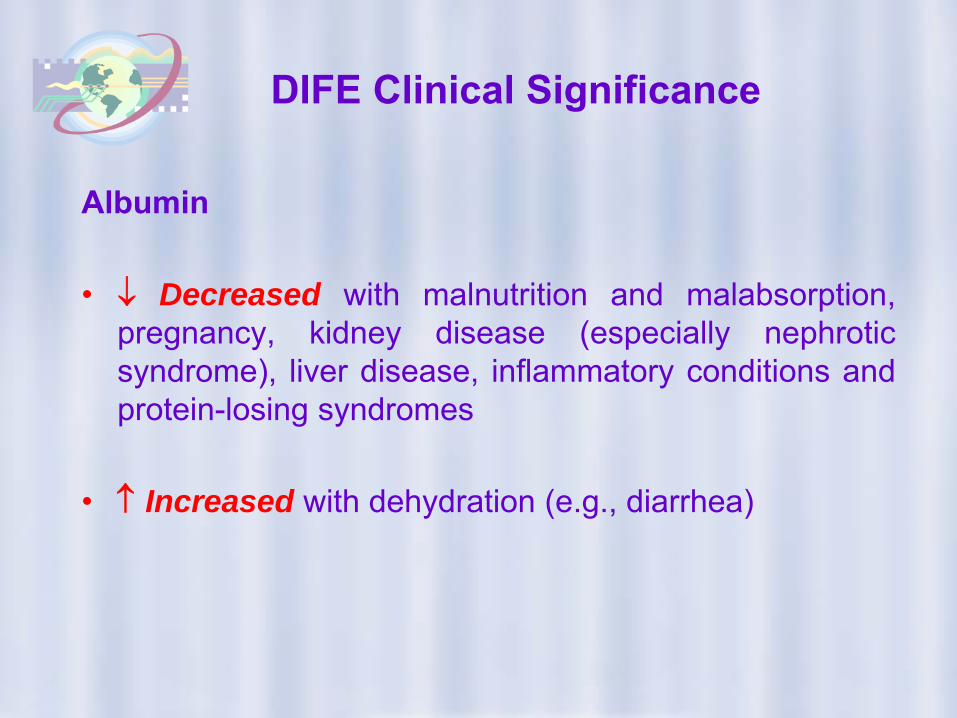
Albumin
• ↓ Decreased with malnutrition and malabsorption,pregnancy, kidney disease (especially nephroticsyndrome), liver disease, inflammatory conditions andprotein-losing syndromes
• ↑ Increased with dehydration (e.g., diarrhea)
DIFE Clinical Significance
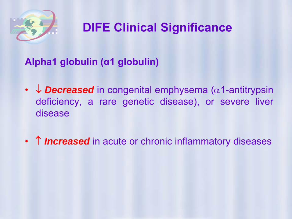
Alpha1 globulin (α1 globulin)
• ↓ Decreased in congenital emphysema (α1-antitrypsindeficiency, a rare genetic disease), or severe liverdisease
• ↑ Increased in acute or chronic inflammatory diseases
DIFE Clinical Significance
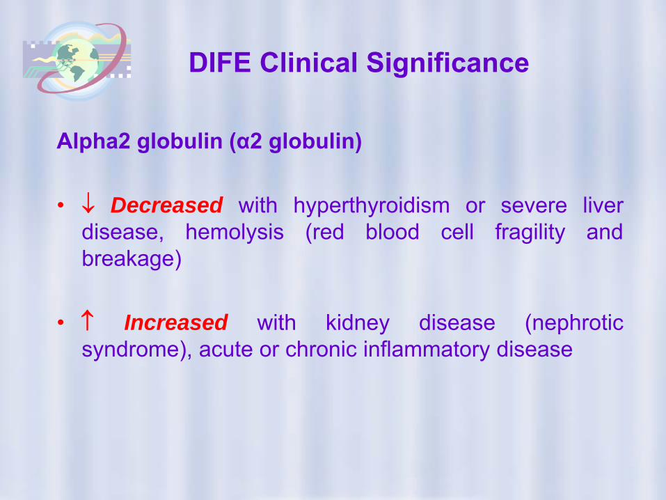
Alpha2 globulin (α2 globulin)
• ↓ Decreased with hyperthyroidism or severe liverdisease, hemolysis (red blood cell fragility andbreakage)
• ↑ Increased with kidney disease (nephroticsyndrome), acute or chronic inflammatory disease
DIFE Clinical Significance
Beta globulin (β globin)
• ↓ Decreased with malnutrition, cirrhosis (e.g.,hepatitis)
• ↑ Increased with hypercholesterolemia, iron deficiencyanemia, some cases of multiple myeloma ormonoclonal gammopathy of undetermined significance(MGUS)
DIFE Clinical Significance
Gamma globulin (γ globulin)
• ↓ Decreased in a variety of genetic immune disorders,and in secondary immune deficiency
• ↑ Increased– Polyclonal: chronic inflammatory disease, rheumatoid arthritis,
systemic lupus erythematosus (SLE), cirrhosis, chronic liverdisease, acute and chronic infection, recent immunization
– Monoclonal: Waldenstrom’s macroglobulinaemia, multiplemyeloma, monoclonal gammopathies of undeterminedsignificance (MGUS)
DIFE Clinical Significance
• Immunizations within the previous six months canincrease immunoglobulins, as can drugs such asphenytoin (Dilantin), procainamide, oralcontraceptives, methadone and therapeutic gammaglobulin.
• Aspirin, bicarbonates, chlorpromazine (Thorazine),corticosteroids and neomycin can affect proteinelectrophoresis results.
The Test Sample
• As indicated, protein electrophoresis is a method forseparating the proteins found in blood (serum) orurine.
• During the test, an electric current is used to move theproteins across a thin layer of agarose/polyacrylamidegel.
• The distances that individual proteins travel depend ontheir size, shape and electrical charge.
The Test Sample
• These separated proteins may be detected by the use of adye that binds to (stains) all of the proteins and reveals acharacteristic pattern of bands.
• Each band indicates the presence of a particular protein,while the size of the band is a rough indication of thequantity.
• This pattern of bands is converted into a visual graph,showing vertical spikes or peaks where there is a lot ofprotein and smaller peaks or valleys where there is less.
The Test Sample
• A newer method called capillary zone electrophoresis (CZE)separates proteins by passing them through a long, thincolumn, producing a graph that is very similar to the onemade by running the protein through a gel.
The Test Sample
• Specific proteins of interest can be identified by firstmixing them into the gel with monoclonal or polyclonalantibodies, then washing away all the other proteinsprior to staining. This procedure is calledimmunofixation electrophoresis (IFE).
• A slightly different method, immuno-electrophoresis,was used in the past to identify specific proteins.However this technique has been largely supercededby IFE because IFE is easier to perform and interpret.
The Test Sample
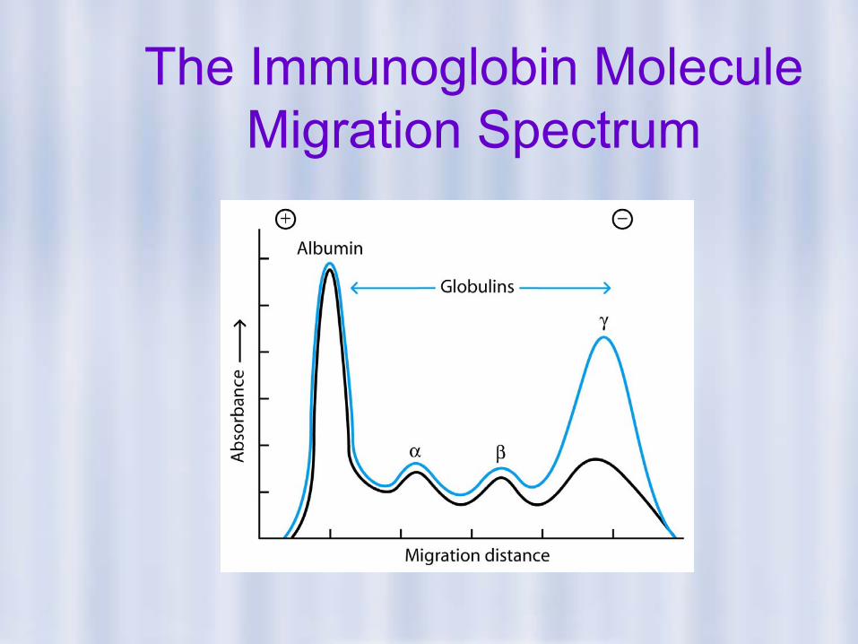
• Normally, serum proteins are separated into five or sixmajor groupings by protein electrophoresis. These fractionsare called albumin, α1, α2, β and γ (the β fraction issometimes divided into β1 and β2).
• Albumin, which is produced in the liver, forms its own groupand accounts for about 60% of proteins in the blood.
• 'Globulins' is a collective term used to refer to proteins otherthan albumin. With the exception of the immunoglobulins(produced by activated B lymphocytes) and somecomplement proteins, most of the globulins are produced inthe liver.
The Immunoglobin MoleculeMigration Spectrum
In the 1930’s, Elvin Kabat and others showed thatproteins in a fraction of serum called γ-globulin hadactivities of antitoxins, precipitins and agglutinins thathad previously been thought to be separate activities.
The active molecules in the γ-globulin fraction weregiven the name, antibodies.
It had been determined by several means that theproteins with antibody activity (immunoglobulins) had amolecular weight of approximately 150 kDa.
The Immunoglobin MoleculeMigration Spectrum
The Test Sample
• The bands seen on protein electrophoresis formcharacteristic patterns. Alterations to these patterns areassociated with a variety of different diseases andconditions.
• For example in multiple myeloma (a cancer of certain typesof white blood cells called plasma cells), the uncontrolledgrowth and division of a malignant plasma cell leads to theproduction of large amounts of a single type ofimmunoglobulin (antibody).
The Test Sample
• In contrast to other proteins in serum, which are typically of asingle type, antibodies (immunoglobulins) must differ from eachother to be able to recognize bacteria, viruses and other 'foreign'substances. Each time the body is exposed to a virus, forexample, one plasma cell replicates and makes a group (or clone)of plasma cells to produce antibody to eliminate it.
• Since our total immunoglobulin represents antibody made bymany clones, we refer to it as a polyclonal pattern. When there isa cancer of plasma cells, only one type of antibody is produced,termed a monoclonal pattern. This abnormal protein can be seenas a characteristic band on the electrophoresis gel.

The Immunoglobin MoleculeMigration Spectrum
Agarose gel immunofixation electrophoresis of
normal serum.
Example
• An agarose gel electrophoresis first separates the proteins in aserum sample. Antiserum against the protein of interest is spreaddirectly on the gel. The protein of interest precipitates in the gelmatrix. After a wash step to remove other proteins, theprecipitated protein is stained. This method is qualitative and isused to identify proteins found in multiple myeloma.
• Below is the immunofixation electrophoresis gel from a serumsample analyzed. After electrophoresis, the precipitated proteinsare stained with Acid Violet, a stain.
Example
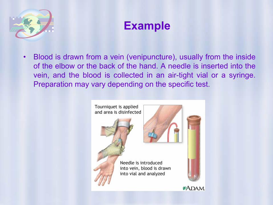
• Blood is drawn from a vein (venipuncture), usually from the insideof the elbow or the back of the hand. A needle is inserted into thevein, and the blood is collected in an air-tight vial or a syringe.Preparation may vary depending on the specific test.
Example
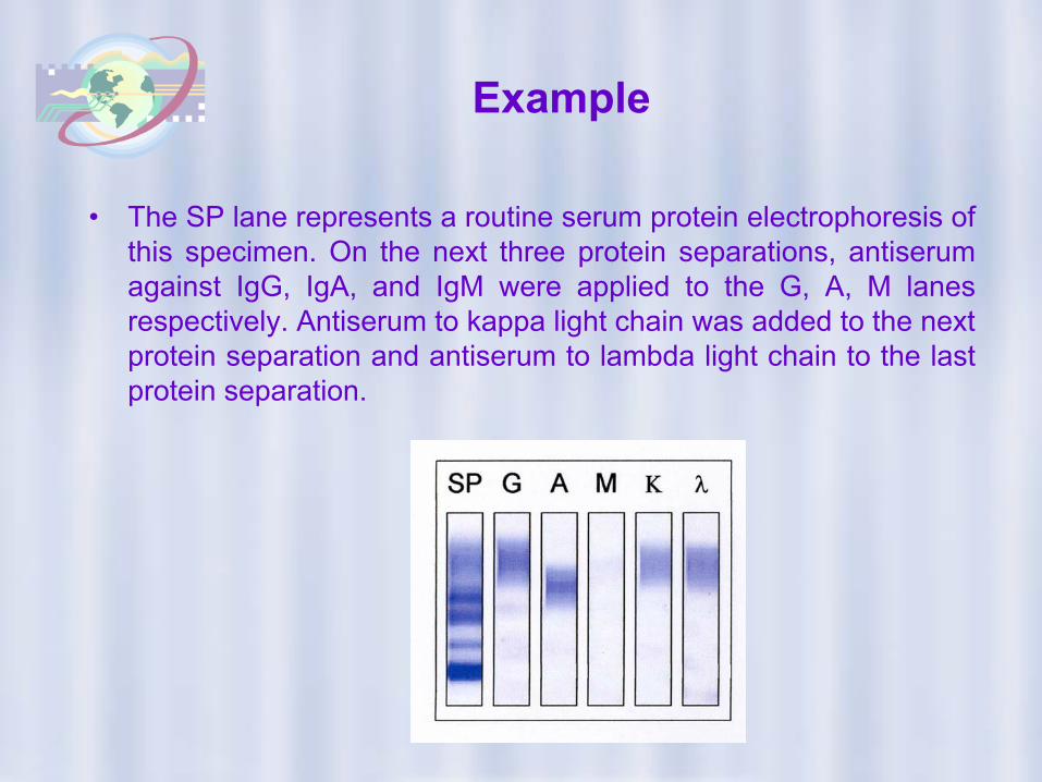
• The SP lane represents a routine serum protein electrophoresis ofthis specimen. On the next three protein separations, antiserumagainst IgG, IgA, and IgM were applied to the G, A, M lanesrespectively. Antiserum to kappa light chain was added to the nextprotein separation and antiserum to lambda light chain to the lastprotein separation.
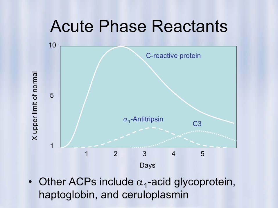
Acute Phase Reactants
• Other ACPs include α1-acid glycoprotein, haptoglobin, and ceruloplasmin
X u
pper
lim
it of
nor
mal
1
5
10
Days
1 2 3 4 5
C-reactive protein
α1-AntitripsinC3
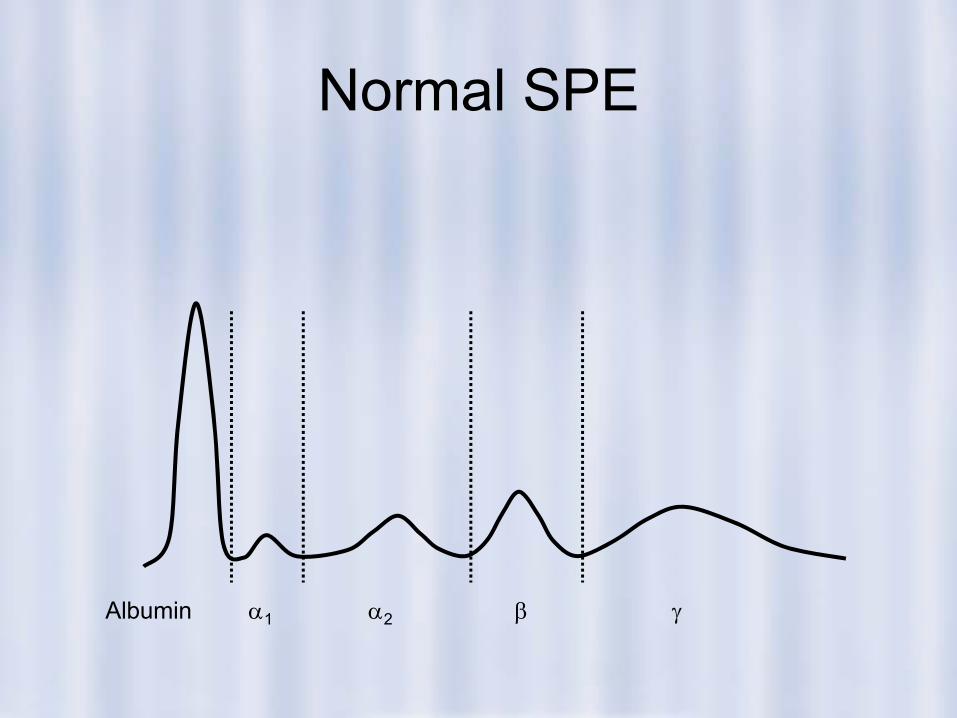
Normal SPE
Albumin α1 α2 β γ
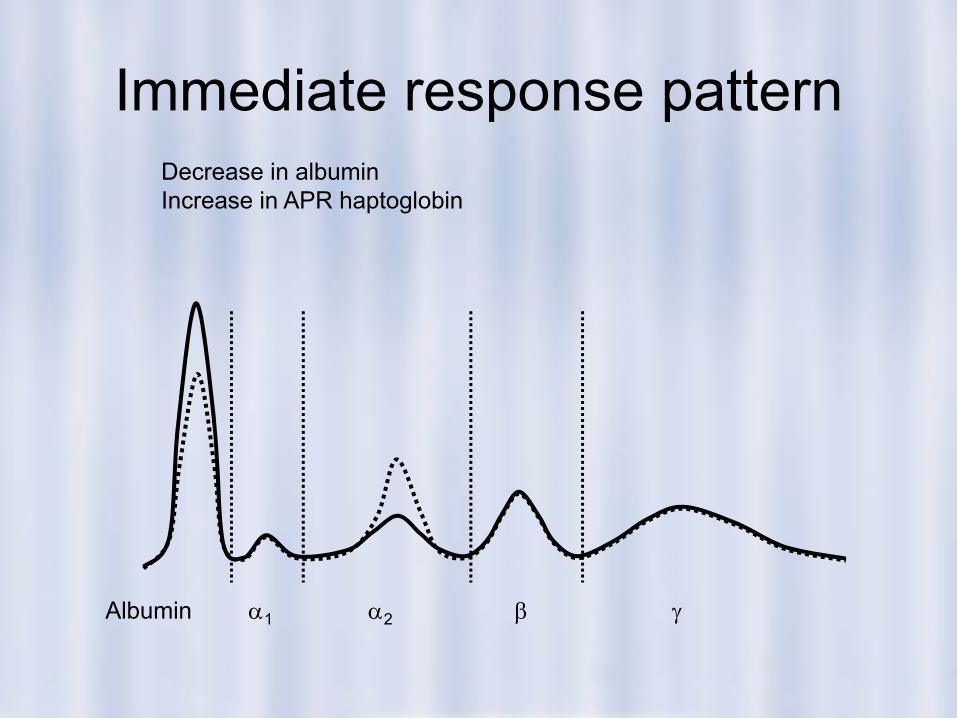
Immediate response pattern
Albumin α1 α2 β γ
Decrease in albuminIncrease in APR haptoglobin
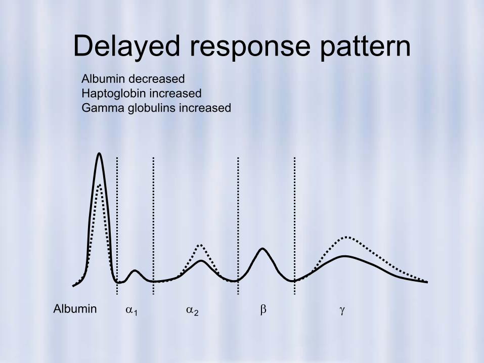
Delayed response pattern
Albumin α1 α2 β γ
Albumin decreasedHaptoglobin increasedGamma globulins increased
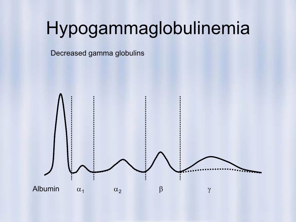
Hypogammaglobulinemia
Albumin α1 α2 β γ
Decreased gamma globulins
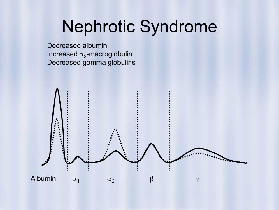
Nephrotic Syndrome
Albumin α1 α2 β γ
Decreased albuminIncreased α2-macroglobulinDecreased gamma globulins
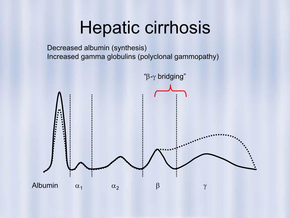
Hepatic cirrhosis
Albumin α1 α2 β γ
Decreased albumin (synthesis)Increased gamma globulins (polyclonal gammopathy)
“β-γ bridging”
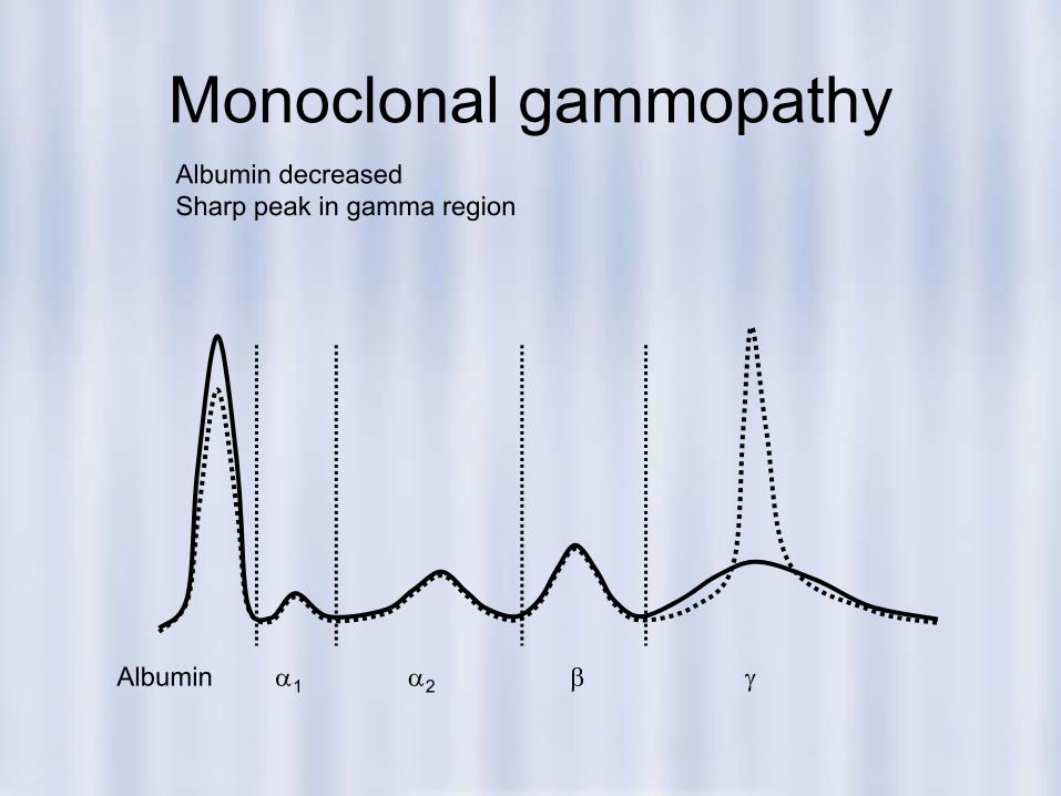
Monoclonal gammopathy
Albumin α1 α2 β γ
Albumin decreasedSharp peak in gamma region
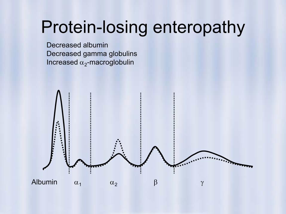
Protein-losing enteropathy
Albumin α1 α2 β γ
Decreased albuminDecreased gamma globulinsIncreased α2-macroglobulin