Embed Size (px)
Citation preview

Diagnostic Medical
SonographyCAAHEP ANNUAL MEETING 2016
Sheryl E. Goss, MS, RT(R)(S), RDMS, RDCS, RVT, FSDMS
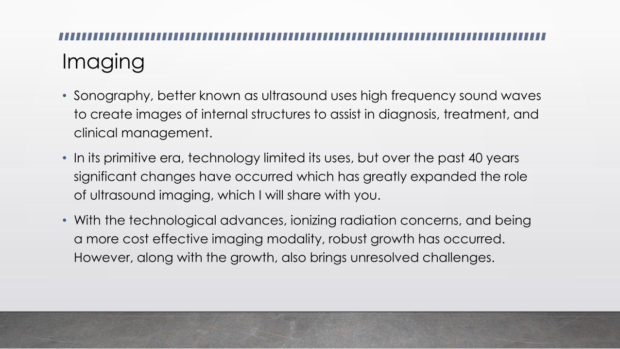
Imaging
• Sonography, better known as ultrasound uses high frequency sound waves
to create images of internal structures to assist in diagnosis, treatment, and
clinical management.
• In its primitive era, technology limited its uses, but over the past 40 years
significant changes have occurred which has greatly expanded the role
of ultrasound imaging, which I will share with you.
• With the technological advances, ionizing radiation concerns, and being
a more cost effective imaging modality, robust growth has occurred.
However, along with the growth, also brings unresolved challenges.

Sonography
Diagnostic
(traditional users)
Abdominal/superficial structures
OB/GYN
Cardiac
Vascular
Invasive/therapeutic guidance
Needle biopsies
Fluid drainage
Musculoskeletal
Point of Care
(non-traditional users)
Emergency room
Midwives
Labor and delivery
Nerve anesthesia

Where are sonographers employed?
SDMS Sonographer Survey 2015 SDMS Educator’s Survey 2015
EducationClinical

Where is the significant growth over the past decade?
Abdominal/superficial structures
OB/GYN
Cardiac
Vascular
Diagnostic

Diagnostic
• Superficial Structures
• Breast Sonography
• Over the past 10-15 years, sonography plays a primary and collaborative role in
breast imaging
• Majority of breast biopsies are performed using ultrasound guidance.
• Allows for live visualization of lesion and needle.
• Breast Density Notification Law
• Increase in volume of patients who have fibrocystic changes as
mammography can be limited in visualizing lesions within the dense tissue

States with Breast Density Notification Laws (July 2015)
http://www.diagnosticimaging.com/breast-imaging/breast-density-notification-laws-state-interactive-
map

Diagnostic
• Obstetrical sonography – once performed predominately in a
radiology department, then moved to an OB/GYN office and
current is high volume in Maternal Fetal Medicine
(Perinatology) centers.
• Technology has greatly enhanced the anatomical structures seen;
therefore, there is an expectation that most all congenital
anomalies will be detected in utero
• Growth of fetal echocardiography (visualization of fetal heart)
• Performance of fetal echocardiography is often shared between OB
sonographers and Pediatric Echocardiographers.

Musculoskeletal
• The role of sonography in musculoskeletal is rapidly growing.
• The advantage over MRI is sonography is dynamic and allows for freedom
of movement during imaging.
• Performance occurs in a diversity of specialties to include, but not limited
to:
• Radiology
• Orthopedics
• Sports Medicine
• Chiropractic

Collaboration
• We are fortunate to have a large network of organizations to collaborate for
the best interests of the profession. Over the past two years, two major
documents underwent review and revision to reflect current practice.
• Scope of Practice
• Sixteen organizations participated in the process. Seventeen organizations have either endorsed or supported.
• National Education Curriculum
• First created in 2008 with copyright signed off to the JRCDMS, over the past two years, the document has underwent review and revision. The JRCDMS has sent the final document out to fifteen sponsoring organizations for endorsement or support.
• Both documents are valuable to the clinical sonographer, educator, and
student.

Unresolved Challenges
• Lack of entry level education requirements
• Lack of national certification requirements
• Certification requirements
• Employer driven
• Lab accreditation
• Variation amongst the organizations that offer lab accreditation
• State licensure
• Oregon, North Dakota, New Mexico
• Clinical education affiliates

Thank you to all the
sponsors through
their commissioners
and board members
that make both
CoA’s successful.
We can’t do it
without you!
Programmatic AccreditationCAAHEP
JRCDMS• General (AB, Superficial
structures, OB/GYN)
• Vascular
• Adult Echocardiography
• Pediatric
Echocardiography
JRCCVT• Adult Echocardiography
• Pediatric Echocardiography
• Vascular Technology
• Invasive Cardiovascular Technology
• Cardiac Electrophysiology
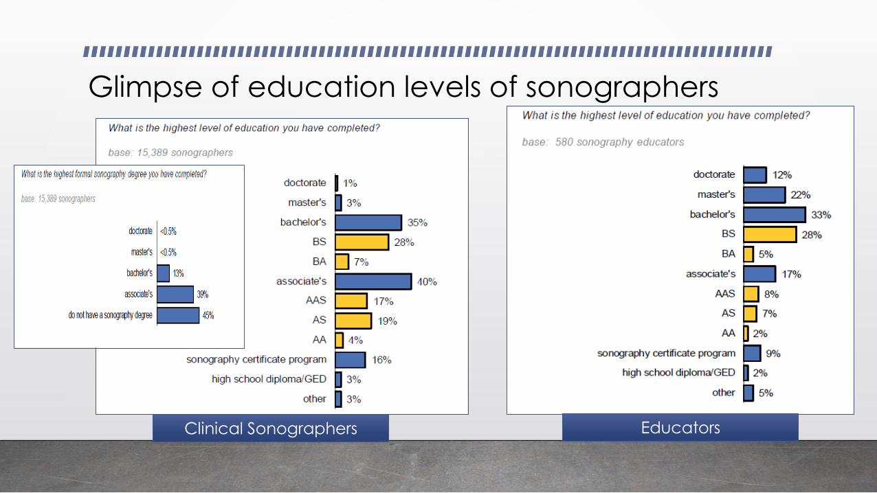
Glimpse of education levels of sonographers
EducatorsClinical Sonographers

Education and National Certification
• Evolution of education from on-the-job training to formal programs
• For entry into the profession, the most common pathway is a formal education
program in at least one concentration.
• On-the-job training continues to occur in different specialties beyond the entry
level concentration(s). Sonographers are eligible to sit for national certification
exams in other specialties without additional formal education.
• Three organizations offer credentials
• Overlap in some specialties as noted in the same colors on the following slide.
• Various educational pathways are accepted to apply for exams.

ARDMS CCI ARRT
RDMSAbdominal
OB/GYN
Breast
Pediatric Sonography
Fetal Echocardiography
RCS
Adult Cardiac
ARRT (S)
Encompasses
abdominal and
OB/GYN
RVT
Vascular Technology
RDCS
Adult Cardiac
Pediatric Cardiac
Fetal
Echocardiography
MSKS
Musculoskeletal
ARRT(V)
Vascular
ARRT (BR)
Breast
RVS
Vascular
RCCS
Congenital Cardiac
RPhS
Phlebology
ACS
Advanced Cardiac
Sonographer
Continuing Education and Recertification
• Continuing education is a vital part of the certification process, but more
important for patient care.
• Coming in 2019:
• ARDMS and ARRT are implementing recertification/continuing qualification
processes.
• Final details have not been provided, but anticipate it to be an assessment to
identify areas of strength and areas that need further study.
• Be nice to your sonographer in 2018 and beyond while they prepare for
multiple recertification exams in our system of testing per specialty.

Hmmm… I wonder
what ultrasound
will be like when I
graduate from
high school.