Embed Size (px)
Citation preview
Vet Education Pty Ltd: Proudly Supported By
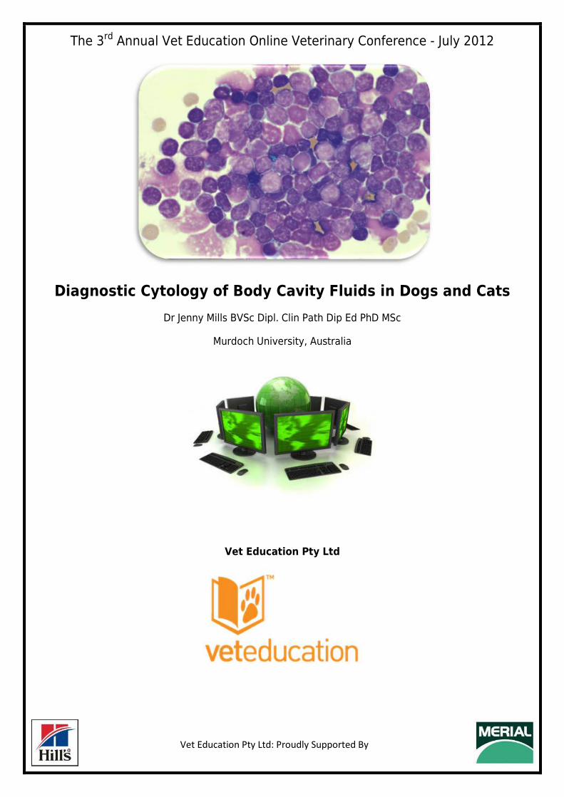
The 3rd Annual Vet Education Online Veterinary Conference - July 2012
Diagnostic Cytology of Body Cavity Fluids in Dogs and Cats
Dr Jenny Mills BVSc Dipl. Clin Path Dip Ed PhD MSc
Murdoch University, Australia
Vet Education Pty Ltd
1
A REVIEW OF
DIAGNOSTIC CYTOLOGY & BODY CAVITY FLUIDS
Webinar Notes
Presenter
Dr Jennifer N Mills, BVSc, Dip.Clin.Path, Dip Ed., MSc, PhD
School of Veterinary and Biomedical Sciences
Murdoch University
J N Mills, Murdoch University, Murdoch, WA 6150 July 2012
This publication is copyright. Except as permitted by the Copyright Act no part of it may in any form or by an
electronic, mechanical, photocopying, recording or any other means be reproduced, stored in a retrieval system or be
broadcast or transmitted without the prior written permission of the publisher.
2
DIAGNOSTIC CYTOLOGY
INDEX
Pages
OBJECTIVES 3
REFERENCES 3
INTRODUCTION 4
SAMPLE COLLECTION & PREPARATION 4
PRINCIPLES OF CYTOLOGICAL INTERPRETATION 5
LYMPH NODE CYTOLOGY 10
BODY CAVITY FLUIDS 11
SYNOVIAL FLUID 16
BODY CAVITY FLUID CASES 17
3
DIAGNOSTIC CYTOLOGY
Objectives
The notes and presentat ion is a refresher, designed for you to:
(1) collect and prepare cytological specimens for analysis
(2) recognise the features of cytopathological change, specif ically
inf lammation and neoplasia, in samples collected from body
cavit ies and solid t issue.
(3) recognise cellular features of malignancy.
(4) classify a neoplasm cytologically as a round cell tumour, or of
mesenchymal (sarcoma) or epithelial (carcinoma) origin.
(5) classify a f luid effusion, and determine its possible pathogenesis
based primarily on cellular content.
References
1. Cow ell A.H., Tyler R.D., Meinkoth J.H. and DeNicola D.B. (2008).
Diagnost ic Cytology and Hematology of the Dog and Cat . 3 rd Edn. St Louis, Mo:
Mosby Elsevier.
2. Cow ell A.H. and Tyler R.D. (2002).
Cytology and Hematology of the Horse. 2nd Edn. St Louis, Mo, London: Mosby.
3. Raskin R and Meyer D.J. (2009). Canine and Feline Cytology: A Color At las and
Interpretat ion Guide. 2nd Edn. St Louis, Mo: Saunders Elsevier.
.
4. Alleman A.R. (2003).Abdoiminal, thoracic and pericardial effusions. Vet Clin Nth
Amer. Small Animal Pract ice. Vol 33: 89-118.
5. Baker R. & Lumsden J.H.(2000). Colour At las of Cytology of the Dog and Cat. St
Louis, Mo: Mosby
4
DIAGNOSTIC CYTOLOGY
Introduction
Cytopathology involves the examinat ion of individual cell populat ions collected from a pat ient , in
order to determine the underlying pathophysiological process.
The purpose of cytology is to provide a diagnost ic method w hich w ill allow the clinician to
determine if a lesion (solid t issue mass, body cavity f luid, or w ash) is primarily inflammatory or
neoplastic; and provide further information on the possible cause (aet iological diagnosis) and
chronicity of the lesion, or the type of neoplasm and w hether it is benign or malignant.
Hyperplast ic, non-neoplast ic and cyst ic lesions are possible. Occasionally the cytological
interpretat ion may be inconclusive, in w hich case surgical biopsy may be required.
Sample Collection and Preparation
a) BODY CAVITY FLUID - Fluids from peritoneal, pleural cavit ies, synovial, cerebrospinal and
pericardial f luids and aqueous humour are preferably collected into EDTA anticoagulant to
preserve cell morphology. Sediment smears are made after gent le centrifugat ion (1500
rpm, 10 min); direct smears can be made if cell count exceeds 10 x10 9/L. Body cavity
f luids requiring biochemical analysis or bacterial culture are collected into a plain serum
tube as EDTA inhibits bacterial grow th.
b) URINE - Fresh urine sediments can be examined for the presence of neoplast ic or
inf lammatory cells and infect ious agents such as bacteria, fungi or yeasts.
c) WASH - Using sterile saline, cells can be encouraged to exfoliate from surfaces such as
bronchi, prostat ic gland, nasal cavity and uterus (mare). Sediment smears are made. If
sample contains macroscopic f loccules, squash preparat ions of these are desirable.
Collect into EDTA anticoagulant .
d) BRUSH/SCRAPE - Flat surfaces, in situ, are amenable to these procedures e.g. colon,
vaginal w all, conjunct iva and nasal cavity. Cells are direct ly transferred onto slides.
Cytobrushes w ith nylon brist les are preferable to cotton sw abs for cytological brush
collect ion, as cells cling to cotton f ibres and distort w hen transferred to slides. Cotton
sw abs are suitable for microbial culture.
e) IMPRESSION/IMPRINT SMEARS - This technique is best for cut surfaces from excised
lesions. It is not part icularly useful for erosive surfaces as only superf icial inf lammatory
cells may be obtained. The freshly cut surface is blotted dry onto an absorbant tow el to
absorb excess f luid and blood, then gently rolled onto a slide. Suct ion artefacts are
avoided by rolling the t issue.
f ) FINE-NEEDLE ASPIRATION BIOPSY (FNAB) - Used for 3-dimensional t issue masses, w hich
can be internal or external and palpable. For non-palpable masses, ultrasound guided
collect ion helps to ensure the needle is sampling the correct site. Use a 22 gauge needle
and 10 ml syringe and pass the needle in at least 3 direct ions in the mass before releasing
vacuum. Collect cells into the needle only. Remove the needle and place air into the
syringe before expelling the sample; smear to make monolayers of the sample. Stop the
procedure if blood enters the syringe, as clot formation ruins the sample. Speed is
required w hen transferring cells to slides. Syringe is often omitted for splenic aspirates to
avoid haemorrhage. For the cytological diagnosis to be reliable, a basic premis of cytology
is that the sample collected must be totally representive of the lesion, and several smears
should be submitted from each lesion.
g) DRILL OR CORE BIOPSIES - Firm connect ive t issue and muscle do not readily yield cells
on aspirat ion, and in these cases an air drill or larger needle (Vim-Silverman) may be
required. Cell Block Preparat ions may also be made from these samples or from FNAB.
5
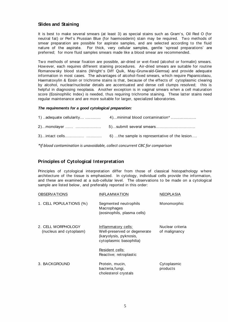
Slides and Staining
It is best to make several smears (at least 3) as special stains such as Gram' s, Oil Red O (for
neutral fat) or Perl' s Prussian Blue (for haemosiderin) stain may be required. Tw o methods of
smear preparat ion are possible for aspirate samples, and are selected according to the f luid
nature of the aspirate. For thick, very cellular samples, gent le ‘spread preparat ions’ are
preferred; for more f luid samples smears made like a blood smear are recommended.
Tw o methods of smear f ixat ion are possible, air-dried or w et-f ixed (alcohol or formalin) smears.
How ever, each requires dif ferent staining procedures. Air-dried smears are suitable for rout ine
Romanow sky blood stains (Wright ' s Dif f Quik, May-Grunw ald-Giemsa) and provide adequate
information in most cases. The advantages of alcohol-f ixed smears, w hich require Papanicolaou,
Haematoxylin & Eosin or trichrome stains is that, because of the effects of cytoplasmic clearing
by alcohol, nuclear/nucleolar details are accentuated and dense cell clumps resolved; this is
helpful in diagnosing neoplasia. Another except ion is in vaginal smears w hen a cell maturat ion
score (Eosinophilic Index) is needed, thus requiring trichrome staining. These latter stains need
regular maintenance and are more suitable for larger, specialized laboratories.
The requirements for a good cytological preparation:
1) ..adequate cellularity… ….…..… 4)…minimal blood contaminat ion* ..……..……….
2)…monolayer …… ……………..… 5)…submit several smears………………………....
3)…intact cells…………… ...……… 6) …the sample is representat ive of the lesion.…
*If blood contamination is unavoidable, collect concurrent CBC for comparison
Principles of Cytological Interpretation
Principles of cytological interpretat ion dif fer from those of classical histopathology w here
architecture of the t issue is emphasized. In cytology, individual cells provide the information,
and these are examined at a sub-cellular level. The observat ions to be made on a cytological
sample are listed below , and preferably reported in this order:
OBSERVATIONS INFLAMMATION NEOPLASIA
1. CELL POPULATIONS (%) Segmented neutrophils Monomorphic
Macrophages
(eosinophils, plasma cells)
2. CELL MORPHOLOGY Inf lammatory cells: Nuclear criteria
(nucleus and cytoplasm) Well-preserved or degenerate of malignancy
(karyolysis, pyknosis,
cytoplasmic basophilia)
Resident cells:
React ive; retroplast ic
3. BACKGROUND Protein, mucin, Cytoplasmic
bacteria,fungi, products
cholesterol crystals
6
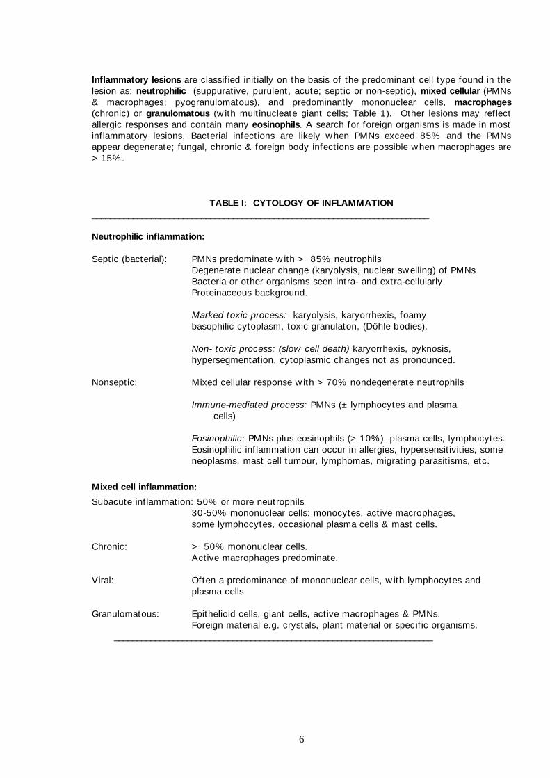
Inflammatory lesions are classif ied init ially on the basis of the predominant cell type found in the
lesion as: neutrophilic (suppurat ive, purulent, acute; sept ic or non-septic), mixed cellular (PMNs
& macrophages; pyogranulomatous), and predominantly mononuclear cells, macrophages
(chronic) or granulomatous (w ith mult inucleate giant cells; Table 1). Other lesions may ref lect
allergic responses and contain many eosinophils. A search for foreign organisms is made in most
inf lammatory lesions. Bacterial infect ions are likely w hen PMNs exceed 85% and the PMNs
appear degenerate; fungal, chronic & foreign body infect ions are possible w hen macrophages are
> 15%.
TABLE I: CYTOLOGY OF INFLAMMATION
__________________________________________________________________________
Neutrophilic inflammation:
Septic (bacterial): PMNs predominate w ith > 85% neutrophils
Degenerate nuclear change (karyolysis, nuclear sw elling) of PMNs
Bacteria or other organisms seen intra- and extra-cellularly.
Proteinaceous background.
Marked toxic process: karyolysis, karyorrhexis, foamy
basophilic cytoplasm, toxic granulaton, (Döhle bodies).
Non- toxic process: (slow cell death) karyorrhexis, pyknosis,
hypersegmentat ion, cytoplasmic changes not as pronounced.
Nonseptic: Mixed cellular response w ith > 70% nondegenerate neutrophils
Immune-mediated process: PMNs (± lymphocytes and plasma
cells)
Eosinophilic: PMNs plus eosinophils (> 10%), plasma cells, lymphocytes.
Eosinophilic inf lammation can occur in allergies, hypersensit ivit ies, some
neoplasms, mast cell tumour, lymphomas, migrat ing parasit isms, etc.
Mixed cell inflammation:
Subacute inf lammation: 50% or more neutrophils
30-50% mononuclear cells: monocytes, act ive macrophages,
some lymphocytes, occasional plasma cells & mast cells.
Chronic: > 50% mononuclear cells.
Act ive macrophages predominate.
Viral: Often a predominance of mononuclear cells, w ith lymphocytes and
plasma cells
Granulomatous: Epithelioid cells, giant cells, act ive macrophages & PMNs.
Foreign material e.g. crystals, plant material or specif ic organisms.
______________________________________________________________________
7
M orphology of Normal and Neoplastic Cells
In observing sub-cellular detail, the basic principle of cytological interpretat ion is that:
The state of grow th act ivit ies recognisable morphologically (by nuclear features) are:
EUPLASIA - A cell w ith normal grow th act ivity has regular, smooth cytoplasmic and nuclear
margins and an even, bland chromatin pattern.
PROPLASIA - A cell w ith increased grow th act ivity, in response to hormonal st imulat ion, injury
or repair has a slight ly higher nuclear/cytoplasmic rat io, an apparent nucleolus and a denser
chromatin pattern.
RETROPLASIA - A cell w ith decreased grow th act ivity may occur in injury, aging or starvat ion.
The nucleus w ill by pyknotic and the cytoplasm more basophilic than normal.
DYSKARYOSIS - A cell w ith an abnormal nucleus is called ` dyskaryot ic' . The change may be
a pre-neoplast ic change or result f rom a toxic insult . These cells present the greatest dif f iculty
in interpretat ion to dist inguish them definit ively from neoplast ic cells. Histological examinat ion
may be required.
NEOPLASIA - A cell w hich has undergone neoplast ic change, w ill show dist inct nuclear and
nucleolar irregularit ies. There is marked variat ion in size, shape and number of nucleoli, w ith
dense chromatin ref lect ing increased ploidy of the cell, and usually a high nuclear to cytoplasmic
rat io. (see Table 2). These features vary w ith the type and degree of malignancy of the
neoplasm and w hether it is of epithelial or mesenchymal origin.
Nuclear features and chromatin patterns change through the cell cycle; nuclei have a f ine sieve-
like (cribriform) chromatin pattern in rest ing (G zero; f igure A), a st ippled chromatin pat tern w ith
a nucleous during rapid grow th (proplasia; f igure B), and coarse clumped chromatin pattern in
the pre-mitot ic phase (f igure C). Chromatin may darken in malignant cells due to polyploidy ,
nucleoli become more prominent and variable, and many malignant cells are found in G zero
phase.
In diagnosing neoplasia a number of cytologically characterist ics of malignant cells must apply
and generally a monomorphism of cell type is present. Mitot ic f igures alone are not necessarily
useful unless mitoses themselves are abnormal or are in large numbers. The frequency of
mitoses is often reported.
Cytoplasmic morphological features init ially help to def ine the lineage of cells in a neoplasm.
How ever poorly dif ferent iated blast ic cells are more challenging, and immunophenotyping using
immunocytochemistry is often recommended to detect ant igenic markers w hich may be in the
cytosol or cytoplasmic membrane.
Molecular based diagnost ic tests are increasingly available to characterise neoplast ic cell
populat ions, by ident ifying mutat ions of oncogenes (e.g., c-kit gene), chromosomal abnormalit ies
and clonality in lymphoma and leukemia cells to dist inguish react ive from neoplast ic
lymphocytes. Tests such as PCR for ant igen receptor rearrangements (PARR) ident ify
homogeneity in gene rearrangaments of neoplast ic lymphoid cells. How ever despite variat ions in
survival t imes found w ith various subsets of lymphoma types, histology & f low cytomet ry for
immunophenotyping is st ill recommended to help establish prognosis in lymphoma pat ients.
a) the nucleus indicates the state of growth activity of the cell (e.g. euplast ic,
proplast ic, retroplast ic and neoplast ic);
b) the cytoplasm indicates the lineage of the cell and its functional differentiation
(e.g. chondrocyte, mast cell, f ibrocyte).
8
TABLE 2: CYTOLOGICAL FEATURES OF MALIGNANCY
General Criteria
Macrocytosis - extremely large cells up to 2 t imes larger in diameter than
normal size. (Size should be compared to the erythrocyte).
Hypercellularity - increased cell exfoliat ion due to decreased cell adherence (this
varies w ith the type of tumour)
Pleomorphism and
anisocytosis
- variable size and shape of the cells of the same family (except
lymphoid t issue)
Nuclear Criteria
Nucleus - anisokaryosis (variat ion in nuclear size)
- high nuclear/cytoplasmic rat io
- nuclear deformation by other nuclei
- macrokaryosis (> 10 microns diameter)
- mult inucleat ion (part icularly if variable in size)
- nuclear moulding (deformation of nuclei by other nuclei)
Nuclear membrane - extreme variat ions in thickness
- sharp angularity and irregularit ies in out line (esp. carcinomas)
- close parallelism of nuclear and cytoplasmic margins over
extended distances
- sharply def ined inner and outer surfaces
Nucleoli - marked variat ions in size, shape and number ( 5)
- irregular shapes w ith sharp angularity
- Macronucleoli
Chromatin - dense, but chromacentres variable, clear parachromatin areas
Mitoses - abnormal mitot ic f igures. (mitoses are rare in normal t issue)
- binucleate cells containing dif ferent sized nuclei
The neoplasm should be classif ied into one of 3 groups on the basis of lineage, as epithelial,
mesenchymal (spindle cell) tumours, and round cell tumours.
Epithelial neoplasms are characterised by cell clusters or sheets w ith obvious close
cytoplasmic attachment (desmosomes), abundant cytoplasm and 1-3 prominent nucleoli.
They include squamous cell carcinoma, adenocarcinoma, etc. Squamous cells are expected
to have a central nucleus and may have a moderate amount of clear cytoplasm.
Adenocarcinomas are characterized by having eccentric nuclei and may appear in papillary,
acinar or tubular structures depending on their t issue of origin; they may be secretory and
have cytoplasmic cyst ic inclusions.
Mesenchymal neoplasms contain cells w ith fusiform (spindle-shaped) to polyhedral cytoplasm
and round to oval nuclei. The cytoplasmic margins are often indist inct. They include
f ibrosarcoma, haemangiosarcoma, osteosarcoma and chrondrosarcoma, haemaniopericytoma,
etc. Histopatology is often required to accurately classify mesenchymal neoplasms.
Round cell tumours contain individual, discrete round cells and include lymphosarcoma,
hist iocytoma, mastocytoma, plasmacytoma, melanoma (can appear round or spindle) and
transmissible veneral tumour (TVT).
TVT can be further subtyped cytologically as lymphoid (w ith > 60% round cells having
central nuclei), plasmacytoid (w ith > 60% ovoid cells w ith eccentric nuclei) and mixed type
9
containing both lymphocytoid and plasmacytoid cells w ith neither exceeding 59% of the total
cells (Florez et al 2012, VCP 41(1) 4.
Another classif icat ion includes a fourth group, of cells w hich readily lose their cytoplasm on
smearing, leaving bare nuclei and making their lineage hard to determine. These need to be
dist inguished from cells of poorly made smears w itrh damaged cells. Bare nuclei neoplasms are
commonly endocrine or neuroendocrine tumours. They include tumours of thyroid gland, islet
cells, paragangliomas, chemodectomas, carcinoids, etc.
Lipomas are one of the most common benign neoplasms diagnosed and are often poorly cellular
and contain oily lipid and very large vacuous adipocytes w ith a very small nucleus and a very
low nuclear to cytoplasmic rat io. Contrasted to this, liposarcomas are quite cellular, may often
contain cells w hich are very poorly dif ferent iated and contain very lit t le free fat either w ithin the
cell or outside the cell.
Non-Inflammatory and Non-Neoplastic Lesions
This category includes cysts, transudates and hyperplast ic lesions. Samples from these lesions
are usually a simple accumulat ion of resident ial normal cells, e.g. epidermal cysts contain
primarily anucleate epithelial squames and cholesterol crystals, although they can occasionally
be accompanied by an inf lammatory inf ilt rate; salivary cysts contain mucinous aggregates, some
phagocytes and salivary epithelial cell clusters. The diagnosis of benign hyperplasia (e.g. in a
lymph node) is a dif f icult one cytologically as the cells are not abnormal and show only euplasia
or proplasia; they may show increased proport ions of intermediate sized lymphoid cells. It is a
diagnosis w hich has to be made w ith the know ledge of t issue/organ enlargement and w ith the
assurity that inf ilt rat ing cells such as in a metastat ic neoplasm have not been missed in
sampling.
Limitations
Cytopathology is an adjunct to clinical diagnosis. It can give a rapid and reliable result if handled
w ith skill, but it does not replace histopathology. In some instances, an answ er of inf lammation vs
neoplasia is all that is possible; but in other cases a more def init ive diagnosis can be given
depending on the experience of the person reading the slides. In commencing this study, it is
desirable to examine parallel cytology and histology preparat ions. Samples may also provide
misleading information if not properly collected and handled. A posit ive cytological diagnosis of
malignancy is meaningful, but a negative f inding does not necesarily rule out malignancy.
Addit ional or alternat ive tests may be required. How ever, the init ial expense of cytological
examinat ion is minimal and the requirements for sample collect ion are minimal.
10
LYMPH NODE CYTOLOGY
Cytological Categories
Lymphadenit is (non-septic & sept ic) - contains increased inf lammatory cells
Neutrophilic lymphadenit is (> 5% neutrophils)
Eosinophilic lymphadenit is (> 3% eosinophils)
Pyogranulomatous lymphadenit is (neutrophils & macrophages)
Immunoreact ive hyperplasia (> plasma cells; > 15% medium and/or large lymphoid cells)
Lymphoma - classify type (> 50% medium or large lymphoblast ic cells; there are exceptions)
Metastat ic neoplasia
(Inconclusive) - i.e. cellular changes not def init ive for lymphoma
Causes of Error in Diagnosis
Inadequate sample - insuff icient cells; blood contaminat ion
Sample not representat ive of ent ire lesion - e.g. early metastat ic neoplasm
Normal lymph nodes are dif f icult to aspirate but contain a heterogeneous populat ion of lymphoid
cells of various stages of maturat ion, w ith a predominance of w ell-dif ferent iated small and
medium sized lymphocytes (approx 90%). There are some prolymphocytes containing a single
nucleolus and a scattering of large lymphoblasts containing several nucleoli; medium and large
lymphoid cells may const itute < 5 to 10% of the cell populat ion. There may be an occasional
plasma cell and mast cell, w hile neutrophils are very rare in normal lymph nodes. The
background should be clear of cell debris and may be light ly proteinaceous.
Lymphadenitis is deemed septic if organisms can be observed on smears. Fungal hyphae may
only show by negative staining on Romanow sky stains, and PAS stain may be required.
Bacteria, fungal spores, Cryptococcus and Aspergillus sp. may be found. Aspirates may be
direct ly cultured onto agar plates if sepsis is suspected. Inf lammatory cells are increased
(neutrophils and macrophages), and some plasma cells may be noted; medium sized
lymphocytes may be mildly increased. Neutrophils may show degenerat ive karyolyt ic change if
foreign organisms are present in the node. Purulent lymphadenit is (or absessed node) may be
diagnosed if neutrophils are excessive. Chronic lymphadenit is may be diagnosed if macrophages
are plent iful, and granulomatous lymphadenit is contains inf lammatory giant cells.
Eosinophilic Lymphadenitis involves an inf lammatory react ion in the node w ith a higher
proport ion of eosinophils w ith occasional mast cells, along w ith a mixed lymphoid populat ion.
This may be associated w ith hypersensit ivity react ions, prurit ic skin disease such as f lea allergy
dermatit is, chronic seborrhoeic dermatit is, generalized allergies and some rare fungal infect ions.
The background may contain plent iful f ine part iculate debris in dermatopathic lymphadenopathy.
Immunoreactive Lymphoid Hyperplasia consists of predominantly small mature lymphocytes w ith
increased medium lymphocytes and plasma cells indicat ing ant igenic response. The node may
also be odematous.
Lymphoma. In lymphoma an homogenous populat ion of lymphoid cells generally replaces the
normal mixed populat ion, and immature (blast ic) cells predominate w ith > 50% medium or large
lymphoid cells. In dogs, the lymphoblast ic form is commonly seen. Full categorisat ion of the
type of lymphoma is best achieved using a combinat ion of morphology, immunocytochemistry
and ant igenic markers using f low cytometry, but this is not readily available in Australia at
present. Lymphoblasts and prolymphocytes are often fragile, and as cells rupture, cytoplasmic
fragments form as t iny spherical, blue-staining bodies, " lymphoglandular" bodies. They are
frequently found in the background of lymphoma smears. Macrophages w ith lymphophagia may
be seen, w ith several nuclear remnants in the cytoplasm.
Metastatic Neoplasia. Basically this show s a mixed lymphoid populat ion w ith scattered foreign
malignant cells and some inf lammatory cells. Metastat ic squamous cell carcinoma and
adenocarcinoma are the common types seen; metastat ic melanoma and mast cell tumours may
also be diagnosed. Metastat ic sarcomas, haemangiosarcoma and poorly dif ferent iated leukemias
may be more dif f icult to diagnose.
11
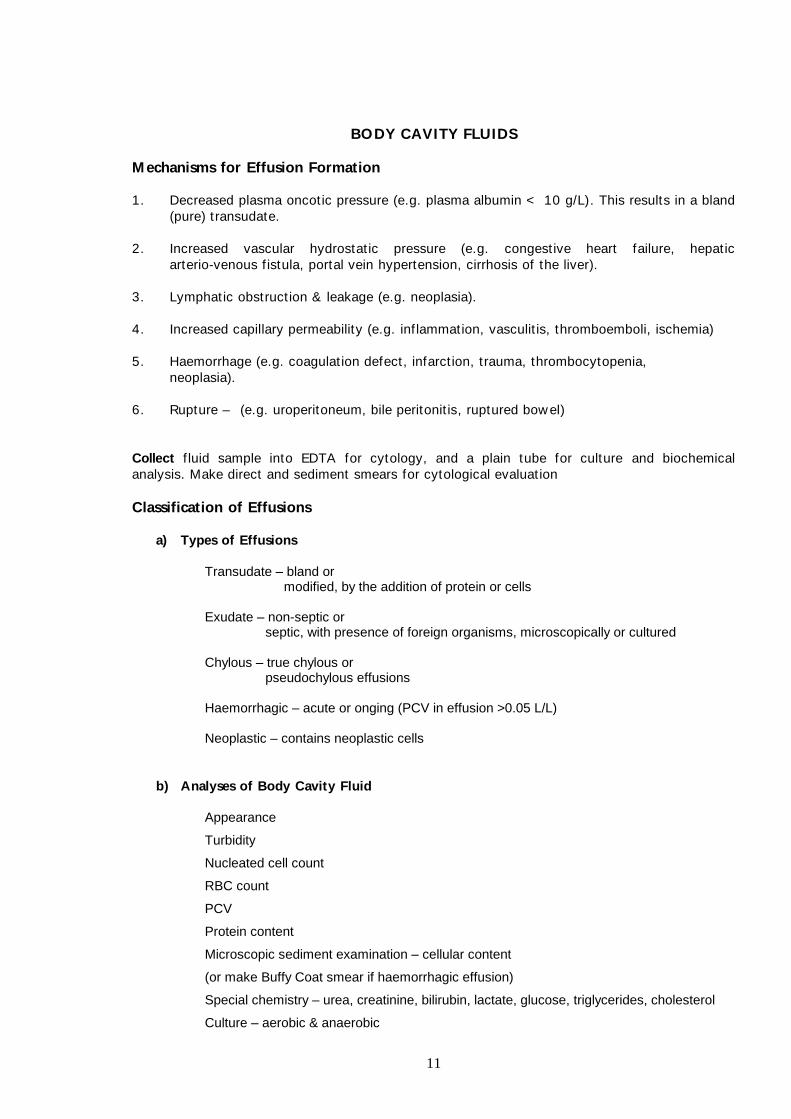
BODY CAVITY FLUIDS
M echanisms for Effusion Formation
1. Decreased plasma oncotic pressure (e.g. plasma albumin < 10 g/L). This results in a bland
(pure) transudate.
2. Increased vascular hydrostat ic pressure (e.g. congest ive heart failure, hepatic
arterio-venous f istula, portal vein hypertension, cirrhosis of the liver).
3. Lymphatic obstruct ion & leakage (e.g. neoplasia).
4. Increased capillary permeability (e.g. inf lammation, vasculit is, thromboemboli, ischemia)
5. Haemorrhage (e.g. coagulat ion defect, infarct ion, trauma, thrombocytopenia,
neoplasia).
6. Rupture – (e.g. uroperitoneum, bile peritonit is, ruptured bow el)
Collect f luid sample into EDTA for cytology, and a plain tube for culture and biochemical
analysis. Make direct and sediment smears for cytological evaluat ion
Classification of Effusions
a) Types of Effusions
Transudate – bland or modified, by the addition of protein or cells Exudate – non-septic or septic, with presence of foreign organisms, microscopically or cultured Chylous – true chylous or pseudochylous effusions Haemorrhagic – acute or onging (PCV in effusion >0.05 L/L) Neoplastic – contains neoplastic cells
b) Analyses of Body Cavity Fluid
Appearance
Turbidity
Nucleated cell count
RBC count
PCV
Protein content
Microscopic sediment examination – cellular content
(or make Buffy Coat smear if haemorrhagic effusion)
Special chemistry – urea, creatinine, bilirubin, lactate, glucose, triglycerides, cholesterol
Culture – aerobic & anaerobic
12
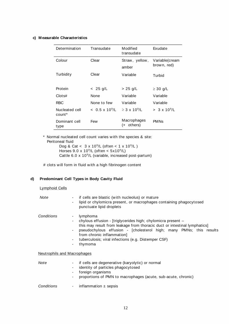
c) M easurable Characteristics
Determinat ion Transudate Modif ied
transudate
Exudate
Colour
Turbidity
Clear
Clear
Straw , yellow ,
amber kl
Variable
Variable(cream
brow n, red)
Turbid
Protein < 25 g/L > 25 g/L 30 g/L
Clots# None Variable Variable
RBC None to few Variable Variable
Nucleated cell
count*
< 0.5 x 10⁹/L ≥ 3 x 10⁹/L > 3 x 10⁹/L
Dominant cell
type
Few Macrophages
(+ others) PMNs
* Normal nucleated cell count varies w ith the species & site:
Peritoneal f luid
Dog & Cat < 3 x 10⁹/L (often < 1 x 10⁹/L )
Horses 9.0 x 10⁹/L (often < 5x10⁹/L)
Catt le 6.0 x 10⁹/L (variable, increased post -partum)
# clots w ill form in f luid w ith a high f ibrinogen content
d) Predominant Cell Types in Body Cavity Fluid
Lymphoid Cells
Note - if cells are blast ic (w ith nucleolus) or mature
- lipid or chylomicra present , or macrophages containing phagocytosed
punctuate lipid droplets
Condit ions - lymphoma
- chylous effusion - [t riglycerides high; chylomicra present –
this may result f rom leakage from thoracic duct or intest inal lymphatics]
- pseudochylous effusion - [cholesterol high; many PMNs; this results
f rom chronic inf lammation]
- tuberculosis; viral infect ions (e.g. Distemper CSF)
- thymoma
Neutrophils and Macrophages
Note - if cells are degenerat ive (karyolyt ic) or normal
- ident ity of part icles phagocytosed
- foreign organisms
- proport ions of PMN to macrophages (acute, sub-acute, chronic)
Condit ions - inf lammation ± sepsis
13
Eosinophils
Condit ions - parasit ic larval migrat ion
- hypersensit ivit ies
- t rauma
A cytological diagnosis of eosinophilic inf lammation may be made if eosinophils are plent iful
Mesothelial Cells
Note - if cells are react ive or neoplast ic
- cells clustered
Condit ions - react ive hyperplasia
- haemopericardium
- low numbers in modif ied transudates
- mesothelioma
RBCs
Note: - erythrophagia/haemosiderophagia
- PCV of f luid (compare to CBC)
- ± platelets
Condit ions - pathological haemorrhage and iatrogenic blood contaminat ion
(i) Iatrogenic blood contaminat ion (at t ime of collect ion)- resembles
peripheral blood, w ith many platelets. PCV of f luid likely to be
< 0.05 L/L.
(ii) Pathological haemorrhage w ithin few hours - intact RBCs w ith fresh
leukocytes and lysing platelets. PCV of f luid > 0.05 L/L in
pathological haemorrhagic effusion.
(iii) Pathological haemorrhage w ithin days - erythrophagia and early
evidence of hemosiderin w ithin macrophages. Neutrophils
hypersegmented, some cytophagia. No platelets if haemorrhage
has stopped.
(iv) Note that the mast cells normally found in body cavity f luids
contain heparin w hich prevents clott ing in vivo of haemorhagic
ef fusions in a body cavity.
Foreign Cells e.g. neoplast ic cells; or intest inal epithelial cells if aspirate enters lumen of bow el
Neoplasms w hich exfoliate w ell include carcinomas, round cell tumours and mesotheliomas. Poorly
exfoliat ive neoplasms are sarcomas & germ cell tumours. Most effusions (93%) w ith a pH greater than
7.0 (using urost icks) or 7.3 (using pH meter) w ere found to be non-inf lammatory or neoplast ic, w hereas
those w ith a low er pH w ere benign or non-neoplast ic.
(i) Squamous cell carcinoma - pleomorphic squamous epithelial cells, w ith act ive inf lammatory
response (e.g. gastric carcinoma in horse).
(ii) Adenocarcinoma - cell aggregates of glandular or tubular epithelial cells, occasionally w ith
secretory product.
(iii) Lymphoma – blast ic or large lymphoid cells in abundance; may f ind lymphophagia
(phagocytosis of lymphocytes).
14
(iv) Mesothelioma – usually extremely cellular, best seen on direct smears, w ith many large
clumps of aberrant mesothelial cells show ing criteria of malignancy. These must be
dist inguished from very react ive and dysplast ic mesothelial cells seen in chronic inf lammation.
Examples of Specific Processes in Body Cavity Effusions
1. Bland Transudate
- hypoalbuminaemia (< 10 g/L)
- pre-sinusoidal obstruct ion to portal blood supply in conjunct ion w ith reduced funct ional
hepatic mass or chronic hepatic disease; excessive IV f luid administrat ion
2. Modif ied Transudate
(commonly associated w ith underlying changes in vascular hydrostat ic pressure or lymphatic
drainage, w ith leakage out of normal or non-inf lammed vessels)
- intrahepatic portal hypertension (some hepatic lymph)
- ascites of congest ive heart failure (protein 25-50 g/L)
- lung lobe torsion, diaphragmatic hernia
- neoplasm e.g. lymphoma, adenocarcinoma
3. Chylous and Pseudochylous Effusions
(opaque milky f luids)
- Chylothorax/chylous effusion (small mature lymphocytes predominate; chylomicra,
triglycerides in f luid are 2 to 20 x that in serum. May be due to leakage from obstruct ion
of thoracic duct, cardiomyopathy in the cat , cardiovascular disease, neoplasia (thymoma,
lymphoma, lymphangiosarcoma), heartw orm, trauma, diaphragmatic hernia, lung torsion,
fungal granulomas, chronic cough, or idiopathic. Chylous ascites: neoplasia, steat it is,
biliary cirrhosis, lymphatic rupture or leakage, ligat ion of thoracic duct . Chronic
accumulat ion of chylous f luid leads to inf lux of inf lammatory cells, large react ive
macrophages show punctate cytoplasmic vacuolat ion of phagocytosed chylomicra.
- Pseudochylous effusions (cholesterol is higher in f luid than serum; contains mixed cell
populat ion w ith inf lammatory cells; usually due to chronic pleural disease). Rare condit ion;
term now out of favour. Chyliform effusions in humans; tuberculosis is most common
cause, rheumatoid lung disease, malignancies, etc – contains inf lammatory cells &
cholesterol from cell membranes.
4. Non-septic Exudate
- uroperitoneum (creat inine and urea in f luid exceeds that in serum, odour; low serum Na:K)
- gall bladder rupture, bile peritonit is (green, bile pigment phagocytosed)
- intest inal infarct/torsion (RBCs)
- sterile foreign body
- acute pancreat it is (should contain lipid droplets, w ith concurrent mild lipaemia)
- feline infect ious peritonit is effusive form (usually low cell count, high protein & globulins)
5. Septic Exudate
- bacteria, fungi
- intest inal rupture (may also follow volvulus, torsion, etc)
- ruptured abscess
6. Haemorrhage (iatrogenic vs pathological)
- coagulopathy (check coagulat ion prof ile)
- t rauma, ruptured organ (liver, spleen)
- neoplasm (spleen)
- infarct
- thrombocytopenia
- blood contaminat ion at collect ion
15
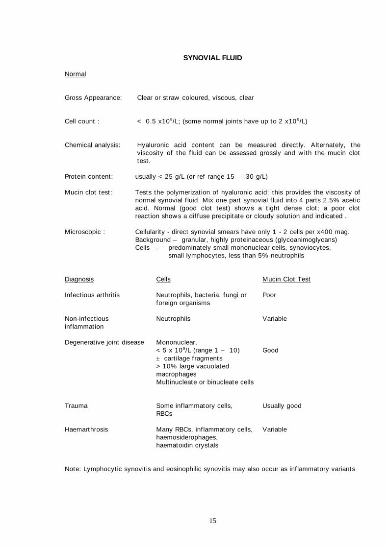
SYNOVIAL FLUID
Normal
Gross Appearance: Clear or straw coloured, viscous, clear
Cell count : < 0.5 x10⁹/L; (some normal joints have up to 2 x10 9/L)
Chemical analysis: Hyaluronic acid content can be measured direct ly. Alternately, the
viscosity of the f luid can be assessed grossly and w ith the mucin clot
test.
Protein content: usually < 25 g/L (or ref range 15 – 30 g/L)
Mucin clot test: Tests the polymerizat ion of hyaluronic acid; this provides the viscosity of
normal synovial f luid. Mix one part synovial f luid into 4 parts 2.5% acetic
acid. Normal (good clot test) show s a t ight dense clot; a poor clot
react ion show s a dif fuse precipitate or cloudy solut ion and indicated .
Microscopic : Cellularity - direct synovial smears have only 1 - 2 cells per x400 mag.
Background – granular, highly proteinaceous (glycoanimoglycans)
Cells - predominately small mononuclear cells, synoviocytes,
small lymphocytes, less than 5% neutrophils
Diagnosis Cells Mucin Clot Test
Infect ious arthrit is Neutrophils, bacteria, fungi or
foreign organisms
Poor
Non-infect ious
inf lammation
Neutrophils Variable
Degenerat ive joint disease Mononuclear,
< 5 x 10⁹/L (range 1 – 10) Good
cart ilage fragments
> 10% large vacuolated
macrophages
Mult inucleate or binucleate cells
Trauma Some inf lammatory cells, Usually good
RBCs
Haemarthrosis Many RBCs, inf lammatory cells,
haemosiderophages,
haematoidin crystals
Variable
Note: Lymphocyt ic synovit is and eosinophilic synovit is may also occur as inf lammatory variants
16
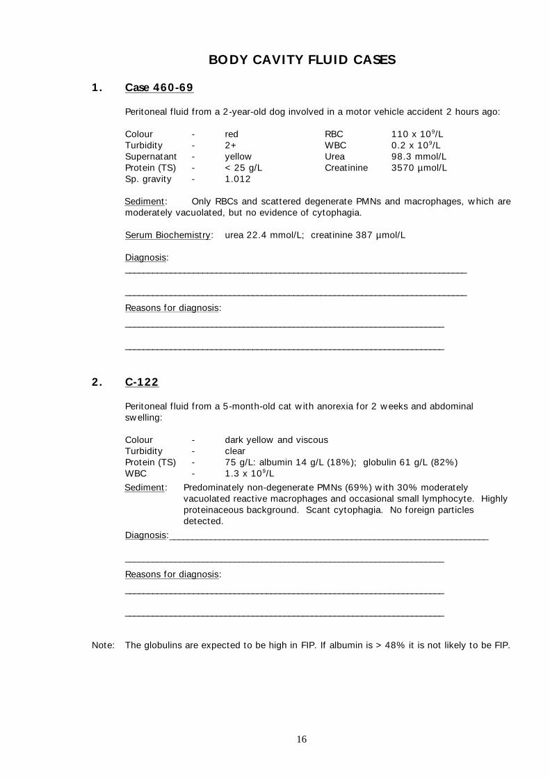
BODY CAVITY FLUID CASES
1. Case 460-69 Peritoneal f luid from a 2-year-old dog involved in a motor vehicle accident 2 hours ago:
Colour - red RBC 110 x 109/L
Turbidity - 2+ WBC 0.2 x 10⁹/L
Supernatant - yellow Urea 98.3 mmol/L
Protein (TS) - < 25 g/L Creat inine 3570 µmol/L
Sp. gravity - 1.012
Sediment: Only RBCs and scattered degenerate PMNs and macrophages, w hich are
moderately vacuolated, but no evidence of cytophagia.
Serum Biochemistry: urea 22.4 mmol/L; creat inine 387 µmol/L
Diagnosis:
___________________________________________________________________________
___________________________________________________________________________
Reasons for diagnosis:
______________________________________________________________________
______________________________________________________________________
2. C-122 Peritoneal f luid from a 5-month-old cat w ith anorexia for 2 w eeks and abdominal
sw elling:
Colour - dark yellow and viscous
Turbidity - clear
Protein (TS) - 75 g/L: albumin 14 g/L (18%); globulin 61 g/L (82%)
WBC - 1.3 x 10⁹/L
Sediment: Predominately non-degenerate PMNs (69%) w ith 30% moderately
vacuolated react ive macrophages and occasional small lymphocyte. Highly
proteinaceous background. Scant cytophagia. No foreign part icles
detected.
Diagnosis:______________________________________________________________________
______________________________________________________________________
Reasons for diagnosis:
______________________________________________________________________
______________________________________________________________________
Note: The globulins are expected to be high in FIP. If albumin is > 48% it is not likely to be FIP.
17
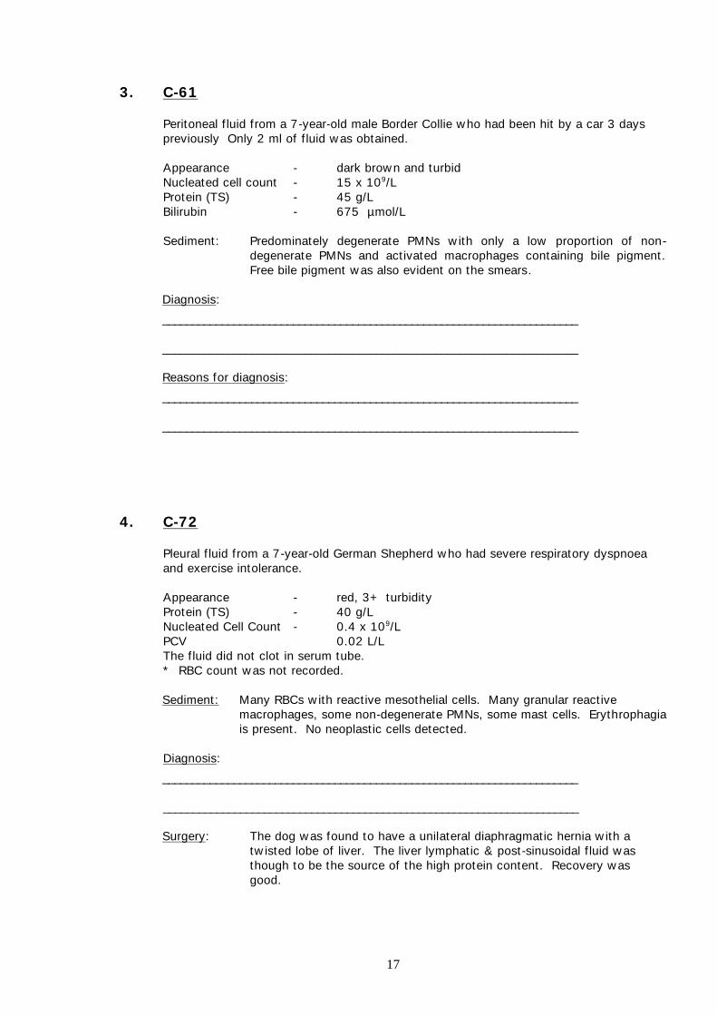
3. C-61
Peritoneal f luid from a 7-year-old male Border Collie w ho had been hit by a car 3 days
previously Only 2 ml of f luid w as obtained.
Appearance - dark brow n and turbid
Nucleated cell count - 15 x 109/L
Protein (TS) - 45 g/L
Bilirubin - 675 µmol/L
Sediment: Predominately degenerate PMNs w ith only a low proport ion of non-
degenerate PMNs and act ivated macrophages containing bile pigment.
Free bile pigment w as also evident on the smears.
Diagnosis:
______________________________________________________________________
______________________________________________________________________
Reasons for diagnosis:
______________________________________________________________________
______________________________________________________________________
4. C-72
Pleural f luid from a 7-year-old German Shepherd w ho had severe respiratory dyspnoea
and exercise intolerance.
Appearance - red, 3+ turbidity
Protein (TS) - 40 g/L
Nucleated Cell Count - 0.4 x 10⁹/L
PCV 0.02 L/L
The f luid did not clot in serum tube.
* RBC count w as not recorded.
Sediment: Many RBCs w ith react ive mesothelial cells. Many granular react ive
macrophages, some non-degenerate PMNs, some mast cells. Erythrophagia
is present. No neoplast ic cells detected.
Diagnosis:
______________________________________________________________________
______________________________________________________________________
Surgery: The dog w as found to have a unilateral diaphragmatic hernia w ith a
tw isted lobe of liver. The liver lymphatic & post-sinusoidal f luid w as
though to be the source of the high protein content. Recovery w as
good.
18
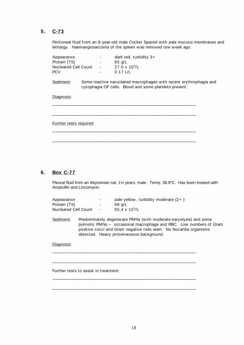
5. C-73 Peritoneal f luid from an 8-year-old male Cocker Spaniel w ith pale mucous membranes and
lethargy. Haemangiosarcoma of the spleen w as removed one w eek ago.
Appearance - dark red, turbidity 3+
Protein (TS) - 65 g/L
Nucleated Cell Count - 27.0 x 10⁹/L
PCV - 0.17 L/L
Sediment: Some react ive vacuolated macrophages w ith recent erythrophagia and
cytophagia OF cells. Blood and some platelets present.
Diagnosis:
_______________________________________________________________________
_______________________________________________________________________
Further tests required:
_______________________________________________________________________
_______________________________________________________________________
6. Box C-77
Pleural fluid from an Abyssinian cat, 1½ years, male. Temp. 38.9ºC. Has been treated with Ampicillin and Lincomycin.
Appearance - pale yellow , turbidity moderate (2+ )
Protein (TS) - 68 g/L
Nucleated Cell Count - 55.4 x 10⁹/L
Sediment: Predominately degenerate PMNs (w ith moderate karyolysis) and some
pyknotic PMNs – occasional macrophage and RBC. Low numbers of Gram
posit ive cocci and Gram negative rods seen. No Nocardia organisms
detected. Heavy proteinaceous background.
Diagnosis:
_______________________________________________________________________
_______________________________________________________________________
Further tests to assist in treatment:
_______________________________________________________________________
_______________________________________________________________________