Embed Size (px)
Citation preview
DISEASES OF AQUATIC ORGANISMSDis Aquat Org
Vol. 72: 19–30, 2006 Published September 14
INTRODUCTION
The International Joint Commission (IJC), comprisedof American and Canadian officials, was formed in1909 to assist their governments in finding solutions tothe problems facing the waters bordering the UnitedStates and Canada. The Great Lakes Water QualityAgreement, in which the United States and Canadaagreed to restore and preserve the biological, physicaland chemical integrity of the Great Lakes Basin Eco-system, was first signed in 1972. In 1987 a protocolwas signed by both governments which defined GreatLakes areas of concern (AOC) as ‘geographic areas thatfail to meet the general or specific objectives of theagreement where such failure has caused or is likely tocause impairment of beneficial use of the area’s abilityto support aquatic life’. The US and Canadian gov-ernments identified 43 such areas; 26 in the US, 12 in
Canada, and 5 shared between the US and Canada onconnecting river systems. The Great Lakes Water Qual-ity Agreement, as amended via the 1987 protocol, di-rects the 2 federal governments to cooperate with stateand provincial governments to develop and implementremedial action plans (RAPs) for each AOC. The proto-col also called for reports on restorative progress andfor the IJC to review RAPs proposed by the 43 AOC. Todate, 2 AOC (Collingwood Harbor and Severn Sound)have been delisted (IJC 1987) and Presque Isle Bay hasbeen designated an area of recovery.
So far, 14 beneficial use impairments have beenidentified and the various AOC have different combi-nations of these impairments. One impairment, listedas ‘fish tumors and other deformities’ is defined asoccurring when ‘the incidence rate of fish tumors andother deformities exceeds rates at unimpacted or con-trol sites or when survey data confirm the presence of
© Inter-Research 2006 · www.int-res.com*Email: [email protected]
Diagnostic criteria for proliferative hepatic lesionsin brown bullhead Ameiurus nebulosus
Vicki S. Blazer1,*, John W. Fournie2, Jeffrey C. Wolf3, Marilyn J. Wolfe3
1National Fish Health Research Laboratory, US Geological Survey, 11649 Leetown Road, Kearneysville, West Virginia 25430, USA2U.S. Environmental Protection Agency, Gulf Ecology Division, 1 Sabine Island Drive, Gulf Breeze, Florida 32561, USA
3The Registry of Tumors in Lower Animals, 22900 Shaw Road, Suite 107, Sterling, Virginia 20166, USA
ABSTRACT: Brown bullhead Ameiurus nebulosus is used as indicator species for contaminant effectsat areas of concern (AOC) in the Great Lakes and other areas. One of the beneficial use impairmentsat numerous AOC is ‘fish tumors and other deformities’. An impairment occurs when the prevalenceof fish tumors and other deformities exceeds those at unimpacted or control sites or when survey dataconfirm the presence of neoplastic or preneoplastic liver lesions in bullhead or white sucker Catosto-mus commersonii. Numerous surveys have been conducted over the years assessing neoplasia inthese fishes, both liver and skin tumors. However, a major problem in comparing the results has beena lack of consistent criteria for evaluating histological changes in bullhead livers. As individual AOCdevelop and implement remedial action plans, realistic and attainable delisting targets need to bespecified. For this to occur and be consistent from site to site there must be standardization of the cri-teria being used to evaluate specific impairments. In this report, specific diagnostic criteria are pro-vided for both non-neoplastic and neoplastic proliferative hepatocellular and biliary lesions. Thesecriteria should assist fish pathologists in describing and categorizing proliferative liver lesions frombrown bullhead.
KEY WORDS: Brown bullhead · Liver · Neoplasia · Proliferative lesions · Diagnostic criteria
Resale or republication not permitted without written consent of the publisher

Dis Aquat Org 72: 19–30, 2006
neoplastic or preneoplastic liver tumors in bullhead orsuckers’ (IJC 1989; www.epa.gov/lakeerie/buia/reports).Deformities are defined as twisted, missing, forked orbulging body parts including fins, barbels, abdomen orskeleton. Currently, 16 of the 41 AOC have the pres-ence of tumors or other deformities listed as one oftheir beneficial use impairments.
A key indicator species for AOC is the brown bull-head Ameiurus nebulosus (IJC 1987), because this is abottom-dwelling fish with a limited home range and isknown to take up contaminants from food and sedi-ments (Maccubbin et al. 1985, Baumann 1989, Bau-mann et al. 1991, 1996, Smith et al. 1994). Both liver(Harshbarger & Clark 1990, Baumann & Harshbarger1995, Pinkney et al. 2001) and skin (Black 1983, Smithet al. 1989, Bowser et al. 1991, Poulet et al. 1994) neo-plasms have been associated with exposure to polynu-clear aromatic hydrocarbons (PAH) and other contam-inants. Although the prevalence of skin neoplasia isoften elevated in fish from chemically polluted, oftenindustrial, sites (Sonstegard 1977, Hayes et al. 1990,Bowser et al. 1991), the association of epidermal papil-lomas and carcinomas with chemical exposure is lessconclusive than with liver neoplasia (Poulet et al.1994). There is also good evidence for the influence ofPAH exposure on the development of liver neoplasmand other microscopic liver lesions in other fish spe-cies, including English sole Parophrys vetulus (Myerset al. 1990), mummichog Fundulus heteroclitus (Vogel-bein et al. 1990) and winter flounder Pseudopleuro-nectes americanus (Gardner et al. 1989).
Numerous surveys have been undertaken to assessthe prevalence of skin and liver tumors, as well asother contaminant-associated lesions at AOC. How-ever, a major problem has been a lack of consistent cri-teria for evaluating histological changes in bullheadlivers. For instance, in some studies there is no distinc-tion between altered foci (preneoplastic) and neoplasticlesions, or inadequate descriptions of individual lesions(Black 1983, Pyron et al. 2001). Baumann et al. (2000)present a compilation of liver neoplasm prevalence atAOC by location and year. However, a comparison ofthe papers/reports from which these data wereobtained demonstrates the lack of consistent criteria.For example, a prevalence of 7% was noted in theAshtabula River in 1991 by Mueller & Mac (1994). Inthat study, neoplasia included only hepatocellular car-cinoma (advanced liver neoplasms) or cholangiocarci-noma (biliary neoplasms), but not hepatocellular ade-noma, cholangioma or altered foci (Mueller & Mac1994). Conversely, the prevalence reported for theBlack River in 1982 (60%) included carcinomas andadenomas (Baumann et al. 1990), while the 9% in theDetroit River in 1985 to 1987 included carcinomas,adenomas and altered foci (Maccubbin & Ersing 1991).
An RAP is required for each AOC. The intent is toprovide a strategic guideline that outlines all phases ofthe remedial effort, from problem definition to activeremediation, through to the recovery stage and, finally,delisting. A key component of these plans is the speci-fication of realistic and attainable delisting targets andthe collection of data necessary to demonstrate that thebeneficial use is no longer impaired. For the ‘fishtumors and other deformities’ impairment, there are2 primary issues that require resolution: (1) the stan-dardization of the criteria being used to evaluate theimpairment, and (2) the institution of realistic delistingtargets. Criteria need to be established for both sam-pling and monitoring this impairment so that datafrom different years, reference sites and AOC can becompared.
Diagnostic criteria already exist for non-neoplastic andneoplastic liver lesions in some fish species such as Eng-lish sole Parophrys vetulus, medaka Oryzias latipes, thecommon dab Limanda limanda and European flounderPlatichthys flesus (Myers et al. 1987, Boorman et al. 1997,Feist et al. 2004). Even though liver lesions in a variety offish species are very similar, it is useful to provide de-tailed descriptions and illustrations for species commonlyused in laboratory studies and monitoring programs.Therefore, the purpose of this report was to address theneed for diagnostic criteria for proliferative liver lesionsin bullhead and to suggest a classification system forthese lesions. Commonly observed non-neoplastic andneoplastic proliferative lesions are described andillustrated to provide guidelines to assist pathologistsevaluating studies utilizing these fish as indicator orsentinel species, particularly in the Great Lakes.
MATERIALS AND METHODS
Material for this review was obtained from numerousfield collections of brown bullheads, primarily fromtributaries of Lake Erie but including other sites suchas the Hudson River (New York), and ponds on CapeCod (Massachusetts), where bullhead have been usedas an indicator species for assessing environmentalstress. These include liver samples from over 1500brown bullhead collected from 1998 to the present, forwhich histologic slide collections are housed at the USGeological Survey’s National Fish Health ResearchLaboratory, Kearneysville, West Virginia. These stud-ies involved random samples of fish from specific sites,and the proliferative lesions observed microscopicallywere not often associated with grossly visible liverlesions. In addition, the National Cancer Institute’sRegistry of Tumors in Lower Animals, Sterling, Vir-ginia, currently has 991 brown bullhead cases fromnumerous surveys dating from 1965 to 2001. Approxi-
20
Blazer et al.: Proliferative hepatic lesions of brown bullhead
mately 680 of these cases had previously been diag-nosed with liver neoplasms and we reviewed 25 casesfor specific lesions. Many of the cases in the Registrywere associated with grossly observable lesions. Mostof the above studies targeted fish 3 yr of age or older,with a total length of 250 mm or greater. Typically, tis-sue was fixed in 10% buffered formalin, embedded inparaffin, sectioned at 3 to 6 µm and stained with hema-toxylin and eosin.
Slides were examined with a Nikon Eclipse E600microscope with Plan Apochromatic lenses and a SpotRT Slider™ (Model 2.3.1) high resolution digital cam-era system (Diagnostic Instruments). Images werecaptured from the system using Spot™ (Version 3.3for Windows®) capture software at 1600 × 1200 dotsper inch (dpi) resolution. Brightness/contrast adjust-ments were performed in Adobe Photoshop® 6.0 forWindows (Adobe Systems).
RESULTS
Normal hepatic histology of brown bullhead
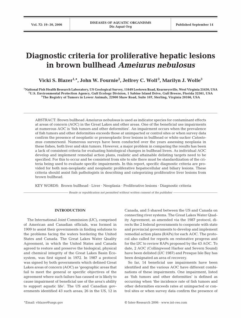
As in most teleosts, the liver of brown bullhead iscomposed of hepatic tubules. The classic hexagonalarchitecture of the mammalian hepatic lobule is notevident in fish livers (Gingerich 1982, Vethaak &Wester 1996, Metcalfe 1998, Hinton et al. 2001). Theappearance of teleost hepatocytes can vary greatly dueto sex, maturity, diet, season, contaminant exposureand other factors (Hinton & Couch 1998, Rocha & Mon-teiro 1999). Normal hepatocytes may have little or noobservable lipid or glycogen storage (vacuolization)(Fig. 1A) or may contain extensive amounts of thesestored energy sources (Fig. 1B). Exocrine pancreatictissue and macrophage aggregates are commonly pre-sent (Fig. 1C,D) and bile ducts may also be observed
21
Fig. 1. Ameiurus nebulosus. Microscopic appearance of normal liver of brown bullhead. (A,B) Normal hepatocyte appearance canvary from (A) relatively unvacuolated to (B) significant vacuolization due to lipid and glycogen storage. Scale bars = 50 µm.(C) Within liver parenchyma (a), exocrine pancreatic tissue (b) often surrounds vessels and bile ducts (arrows). Scale bar =100 µm. (D) Bile ducts (a) have a low columnar epithelium and are surrounded by a thin band of connective tissue (arrow); pan-creatic tissue (b) and macrophage aggregates (c) are often observed in this area. Scale bar = 25 µm. Hematoxylin & eosin

Dis Aquat Org 72: 19–30, 2006
within this tissue. In liver sections from bullheads col-lected at reference sites, bile ducts are often sparselydistributed in sections examined by light microscopy,although numerous bile preductules and ductules arelocated within the hepatic parenchyma (Hinton &Couch 1998). Small and intermediate bile ducts havecolumnar epithelium and the ducts are encircled bya thin band of connective tissue (Fig. 1D). Large bileducts are surrounded by connective tissue and areprominent near the common bile duct.
Proliferative lesions in brown bullhead liver
As in higher vertebrates, proliferative lesions of eitherhepatocellular or biliary origin can be categorized asputative pre-neoplastic, non-neoplastic or neoplastic.
Putative pre-neoplastic hepatocellular lesions: foci of cellular alteration
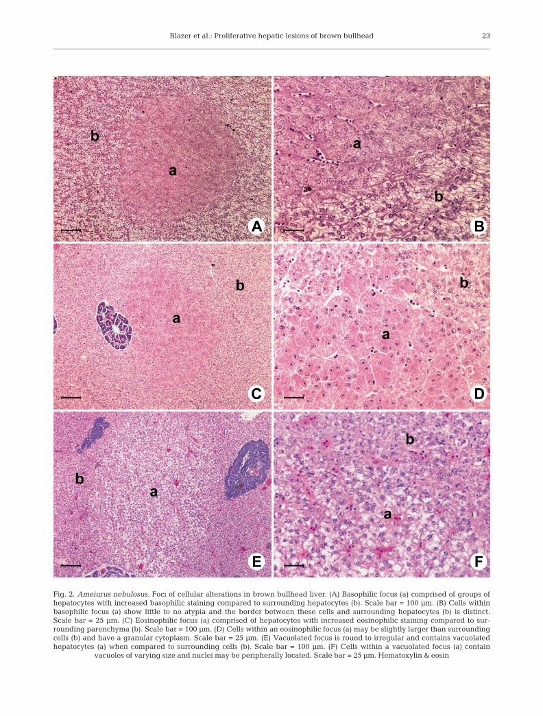
Based on tinctorial characteristics of the hepatocytecytoplasm, 4 categories of altered foci can be re-cognized in hematoxylin and eosin-stained sections.These include basophilic foci, eosinophilic foci, vacuo-lated cell foci and clear cell foci. Foci are generally dis-tinct in terms of coloration, but the hepatic tubules arearranged in a relatively normal pattern, and theymerge imperceptibly with the surrounding paren-chyma with little to no compression. Features of cellu-lar atypia (increased nuclear to cytoplasmic ratio,nuclear pleomorphism, nucleolar enlargement, or thepresence of coarsely clumped chromatin) are notgenerally evident.• Basophilic foci are round to irregular clusters of
hepatocytes with increased basophilic staining com-pared to adjacent cells (Fig. 2A). There is no evi-dence of compression of adjacent unaffected hepatictissues, little to no atypia of hepatocytes, and mitoticfigures are generally not present. In some lesions,constituent cells may be smaller than adjacent hepa-tocytes (Fig. 2B).
• Eosinophilic foci are round to irregular areas ofhepatocytes with increased eosinophilic stainingcompared to adjacent cells (Fig. 2C). There is little tono evidence of compression; however, the cells maybe slightly enlarged with granular eosinophilic cyto-plasm (Fig. 2D).
• Vacuolated cell foci are round to irregular, blend intothe surrounding parenchyma without compression,and contain hepatocytes with clear cytoplasmicvacuoles of varying sizes (Fig. 2E). Nuclei are ofteneccentrically located within the cells (Fig. 2F). Be-cause the cytoplasmic lipid is often present in a micro-
vesicular form, at low magnification the vacuolatedfoci may resemble clear cell foci. Clear cell foci, as described in other fish species, with
a ‘ground glass’ appearance of the cytoplasm, indica-tive of glycogen storage, and centrally located nuclei(Feist et al. 2004, Köhler 2004) were not observed inthe bullhead livers examined. Occasionally individualcells within a vacuolated focus appeared to containboth lipid and glycogen and it is possible that bullheadfoci may contain both storage products.
Neoplastic hepatocellular lesions
Hepatocellular adenoma
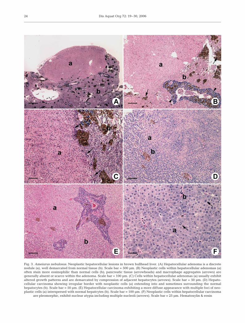
Adenomas are usually single discrete lesions withdistinct borders (Fig. 3A).The cells often exhibit alteredstaining properties, either more eosinophilic or morebasophilic than the non-neoplastic liver, and usuallyexhibit altered growth patterns (Fig. 3B). Macrophageaggregates, pancreatic tissue and other structures areoften missing or sparse within adenomas (Fig. 3A–C).Adenomas are often well demarcated by compressionof the adjacent parenchymal cells (Fig. 3C) and mitoticfigures are rarely observed.
Hepatocellular carcinoma
Hepatocellular carcinomas are usually distinctly dif-ferent from the surrounding liver tissue and haveirregular borders due to invasion of neoplastic cellsinto the adjacent hepatic parenchyma (Fig. 3D). Carci-nomas may be small or large lesions and may featuretumor giant cell formation. In some cases, foci ofneoplastic cells are diffusely spread throughout thehepatic parenchyma (Fig. 3E). Cellular pleomorphismand nuclear atypia are key features, and there may bean increase in the number of mitotic figures and/orcells with multiple nucleoli (Fig. 3F).
Non-neoplastic biliary lesions: bile duct hyperplasia
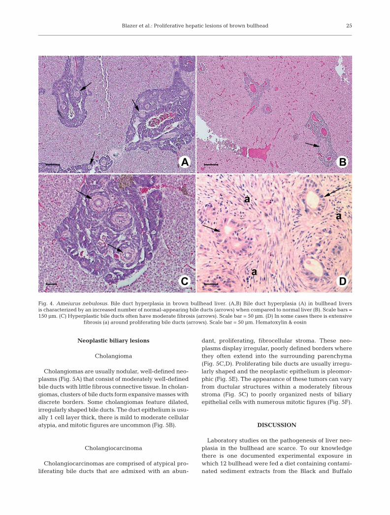
Bile duct hyperplasia is characterized by an in-creased number of bile ducts (Fig. 4A) compared tonormal liver (Fig. 4B). Hyperplastic bile ducts are oftenscattered and do not necessarily form discrete masses.The biliary epithelium is always well-differentiated;however, there may be moderate fibrosis associatedwith proliferating bile ducts (Fig. 4C), and the ductstend to display substantial variation in size and shape.Occasionally the periductal fibrosis can be extensive(Fig. 4D).
22
Blazer et al.: Proliferative hepatic lesions of brown bullhead 23
Fig. 2. Ameiurus nebulosus. Foci of cellular alterations in brown bullhead liver. (A) Basophilic focus (a) comprised of groups ofhepatocytes with increased basophilic staining compared to surrounding hepatocytes (b). Scale bar = 100 µm. (B) Cells withinbasophilic focus (a) show little to no atypia and the border between these cells and surrounding hepatocytes (b) is distinct.Scale bar = 25 µm. (C) Eosinophilic focus (a) comprised of hepatocytes with increased eosinophilic staining compared to sur-rounding parenchyma (b). Scale bar = 100 µm. (D) Cells within an eosinophilic focus (a) may be slightly larger than surroundingcells (b) and have a granular cytoplasm. Scale bar = 25 µm. (E) Vacuolated focus is round to irregular and contains vacuolatedhepatocytes (a) when compared to surrounding cells (b). Scale bar = 100 µm. (F) Cells within a vacuolated focus (a) contain
vacuoles of varying size and nuclei may be peripherally located. Scale bar = 25 µm. Hematoxylin & eosin
Dis Aquat Org 72: 19–30, 200624
Fig. 3. Ameiurus nebulosus. Neoplastic hepatocellular lesions in brown bullhead liver. (A) Hepatocellular adenoma is a discretenodule (a), well demarcated from normal tissue (b). Scale bar = 500 µm. (B) Neoplastic cells within hepatocellular adenomas (a)often stain more eosinophilic than normal cells (b); pancreatic tissue (arrowheads) and macrophage aggregates (arrows) aregenerally absent or scarce within the adenoma. Scale bar = 100 µm. (C) Cells within hepatocellular adenomas (a) usually exhibitaltered growth patterns and are demarcated by compression of adjacent hepatocytes (arrows). Scale bar = 50 µm. (D) Hepato-cellular carcinoma showing irregular border with neoplastic cells (a) extending into and sometimes surrounding the normalhepatocytes (b). Scale bar = 50 µm. (E) Hepatocellular carcinoma exhibiting a more diffuse appearance with multiple foci of neo-plastic cells (a) interspersed with normal hepatcytes (b). Scale bar = 100 µm. (F) Neoplastic cells within hepatocellular carcinoma
are pleomorphic, exhibit nuclear atypia including multiple nucleoli (arrows). Scale bar = 25 µm. Hematoxylin & eosin
Blazer et al.: Proliferative hepatic lesions of brown bullhead
Neoplastic biliary lesions
Cholangioma
Cholangiomas are usually nodular, well-defined neo-plasms (Fig. 5A) that consist of moderately well-definedbile ducts with little fibrous connective tissue. In cholan-giomas, clusters of bile ducts form expansive masses withdiscrete borders. Some cholangiomas feature dilated,irregularly shaped bile ducts. The duct epithelium is usu-ally 1 cell layer thick, there is mild to moderate cellularatypia, and mitotic figures are uncommon (Fig. 5B).
Cholangiocarcinoma
Cholangiocarcinomas are comprised of atypical pro-liferating bile ducts that are admixed with an abun-
dant, proliferating, fibrocellular stroma. These neo-plasms display irregular, poorly defined borders wherethey often extend into the surrounding parenchyma(Fig. 5C,D). Proliferating bile ducts are usually irregu-larly shaped and the neoplastic epithelium is pleomor-phic (Fig. 5E). The appearance of these tumors can varyfrom ductular structures within a moderately fibrousstroma (Fig. 5C) to poorly organized nests of biliaryepithelial cells with numerous mitotic figures (Fig. 5F).
DISCUSSION
Laboratory studies on the pathogenesis of liver neo-plasia in the bullhead are scarce. To our knowledgethere is one documented experimental exposure inwhich 12 bullhead were fed a diet containing contami-nated sediment extracts from the Black and Buffalo
25
Fig. 4. Ameiurus nebulosus. Bile duct hyperplasia in brown bullhead liver. (A,B) Bile duct hyperplasia (A) in bullhead liversis characterized by an increased number of normal-appearing bile ducts (arrows) when compared to normal liver (B). Scale bars =150 µm. (C) Hyperplastic bile ducts often have moderate fibrosis (arrows). Scale bar = 50 µm. (D) In some cases there is extensive
fibrosis (a) around proliferating bile ducts (arrows). Scale bar = 50 µm. Hematoxylin & eosin

Dis Aquat Org 72: 19–30, 200626
Fig. 5. Ameiurus nebulosus. Neoplastic biliary lesions in brown bullhead liver. (A) Cholangioma (a) is generally nodular and sepa-rated from normal hepatic tissue (b) by a well-defined border. Scale bar = 100 µm. (B) Higher magnification of (A) showing well-defined border (arrows) and epithelium of bile ducts (a), usually 1 cell thick but may be dilated and irregularly shaped. Scale bar =25 µm. (C) Cholangiocarcinoma composed of atypical proliferating bile ducts (a) with irregular, poorly defined border extendinginto surrounding parenchyma (b). Scale bar = 100 µm. (D) Higher magnification of (C) illustrating neoplastic bile ducts extending intonormal hepatic parenchyma (b), eliciting an inflammatory reaction (a) at the border; normal-appearing ducts can still be observedin some areas (arrows). Scale bar = 50 µm. (E) Cholangiocarcinoma composed of irregularly shaped ducts (a) within a fibrocellularstroma (arrows). Scale bar = 25 µm. (F) In some cases, cholangiocarcinomas are composed of poorly organized nests of biliaryepithelial cells (a) within a fibrocellular stroma (b); mitotic figure (arrow) can be observed. Scale bar = 25 µm. Hematoxylin & eosin
Blazer et al.: Proliferative hepatic lesions of brown bullhead
rivers (Black et al. 1985). In that experiment, 6 of thefish on the experimental diet were sacrificed after 4 moand 6 were terminated after 7 mo. Lesions identified inthe fish sacrificied at 4 mo included a basophilic focus,a small focus of bile duct proliferation, and a largerlesion consisting of mixed fibrosis and duct-likeformations of hepatocytes. At 7 mo, findings includednumerous clear cell and eosinophilic foci, bile ductproliferation and fibrosis, and a cholangioma. In asecond study, brown bullhead were injected with asingle intraperitoneal dose of 0, 5, 25, or 125 mg kg–1
benzo[a]pyrene and evaluated over 18 mo (Grady etal. 1992); one focus of biliary hyperplasia and fibrosisin a bullhead exposed to 25 mg kg–1 and basophilic fociat all exposure levels were observed at Day 540. Noneof these lesions were observed in control fish. Un-fortunately, none of the lesions in either study weredescribed in detail or illustrated, nor was there anymention of control fish in the first study. Hence, fur-ther, more definitive studies are necessary to deter-mine whether non-neoplastic proliferative lesionsshould be regarded as preneoplastic precursors ofhepatocellular or biliary neoplasms in bullhead.
Foci of cellular alteration (basophilic, eosinophilic,vacuolated and clear cell) have been reported inneoplastic epizootics as well as carcinogenesisstudies. There is evidence for the progression ofbasophilic foci to hepatocellular carcinoma in rain-bow trout Oncorhynchus mykiss (Hendricks et al.1984), medaka Oryzias latipes, guppies Poecilia retic-ulata (Hawkins et al. 1990), mosquitofish Gambusiaaffinis (Law et al. 1994), and sheepshead minnowCyprinodon variegatus (Hinton et al. 1988), whereasthere is no clear indication that other altered fociprogress to neoplasia (Bunton 1996). However, sev-eral types of foci (vacuolated cell, eosinophilic andbasophilic) that were observed in rivulus Rivulusocellatus marmoratus exposed to diethylnitrosaminewere considered preneoplastic (Grizzle & Thiyagarajah1988). Altered foci are also considered preneoplasticin wild English sole exposed to PAHs in Puget Sound(Myers et al. 1987, 1991), winter flounder from conta-minated sites in Boston Harbor (Murchelano & Wolke1991), flounder from Dutch coastal areas (Vethaak &Wester 1996), and British estuaries impacted by cont-aminants, including PAHs (Stentiford et al. 2003).Indeed, there is substantial evidence that increasedprevalences of altered foci are associated with conta-minant exposure, and should therefore be consideredpotential indications of such exposure and putativelypre-neoplastic.
Cholangiolar neoplasia has also been induced innumerous species through experimental contaminantexposures (Hendricks et al. 1984, Parland & Baumann1985, Couch & Courtney 1987, Grizzle & Thiyagarajah
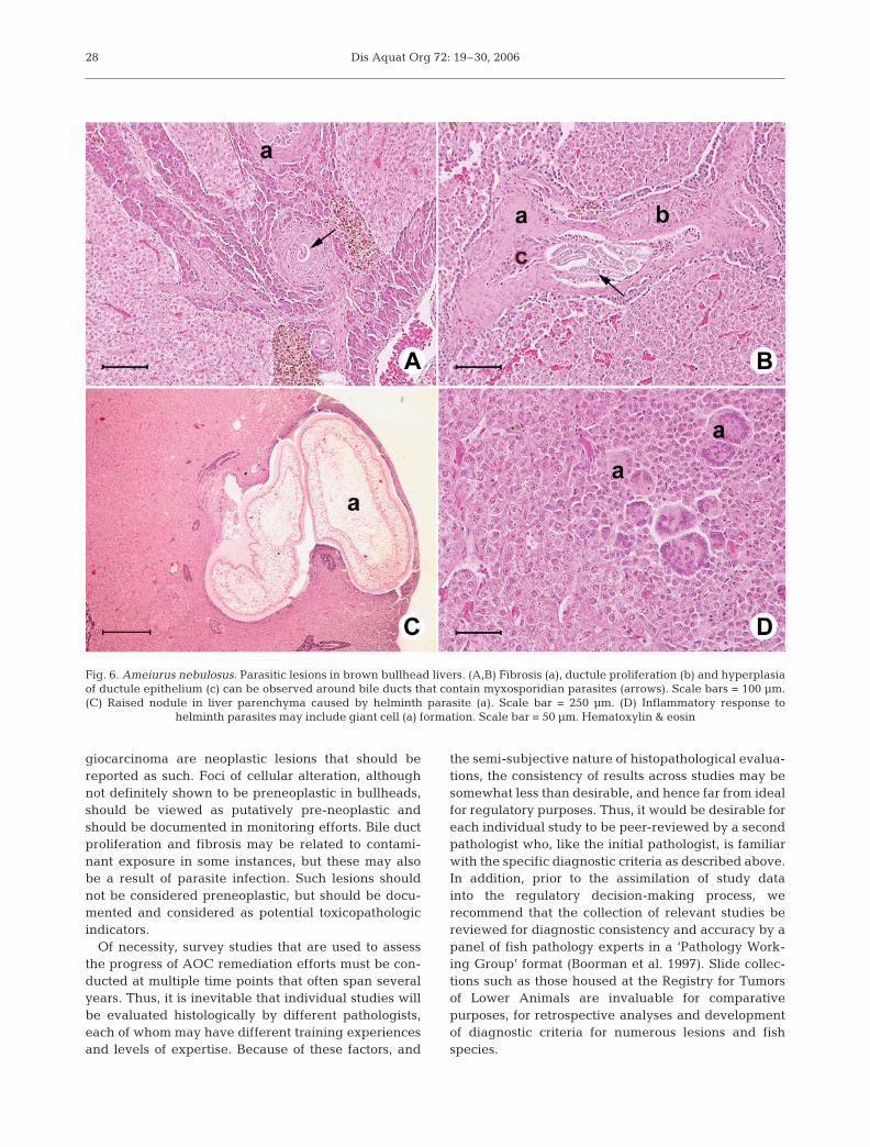
1988, Hawkins et al. 1988, Law et al. 1994, Boormanet al. 1997). As with hepatic lesions, it has not beenexperimentally demonstrated which, if any, non-neo-plastic cholangiolar changes may be considered pre-neoplastic in the bullhead. Proliferation of bile ductularepithelial cells has been observed during experimentalchemical exposures in a number of species (Hendrickset al. 1984, Parland & Baumann 1985, Couch & Court-ney 1987), and in such fishes the cells may be consid-ered preneoplastic (Bunton 1996). Bile duct prolifera-tion and fibrosis have also been reported in associationwith hepatic neoplasia in other fish species at a num-ber of contaminated sites and are considered toxico-pathic, and in some cases preneoplastic (May et al.1987, Moore et al. 1989, Vogelbein et al. 1990, Bau-mann et al. 1991, Stehr et al. 2003). However, in bull-heads, a myxosporidian parasite can commonly beobserved within the lumen of bile ducts (Fig. 6A,B) andit is possible that bile duct proliferation and fibrosismay be related to the presence of this or other para-sites. Similarly, a proliferative inflammatory responseand giant cell formation has been observed in associa-tion with helminth parasites in the parenchyma of bull-heads (Fig. 6C,D) and should not be confused withtoxicopathic lesions.
In addition to the need for consistent diagnostic crite-ria, there is a need to establish consistent samplingprocedures. A quality control issue for detection ofneoplastic and other proliferative liver lesions con-cerns the number of histologic sections or levels in aliver that are necessary to accurately estimate the pres-ence and extent in an individual liver. Stine et al.(2004) evaluated 6 livers from adult mummichog(a much smaller fish species than bullhead), 5 of whichhad grossly visible nodules. The authors discoveredthat: cancerous and precancerous lesions are nothomogenously distributed; the presence of one lesiontype may influence the extent and distribution of otherlesions; multiple sections are needed (an average of 6sections for 50% of lesions to be identified or 7 sectionsfor 95% of lesion estimates to be included); and tissuessectioned in different planes and observations fromdifferent sections will generate different lesion obser-vations and conclusions. In our studies, very few ofthe proliferative lesions diagnosed in bullhead weregrossly apparent. Hence, it must be recognized that forlarge fish species such as adult brown bullhead morethan 10 sections at various locations throughout theliver may be necessary to adequately estimate lesionprevalence.
One problem with the IJC ‘fish tumors and other de-formities’ beneficial use impairment is that the phrase‘neoplastic or preneoplastic liver tumors’ was neverclearly defined. Certainly, hepatocellular adenoma,hepatocellular carcinoma, cholangioma and cholan-
27
Dis Aquat Org 72: 19–30, 2006
giocarcinoma are neoplastic lesions that should bereported as such. Foci of cellular alteration, althoughnot definitely shown to be preneoplastic in bullheads,should be viewed as putatively pre-neoplastic andshould be documented in monitoring efforts. Bile ductproliferation and fibrosis may be related to contami-nant exposure in some instances, but these may alsobe a result of parasite infection. Such lesions shouldnot be considered preneoplastic, but should be docu-mented and considered as potential toxicopathologicindicators.
Of necessity, survey studies that are used to assessthe progress of AOC remediation efforts must be con-ducted at multiple time points that often span severalyears. Thus, it is inevitable that individual studies willbe evaluated histologically by different pathologists,each of whom may have different training experiencesand levels of expertise. Because of these factors, and
the semi-subjective nature of histopathological evalua-tions, the consistency of results across studies may besomewhat less than desirable, and hence far from idealfor regulatory purposes. Thus, it would be desirable foreach individual study to be peer-reviewed by a secondpathologist who, like the initial pathologist, is familiarwith the specific diagnostic criteria as described above.In addition, prior to the assimilation of study datainto the regulatory decision-making process, werecommend that the collection of relevant studies bereviewed for diagnostic consistency and accuracy by apanel of fish pathology experts in a ‘Pathology Work-ing Group’ format (Boorman et al. 1997). Slide collec-tions such as those housed at the Registry for Tumorsof Lower Animals are invaluable for comparativepurposes, for retrospective analyses and developmentof diagnostic criteria for numerous lesions and fishspecies.
28
Fig. 6. Ameiurus nebulosus. Parasitic lesions in brown bullhead livers. (A,B) Fibrosis (a), ductule proliferation (b) and hyperplasiaof ductule epithelium (c) can be observed around bile ducts that contain myxosporidian parasites (arrows). Scale bars = 100 µm.(C) Raised nodule in liver parenchyma caused by helminth parasite (a). Scale bar = 250 µm. (D) Inflammatory response to
helminth parasites may include giant cell (a) formation. Scale bar = 50 µm. Hematoxylin & eosin
Blazer et al.: Proliferative hepatic lesions of brown bullhead
Acknowledgements. Funding for this project was providedby Pennsylvania Sea Grant, US Geological Survey and theUS Environmental Protection Agency. This project wasperformed, in part, by using the services provided by theNational Cancer Institute’s Registry of Tumors in Lower Ani-mals, operated under contract by Experimental PathologyLaboratories, N02-CB-27034. We thank Lee Courtney forassistance in preparing the figures and Dr. William E.Hawkins for critically reviewing the manuscript.
LITERATURE CITED
Baumann PC (1989) PAH, metabolites, and neoplasia in feralfish populations. In: Varansi U (ed) Metabolism of poly-cyclic aromatic hydrocarbons in the aquatic environment.CRC Press, Boca Raton, FL, p 69–92
Baumann PC, Harshbarger JC (1995) Decline in liver neo-plasms in the wild brown bullhead catfish after the cokingplant closes and environmental PAHs plummet. EnvironHealth Perspect 103:168–170
Baumann PC, Harshbarger JC, Hartman KJ (1990) Relation-ship between liver tumors and age in brown bullhead pop-ulations from two Lake Erie tributaries. Sci Total Environ94:71–87
Baumann PC, Mac MJ, Smith SB, Harshbarger JC (1991)Tumor frequencies in walleye (Stizostedion vitreum) andbrown bullhead (Ictalurus nebulosus) and sediment conta-minants in tributaries of the Laurentian Great Lakes. CanJ Fish Aquat Sci 48:1804–1810
Baumann PC, Smith IR, Metcalfe CD (1996) Linkages be-tween chemical contaminants and tumors in benthic GreatLakes fish. J Gt Lakes Res 22:131–152
Baumann PC, Cairns V, Kurey W, Lambert L, Smith I, ThomaR (2000) Fish tumors or other deformities. Lake ErieLakewide Management Plan (LaMP) Tech Rep Ser No. 6.Great Lakes National Program Office, Chicago, IL
Black JJ (1983) Epidermal hyperplasia and neoplasia inbrown bullheads (Ictalurus nebulosus) in response torepeated applications of a PAH containing extract ofpolluted river sediment. In: Cooke M, Dennis AJ (eds)Polynuclear aromatic hydrocarbons: formation, metabo-lism, and measurement. Battelle Press, Columbus, OH,p 99–111
Black JJ, Fox H, Black P, Bock F (1985) Carcinogenic effectsof river sediment extracts in fish and mice. In: Jolley RL,Bull RJ, Davis WP, Katz S, Roberts MH, Jacobs VA (eds)Water chlorination chemistry: environmental impact andhealth effects. Lewis Publishers, Chelsea, MI, p 415–427
Boorman GA, Botts S, Bunton TE, Fournie JW and 6 others(1997) Diagnostic criteria for degenerative, inflammatory,proliferative nonneoplastic and neoplastic liver lesions inmedaka (Oryzias latipes): consensus of a National Toxico-logy Program pathology working group. Toxicol Pathol 25:202–210
Bowser PR, Wolfe MJ, Reimer J, Shane BS (1991) Epizooticpapillomas in brown bullheads Ictalurus nebulosus fromSilver Stream Reservoir, New York. Dis Aquat Org 11:117–127
Bunton TE (1996) Experimental chemical carcinogenesis infish. Toxicol Pathol 24:603–618
Couch JA, Courtney LA (1987) N-nitrosodiethylamine-induced hepatocarcinogenesis in estuarine sheepsheadminnow (Cyprinodon variegatus): neoplasms and relatedlesions compared with mammalian lesions. J Natl CancerInst 79:297–321
Feist SW, Lang T, Stentiford GD, Köhler A (2004) Biological
effects of contaminants: use of liver pathology of the Euro-pean flatfish dab (Limanda limanda L.) and flounder(Platichthys flesus L.) for monitoring. ICES Tech MarEnviron Sci 38:42
Gardner GR, Pruell RJ, Folmer LC (1989) A comparison ofboth neoplastic and non-neoplastic disorders of winterflounder (Pseudopleuronectes americanus) from eightareas in New England. Mar Environ Res 28:393–397
Gingerich WH (1982) Hepatic toxicology of fishes. In: WeberLJ (ed) Aquatic toxicology. Raven Press, New York,p 55–105
Grady AW, McLaughlin RM, Caldwell CW, Schmitt CJ,Stalling DL (1992) Flow cytometry, morphometry andhistopathology as biomarkers of benz[a]pyrene exposurein brown bullhead (Ameiurus nebulosus). J Appl Toxicol12:165–177
Grizzle JM, Thiyagarajah A (1988) Diethylnitrosamine-induced hepatic neoplasms in the fish Rivulus ocellatusmarmoratus. Dis Aquat Org 5:39–50
Harshbarger JC, Clark JB (1990) Epizootiology of neoplasmsin bony fish of North America. Sci Total Environ 94:1–32
Hawkins WE, Walker WW, Overstreet RM, Lytle JS, Lytle TF(1988) Dose-related carcinogenic effects of water-bornebenzo[a]pyrene on livers of two small fish species. Eco-toxicol Environ Saf 16:219–231
Hawkins WE, Walker WW, Overstreet RM, Lytle JS, Lytle TF(1990) Carcinogenic effects of some polycyclic aromatichydrocarbons on the Japanese medaka and guppy inwaterborne exposures. Sci Total Environ 94:155–167
Hayes MA, Smith IR, Rushmore TH, Crane TL, Thorn C,Kocal TE, Ferguson HW (1990) Pathogenesis of skin andliver neoplasms in white suckers from industrially pollutedareas in Lake Ontario. Sci Total Environ 94:105–123
Hendricks JD, Meyers TR, Shelton DW (1984) Histologicalprogression of hepatic neoplasia in rainbow trout (Salmogairdneri). Natl Cancer Inst Monogr 65:321–336
Hinton DE, Couch JA (1998) Architectural pattern, tissueand cellular morphology in livers of fishes: relationship toexperimentally-induced neoplastic responses. In: Braun-beck T, Hinton DE, Streit B (eds) Fish ecotoxicology.Birkhäuser, Basel, p 141–164
Hinton DE, Couch JA, Teh SJ, Courtney LA (1988) Cyto-logical changes during the progression of neoplasia inselected fish species. Aquat Toxicol (Amst) 11:77–112
Hinton DE, Segner H, Braunbeck T (2001) Toxic responses ofthe liver. In: Schlenk D, Benson WH (eds) Target organtoxicity in marine and freshwater teleosts. Taylor &Francis, London, p 224–268
IJC (International Joint Commission) (1987) Guidance oncharacterization of toxic substance problems in areas ofconcern in the Great Lakes Basin. Surveillance WorkGroup. International Joint Commission, Windsor, ON
IJC (International Joint Commission) (1989) Proposed listing/delisting criteria for the Great Lakes areas of concern.Focus on international joint commission activities, Vol 14,Issue 1. International Joint Commission Windsor, ON
Köhler A (2004) The gender-specific risk to liver toxicity andcancer of flounder (Platichthys flesus (L.)) at the GermanWadden Sea coast. Aquat Toxicol 70:257–276
Law JM, Hawkins WE, Overstreet RM, Walker WW (1994)Hepatocarcinogenesis in western mosquitofish (Gambusiaaffinis) exposed to methylazoxymethanol acetate. J CompPathol 110:117–127
Maccubbin AE, Ersing N (1991) Tumors in fish from theDetroit River. Hydrobiologia 219:301–306
Maccubbin AE, Black P, Trzeciak L, Black JJ (1985) Evidencefor polynuclear aromatic hydrocarbons in the diet of
29

Dis Aquat Org 72: 19–30, 2006
bottom-feeding fish. Bull Environ Contam Toxicol 34:876–882
May EB, Lukacovic R, King H, Lipsky MM (1987) Hyperplas-tic and neoplastic alterations in the livers of white perch(Morone americana) from the Chesapeake Bay. J NatlCancer Inst 79:137–143
Metcalfe CD (1998) Toxicopathic responses to organic com-pounds. In: Leatherland JF, Woo PTK (eds) Fish diseasesand disorders, Vol 2. Non-infectious disorders. CABIPublishing, Oxon, p 132–162
Moore MJ, Smolowitz R, Stegeman JJ (1989) Cellular alter-ations preceding neoplasia in Pseudopleuronectes ameri-canus from Boston Harbor. Mar Environ Res 28:425–429
Mueller ME, Mac MJ (1994) Fish tumors and abnormalities.Assessment and remediation of contaminated sediments(ARCS) program: assessment guidance document. USEnvironmental Protection Agency, Chicago, IL
Murchelano RA, Wolke RE (1991) Neoplasms and nonneo-plastic liver lesions in winter flounder, Pseudopleuro-nectes americanus, from Boston Harbor, Massachusettes.Environ Health Perspect 90:17–26
Myers MS, Rhodes LD, McCain BB (1987) Pathologic anatomyand patterns of occurrence of hepatic neoplasms, putativepreneoplastic lesions, and other idiopathic hepatic con-ditions in English sole (Parophrys vetulus) from PugetSound, Washington. J Natl Cancer Inst 78:333–363
Myers MS, Landahl JT, Kahn MK, Johnson LL, McCain BB(1990) Overview of studies on liver carcinogenesis inEnglish sole from Puget Sound; evidence for a xenobioticchemical etiology I. Pathology and epizootiology. Sci TotalEnviron 94:33–50
Myers MS, Landahl JT, Kahn MK, McCain BB (1991) Rela-tionships between hepatic neoplasms and related lesionsand exposure to toxic chemicals in marine fish from the USWest coast. Environ Health Perspect 90:7–15
Parland WK, Baumann PC (1985) Pathology and tumordevelopment through time in guppies dosed with diethyl-nitrosamine (DEN). J Appl Toxicol 5:265–272
Pinkney AE, Harshbarger JC, May EB, Melancon MJ (2001)Tumor prevalence and biomarkers of exposure in brownbullheads (Ameiurus nebulosus) from the tidal PotomacRiver, USA, watershed. Environ Toxicol Chem 20:1196–1205
Poulet FM, Wolfe MJ, Spitsbergen JM (1994) Naturally occur-ring orocutaneous papillomas and carcinomas of brownbullheads (Ictalurus nebulosus) in New York state. VetPathol 31:8–18
Pyron M, Obert EC, Wellington R (2001) Tumor rates and pop-ulation estimates of brown bullhead (Ameiurus nebulosus)in Presque Isle Bay, Lake Erie. J Great Lakes Res 27:185–190
Rocha E, Monteiro RAF (1999) Histology and cytology offish liver: a review. In: Saksena DN (ed) Ichthyology:recent research advances. Science Publishers, Enfield,NH, p 321–344
Smith IR, Ferguson HW, Hayes MA (1989) Histopathologyand prevalence of epidermal papillomas epidmic inbrown bullhead, Ictalurus nebulosus (Lesueur), and whitesucker, Catostomus commersoni (Lacépède), populationfrom Ontario. Canada. J Fish Dis 12:373–388
Smith SB, Bouin MA, Mac MJ (1994) Ecological comparisonsof Lake Erie tributaries with elevated incidences of fishtumors. J Great Lakes Res 20:701–716
Sonstegard RA (1977) Environmental carcinogenesis studiesin fishes of the Great Lakes of North America. Ann NYAcad Sci 298:261–269
Stehr CM, Myers MS, Johnson LL, Spencer S, Stein JE (2003)Toxicopathic liver lesions in English sole and chemicalcontaminant exposure in Vancouver Harbour, Canada.Mar Environ Res 57:55–74
Stentiford GD, Longshaw M, Lyons BP, Jones G, Green M,Feist SW (2003) Histopathological biomarkers in estuarinefish species for the assessment of biological effects ofcontaminants. Mar Environ Res 55:137–159
Stine CB, Smith DL, Vogelbein WK, Harshbarger JC, GudlaPR, Lipsky MM, Kane AS (2004) Morphometry of hepaticneoplasms and altered foci in the mummichog, Fundulusheteroclitus. Toxicol Pathol 32:375–383
Vethaak AD, Wester PW (1996) Diseases of flounder Platich-thys flesus in Dutch coastal and estuarine waters withparticular reference to environmental stress factors. II.Liver histopathology. Dis Aquat Org 26:99–116
Vogelbein, WK, Fournie JW, Van Veld PA, Huggett RJ (1990)Hepatic neoplasms in the mummichog Fundulus hetero-clitus from a creosote-contaminated site. Can J Res 50:5978–5986
30
Editorial responsibility: Thomas Lang,Cuxhaven, Germany
Submitted: October 26, 2005; Accepted: May 12, 2006Proofs received from author(s): August 29, 2006


![Metastatic Lesions to the Liverdownloads.hindawi.com/journals/specialissues/258563.pdffact that most metastatic liver tumors are supplied by the hepatic artery [6, 7], hepatic artery](https://img.pdfslide.us/doc/110x75/601645b97fef143ef6536e4f/metastatic-lesions-to-the-fact-that-most-metastatic-liver-tumors-are-supplied-by.jpg)